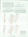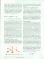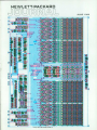
1
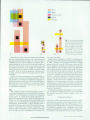
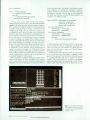
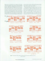
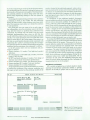
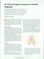
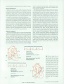
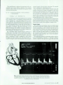
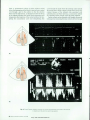
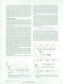
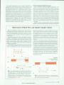
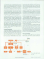
limited number of applications, and its relative newness.
Recent Developments
Within the past two years, cardiac Doppler ultrasound
technology has been recognized as an important tool in
evaluation of cardiac blood flow rates. Although it has been
available for many years, it has not been considered as
having clinical utility until the work of Hatle and Holen,3'4
which demonstrated that valve pressure gradients could
be quantified using Doppler ultrasound techniques.
In 1982, radionuclide and contrast angiography were pre
ferred tools for the diagnosis of global ventricular function,
identification of regurgitan! valvular cardiac lesions, iden
tification of intracardiac shunts, and assessment of coro
nary artery disease.2 Today, ultrasound techniques are ex
tremely successful in aiding the diagnosis of many cardiac
abnormalities. The use of ultrasound in the diagnosis of
coronary artery disease has had little success, except in
pediatrics. Coronary angiography still provides the critical
information for assessment of coronary function.
Technical Limitations
The use of the Doppler effect in ultrasound measurement
of blood flow has some limitations. There are two important
aspects to the Doppler equation (see article, page 26) that
must be considered where evaluation of cardiac disease is
concerned. The first is the angle between the flow velocity
of interest and the incident ultrasound beam. The most
accurate velocities are measured when the angle is very
small. However, when searching for certain cardiac anoma
lies such as high-velocity jets caused by stenotic, regurgi
tan!, or shunt lesions, or defects in the heart, the exact angle
of flow is unknown and movement or rotation of the trans
ducer is necessary until the location of the highest maxi
mum velocity is obtained.
The other important aspect of the equation is the propor
tional relationship between the frequency used to interro
gate the blood flow and the resultant frequency shift. In
pulsed Doppler systems, the maximum measurable shift is
limited by the rate of the ultrasound pulses sent out. For
example, assume that a pulsed Doppler system is sampling
flow from a vessel or heart chamber at a depth of 12 cm.
Further assume that the speed of sound in body tissue is
approximately 1540 m/s and the frequency of the beam is
2.5 MHz. Given the depth, the maximum pulse rate is 6.4
kHz. This means that the maximum measurable frequency
shift is 3.2 kHz if the Nyquist criteria is observed. This
shift corresponds to a blood flow velocity of about one
meter/second, assuming an angle of zero degrees between
the transducer and the flow direction. However, the veloc
ities associated with many valvular defects are much
higher, 3 to 5 m/s in some cases. For this reason, continu
ous-wave (CW) Doppler ultrasound is used. The trade-off
here is between the measurement of flow at a selected
depth, available from pulsed Doppler measurements, and
the maximum velocities obtained from CW Doppler tech
niques. Use of both techniques during an examination has
become an accepted practice because of the importance of
determining severity as well as location of the disease. Fig.
1 shows common cardiac "windows" used in obtaining
signals from the numerous areas of interest of the heart.
Fig. 2 illustrates how the two sides of the heart function
normally.
Systole Diastole Systole Diastole Systole Diastole
Diastolic Flow
Right Atrium (RA)
Tricuspid Valve (TV)
Right Ventricular Inflow Tract (RVIT)
Systolic Flow
RVOT
Right Ventricular Outflow Tract (RVOT)
Pulmonic Valve (PV)
Pulmonary Artery (PA)
Systole Diastole Systole Diastole Systole Diastole
Diastolic Flow
LVIT
Left Atrium (LA)
Mitral Valve (MV)
Left Ventricular Inflow Tract (LVIT)
MV
Systolic Flow
(b)
Left Ventricular Outflow Tract (LVOT)
Aortic Valve (AOV)
Aorta (AO)
AOV
LVOT
Fig. 2. Normal cardiac flow, (a)
The right heart. Blood enters the
heart through the vena cavae,
which empty into the right atrium.
Flow then proceeds from the right
atrium through the tricuspid valve
to the right ventricle during dia
stole. Systolic flow occurs when
blood is ejected from the right ven
tricle through the pulmonic valve
to the main pulmonary artery, (b)
The left heart. Freshly oxygenated
blood from the lungs returns to
the heart via the left atrium. Flow
then proceeds from the left atrium
through the mitral valve to the left
ventricle during diastole. Systolic
flow occurs when the left ventricle
pumps blood through the aortic
valve back into the circulation
system.
JUNE 1986 HEWLETT-PACKARD JOURNAL 21
© Copr. 1949-1998 Hewlett-Packard Co.