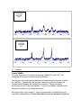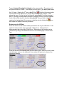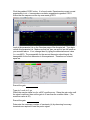
1
Nuclear Magnetic Resonance Facility
User’s Manual
Department of Chemistry and Biochemistry
University of Oklahoma
Version 2.1.03
March 16, 2011
Stephenson Life Center, Room 1700
Acknowledgements
This manual was constructed using VNMRJ2.2C and VNMRJ2.2D with a Red
Hat Linux 5.1 operating system on the computers within the NMR facility at the
University of Oklahoma. Material was also incorporated from the Structural
Biology NMR lab User’s Manual from the University of Minnesota with the
express permission and review of Dr. Beverly Ostrowski.
The NMR Facility of the Department of Chemistry and Biochemistry at the
University of Oklahoma would like to express gratitude to the Structural Biology
NMR Resource at the University of Minnesota for their willingness share their
knowledge, resources and training.
Suggested Reference Materials
Books
Modern NMR Spectroscopy by Sanders and Hunter
High-resolution NMR techniques in organic chemistry by Claridge, Timothy D. W
Spectrometric Identification of Organic Compounds by Silverstein and Webster
200 and More NMR Experiments by Berger, and Braum
Article
Reynolds, W.F.; Enriquez, Raul G.; “Choosing the Best Pulse Sequences,
Acquisition Parameters, Postacquisisiton Processing Strategies, and Probes for
Natural Product Structure Elucidation by NMR Spectroscopy”, J. Nat. Proc.,
2002, 65, 221-224.
Web Resource
Basic NMR Concepts: A Guide for the Modern Laboratory by Dr. Daniel Holmes.
http://www.chemistry.msu.edu/facilities/nmr/
Contents
I.
II.
III.
IV.
V.
VI.
VII.
VIII.
IX.
X.
XI.
Abbreviations and Terms
NMR Laboratory Policies
NMR Training Policies
Preparing Samples
Tubes
Handling and Preparation
Sources of Contamination
Equipment
Spectrometers
Probes
Choosing the Spectrometer
Instrument specifications and set-up
Sensitivity
Resolution
Dynamic Molecules
Software
Intro to VNMRJ
Data Files
Creating Data Directories
Creating a Desktop Shortcut to the Data Directory
Creating Data Transfer Shortcuts on the Red Hat Desktop
Transferring the data
Probe Files
Instrument Calibration
Probe Files
Changing the Probe File
Gradient Shimmaps
Using the Varian Mercury VX-300 MHz Spectrometer
300 NMR sign-up rules
Mercury 300 Tests and Assignment Certification
Collecting a 1D proton on the Mercury VX-300 MHz NMR
Using the Varian VNMRS-400 MHz Spectrometer
400 NMR sign-up rules
VNMRS- 400 Tests and Assignment Certification
Collecting a 1D proton on the VNMRS-400 MHz NMR
Tuning the broadband probe using protune(‘calibrate’)
Using the Varian VNMRS-500 MHz Spectrometer
500 NMR sign-up rules
VNMRS- 500 Tests and Assignment Certification
Tuning the 500 probes
Using mtune
Changing Basic 1D parameters
Setting the frequency window (adjusting sw and tof)
Setting the first delay (d1) and acquisition time (at)
5
6
7
7
7
8
8
9
9
10
10
10
11
12
12
13
13
15
15
16
17
19
21
21
21
22
23
25
25
26
27
33
33
34
35
42
44
44
45
46
47
48
49
50
XII.
Basic 1D processing
Opening Saved Data
Toolbar Icons
Phasing
Referencing the Spectrum
Weighting Functions
Zero Filling
Measuring Signal-to-noise
Finding digital resolution
Integration
Baseline Correction
Peak Picking
Basic 1D Plotting
Making PDF Files
Converting 1D Data to ascii Format
XIII. Finding a 90 degree pulse width
XIV. Basic 13C 1-D NMR
XV. 2D- Experiments
2D Parameter Sets
General Considerations for doing 2D Experiments
COSY
NOESY
HSQC
HMBC
XVI. 2D Processing and Printing
2D Tool Bar
Manipulating 2D Data
Processing 2D data sets manually
Manual Phase correction
Referencing the 2D data
Plotting the 2D data
Appendix
A.1 Mercury VX-300 Practical Assignment #1
A.2
Mercury VX-300 Practical Assignment #2
A.3 VNMRS-400 Practical Assignment #1
A.4 VNMRS-400 Practical Assignment #2
A.5 VNMRS-500 Equipment components
A.6 Transferring Data to your pc using Winscp
A.7 Remotely accessing the 400 NMR via PUTTY and REALVNC
A.8 Glossary of Common NMR Commands and Terms
A.9 A few useful Linux commands.
51
51
53
54
55
56
58
58
59
59
62
63
64
64
65
66
71
74
74
75
76
78
80
81
83
83
83
84
85
86
87
88
88
89
91
96
99
105
107
113
120
Abbreviations and Terms
COSY
DQF-COSY
f1
f2
FID
HMBC
HMQC
HSQC
Hz
MHz
MW
NMR
NOE
NOESY
PFG
ppm
probe
Proton
RF
ROESY
T1
T2
TOCSY
VNMRj
VT
X-nucleus
Correlation Spectroscopy
Double-quantum filtered correlation spectroscopy
“Indirectly” detected dimension
“Directly” detected dimension
Free Induction Decay
Heteronuclear Multiple Bond Coherence
Heteronuclear Multiple Quantum Coherence
Heteronuclear Single Quantum Coherence
Hertz
Megahertz
Molecular Weight
Nuclear Magnetic Resonance
Nuclear Overhauser Effect
Nuclear Overhauser Effect Spectroscopy
Pulsed field gradient
parts per million
part of the spectrometer where RF signal is sent and received
used interchangeably for 1H
radio frequency
Rotating frame Overhauser effect spectroscopy
Longitudinal Relaxation
Transverse Relaxation
Total Correlation Spectroscopy
Varian’s software used to interface with console
variable temperature control
used to refer to anything not 1H
NMR Laboratory Policies
Laboratory Information
NMR Facility Manager:
Office:
Phone:
Email:
Dr. Susan L. Nimmo
SLSC 1700
627-7044
[email protected]
Instrument Problems
Please contact Dr. Nimmo by cell phone and email with details of the problem.
Instrument Log Book
Please record your name, group, general experimental details and any problems
that are encountered when you use the instrument.
Sample Breakage
In the event of a sample tube breaking in the magnet please contact the NMR
facility manager immediately. This should be done both by phone and email. It
is imperative that no one use the instrument until after Dr. Nimmo has accessed
the situation. The student should place a “broken sample” note on the keyboard
and lock the computer screen.
Faces On-Line Signup
The faces on-line scheduling program hosted by the Complex Carbohydrate
Research Center at the University of Georgia is used for instrument sign-up for
all instruments our laboratory. The page is found at http://faces.ccrc.uga.edu/
It can be accessed from any computer which is attached to the internet.
Individual login and passwords will be set up for users at the initial training
sessions. You will only be allowed to sign-up for instruments that you have been
trained to use. Please read the news page of the sign-up for instrument status
and special instructions for each instruments. All users will be held responsible
for following specific instructions found on this news page.
Funding Acknowledgements
Please acknowledge the instrument funding and notify the NMR facility manager
by email when publishing data collected on the 300 and 400 spectrometers. It is
important for us to keep statistics for both past and future proposals. The Varian
Mercury VX-300 NMR Spectrometer was purchased in 2000 by the multi-user
NSF grant CHE#0077707. The Varian VNMRS-400 NMR Spectrometer was
purchased in 2007 by the multi-user NSF grant CHE#0639199
NMR Training Policies
All students will begin training on the 300 NMR. Training sessions will be
approximately 45 minutes in duration and will be held as many times as
necessary for the student to gain competency. More advanced students and
post-docs may gain certification by demonstrating their ability to use the
instruments and by completing the assignments.
Certification to use the 300 NMR will be given as NMR Test #300-1, Test #300-2
and Practical Assignments #300-1 and #300-2 are completed as approved by Dr.
Nimmo.
Training will begin on the 400 upon successful completion of 300 training.
Certification to use the 400 NMR will be given as NMR Test #400-1, and
Practical Assignments #400-1 and #400-2 are completed as approved by Dr.
Nimmo.
Training will begin on the 500 upon successful completion of 400 training and
Test #500-1. Certification to use the 500 NMR between 8-5 Monday-Friday will
be given as NMR Test #500-2 is completed as approved by Dr. Nimmo.
Unlimited access will be given upon the completion of NMR Test #500-3
Additional training may be requested on an individual basis. But please
understand that training sessions will not last longer than 1 hour per session.
Variable temperature training will be given as needed. Supervision of variable
temperature experiments will continue until both the NMR facility manager and
the student are completely confident of the student’s ability to safely carry out
these experiments independently.
I. Preparing Samples
Tubes
For a standard samples, use the equivalent of 5 mm tubes Wilmad 528-pp-8
(available from the stockroom) (rated 500 MHz and 8 inches long). Eight inch
tubes are required for the robotic insertion on the 400. The sample volume
should be at least 500 microliters, typically 650-800 microliters. Minimum sample
height rule of thumb: coil + 3X diameter of the sample. The probe coils are 16
mm (18mm in the 500 MHz triple resonance and 500 MHz indirect detection
probes). Standard NMR tubes are 5mm. Samples shorter than 500 microliters
are very difficult to shim. Users running samples containing hazardous materials
are encouraged to use thick-walled tubes for safety and should notify facility staff.
Do not use chipped, cracked or scratched tubes. Labels for NMR tubes can
also be purchased from most tube vendors. Alternatives for smaller volume
samples include Shigemi tubes or susceptibility plugs that can be purchased
from Wilmad and generally work better for small volumes than thick-walled tubes.
Shigemi tubes, 5mm, use volumes of 270-300 microliters and are made of glass
that is matched to the magnetic susceptibility of the solvent to be used. Using
more than 800 microliters in a regular tube is not recommended because the
sample height will extend past the controlled variable temperature range and a
temperature gradient can be created in the sample. Some vendors also sell
thick-walled tubes that are useful when you have a hazardous sample and are
concerned about tube breakage.
Handling and Preparation
Samples should be clear of precipitate and particulates if possible. Many options
are available for filtering samples. Centrifugation can also eliminate precipitate
from samples. Be sure to wipe off the NMR tube with a Kimwipe around the
bottom where the sample may have been touched. Samples require deuterium
solvent for a lock signal. Higher quality deuterium solvents, purchased in
ampules, will have a smaller residual solvent peak. Samples can be run without
deuterium, no lock, for very short runs if necessary. Store deuterated solvents in
well-sealed containers on dessicant. Samples should be transferred into and out
of NMR tubes using long pipettes that reach the bottom of the NMR tube. Extra
long glass pipettes are available from Wilmad. Transfer samples smoothly to
avoid losing liquid to the sides of the tube or adding air bubbles to the sample.
Water (H2O) samples require approximately 10% D2O for a lock signal.
Controlling pH in biological samples and in many water samples is highly
recommended. pH can change chemical shifts drastically in many aqueous
samples. For example, phosphorous shifts are changed dramatically with pH.
Cleaning tubes can be accomplished using a tube cleaner which attaches to an
aspirator. These are available from several companies. Rinsing the tube several
times with water or another solvent if necessary, then with isopropanol or
acetone. Do not dry tubes in an oven. The heat in the oven will warp the
glass. Blowing nitrogen or other dry gas through the tube will help remove
residual solvent, alternatively, pull a vacuum on the tube for several minutes.
Another source of cleaning information can be found on the Wilmad website at:
http://www.wilmad-labglass.com/services/NMR_010.jsp
Sources of Contamination
Tables of chemical shifts of common contaminants are readily available online or
in books. Membranes and membranes in concentrators, dialysis bags, buffers
(i.e. Tris), detergents, and dirty tubes can all be sources of contamination.
Contamination peaks are usually easy to spot because they are often of a
different linewidth and different ratio to the sample being observed. Buffers such
as Tris contain proton signals that contaminate spectra. Tris is difficult to remove
from a protein sample and should be avoided or deuterated if possible. Many
commercial membranes are stored in glycerol and should be washed thoroughly
before use with a protein sample. Samples prepped in fully deuterated solvent
can pick up water from the air very easily and should be sealed tightly. Often the
cap can be sealed with a very small piece of Parafilm and samples stored on
dessicant in the refrigerator. Samples prepared in “100%” deuterated solvent
should be prepared with minimal air exposure or prepared in a dry box or dry
bag. Keep bottled solvents on dessicant or, preferably, use sealed ampules of
solvent. Solvents from a general use bottle can also become easily
contaminated. Occasionally, even newly purchased solvents can contain
contamination that is detected when working at low concentrations in high field
magnets. To determine if the solvent is the source, run a sample of solvent only.
Often a peak near zero is also often seen in samples that can be due to grease
used on glassware. Drying tubes with cans of compressed air can also generate
a contaminate peak from the propellants and should be avoided. One useful
reference in identifying impurities is J Org Chem, 62, 7512-7515 (1997), “NMR
Chemical Shifts of Common Laboratory Solvents as Trace Impurities”.
II. Equipment
Spectrometers
There are three spectrometers located in the NMR laboratory: Varian Mercury
VX-300 MHz, Varian VNMRS-400 MHz and VNMRS-500 MHz. All spectrometers
are equipped with wave form generators and pulse field gradients. The
frequency at which protons precess in the particular strength of magnetic field is
used to designate the magnets. For example, protons precess at 300 MHz in the
300 MHz magnet, but the magnet strength is actually 7 Tesla. Further, carbon
precesses at about 75 MHz in a 300 MHz magnet. Nuclei such as carbon,
nitrogen and phosphorous resonate at much lower frequencies than proton.
Often, nuclei that resonant at the higher frequencies such as proton and fluorine
are called “high band” and other nuclei like carbon and nitrogen are called “low
band”. The basic components of the spectrometer include: workstation, console,
magnet and probe.
Workstation
The computer workstation is where most of the operation of the instrument
occurs including data collection and simple processing. Extensive processing
should be done on offline workstations that do not absorb instrument time. The
workstation communicates with the console that, in turn, controls the console and
the probe in the magnet. The workstations are Dell PC’s running under Red Hat
Linux 5.1.
Console
The console contains the radio frequency generators, amplifiers, a variable
temperature controller, pulsed-field gradient generator, waveform generators,
and other computer components. In typical operation, a user will very rarely, if
ever, need to interact with the console.
Magnet
The magnetic field in all of the instruments is generated by a current flowing
through a solenoid of superconducting wire. For the wire to be superconducting,
the wire must stay at liquid helium temperature (4 K) or below. Therefore, the
cryostats are filled with liquid helium and outer liquid nitrogen to keep the
magnets cold. If the magnets warm up above that temperature, a quench can
occur. A quench is when the current in the magnet coil is lost. If a quench
occurs, it is usually accompanied by a loud noise followed by fast release of
helium gas from the cryostat. If this occurs, please leave the lab as quickly as
possible. The magnet is contained inside the silver dewar. The magnets are
mounted on vibration legs. The air legs maintain level and stable against small
vibrations by air pressure. Therefore, do not lean against the magnets because
the magnet will rock. Try to avoid walking around near the magnet during an
experiment because it can contribute to vibrations. Typically, the only time a
user needs to go near the magnets is to insert the sample and tune the probe.
Never take metal or magnetic objects near the magnets. Always check pockets
and person for these things before approaching the magnet. Non-digital
watches, cards with magnetic strips, and magnetic media (such as disks) will
also be affected by the magnetic field. Also inside the magnet are the shim coils.
Shim coils are a collection of electrical coils used to remove residual magnet field
inhomogeneities. The temperature in the bore of the magnet, where the sample
will sit, is controlled by a variable temperature controller and is typically set at 25
deg. C.
Probes
The probe is inside the bore of the magnet. The probe contains the
transmitter/receiver coils on where pulses go into the sample and RF frequencies
come out. Probes can be changed in a few minutes by facility personnel and
have different configurations depending on the application intended. Probes are
only changed by facility personnel or by specially trained users. The type of
probe selected is determined by the nucleus to be detected and the specific
experiment. The 400 and 500 spectrometers have indirect detection probes.
These probes are the best choice for direct proton detection or indirect detect
experiments. This probe has the proton transmitter/receiver coil closest to the
sample and is, therefore, most sensitive for proton detection. The 300 and 400
spectrometers have broadband probes which typically have the X-nucleus coil
closer and are, therefore, more sensitive for nuclei like carbon. Carbon can still
be detected directly using an indirect detection probe, but it will have a much
lower signal-to-noise. Broadband probes or probes that include a X-nucleus can
be tuned to a different nucleus depending on the tuning range of the probe.
Direct detection of an X-nucleus is best done with a broadband probe.
III. Choosing the Spectrometer
Instrument Specifications and Set-up
It is important to choose the appropriate instrument in the laboratory to answer
the relevant experimental questions. Important things to consider include type of
probe on the magnet, sensitivity of the system in regards to sample
concentration, variable temperature set up, and magnetic field strength.
Varian Mercury 300 MHz NMR Spectrometer
The 300 NMR is especially easy to use. The afternoons are set up in 15 minute
time slots because it is possible for anyone to collect a 1H NMR spectrum in a
very short amount of time. The spectrometer has a 4-nuclei auto-switchable
probe. This means that 1H, 19F, 13C, and 31P experiments can be collected
without changing cables or tuning the probe. This instrument is also capable of
running more advanced 1D and 2D experiments, but the sensitivity is less than
on the other spectrometers and therefore requires higher sample concentration.
The spectrometer is running VNMRJ 2.2D software with chempack 4.1.
Varian VNMRS 400 MHz NMR Spectrometer
The 400 NMR is equipped with a 50 sample autosampler. Therefore this is the
best spectrometer for running several samples. It can be set up to automatically
run multiple experiments on different samples. It is more sensitive than the 300
NMR and requires less sample concentration. It is equipped with dual broad
band probe and automatic tuning. As a result this is the best spectrometer for
collecting direct detect broad band spectra. This spectrometer has extended
variable temperature accessories and can conveniently collect variable
temperature experiments. It also has an indirect detect 1H probe for running
indirect detection experiments with lower sample concentration. The
spectrometer is running VNMRJ 2.2C software with chempack 4.1.
Varian VNMRS 500 MHz NMR Spectrometer
The 500 NMR is three channel system with both a triple resonance probe and an
two channel indirect detection probe. This system has the highest resolution,
best sensitivity in the laboratory. Longer 2D and 3D experiments have
preference on this instrument over short 1D 1H experiments. The spectrometer is
running VNMRJ 2.2 c software with an chempack 4.1 and Biopack options.
Sensitivity
Signal to noise increases as field strength is increased. Signal to noise is also
very dependent on the probe. Indirect detection probes are constructed to
maximize the proton sensitivity, while direct detect probes are constructed to
maximize broad band signal intensity. The table below lists the signal to noise
values for the NMR instruments according to the field strength and probe. These
measurements were made after reinstallation in our new laboratory with the
Varian standard sensitivity standards.
1H
F19
13C
P31
N15
300 MHz NMR
4-nuclei probe
125:1
85:1
90:1
72:1
-400 MHz NMR
Broad-band probe
300:1
325:1
180:1
180:1
20:1
Indirect Detection Probe
725:1
-----
500 MHz NMR
Indirect Detection Probe
Triple Resonance Probe
965:1
1070:1
---
75:1
--
---
---
Resolution
Resolution is normally increased as the field strength of the magnet is increased.
Overlapped peaks on the 300 NMR may be resolved at higher field strength.
The partial 1H spectra of a natural product are shown below. The spectra were
collected on the 400 and 500 spectrometers within our NMR facility. The 800 Mz
spectrum was collected at the University of Minnesota. The three peaks are
completely resolved from each other at 800 MHz.
800 MHz
500 MHz
400 MHz
Dynamic Molecules
Molecules which are dynamic on the NMR time scale have a lower coalescence
temperature on a lower field magnet. A single set of sharp lines is observed
above the coalescence temperature. In the example below, the dynamic
catenane compound had broad lines in the 13C spectra at room temperature.
Due to sample constraints 50oC was the upper limit that the sample could be
heated. On the 400 MHz NMR spectrometer, this temperature was not far
enough above the coalescence temperature to result in sharp lines. However,
considerably sharper lines were observed on the 300 MHz NMR spectrometer at
50oC due to the lower coalescence temperature.
13
C{1H}
100.58 MHz
(400 NMR)
50oC
150
149
148
147
146
145
144
143
148
147
146
145
144
143
13
C{1H}
75.46 MHz
(300 NMR)
50oC
150
149
IV. Software
Intro to VNMRJ
All of the computers in the facility are running VNMRj 2.2C and 2.2D. The
interfaces of these two versions are nearly identical.
The program that communicates between the workstation and console is called
the “acqproc”. Occasionally this program loses communication between the
console and workstation and needs to be re-started. Please ask facility
personnel to do this if they are available. If no one is available, instructions are
posted in the facility for re-setting the console.
Every group has a login account. They are responsible for remembering their
user name and password. Do not allow others to use the account or give out
the password to anyone. If the facility determines that a user is allowing
unauthorized users to use the account, privileges will be suspended.
The VNMR software currently uses linux for many things. Knowing a few simple
linux commands can be very helpful (See Appendix for useful linux commands).
In a user account, there is a directory called "vnmrsys". This is a VNMR system
directory. In this directory are many subdirectories. These directories are
probably empty unless someone has put in a new macro, pulse sequence, shim
file, parameter file, etc.
The most significant of the subdirectories:
psglib - pulse sequence library, contains uncompiled pulse sequences
seqlib - sequence library, contains the compiled, executable versions of pulse
sequences
maclib - macro library
shims - stores personal saved shim files
parlib - parameter files
probes – probe files (created with an addprobe command)
shapelib – shaped pulse files (filename.RF)
gshimlib – gradient shimming maps and files (created from gmapsys)
Global directories are found in the /vnmr directory (a few directories up from the
personal directories). The global directories, like psglib, contain all the pulse
sequences, macros, parameters, shims etc. available to all users. To see the
pulse sequences available on a specific machine, look in the psglib in the VNMR
directory. The main VNMR subdirectories can be altered by facility staff only.
Files may be copied into a personal vnmrsys and altered there. Any macros,
pulse sequences, parameter files, etc. in a personal vnmrsys will be accessed
preferentially to the global vnmrsys during operation.
To put a new pulse sequence into the vnmrsys, copy the uncompiled sequence
(seqencename.c) into the psglib. Be sure to copy any necessary macros into
the maclib, any parameters into the parlib, shaped pulses into shapelib etc.
To compile a sequence copied into the psglib,
>seqgen pulseq.c
This will compile the sequence and put the executable into the seqlib. Pulse
sequences can also be compiled from the VNMR command line as
>seqgen(‘sequencename.c’).
VNMR also has various packages for specific applications. The most common
one in the facility is called BioPack and includes many pulse sequences for
applications to biomolecules. These packages can be installed or activated in
individual user accounts.
V. Data Files
Creating Data Directories
We ask that each group save its data within the data directory of the vnmrsys
folder. The data directory is found at /home/usergroup/vnmrsys/data. The
usergroup is designated by the group login. For example, if your group login is
vnmr1, then the group data directory is found at /home/vnmr1/vnmrsys/data, but
if your login in rhc, then the group data directory is found at
/home/rhc/vnmrsys/data. Please maintain individual directories for each member
of the group. Directories can be made using the file browser. A file browser can
be accessed under the Applications>System Tools.
After the file browser is opened, type the location of your group data directory in
the location bar and create a new folder under the File menu.
The NMR facility automatically sends all files within the data directories of each
group to a local server at the end of every day. Files that are not saved in the
data directory are not sent to the server. However, the facility does not
assume any responsibility for backing up user data files. Users must back
up their own data. Each spectrometer and data station computer has a flash
drive. It is suggested that users transfer data via flash drives to their own
computers where they can maintain a CD back-up of all data. Alternatively, files
can be copied from spectrometer to workstation within the facility via ssh. See
appendix A.6 for details in using the freeware Winscp to transfer data.
Creating a Desktop Shortcut to the Data Directory
It will be convenient to have a link on the desktop that will navigate directly to the
data directory of your group.
Using the file browser, open the vnmrsys directory within the home directory and
right click on the data folder. Click Make Link.
This creates a link inside the vnmrsys folder named link to data. Drag the link to
data shortcut to the desktop.
Right click on the link to data and rename it to Data Directory.
Creating the shortcut shown below:
Creating Data Transfer Shortcuts on the Red Hat Linux 5.1 Desktop
Click on Places
Click Connect to Server ……
The Connect to Server box will open:
Set the Service type to SSH and fill in the Server, Folder and Name to user for
connection
Server-Internal IP
Server-External IP
Name for Connection
10.254.219.26
129.15.22.45
300 NMR
10.254.219.27
129.15.22.46
400 NMR
10.254.219.28
129.15.22.47
500 NMR
10.254.219.30
129.15.22.49
Data Station 1
10.254.219.31
129.15.22.50
Data Station 2
Note: Use the Internal IP address for the Server number if you are transferring
between spectrometer and data stations. Use external IP address for the Server
Number if you are transferring data to a computer outside of this network.
Folder: Location of your group’s data directory
Generally: /home/login/vnmrsys/data
Specifically if your group login is rhc then the folder is /home/rhc/vnmrsys/data
Example: This is a shortcut made to Data Station 1 by the vnmr1 group.
The above information created the following shortcut:
Transferring the Data
To transfer the data double click on the desired short cut. This will open up box
requiring your group password. Put in the password and click connect. Copy
and paste your files from the local computer to the destination computer.
Example: Transferring data from the 300 to Data Station 1 using the vnmr1
account: Click on the data directory shortcut on the desktop and navigate to the
files that you wish to copy. NMR data have a .fid extension. These are actually
folders containing four files. You must copy the entire .fid folder.
Open the appropriate folder to find the files that you wish to transfer.
Click on the Data Station 1 shortcut to open a connection to the data station.
Enter in the password and click the connect button.
This will open a file browser on Data Station 1 (destination folder)
Local Folder
Destination Folder
Transfer the data from the local folder to the destination folder using the copy
and paste functions under the edit menu. You may also “drag and drop” but be
aware that you may be moving your files instead of copying them.
VI. Probe Files
Instrument Calibration
Proper operation of both chempack and biopack within the VNMRJ 2.2C and
VNMRJ2.2D software requires the use of up-to-date and calibrated probe files.
Setting up experiments from the software menu as described in the experimental
instructions of this manual should result in the most recently calibrated values
being automatically loaded into the experimental data sets. This can be checked
by comparing the data parameter sets to the calibrated values within the probe
files on the computer and/or the hard copies in the red calibration notebooks
located next to the computer of each spectrometer.
Probe Files
Probe files are specific to each instrument probe. They contain all of the
necessary calibration information. The actual calibration values can be found in
the red calibration notebooks located next to each spectrometer and inside the
text of the probe file itself. The location of the system probe files is in the
/vnmr/probes/ directory of each instrument and can be viewed by any user. The
system probe files can only be changed by the system administrator. Groups
may manage their own local probe files in desired, but be aware that only the
system probe files will be updated with current calibration data. Groups that
create their own probe files will be responsible for their maintenance.
It is important for the user to be aware of which probe is on the instrument and
which probe file is loaded in the software. The 400 and 500 spectrometers each
have two probes. The news page of the faces on-line sign-up, and the first page
of the red calibration notebooks next to each spectrometer have current probe
installation information. The table below shows the name of the probe file that
corresponds to each available probe in the laboratory.
Instrument
Mercury VX-300
VNMRS-400
VNMRS-400
VNMRS-500
Probe
4-nuclei autoswitchable
PFG probe
Auto-X-Indirect Detection
probe
Auto-X-Dual Broadband
probe
Triple Resonance Probe
Probe File
asw4405
AutoX_ID_8726
AutoX_DB_8790
TR_8064 (for chempack
use)
HCN (for biopack use)
VNMRS-500
Indirect Detection Probe
id8297
Changing the Probe File
The loaded probe file can be seen and accessed at the bottom of the VNMRJ
window.
Click on the Probe button to access the probe files.
Click on the down arrow to view the probe file options. Select the desired file and
close the box. 6
VII. Gradient Shim Maps
All three spectrometers utilize gradient shimming. Gradient shim maps are made
by the NMR facility staff and stored in the /vnmr/ gshimlib/shimmaps/ directory.
Users are welcome to make and store their own gradient shimmaps within their
users gshimlib directory, but the details of making these maps is not found in this
manual.
It is important that you are using a shimmap which corresponds to the probe
installed on the instrument. You may also want to change shim maps to use the
most current map. The name of the most current gradient shim map can be
found on the instrument status report located in the three ring calibration
notebook next to each of the spectrometer’s host computers.
Loading a gradient shim map
The currently loaded gradient shimmap name can be found on the Standard Tab
of the Acquire Page. The maps are named by the corresponding probe and the
date that the map was made. In this case, the map name is
AutoX_DB_8790_lk_2011-02-25, meaning that it was generated on February 25,
2011 with the AutoX_DB_8790 probe on the magnet.
To change the gradient shimmap, first type gmapsys on the vnmrj command line.
This will bring up the gradient shimmap pages. The option to change the map
can be found on the Gradient Shim Tab of the Acquire Page.
Click the down arrow to see all of the available maps, highlight the desired map
and it will load into the current mapname box.
Click the Quit Gradient AutoShim button to return to the regular VNMRJ pages.
VIII. Using the Varian Mercury VX-300 MHz Spectrometer
The 300 MHz NMR was purchased in 2000 under a NSF multi-user grant (CHE
0077707). It has a 4-nuclei autoswitchable PFG probe. It can collect
1H/19F/13C/P31 without retuning the probe or changing cables. This instrument
is designed for walk-up use and is primarily used by the synthetic chemistry
groups. Mornings, Evenings and Overnight time can be reserved in advance in
large blocks, but daytime can only be reserved in 15 minute blocks.
300 NMR Sign-up Rules:
Mornings ( 8 am -12:00 pm)
Sign-up is allowed one day in advance for a maximum of 1 consecutive hour. Do
not sign-up for one hour to use the instrument for 5 minutes. This time is
reserved for longer experiments and inexperienced users.
Afternoons (12-noon to 6:00 pm)
Sign-up is allowed only on the day of use and is restricted to a maximum of 15
consecutive minutes.
Evenings (6:00 pm to 8:00 pm)
Sign-up is allowed one day in advance for a maximum of 2 consecutive hour
Overnight (8:00 pm to 8:00 am):
Sign-up is anytime in advance for a maximum of 12 consecutive hours
Mercury 300 Tests and Assignments Certification
Student Name: ________________________
300-Test #1:
Student gives demonstration of properly operating the instrument. The student
must log into the spectrometer, insert sample, lock, shim, collect a 1H
experiment, save the data and transfer the data to the data station. Speed is not
required. At the completion of this test, the student is allowed to use the
instrument Monday-Friday between the hours of 8 and Noon.
Date Completed: ________________Supervisor: _____________________
Practical Assignment #1
Collect 2 1H NMR spectra of 2-Ethyl-1-indanone on the 300. This sample is
located in the NMR laboratory. Please shim the sample as well as you can.
Follow the instructions located in Appendix A.1 of this manual. Upon completion
copy the assignment and give to the NMR facility staff.
Date Completed: ________________Supervisor: _____________________
Practical Assignment #2
Collect a 13C NMR spectrum of 2-Ethyl-1-indanone. Follow the instructions
located in Appendix A.2 of this manual. Upon completion copy the assignment
and give to the NMR facility staff.
Date Completed: ________________Supervisor: _____________________
300-Test #2:
Student gives demonstration of properly operating the instrument. The student
must log into the spectrometer, insert sample, lock, shim, collect a 1H
experiment, save the data and transfer the data to the data station. The test must
be completed within 15 minutes. Upon completion of this test, the student is
permitted to use the instrument at any time.
Date Completed: ________________Supervisor: _____________________
Collecting a 1D-proton on the Mercury VX-300 MHz NMR Spectrometer
Log in and double click on the VNMRJ icon.
Insert your sample clicking the Eject and Insert buttons on the Start> Standard
Page.
Set up a proton experiment by choosing proton under the experiment menu. If
there are no experiments under the Experiment menu, uncheck the Use Study
Queue option under the Acquisition menu.
Select the desired solvent on the standard tab of the Start page.
Lock:
Click the Find z0 button on the Lock tab of the Start page.
Click the Lock Scan, Lock Off and the Spin on buttons. Adjust (raise or lower as
necessary) the Power and Gain until the signal completely appears on the
screen.
Adjust z0 to maximize the lock signal. For best results adjust z0 until signal is
maximized, then adjust phase until the signal is maximized. Clicking the middle
wheel of the mouse will change the steps of z0, power, gain and phase to 1, 10
or 100. If the lock level is fluctuating at a low power level, then lower the gain.
Turn the Lock On when you are finished locking. You must unclick the Lock
Scan button for the other buttons to become available.
Shim by clicking the Gradient Shim button on the Lock tab of the Start page. If
you want to manually shim z1-z5, click the shim tab. It should not be necessary
to shim the higher order shims.
Alternatively, you may manually shim using the buttons on the Start>Shim Page
to maximize the lock signal.
Start your experiment by clicking the Acquire button. Parameters may be
accessed under the Acquire pages. Basic parameters such as sweep width,
number of scans, relaxation delay, etc are found in the default 1H tab. Automatic
plotting and integration can also be set to on or off here.
An “old style” parameter page can be accessed by typing dg in the command line
and clicking on the Process>Text Output Page
More instructions regarding changing the parameters of a basic 1D experiment
can be found in section X of the this manual: Changing Basic 1D Parameters.
Save your data by choosing File > Save As from the drop down menu.
This opens a box in which you can type your filename. Clicking the Home icon
will take you to the data directory within your account. You can create folders
here using the create folder icon. Double click on your folder.
Create New
Folder
Go up one
directory
Go To Home
Directory
Eject your sample clicking the Eject and Insert buttons on the Start> Standard
Page.
Close VNMRJ software
Type exit in the command line to close the VnmrJ software.
Log off Computer
Click Log Out from the Systems Menu.
IX. Using the Varian VNMRS-400 MHz Spectrometer
The Varian VNMRS-400 NMR Spectrometer was purchased in 2007 by the multiuser NSF grant CHE#0639199. It is equipped with two probes: Auto-X indirect
detection probe and Auto-X dual broad band probe. Please request a probe
change at least 2 working days in advance.
Training will begin on the 400 upon successful completion of 300 training.
Certification to use the 400 NMR will be given as NMR Test #400-1, and
Practical Assignments #400-1 and #400-2 are completed as approved by Dr.
Nimmo.
400 NMR Sign-up Rules:
Rules for Advanced Signup
Advanced signup is defined as signing up for time more than 24 hours in
advance. These rules do not apply if this time has not been taken within 24 hours
of the start of the experiment.
Weekdays (Monday – Friday 8:00-4:00):
Advanced sign-up of this time is reserved for experiments which must be
monitored, 400 beginning students who are only allowed to use the instrument
during this time and experiments which require help from the NMR facility staff.
Weekends: (Friday 6 pm – Monday 8 AM)
Please limit advanced sign-up to 24 hours during the weekend unless approved
by the NMR facility staff.
400 NMR Tests and Assignment Certification
Student Name: ________________________
400-Test #1:
Student must give demonstration of properly operating the instrument including
sending the data to the data station. This test includes operation of the robot,
automated tuning unit and shimming the standard line shape sample to
instrument specifications.
Date Completed: ________________Supervisor: _____________________
400- Practical Assignment # 1
Measure the 1H pw90 for any sample. Follow the instructions located in
Appendix A.3 of this manual. Make a copy of the assignment and hand it in to
the NMR facility staff.
Date Completed: ________________Supervisor: _____________________
400- Practical Assignment # 2
Collect a gCOSY experiment using any sample. Follow the instructions located
in Appendix A.4 of this manual. Make a copy of the assignment and hand it in to
the NMR facility staff.
Date Completed: ________________Supervisor: _____________________
Upon completion of 400- test #1, practical assignment #1 and practical
assignment #2 , the student is permitted to use the instrument at any time.
Collecting a 1D-proton on the Varian VNMRS-400 MHz NMR Spectrometer
Put your sample tube in a spinner turbine and set the depth using the depth
gauge. Put the sample into one of the numbered slots in the sample tray and
take note of the slot number. Do not use slot #1 (upper left corner) or slot #0
(lower right corner). A standard sample should be in the magnet which will be
taken out and put into slot #1 when your sample is inserted. Slot #0 must be left
empty. If the robot takes your sample out and places it in slot #0, you must
remove it.
In this example, the sample is placed in slot #19.
Login and start VNMRJ by left double clicking on the VnmrJ icon on the desktop.
Set up a proton experiment by choosing proton under the experiment menu.
If there are no experiments under the Experiment menu, uncheck the Use
Study Queue option under the Acquisition menu.
Select the desired solvent on the standard tab of the Start page.
Insert your sample by typing loc=slot# change. In this example the sample
was placed into slot #19, so loc=19 change will cause the robot to eject the
standard sample, replace it into slot #1 and then insert the sample from slot
#19 into the magnet.
Lock:
Click the Find z0 button on the Lock tab of the Start page.
Click the Lock Scan, Lock Off and the Spin on buttons. Lower the Power
and Gain until the signal completely appears on the screen.
Adjust z0 and Phase to maximize the lock signal. The lock signal is gold
and the phase difference is blue. For best results adjust z0 until signal is
maximized, then adjust phase until the signal is maximized. Continue
alternating between z0 and phase until the signal doesn’t change when the
Lock is turned on. The phase difference (blue) should be zero (flat line)
when the lock signal (gold) is maximized. Clicking the middle wheel of the
mouse will change the steps of z0, power, gain and phase to 1, 10 or 100.
Turn the Lock On when you are finished locking. You must unclick the Lock
Scan button for the other buttons to become available.
Shim by clicking the Gradient Shim button on the Lock tab of the Start page.
If you want to manually shim z1-z5, click the shim tab. It should not be
necessary to shim the higher order shims.
Tune your sample by typing protune in the command line. (Alternately you
may use the protune(‘calibrate’) macro: see page 42.)
Start by tuning the high band to H1 (or F19). If tuning proton, then type H1
in the nucleus box under Advanced Tune and choose Fine for the Tune
Criterion. Click Tune to Criterion to begin the tuning.
The acquisition status will return to idle and a Tuning done ok message will
appear when tuning is completed.
You may also tune the low band at this time (i.e. C13, P31, etc) by the same
method described above. Click the close button at the bottom of the
window when finished.
Start your experiment by clicking the Acquire button or typing ga in the
command line. Parameters may be accessed under the Acquire pages.
Basic parameters such as sweep width, number of scans, relaxation delay,
etc are found in the default 1H tab. Automatic plotting and integration can
also be set to on or off here.
Save your data by choosing File > Save As from the drop down menu.
This opens a box in which you can type your filename.
To eject your sample, type loc=1 change in the command line. This will
eject your sample, replace it into its slot (slot#19 in this example) and put
the standard sample from slot #1 into the magnet.
Type exit in the command line to close the VnmrJ software.
To completely log off, choose Log Out from the Actions Menu.
Tuning the Broad-Band Probe Using protune(‘calibrate’)
The protune macro will automatically tune 1H/13C/19F/31P. However, the
broadband probe is tunable to many additional nuclei. In this example, we will be
tuning the probe to 7Li which is at 155.440 MHz on the 400 MHz NMR.
Type protune(‘calibrate’) in the command window of VNMRJ.
This will open the manual calibrating program as shown below.
Type the desired frequency in the ‘Tune to’ box and then click the ‘Tune to’
button. (155.440 for this example).
The tuning will begin and continue until the bottom of the frequency dip reaches
the bulls-eye.
Click the Quit button when the tuning is finished.
X. Using the Varian VNMRS-500 MHz Spectrometer
The 500 NMR spectrometer was originally purchased as a VXR model in 1987
and was completed upgraded to a VNMRS model by the University in 2006 with
the addition of Dr. Susan Schroeder and Dr. Robert Cichewicz to the Department
of Chemistry and Biochemistry Faculty.
The spectrometer is a three channel, 28 shims Varian VNMRS-500 equipped
with two probes: triplet resonance H{13C/15N} PFG probe, and a tunable indirect
detection 1H{15N-31P} PFG probe.
Training will begin on the 500 upon successful completion of 400 training and
Test #500-1. Certification to use the 500 NMR between 8-5 Monday-Friday will
be given as NMR Test #500-2 is completed as approved by Dr. Nimmo.
Unlimited access will be given upon the completion of NMR Test #500-3
Advanced sign-up rules
Advanced sign-up for less than two hours will be limited to Wednesday
afternoons. No one person is allowed to sign up in advance for more than 18
consecutive hours, with this exception of Dr. Schroeder’s group which has
ownership of 50% of all the available NMR time.
Operation Instructions
The 500 NMR is operated using VNMRJ2.2C software which is identical to the
400 NMR. With the exception of the autosampler and tuning, operation of the
500 NMR is consistent with the instructions presented in the 400 NMR operation
section.
VNMRS 500 Tests and Certification
Student Name: ________________________
500-Test #1:
The student will be given a written test given by Dr. Nimmo. This test will be
similar to the test at the end of the appendix. Upon the successful completion of
this written test the practical training sessions will begin.
Date Completed: ________________Supervisor: _____________________
500-Test #2
The student will demonstrate the proper use of the instrument. This will include
setting the depth of the sample and inserting the sample into the magnet,
changing the probe file, setting up a 1H experiment and tuning the probe. The
student will be expected to properly identify all probe cables and connections and
equipment components. At the successful completion of this test the student will
be allowed to use the NMR from 8am – 5 pm, Monday –Friday.
Date Completed: ________________Supervisor: _____________________
500-Test #3
The student will demonstrate the proper use of the instrument to a NMR faculty
member. This will include setting the depth of the sample and inserting the
sample into the magnet, changing the probe file, setting up an 1H experiment
and tuning the probe. The student will be expected to properly identify all probe
cables and connections and equipment components. This will also require the
signature of the student’s research director. At the successful completion of this
test the student will be allowed to use the NMR at any time.
Date : ________________
NMR Faculty: _______________________
Date: _________________
__
Research Director: ___________________
Tuning the VNMRS-500 MHz NMR probes
WARNING: If you are a new user and you don't know how to do this or are
unsure, GET HELP. Do not attempt to just “figure this out”. Equipment can be
damaged. Tuning probes may not be allowed in other NMR labs so please
check with the lab manager before attempting to tune probes elsewhere.
The VNMRS model has some differences to earlier models. These differences
result in some differences in tuning of this instrument and other models of
instruments. There is no need to move any cables for the tuning process.
Tuning the probe reduces the reflected power. Probe tuning requires adjusting
the tune wands very carefully to reduce the reflected power to the 50 range on
the tune interface box. Tuning the probe will insure that the 90-degree pulse
width determination will be accurate and the pulse width will be as short as
possible. If the sample is high in salt, the probe may not tune down to 50.
Tuning Tips: The wands can be very sensitive. Try to make small adjustments.
If minimizing both the tune and the match and the reading doesn’t go any lower,
try moving the match part either clockwise or counter-clockwise to slightly
increase the signal. Then try to minimize the signal again using the tune part. If
the signal improves, continue to move the tune part in the same direction and
repeat. If the signal does not improve, try adjusting the tune part slightly in the
opposite direction to increase the signal and try to minimize again with the match
part. This process should helps get out of any local minimum it is trapped in. Do
not force the tuning rods, if they feel “stuck” stop and ask for help.
To tune the probe, first setup the correct frequencies through the computer. To
do this, load the experimental parameters, i.e. setup a proton experiment then
type su on the command line. Look at the channels page of the Acquire tab to
see which nuclei is set up on each channel. Channel 1 is 1H, and channel 2 is
carbon.
Set the tuning box to channel 1 and check that the attenuation is set at 9. If the
meter reading is very large or off-scale, lower the attenuation. Tune the proton
wand (top part is tune, bottom is match).
Channel
Attenuation
Adjust the wands to minimize the value of the reflectance power. Raise the
attenuation back to nine (if necessary) as the reading decreases. Users should
be able to reach a reading of 50. Always end with the attenuation at 9. Change
the channel to 2 and tune the carbon wand if desired. When you are finished the
tuning box must read channel 0 attenuation 9. The tuning box is only accurate to
a value of 50; it may be helpful to use mtune to achieve a better tune.
Using mtune
To start the mtune program, type mtune on the command line. To observe the
high band tune choose Tune RF channel 1; center frequency H1 and click Start
Probe Tune.
To change to low band, click Stop Probe Tune, change Tune RF Chanel to
2 and center frequency to desired nucleus (C13 in this case). Click Start
Probe Tune. Chanel 3 can be tuned to 15N in the same manner.
XI. Changing Basic 1D Parameters
A list of parameters can be accessed on the text output page of the process
panel after typing dg in the command window. A short description of each
parameter listed can by found in the Command and Parameter Reference
Manual. Each parameter can be changed by typing the abbreviation = value in
the command window. For example to change the number of scans from 1 to 16,
simply type nt=16 in the command window. It is necessary to type dg for the list
to refresh, showing that the change has been made.
Selected parameter list
at
Acquisition time
d1
Delay time between scans in seconds –recycle delay
ni
Number of increments
np
Number of points
nt
Number of transients
pw
Pulse width
pw90
90o pulse width
seqfil
Pulse sequence being used
sfrq
sw
tn
tof
tpwr
dfrq
dmf
dn
dof
dpwr
Observed frequency (frequency of tn)
Sweep width (of tn)
Transmitting nuclei (observed nuclei)
Tuner offset frequency—center of the spectrum
Transmitting nuclei power level
Decoupling frequency
Decoupling Modulation Frequency
Decoupling Nuclei
Decoupling Offset Frequency
Decoupler Power
Setting the frequency window (adjusting sw and tof)
The transmitter offset (tof) will always be in the center of the spectral window
(sw). The spectral width (sw) of a spectrum may need to be altered and with it,
the transmitter offset (tof). Use the smallest spectral width that accommodates
the expected chemical shifts of the sample. Beware of using one too small
though because peaks that are cut off will result in folded peaks.
When using water samples, set tof on the water peak. To do this, set the cursor
on the water peak and type movetof. To move the spectral window, enclose the
spectrum with the cursors, expand, and type movesw. This command moves
the tof to the center of the new spectral window so tof will change. Manually
reset the sw by using >sw=# if the tof needs to stay the same. The sw should
be held constant for a series of experiments.
To select a spectral window, decide the approximate range where the signals will
fall and select a slightly larger window. If all the signals can be observed in one
scan, the window can be set manually using the movesw command. The sw is
in Hertz. ppm = sw/sfrq. Therefore on the 300 NMR spectrometer, sw=3000 is a
window of approximately 10 ppm for a 1H spectrum. For a carbon spectrum on
the 300 NMR spectrometer (carbon sfrq ~ 75 MHz), sw=3000, sfrq=75, is a
window of approximately 40 ppm. Remember the spectral window is not a
specific frequency range, so sw=3000 could be 0-10ppm or –1 to 9 ppm or 1-11
ppm etc. The specific frequency range is set by the location of the transmitter
offset (tof). Tables of tof values are found in the red calibration notebooks next to
each spectrometer.
Changing the sweepwidth using the movesw command:
Simply place the cursors around the peaks and type movesw. This will
automatically change the sw to the position of the cursors and adjust tof to the
half-way point between the cursors. In the spectrum below, the sweepwidth was
changed to 0 – 9 ppm with the center at 4.5 ppm. This corresponds to a sw of
2700 Hz and a tof of -184.9 on the Mercury VX-300 NMR Spectrometer.
Changing the sweepwidth and tof manually:
The transmitter offset (tof) will always be in the center of the spectral window
(sw). The tof is a distance in Hertz, plus or minus, from the original center and
will not match the scale when swapped from ppm to Hz. The tof parameter is
spectrometer dependent and will therefore vary from instrument to instrument.
Therefore, two different 500 NMR spectrometers will have different tof values.
Tables of values for tof values with their corresponding ppm values are found in
the red calibration notebooks next to each spectrometer.
To manually set tof, place the cursor at the desired location of the spectrum
center and type movetof. Then type sw=#(hertz) for the desired window size.
For example, if I wanted to have a 10 ppm window on the 300, with the center at
8 ppm, then I would set the cursor at 8 ppm and type movetof sw=3000. I would
then collect the spectrum and reference the peaks. Alternatively, I could look at
the tof value in the calibration notebook and type tof=# sw=# on the command
line.
Setting the first delay (d1) and acquisition time (at).
The delay time, d1, is the time between the end of the acquisition time, at, and
the next element in the pulse sequence. The total recycle time is the time
between the last pulse and the next pulse at the beginning of the pulse
sequence, typically is d1 + at. The total recycle time usually reflects the T1,
longitudinal relaxation time, of the sample. Typically, a value of 1-2 seconds will
suffice. If the T1 is unknown, a quick T1 experiment should be done to estimate
it. The T1 can be long especially in cases of heteronucleus detection such as
carbon or other X nucleus. T1 values for quaternary carbons can be very long
and the total recycle time should reflect this.
The acquisition time, at, is usually set based on a rough approximation of T2,
transverse relaxation time. The T2 indicates how long the signal in the FID will
last. As a general rule, large molecules such as proteins relax very quickly and
require a short at (such as at=0.5). Small molecules relax much more slowly and
the FID can last several seconds so often using a longer at is favorable (such as
at=3 to 10 seconds). If you type dps on the command line the pulse sequence
will be displayed. This is a graphical representation of the pulses that are taking
place during each scan.
d1
at
To change d1 or at, type d1=# and at=# on the command line. Type dg to
refresh the parameter list and dps to refresh the pulse sequence.
XII. Basic 1D Processing
Opening Saved Data
Click Open..(under File in the Main Menu).
This will open a pop-up window. Clicking Home will take you to the data
directory within your account. You can create folders here using the create folder
icon. Double click on your folder.
Go To Home
Directory
Go up one
directory
Create New
Folder
Highlight your
spectrum and
click Open.
Spectra have a .fid extension.
The spectrum will processes automatically and appear on the screen. Use the
toolbar icons to access cursors. If the spectrum seems to “freeze”, either click
the redraw icon on the toolbar or type ds in the command window. The vertical
scale can be changed with the mouse wheel or by typing in a numerical value for
vertical scale (i.e. vs=5000).
Toolbar Icons
Access one cursor, click again to access a second cursor
Show the full spectrum
Reset to full display
Zoom In
Zoom Out
Zoom More
Pan and Stretch More
Integration
Show/Hide Scale
Show/Hide Threshold
Phase Mode
Redraw Spectrum
Phasing
To display the spectrum on the screen, type ds
Type f full to see the full spectrum in the full screen.
The spectrum will likely need to be phased. To phase the spectrum, click on the
phase icon on the toolbar
. Use the left mouse button to click on a portion of
the spectrum. Hold down the left mouse button and move the mouse to flatten
the spectrum's baseline and makes the peaks symmetrical. The right mouse
button will make fine adjustments and the center mouse button will increase or
decrease the vertical scale. Simple spectra can be phased crudely with by typing
aph (automatic phasing).
The spectrum below is a newly acquired spectrum which needs to be phased.
This spectrum has been “wrapped around” by bad phasing. It can be corrected
by setting lp=0 and rp=0 and then phasing again.
Beware of becoming dependent on aph since autophasing does not always work
and often leaves small errors in the phase that are better corrected manually.
The lp, or left phase, value corresponds to a first order phase correction that is
frequency dependent, meaning it affects peaks differently. The rp, or right
phase, value corresponds to the zero-order phase correction and is an error seen
in all peaks.
Referencing the Spectrum
The spectrum may be manually referenced using the cursor. Expand the region
around the peak you wish to reference by placing a cursor on either side of the
peaks and clicking the zoom in icon.
Lower the vertical scale to bring the desired peak to scale. This can be done by
clicking the mouse wheel under the scale (To raise the vertical scale click the
mouse wheel at the top of the black part of the screen).
Put the cursor on the peak. In this example, it is the chloroform peak.
Type in the reference number on the Display page of the Process Tab.
Weighting Functions
To add a weighting function to a FID, type wti on the command line. This opens
the weighting function screen. The mouse controls are shown on the screen.
The right mouse button toggles the spectrum (top box) off and on, the center
button controls the vertical scale of the FID (bottom box) and the spectrum, and
the left mouse button controls the adjustment of the weighting function (green
line in the center box).
Start by clicking on line broadening icon on the toolbar.
This should bring up
an exponential function in the middle window (green line). Click on the right
mouse button in the top box to see the spectrum. Use the left mouse button to
adjust the green line (weighting function) and observe the change in the
spectrum. The value of the line broadening (lb) is shown on the bottom of the
screen.
The interactive weighting screen displays the FID, spectrum, and the weighting
function applied. Above, an exponential function, line broadening (lb), is applied
to the FID. Line broadening improves signal-to-noise at the expense of
resolution. Larger values also can improve FID truncation artifacts. Negative
values of lb can improve resolution, compared to the unweighted spectrum, at
the expense of signal-to-noise.
After a good adjustment is found, type wft in the command line. This performs a
weighted Fourier transform and applies the weighting function. The spectrum
may need to re-phased slightly after this.
A sinebell function can also be used for resolution enhancement. To use a
weighted function like a shifted sinebell, go to the interactive weighting screen by
typing wti on the command line. Turn off any weighting functions by clicking the
lb, sb, and gf toolbar icons until all of the weighting functions say “unused”.
Set the sinebell function to be about double the width of the data in the FID to be
kept.
Next click the sinebell shifted icon on the toolbar and shift the sinebell over so
that it just covers the FID.
After a good adjustment is found, type wft in the command line. This performs a
weighted Fourier transform and applies the weighting function. The spectrum
may need to re-phased slightly after this.
Zero-Filling
Zero-filling is handled through the fn parameter. The Fourier number, fn, is
normally set to 'n' or not used. In this case, the number of points (np) are the
actual data points. To use zero-filling, setting the fn to a number larger than the
np will zero-fill. This number is typically a power of 2. For example type fn=16k
or fn=32k or fn=np*2 on the command line and the computer will automatically
adjust to the correct number. Re-transform (wft) the data after changing this
parameter to see the result. Using a fn smaller than np will use fewer than the
actual number acquired.
Measuring Signal-to-noise
Measure the intensity of the largest peak in the spectrum. (Use the cursor to
move onto the line and type nl.) Then move the cursor over and enclose an area
of noise with two cursors. Type dsn on the command line for a display of signalto-noise. This command can be found in the Command and Parameter Guide.
For this to be an accurate measure between spectra, use the same peak and the
same window of noise each time.
Finding Digital Resolution
Place the cursor near the maximum of the peak to measure. Type nl dres on the
command line for a display of the digital resolution (width at approx. half height).
Integration
Display the spectrum of interest on the screen.
Clear any existing integrals by clicking Clear Integrals on the Cursors/Integration
Page of the Process tab or by typing cz in the command window.
Click the integral icon on the tool bar to access the integration mode.
This will cause a green integration line to appear on the screen.
The different integration icons are shown below.
Click the define integrals icon on the toolbar, then click the left mouse button to
cut the green line on either side of the peaks of interest. Clicking the right button
will cause the cut to be erased.
To correct leveling and tilt errors in the integral click the level/tilt integration icon
and adjust the integral similar to phasing the spectrum. To set the integral values,
put the cursor on one of the peaks and type a numerical value into the Integral
Area Box.
Click Set Integral Value and Show integral value to see the integral values on the
screen.
Baseline Correction
After integral regions have been specified on a spectrum, the baseline can be
corrected to reflect these regions as peaks. To baseline correct, type bc on the
command line or click the BC Correct button found on the Display page of the
Process Tab. This baseline correction assumes everything in a defined integral
region is a peak and flattens the rest. To work properly, everything that is or
might be a peak must be in an integral region. To massage this further, see the
Varian parameter guide on bc. This command can have many modifiers to
produce a better correction.
The spectrum below illustrates the use of the baseline correction in a 13C 1D
spectrum.
Peak Picking
Use the Show/Hide threshold icon to display the threshold level.
Click the Find Peaks button on the Display page of the Process Tab.
This will display the peak frequencies on all of the peaks which cross the
threshold.
Readjust the threshold by clicking the Show/Hide Threshold icon and moving the
yellow line with the left mouse button.
Click the Find Peaks button to display the peak frequencies.
Basic 1D Plotting
The simpliest way to print is with a list of commands. These commands are
listed below. The following commands should be typed in the vnmr command
window to print the following options. They should be typed in a string separated
by spaces ending with the command page. All commands are optional except for
page. The spectrum will be plotted at a width of 250 mm, unless you specify
different. You can change the width of the plot by typing wc=200. This will
change it to 200 mm.
pl
pscale
pap
ppa
pir
piv
ppf
page
prints spectrum
prints scale
prints all parameters (long list)
prints primary parameters (short list)
prints integration ratios horizontally
prints integration ratios vertically
prints peak frequencies
sends file to printer
You may also use the buttons on the Process>Plot page. Simply click the
options that you would like to print and then click Plot Page.
Making a PDF Files
To make a pdf file from your spectra, choose the features that you would like to
print, then simply click Plot Preview instead of Plot Page.
Adobe Acrobat will open and show you your pdf file. Click File>Save As> to save
the pdf file.
Converting 1D Data to ascii Format
Spectra can be plotted by excel or other programs if it is first converted to ascii
format. To convert the FID, first process, phase and reference the data. Take
note of the frequency at which it was collected. (Type sfrq? on the command line
and write down the number). Type writexy on the command line. There will be
no indication or message that anything was written, however, the file should have
been created inside of the experiment directory. For example, if the vnmr1 group
was working inside of exp1, then the output file named xytrace.1 would be found
inside of /home/vnmr1/vnmrsys/exp1/ directory. The file will have two columns.
The first column is frequency and the second column is intensity. After opening
the file in excel, divide the first column by the sfrq value noted earlier to convert it
into ppm. Plot data as desired.
XIII. Finding a 90 Degree Pulse Width
Introduction
What is the 90 degree pulse width? The radio frequency pulse is described by its
power and duration (time). Before the pulse the proton “spin” is oriented in the z
direction and it cannot be observed in the spectrum. The rf pulse “flips” the spin
into the xy plane so that it can be observed. The maximum signal is seen when
the spin is completely in the xy plane without any z component. The length of
time that this takes is called the 90 degree pulse. In the spectra below the length
of the pulse is varied from 1 to 40 microseconds. The most accurate way to
determine the 90 degree pulse width is to find the 360 and divide it by 4. This
value is directly affected by the pulse power. As you increase the power, then the
90 degree pulse will decrease. Normally a power (tpwr) between 54-60 is used.
90 o
180 o
360 o
270 o
Instructions
Collect a 1D 1H spectrum. Expand the region around one peak as shown in the
spectrum below.
Take note of the previously calibrated 90o pw and power level and record them
below. These values can be found on the acquisition page of the Acquire tab.
Create an array of values for the pulse width (pw). Open the array pop-up
window by clicking the arrays button found on the Default 1H tab of the Acquire
panel. (Alternatively type array on the command line).
The array pop-up window is shown below:
Create the array by doing the following:
Type pw in the Param Name Box
Click New Array
Fill in the Array Size, First Value, and Increment Boxes
Click enter on the keyboard.
The array needs to go past the 360o pw. This number can be estimated by
taking the previously calibrated 90o pw value and multiplying it by 4. You will
want your array to go past this point. In this example the 90o pw is 11.25
microseconds. The array must go past 11.25*4 (45 μseconds), therefore
pw is arrayed from 1 to 55 μsecond in steps of 1 μsecond. After the array is
created click the Close button at the bottom of the box.
An appropriate delay, number of scans, absolute intensity must be now set and
the experiment started. A delay of 2 seconds with 1 scan is chosen in the
example. These are set by typing:
d1=2 nt=1 ai ga on the command line.
When the experiment is completed display one spectrum from the array and
phase it. In this example the third spectrum is displayed and phased. This is
done by typing ds(3) full aph on the command line.
To view the entire array and display the array values in the bottom text box, type
dssh da on the command line. It may be necessary to adjust the vertical scale
(vs) and the vertical position (vp) to make all spectra display completely on the
screen. The array values are found in the Text Output tab of the Process panel.
The 90o pw is calculated by taking the 360o pw and dividing it by 4. The 360o pw
is identified as the second null point in the curve. The numbers can be displayed
under each spectrum by typing dssl (not shown). In this example, the 45th
spectra is the 360o pw. This corresponds to 45 μseconds.
90o
180o
360o
270o
To find a more accurate 90o pw create a second array that spans 4 μseconds
around the 360o pw with 0.25 μseconds increments. In this example, the second
array would be from 43 – 47 μseconds with an increment of .25 μseconds.
XIV. Basic 13C 1-D NMR
Proton Decoupled 13C Experiment:
13
C{1H}
Experimental Set-up:
VNMRS-400 and VNMRS-500: Be sure that the probe is tuned to 1H on channel
1 and 13C on channel 2. Mercury VX-300: the probe is already tuned.
If the proton signals of your compound are either all aromatic or all aliphatic, the
default parameters for 13C 1-D NMR may not give good enough signal to noise.
In this case, the instructions below can be followed to improve your data.
First acquire a proton spectrum of the sample in exp1. Move the transmitter
offset to the center of the proton signals by placing the cursor in the middle of the
peaks and typing movetof in the command window. This value may then be
found by typing tof? in the command window.
Note the tof (transmitter offset). This will become the set value of dof in the
carbon experiment.
Join experiment 2 by typing jexp2 in the command window. If experiment 2
doesn’t exist, create it by typing cexp(2). If experiment 2 is locked, unlock it by
typing unlock(2). Set up the carbon experiment by choosing the carbon
experiment under the Experiments drop down menu.
Set dof to the center of the proton peaks (tof) that was determined above. Type
dg and the parameters will be shown in the text output (or overview) window of
the process tab.
To determine the number of scans for your available time, type
time(hours,minutes). For example, if I have 1 hour and 15 minutes, type
time(1,15) dg. This will set the number of scans (nt) to the appropriate number.
Type bs=16 ga wbs(‘wft’) in the command window. This will start the experiment
and show you the carbon experiment at the completion of every 16 scans.
If you would like to stop the experiment before it is complete, click the Stop
button or type aa. Save the data, send it to the data station and print it.
Important Parameters:
The parameters dpwr, and dmf are essential for good results. These parameters
are calibrated regularly and correct values should load into the parameter set
when the experiment is set up. These parameter values along with their
calibration dates can be found in the red calibration notebook located next to
each spectrometer. The decoupler power (dpwr) should stay at or below 40db
for safety. The decoupler modulation mode (dmm) is the mode the decoupler is
using. To decouple protons, use waltz decoupling (dmm='w'). When using
waltz or garp decoupling, the decoupler modulation frequency (dmf) is set to 4X
γH2. The γH2 value is regularly calibrated and updated in the probe file.
The spectrum below was collected with 32 scans. The decoupler was on
(dm=’yyy’). The spectrum shows three singlets at 124.0, 126.7 and 127.5 ppm.
Integrating the Carbon Spectra
In a proton decoupled 13C spectrum all the coupling information between the 1H
and 13C has been removed and all the carbons peaks should be singlets.
Integration of this spectrum may not be accurate due to the carbon-proton NOE
enhancement. The default setting in the carbon pulse sequence accessible by
the drop-down menu for the decoupler is “on” for the entire length of the
experiment. This is designated by dm=’yyy’. Use dps (display pulse sequence)
to see when the decoupler is off and on. Decoupling the protons in the carbon
experiment significantly increases signal intensity, but removes coupling
information and the possibility of integrating the spectrum.
Proton Coupled 13C Experiment
Carbon-13 1D NMR can also collected with the C-H coupling observed. This is
referred to as a proton-coupled carbon-13 experiment: Different decoupling
settings will allow for better or worse signal-to-noise and determines whether the
spectrum can be accurately integrated and/or whether the coupling information
(splitting) is retained.
Decoupling Mode
‘nnn’
‘nny’
‘yyn’
S/N
poor
fair
fair
Integration
yes
yes
no
Splitting
yes
no
yes
‘yyy’
best
no
no
Set the decoupler to the desired value. The spectrum below was collected with
the decoupler off (dm='nnn'). This change in decoupler settings resulted in a
large decrease in signal-to-noise but contains the coupling information. Many
transients were needed to see carbon peaks (1798 scans). Carbon peaks from
carbons attached to protons are split. This spectrum can be integrated. The
spectrum shows three doublet of doubles at 124.0, 126.7 and 127.5 ppm (J1=163
Hz, J2=9 Hz).
XV. 2D- Experiments
2-D Parameter Sets
Typical operation usually collects the normal 1-D in exp1 and 2-D experiments in
exp2 or higher. Virtually all 2-D experiments will require a 90 deg. pulse width
calibration for setting up the experiment. Also move the spectral window as
necessary to observe only the area of interest and set up any water suppression
needed.
For homonuclear 2-D experiments like COSY and NOESY, the normal 1-D
parameters control the f2 dimension (direct dimension) while the f1 axis (indirect
dimension) is controlled by parameters identified with a 1 (i.e. sw1, lb1, fn1). In
homonuclear experiments, be sure sw=sw1. One other very important
parameter in 2-D experiments is the number of increments (ni). The ni specifies
the number of "points" in the indirect dimension. Typical values for ni are 128,
256, or 512. Steady state scans are also important in long experiments so be
sure to set ss. The suggested value is ss=32. A manual page (man) for the
experiment will also point out parameters that need to be set. To access these
type the following on the command line:
man('pulsesequence name') or
printon man('pulsesequence name') printoff
i.e. man('NOESY')
To determine the length of time the experiment will run, type time on the
command line. Be sure the experiment fits into the time block available. To
change the length of time the experiment will run, options include increasing or
decreasing ni, nt or d1 (or satdly in a presat experiment). Check the at
(acquisition time) to check that a typical number of points is being taken. The
typical number of points (np) taken is 2048 or 4096. These correspond to at
times of less than 0.5 seconds. The at time in some 2-D experiments must be
kept short due to high power decoupling during this time period.
General Considerations for doing 2D Experiments (Varian inc, “TwoDimensional NMR 5 days Series”).
1. All 2D experiments will give better results (less t1 noise) if they are run with
the spinner turned off. FOR HMQC, HMBC, INADEQUATE and ALL water
suppression of gradient experiments it is mandatory that the spinner be turned
off.
2. All 2D experiments will give better results if sample temperature is regulated
during data acquisition.
3. All 2D experiments will give better results if lock conditions are optimized.
This involves setting Z0 more or less exactly on resonance for the lock solvent,
adjusting lock power to just below saturation (varies from solvent to solvent),
optimizing lock phase and setting lock gain as low as possible (after shimming).
4. The S/N of individual F2 spectra (before second FT) needs to be only about
5:1 in many cases. In partitioning total experiment time between scans per
increment (nt) and number of increments (ni), it is probably better to emphasize
S/N of individual increments a little more heavily, bearing in mind the square root
dependence of S/N on number of scans. Increasing the number of increments
(ni) excessively may make linear prediction less advantageous, reducing overall
efficiency.
5. It is always advantageous to reduce the spectra width in both dimensions to
the minimum practical value, keeping in mind how peaks may fold in.
6. Zero-filling in F1 is universally done. Linear prediction is also quite common,
typically being used to extend F1 interferograms by a factor of 2 in length. (If
linear prediction is used, the added points must be included in determining how
many zero-filling points to add. The F1 window function must also be adjusted to
include the added points).
COSY-(Correlation Spectroscopy) spectra are 2D proton homonuclear spectra
that correlate one-bond J couplings. A basic COSY sequence is comprised of
two 90-degree pulses.
Versions of COSY
gCOSY – gradient version of the upper case COSY, faster if abundant sample,
allows down to nt=1. With nt=1 and ni=128, a gCOSY can be run in as little as
three minutes. Gradient COSY setup is detailed below. In gCOSY, phase=1.
No presaturation option available.
cosy (lower case) – older version of COSY, no gradients, sets up relay cosy
(relayh)
Set up by typing cosy on the command line and setting pw, sw=sw1, nt=4
(min.), d1, and check >time. relay=0 for absolute value cosy. (set relay and tau
for relay or long-range COSY)
cosyps (lower case) – phase-sensitive COSY, no gradients, similar to cosy, but
allows solvent presaturation option and data is phase sensitive. Minimum nt=4.
DQF-COSY
The double-quantum filtered COSY is used to examine coupling constants in
detail. In general, this experiment takes much longer than a regular COSY but
can provide much more information when measuring coupling constants in
multiplets.
gCOSY—Experimental Set-up
Put your sample into the magnet. Lock, shim, tune and collect a proton
experiment in experiment 1. Calibrate a 90 degree pulse width.
Join experiment 2 by typing jexp2 in the vnmrj command window. Collect a
proton spectrum and adjust the sweep width, gain, tof, and set the pw90 to the
calibrated 90 degree pulse width.
Change the experiment into a gradient COSY experiment by using the
Experiment drop down menu at the top of the page.
Click the gradient COSY button. Notice that the sequence at the top now reads
gCOSY.
Look at the parameter list on the Overview page of the Acquire tab. Type dg to
refresh this parameter list. Make sure that pw, tpwr, sw and tof are the same as
previously noted. If not, change them by typing the abbreviation and value (i.e.
sw=4803). The sweepwidth for the second dimension should equal the
sweepwidth for the first dimension in this experiment. Therefore sw1 should
equal sw.
Type nt=1 ss=4 ni=1 ga and watch the remote status box for a ADC overflow
error. Raise the gain value until the signal overflows, then set the gain to 5 less
than the overflow value. (The maximum value is 60).
Determine the necessary number of transients (nt) by observing how many
transients are required to see the proton signal.
Type nt = required number as determined above, ss=32 ni=128 Type time to see
how long the experiment will be. Click the green Acquire button.
Save the experiment when it is completed.
NOESY (Nuclear Overhauser Effect Spectroscopy) spectra provide information
about protons that are 5 Angstroms or less apart in space. The information is
through space and not through bond, like a COSY. The presence of a NOE peak
is direct evidence that two protons are within 5 Angstroms through space. The
absence of a NOE peak between protons does not necessarily mean that they
are not within 5 Angstroms since other factors can reduce a NOE peak even if
the protons are close in space. A mid-size molecule (~1000-1500MW range) may
have NOEs that are close to zero and a ROESY may be required to see them.
Large molecules generally give better NOEs at higher field, but small molecules
may actually give better NOEs at lower field. A 2-D NOESY of a small molecule
will have cross peaks of opposite phase to the diagonal. A 2-D NOESY of a
large molecule will have cross peaks of the same phase as the diagonal.
Theoretically, these experiments should be symmetrical, but it is typical to see
more intense peaks on one side of the diagonal than the other.
Experimental Set-up
Put your sample into the magnet. Lock, shim, tune and collect a proton
experiment. Calibrate a 90 degree pulse width; adjust the sweep width and tof.
Convert the proton data experiment into a NOESY experiment using the drop
down menu.
Check that pw is set to the calibrated 90-degree pulse width, set d1 and np. Set
the sw1=sw. Set phase=1,2. Type nt=1 ss=4 ni=1 ga and watch the remote
status box for a ADC overflow error. Raise the gain value until the signal
overflows, then set the gain to 5 less than the overflow value. (The maximum
value is 60).
Determine the necessary number of transients (nt) by observing how many
transients are required to see the proton signal. The minimum nt in this
experiment is 2.
One parameter requiring forethought is the mix time. The mix is usually
determined by the size of the molecule under study. Small molecules require
longer mix times, 0.5 to 0.8 seconds (suggested mix=0.5). Large molecules
generally range from 0.05-0.3 seconds (suggested mix=0.15).
Set the ni and ss=32 then type time to determine experimental time. Adjust the
delays, transients and/or increments to fit the experiment into the time available.
Click the acquire button to start the experiment.
To do simple processing of these spectra type setLP1 gaussian wft2da on the
command line. Further phase correction and manually processing may be
required to improve the spectral quality. These corrections are described in the
2D processing and printing section of this manual.
Indirect Detection Experiments
Indirect detection is the detection of a heteronucleus through direct detection on
proton signals. Detection on proton allows for a much higher sensitivity. Keep in
mind that the amount of sample required for a heteronuclear experiment is still
much larger than a purely proton experiment.
HSQC (Heteronuclear Single Quantum Coherence) A proton-carbon HSQC
will detect all carbons with a proton attached. Carbons with no proton attached
will not appear in the 2-D spectrum.
Experimental Set-Up
Put your sample into the magnet. Lock, shim, tune (to both proton and carbon)
and collect a proton experiment. Calibrate a 90 degree pulse width; adjust the
sweep width (sw) and tof. Take note of the parameters pw90, tpwr, sw and tof
and check that they are carried over into the HSQC experiment. Convert the
proton data experiment into a HSQC experiment using the drop down menu.
Parameters:
Set sw1=carbon sweep width. Use a carbon sweep width large enough to
encompass all the carbons attached to protons (don’t worry about quaternary
shifts). Set the dof=decoupler offset for carbon. If certain what dof to use, a
chart of transmitter offsets for carbon is found in the red calibration notebooks
located next to the spectrometer’s computer. pwx is the 90 degree pulse width
of carbon at the power level pwxlvl. This is normally calibrated with a standard
sample. The value is found in the probe file. The decoupling modulation
frequency, dmf, should be equal to the necessary window for decoupling. dmf is
calibrated with a standard sample and is calibrated at power level dpwr. dmf is
equal to 1/(90 degree pulse width). These values are found in the probe file. Set
the at= acquisition time. Make this time short (<0.2 seconds) due to high
decoupling power on carbon during this time. Set phase=1,2 for HSQC. The
one-bond coupling is selected by j1xh. Type nt=1 ss=4 ni=1 ga and watch the
remote status box for a ADC overflow error. Raise the gain value until the signal
overflows, then set the gain to 5 less than the overflow value. (The maximum
value is 60).Set the ni and ss=32 then type time to determine experimental time.
Adjust the delays, transients and/or increments to fit the experiment into the time
available. Click the acquire button to start the experiment.
To do simple processing of these spectra type setLP1 gaussian wft2da on the
command line. Further phase correction and manually processing may be
required to improve the spectral quality. These corrections are described in the
2D processing and printing section of this manual.
HMBC (Heteronuclear Multiple Bond Coherence)
HMBC is a heteronuclear 2-D experiment that will pick up carbons (non-isolated)
without a proton attached. This experiment can be very useful to see quaternary
peaks when there isn’t enough sample for a 1-D carbon. The HMBC set up is
very similar to HSQC except there is no decoupling on carbon.
Experimental Set-Up
Put your sample into the magnet. Lock, shim, tune (to both proton and carbon)
and collect a proton experiment. Calibrate a 90 degree pulse width; adjust the
sweep width (sw) and tof. Take note of the parameters pw90, tpwr, sw and tof
and check that they are carried over into the gHMBC experiment. Convert the
proton data experiment into a gHMBC experiment using the drop down menu.
Parameters:
Set sw1=carbon sweep width. Use a carbon sweep width large enough to
encompass all possible carbons in the molecule. Set the dof=decoupler offset
for carbon. If certain what dof to use, a chart of transmitter offsets for carbon is
found in the red calibration notebooks located next to the spectrometer’s
computer. pwx is the 90 degree pulse width of carbon at the power level pwxlvl.
This is normally calibrated with a standard sample. The value is found in the
probe file. Set the at= acquisition time. Acquisition time can be longer here
since the decoupler is off during this experiment. Set phase=1,2 for gHMBC.
The one-bond coupling is selected by j1min and j1max and the multiple bond by
jnxh. Type nt=1 ss=4 ni=1 ga and watch the remote status box for a ADC
overflow error. Raise the gain value until the signal overflows, then set the gain
to 5 less than the overflow value. (The maximum value is 60).Set the ni and
ss=32 then type time to determine experimental time. Adjust the delays,
transients and/or increments to fit the experiment into the time available. Click
the acquire button to start the experiment.
To do simple processing of these spectra type setLP1 gaussian wft2da on the
command line. Further phase correction and manually processing may be
required to improve the spectral quality. These corrections are described in the
2D processing and printing section of this manual.
XVI. 2D Processing and Printing
2D Tool Bar
Icon
Description
Box/Cursor
Show Full spectra
Zoom In
Zoom Out
Zoom Mode
Pan/Stretch Mode
Trace
Scale
Projections
Redraw
Rotate
Raise vertical scale by 20%
Lower vertical scale by 20%
Phase Mode
Peak Picking
Return
Manipulating the 2D data
Click on the appropriate icon on the tool bar to expand, zoom, and change the
vertical scale of the spectra. Type dconi on the command line to display the
peaks using the color map in the interactive mode. Type dpcon to display the
peaks in contours. To show more contours than is automatically displayed, use a
command that specifies the number of contours and spacing. To display this,
dpcon(20,1.3) will display 20 contours at a spacing of 1.3. Other variations of
this include dpcon(‘pos’,20,1.2). The modifiers ‘pos’ and ‘neg’ will display only
the positive or only the negative peaks in a 2-D spectrum. To make the contour
display interactive, use a variation of dconi such as dconi(‘dpcon’,’pos’,20,1.3).
To plot more contours, pcon(20,1.3) pltext page. Adjust the number of contours
and spacing for the data. The ‘pos’ and ‘neg’ modifiers can also be used in the
plotting.
Processing 2D Data Sets Manually
This routine applies to most phase-sensitive 2-D data sets. COSY requires a
slightly different approach and often uses a pure sinebell function.
Start by processing the first increment with by typing wft(1) Phase this spectrum
as any 1-D.Then add a weighting function to this data by typing wti on the
command line enabling the use of the interactive weighting screen to add a
weighting function. The most commonly used function at this step is a gaussian
function [gf].
Then process the data with by typing wft1da. After the data is processed, the f1
traces will be on the screen. Select a trace with the cursor. Try to use something
other than solvent. Then weight this trace with wti.
This weighting function can also be a gaussian function [gf], but often a shifted
sinebell is used. To put in a shifted sinebell, start with [sb] and move the cursor
to produce a sinebell curve that is about twice the width of the interferogram.
Then select [sbs] . Using the cursor, shift the sinebell back so the maximum
starts at the left side of the interferogram. Process the 2-D data with >wft2da
Manual Phase Correction
Phase errors in phase-sensitive 2-D data sets can often be seen near the
diagonal where the peaks may be streaked positive and negative. Display the
entire 2-D spectrum. Choose 3 traces containing cross peaks near the top,
middle and bottom of the spectrum and note the index #’s of each trace. The
index # of the trace can be seen in the top window of VNMR next to the seq. and
exp #.
Type r1=index# r2=index# r3=index# on the command line. The values r1-r3
are place holders in VNMR. Then display the first trace: Type ds(r1) and Phase
the 1-D trace. Display the 3rd trace. ds(r3) Click
. Click the left mouse button
on both sides of the spectrum to accept the previous phase changes. Then
phase this trace. Go back to the first trace by typing ds(r1) Continue phasing r1
and r3, clicking to accept phase changes in-between until they are both phased.
Look at the middle trace to check by typing ds(r2) Then go back to the 2-D
spectrum by typing dconi. If necessary, rotate the axis by clicking the
and repeat the phase correction procedure.
icon
Referencing the 2D Data
Place the cursor on the contour which you wish to use as your reference. If the
spectra is homonuclear, then type rl(#p) rl1(#p) dconi. If the spectra is
heteronuclear then type rl(#p) rl1(#d) dconi. Alternatively, the spectra can be
referenced by filling in the reference boxes on the Default page of the Process
Tab.
Place the cursor of the desired peak, set the units to ppm, enter the correct value
into the boxes, enter on the keyboard, then redraw the spectra to observe the
referenced spectra.
Plotting the 2D Data
The data can be easily plotted using the buttons on the Process >Basic page or
the Process>Plot page. Choosing Plot Preview will allow the option of saving the
file as a pdf, while choosing Plot Page will send the printout to the printer.
XVII. Appendix
A.1. 300 NMR Practical Assignment #1
Collecting a 1D proton NMR spectrum
Student Name: _________________________________
1. Collect a 1D 1H spectrum of 2-Ethyl-1-indanone. Follow the instructions in
Section VIII of this manual. Use all default parameters.
2. Reference the chloroform peak to 7.27 ppm. Integrate each peak and set the
peak at 7.76 ppm to 1 proton. Print the spectrum from -1 ppm to 10 ppm with the
integration values at the bottom of the page. Print a second spectrum from -1
ppm to 10 ppm with the peak frequencies listed on the page.
3. Save this spectrum into your personal directory on the 300 NMR computer.
File Name:
.
4. Transfer this file to Data Station 1. Make sure that it is put inside of your
personal directory of Data Station 1. The instructions for transferring data are
found within Section V of this manual.
5. Collect a 1D 1H spectrum of 2-Ethyl-1-indanone. Use a delay (d1) of 2
seconds, an acquisition time (at) of 4 seconds, and a collection window
(sweepwidth) of 0 – 9 ppm and 32 scans. The instructions for changing these
parameters are found in section XI of this manual.
6. Reference the chloroform peak to 7.27 ppm. Integrate each peak and set the
peak at 7.76 ppm to 1 proton. Print the full spectrum with the integration values
at the bottom of the page. Print a second spectrum with the peak frequencies
listed on the page.
7. Save this spectrum into your personal directory on the 300 NMR computer.
File Name:
.
8. Transfer this file to Data Station 2. Make sure that it is put inside of your
personal directory of Data Station 1.
9. Make a copy of this page, attach the four requested printouts to it and hand it
into the NMR facility staff.
A.2. 300 NMR Practical Assignment #2
Collecting a 1D 13C{1H} NMR spectrum
Student Name: _________________________________
1. Collect a proton decoupled 13C spectrum of 2-Ethyl-1-indanone following the
instructions below.
a. Collect a 1D 1H spectrum of 2-Ethyl-1-indanone. Follow the instructions in
Section VI of this manual. Use all default parameters.
b. Change the 1H experiment into a carbon experiment by clicking the carbon
experiment in the Experiments drop down menu.
c. Set a large number of scans by typing nt=100000 bs=16.
Start the experiment by clicking the green acquire button.
The data may be processed at the completion of every block of data. This was
defined to be 16 scans when bs=16 was typed in the command line above. Type
wft on the command line to process the data so that the carbon peaks can be
observed. Typing the command wbs(‘wft’) on the command line should cause
the spectra to process automatically at the end of each block. Click the red Stop
button when the signal to noise is satisfactory.
2. Reference the chloroform peak to 77.23 ppm. (See Section XII of this
manual)
3. Save this spectrum into your personal directory on the 300 NMR computer.
File Name:
.
4. Make a pdf file of the spectrum with the peak frequencies printed on the
spectrum. Save the pdf file in your personal directory on the 300 NMR computer.
(See Section XII of this manual)
PDF File Name:
.
5. Make a copy of this page and hand it into the NMR facility staff.
A.3. 400 NMR Practical Assignment #1
1
H 90o pw calibration
Introduction
What is the 90 degree pulse width? The radio frequency pulse is described by its
power and duration (time). Before the pulse the proton “spin” is oriented in the z
direction and it cannot be observed in the spectrum. The rf pulse “flips” the spin
into the xy plane so that it can be observed. The maximum signal is seen when
the spin is completely in the xy plane without any z component. The length of
time that this takes is called the 90 degree pulse. In the spectra below the length
of the pulse is varied from 1 to 40 microseconds. The most accurate way to
determine the 90 degree pulse width is to find the 360 and divide it by 4. This
value is directly affected by the pulse power. As you increase the power, then the
90 degree pulse will decrease. Normally a power (tpwr) between 54-60 is used.
90 o
180 o
360 o
270 o
Instructions
1. Collect a 1D 1H spectrum. Expand the region around one peak as shown in
the spectrum below. Save this spectrum. File Name:
.
Take note of the previously calibrated 90o pw and power level and record them
below. These values can be found on the acquisition page of the Acquire tab.
Previously calibrated 90opw
at power
.
2. Create an array of values for the pulse width (pw). Open the array pop-up
window by clicking the arrays button found on the Default 1H tab of the Acquire
panel. (Alternatively type array on the command line).
The array pop-up window is shown below:
Create the array by doing the following:
Type pw in the Param Name Box
Click New Array
Fill in the Array Size, First Value, and Increment Boxes
Click enter on the keyboard.
The array needs to go past the 360o pw. This number can be estimated by
taking the previously calibrated 90o pw value and multiplying it by 4. You will
want your array to go past this point. In this example the 90o pw is 11.25
microseconds. The array must go past 11.25*4 (45 μseconds), therefore
pw is arrayed from 1 to 55 μsecond in steps of 1 μsecond. After the array is
created click the Close button at the bottom of the box.
An appropriate delay, number of scans, absolute intensity must be now set and
the experiment started. A delay of 2 seconds with 1 scan is chosen in the
example. These are set by typing:
d1=2 nt=1 ai ga on the command line.
When the experiment is completed display one spectrum from the array and
phase it. In this example the third spectrum is displayed and phased. This is
done by typing
ds(3) full aph on the command line.
To view the entire array and display the array values in the bottom text box, type
dssh da on the command line. It may be necessary to adjust the vertical scale
(vs) and the vertical position (vp) to make all spectra display completely on the
screen. The text box with the array values is found in the Text Output tab of the
Process panel.
The 90o pw is calculated by taking the 360o pw and dividing it by 4. The 360o pw
is identified as the second null point in the curve. The numbers can be displayed
under each spectrum by typing dssl (not shown). In this example, the 45th
spectra is the 360o pw. This corresponds to 45 μseconds. Save the file.
File name:
.
Print the spectra and array parameters by using the commands:
pl(‘all’) pssl page printon dg da printoff
Label the printout with the 90, 180, 270 and 360 degree pulsewidth.
Record the 90o pw
and power (tpwr):
.
3. To find a more precise 90o pw create a second array that spans 4 μseconds
around the 360o pw with 0.25 μseconds increments. In this example, the second
array would be from 43 – 47 μseconds with an increment of .25 μseconds.
Set up the array, collect the spectra and save the file.
Filenaname:
.
Print the spectra and array parameters by using the commands:
pl(‘all’) pssl page printon dg da printoff
Record the 90o pw
and power (tpwr):
.
A.4. 400 NMR Practical Assignment #2
gCOSY
Put your sample into the magnet. Lock, shim, tune and collect a proton
experiment. Save the data and record the file name:
.
Calibrate a 90 degree pulse width. Record the calibrated value with the power at
which is was collected:
90opw:
tpwr:
.
Create a new experiment by typing cexp(10) in the vnmrj command window.
This will create experiment number 10. Go to experiment 10 by typing jexp10 in
the command window.
Open the proton that was saved above. Set the 90 degree pulse width and the
power to the calibrated value. You may fill in the boxes on the Acquisition page
of the Acquire tab.
Minimize the sweepwidth by putting the cursors around the peaks and typing
movesw. Collect a spectrum.
Record the sweepwidth (sw):
Record the tuner offset frequency (tof):
.
.
Save the file and record the file name:
Change the experiment into a gradient COSY experiment by using the
Experiment drop down menu at the top of the page.
.
Click the gradient COSY button. It is found under Experiments>convert current
experiments to do..> homonuclear correlation experiments>gradient COSY.
Notice that the sequence at the top now reads gCOSY.
Look at the parameter list on the Overview page of the Acquire tab. Type dg to
refresh this parameter list. Make sure that pw, tpwr, sw and tof are the same as
you recorded above. If not, change them by typing the abbreviation and value
(i.e. sw=4803). The sweepwidth for the second dimension should equal the
sweepwidth for the first dimension in this experiment. Therefore sw1 should
equal sw.
Record the gain:
.
Type nt=1 ss=4 ni=1 ga
Watch the remote status box for a ADC overflow error. Raise the gain value until
the signal overflows, then set the gain to 5 less than the overflow value. (The
maximum value is 60).
Record the gain:
.
Determine the necessary number of transients (nt) by observing how many
transients are required to see the proton signal.
Record required nt:
.
Type nt = required number as determined above, ss=32 ni=128 Type time to see
how long the experiment will be.
Click the green Acquire button.
Save the experiment when it is completed. Record the filename:
.
A.5. VNMRS-500 NMR testing information
Equipment Components
Probes
Triple Resonance Probe (TR_8064)
Varian High-Field Triple-Resonance Probe Manual, Publication: 01-999132-00E,
page 7.
Varian High-Field Triple-Resonance Probe Manual, Publication: 01-999132-00E,
page 5.
.
Varian High-Field Triple-Resonance Probe Manual, Publication: 01-999132-00E,
page 8.
Indirect Detection Probe (id-8297)
Varian High-Field Indirect Detection Probe Manual, Publication: 01-999175-00E,
page 6
Varian High-Field Indirect Detection Probe Manual, Publication: 01-999175-00E,
page 4
Varian High-Field Indirect Detection Probe Manual, Publication: 01-999175-00E,
page 8
Tune Interface Box
This box is located on the floor in front of the magnet. Set the channel to either
1, 2, or 3 as appropriate for the nuclei that is being tuned. Tune the probe until
the readout is at least 50. The mtune program can also be used for tuning.
Remote Status Box:
The Remote Status Box sits on the table next to the computer. Do not leave the
spin light flashing when you log off the computer. If the spin light is flashing, log
back into VNMRJ and turn the spinner off.
FTS Sample Cooler
This is located behind the 500 console. It is usedShould
to regulate
the sample
be
temperature. The air flow must be 100 psi into the
NMR
laboratory
to turn on the
100 psi
compressor. Operation of this unit requires Variable temperature training and
certification.
Pneumatics Router
The router is located on the east wall of the NMR laboratory. Air flow is regulated
here.
Gas flow sensor must be
within green LED lights
(adjusted within VNMRJ
software)
Varian Pneumatics Router Manual, Publication: 01-999302-00B
A.6. Transferring Data to your pc using Winscp
Data can be transferred using a sftp client. Winscp is freeware which can be
downloaded from: www.winscp.com/
Host name: is the ip address of the computer which you would like to access.
The 400 computer is 129.15.22.46. The User name and Password are your
NMR username and password. See Section V of this manual for a list of IP
addresses for the computers in the NMR facility.
After the host name, user name and password is entered, click Login to access
the data. The left panel show the files on your computer, the right panel shows
the files on the computer that you have logged into.
Find the file which you would like to transfer, highlight and click copy.
A.9. Remote access to the 400 NMR via Putty and Real VNC
Putty and Real VNC are both freeware which can be downloaded from the
internet.
CONFIGURE PUTTY
Open the Putty software
Click on the SSH
Click on tunnel
Put 5901 into the Source port and localhost:5901 into the destination
Click Add
Click Session (it is at the very top)
Put the IP address of the computer that you wish to connect to into Host Name and a name in the
saved Session and click Save. The IP address of the 400 NMR is 129.15.22.46. In this case, I
saved it as “400 NMR”.
Using PUTTY:
Open the Putty software. Highlight your saved Session and Click Load.
Click Open
A terminal window will appear
Log in with your NMR login and password
Just return when prompted for the input display server name.
Type vncserver at the prompt. The session number must be 400Computer:1 or the vncviewer
won’t open in the next step.
Open vnc viewer
type localhost:5901 in the Server Blank. Then click OK
Optional: If you want to share the screen, then you must click Shared connection under the Misc
tab of the Options menu
Put your remote password in the password blank
This will open the 400 desktop.
Run your experiments.
When you are finished, log out of the desktop under actions at then top. Then type pkill
vncserver pkill vnc at the command prompt. This is extremely important. These are the
commands that will reset the session number to :1.
A.8. Glossary of Common NMR Commands and Terms
aa
abort acquisition, hard stop
acqi
Open acqi window if the button has been lost
ai
absolute intensity mode
alock
autolocking routine, alock=’y’ for autolocking, alock=’n’ for typical
manual locking
aph
autophasing, not recommended for most spectra
array
macro for setting up an arrayed experiment
at
points (np)
acquisition time, set by spectral width (sw) and number of data
axis='p'
axis=’pd’
specify ppm or hertz for the axis, i.e. axis='p' or axis='h', use
referencing in an indirect detect experiment with >decref
BPsvf
BioPack save file command: saves all pieces associated with an
Experiment including any shape pulses, global file, probe file, etc.
Usage: >BPsvf(‘filename’)
bc
baseline correct
bs
block size, data is stored to the disk every time an increment of bs
is reached, i.e. bs=16, every 16 scans data is saved and can be
transformed
cd
directory
change directory, changes the directory back to home default
cexp(#)
create the experiment #
COSY
correlation spectroscopy, a 2-D experiment, homonuclear one-bond
J coupling
center
re-size a 2-D spectrum to a centered square, same as [DispMenu]
[Size][Center]
ct
completed transients
cz
clear all integral reset points
d1
seconds
delay time between scans, required to allow for T1 relaxation, in
da
display arrays
dc
drift correct
dconi
display interactive color map (2-D)
df
display FID
dfrq
decoupler frequency (2nd channel)
dg
display first text screen of parameters
dli
display list of integrals
dm
decoupler mode
dmf
decoupler modulation frequency
dmm
decoupler modulation mode (c, g or w)
dn
decoupler nucleus (2nd channel)
dof
decoupler offset (Channel 2 transmitter offset) in Hertz
dp
double precision, set to 'y'
dpcon
display contours in 2-D spectra
dpf
display peak frequencies
dpir
display integrals on screen (requires vp=12)
dps
display pulse sequence
dpwr
decoupler power
dres
display digitial resolution
ds
display spectrum
dscale
display the scale (in ppm or Hertz)
dsn
display signal to noise
dss
display stacked spectra
dssa
display stacked spectra automatically
dssh
display a series of spectra in an arrayed experiment
dssl
display corresponding numbers in arrayed experiment
explib
display experiment library (or list of current exps.)
f
display the whole spectrum
fb
filter bandwidth
fn/fn1
Fourier number for direct (fn) detected dimension, indirect (fn1)
foldt
fold COSY type spectrum along diagonal
ft
fourier transform the data (no weighting functions)
full
display over the whole screen
fullt
re-size a 2-D spectrum for full with traces, same as
[DispMenu][Size][Full with Traces]
ga
get acquisition (start acquisition and transform data)
gain
the receiver gain, to see the value of >gain?
to set the value >gain=40
gCOSY
gradient COSY, same as a COSY, fewer scans required
gettext
will bring up a very simple editing window to type text into
gf
gaussian weighting function
gmapsys
start gradient shimming routine and open menu
go
acquire the spectrum, don't transform
gzsize
number of Z shims to use in gradient shimming
HMBC
heteronuclear multiple bond coherence, 2-D heteronuclear
experiment
HMQC
heteronuclear multiple quantum coherence, 2-D heteronuclear
experiment,
One-bond correlations
jexp#
join a particular experiment, jexp2
lb
line broadening weighting function (exponential)
left
set display to left side of screen
LOCK
The deuterium nuclei in the sample are used to maintain a "lock" on
the sample. The nuclei are used to monitor and correct for any drift
in the magnetic field. If the field "drifts" or changes in strength, the
precessional frequency of a nucleus will change accordingly. In a
pulsed lock system, the field is monitored by observing the
resonance frequency of the deuterium nucleus of the solvent (i.e.
D2O). The resonance frequency of the nucleus is compared to a
reference frequency in the spectrometer and any changes are
corrected by adjusting Z0.
Lock gain
the amplification of the deuterium NMR signal, increases the size of
the signal, but also increases any other signals or noise that may
be present.
Lock phase the phase angle used to control the phase of the deuterium NMR
signal and the phase of the reference signal for the deuterium lock,
normally needs very little if any adjustment.
Lock power The quantity of rf energy used to irradiate the deuterium nucleus,
controls the amplitude of the rf pulse at deuterium frequencies.
Must be large enough to produce a signal for the deuterium but still
below the saturation limit. If the power is too high, the lock signal
may decrease in intensity.
lp
left phase, first – order phase correction
man
a very useful command to access the manual on an experiment
i.e.>man('noesy')
movesw
move sweep width, first enclose the region for the sweep
width with the cursors, then type movesw, this will move the tof
movetof
move transmitter offset, place cursor on peak or position to the
center point of the spectrum, type movetof, does not change sw
value.
ni
number of increments
nl
nearest line
np
number of data points acquired in the FID
nt
number of transients or scans
p1
another pulse that can be used in certain experiments
pad
pre-acquisition delay
page
sends plotting commands to the printer
pap
print parameters on plot, long version
phase
used to set phase selection in multi-dimensional experiments
phase(180) phase the spectrum - 180 degree flip
pl
plot the spectrum
plfid
plot the FID
pll
plot line list
pltext
plot the text
plww
plot arrayed spectra in whitewash mode
ppa
print parameters, written out on plot
ppf
print peak frequencies
printon/
printoff
starts the printer job and ends the job
pw
pulse width measured in microseconds
pw90
the 90-degree pulse width, corresponds to the amount of time the
transmitter is on in order to achieve a 90 degree tip angle
ra
resume acquisition stopped with sa
rl(4.6p)
reference a line to 4.6 ppm
rl1(4.6p)
reference a peak in a 2-D homonuclear experiment in f1 to 4.6 ppm
rl1(77d)
reference a peak in 2-D indirect detection in f1 to 77 ppm
rp
right phase, zero-order phase correction
rts
retrieve shims
sa
stop acquisition, this is a soft stop which means it will stop after the
next FID
sb
sinebell weighting function
sd
set decoupler
sfrq
spectrometer frequency in MHz
SHIM
field
The process of "shimming" a sample is to minimize or eliminate any
differences across a sample. Eliminating these differences will lead
to narrower lines and increased intensity.
ss
steady state scans, scans put in before acquisition really begins to
create a steady state
su
set up the experiment, must be used when retrieving shims, setting
nucleus for tuning, changing the temperature, etc.
svf
save file
svp
save parameters
svs
save shims
sw
the spectral width used to sample NMR signals, directly related to
the chemical shift range for a given nucleus, given in hertz, sets the
rate at which data is sampled.
tn
transmitter nucleus (i.e. H1, channel 1 on tune box)
TOCSY
total correlation spectroscopy, 2-D homonuclear proton experiment,
through bond couplings, multiple bonds
tof
transmitter offset (Channel 1) in Hertz
tpwr
transmitter power in dB
trace
mode for 2-D or greater display (trace=’f1’ or trace=’f2’)
TUNE
Tuning a sample reduces the amount of power reflected back to the
transmitter
vp
vertical position
vs
vertical scale
vs2d
vertical scale for a 2-D spectrum
vsadj
vertical scale adjust, adjusts to tallest peak in display
vttype
changes,
setting for temperature control, vttype=2 allows temperature
vttype=0 does not allow temperature changes
wbs('wft')
with the next block store, transform the data
wft
weighted Fourier transform
wti
open interactive weighting
Z0
The Z0 allows the operator to match the resonance frequency of
the deuterium to the reference frequency for the deuterium lock.
A.9. A few useful linux commands
To open a terminal window on facility workstations:
Hold down the right mouse button on the wallpaper of the screen. The menu will
appear. Select "Programs" and then select "Terminal".
Commands that can be used in the terminal window:
Note: .fid files are directories in UNIX
cd - change directory
>cd
goes back to the home directory
>cd ..
go up one directory to the parent directory
>cd /net/inova6001
for changing directories from one facility workstation to
another facility workstation
compress - compress a file to take up less disk space
>compress data.tar
make a tar file (see tar command) from a .fid directory and
then compress the data using this command. This results in a
file like: data.tar.Z
to reverse the compression:
>uncompress data.tar.Z
cp - copy files
>cp thisfile thatfile
copies the contents of "thisfile" into a file called "thatfile"
>cp -r directory1 directory2
same thing only for directories
>cp filename directory1/subdirectory1/.
copy a file into another directory's subdirectory
du - disk usage
>du -k mydirectory
Be in the parent directory of the directory to be checked.
Put in the username (usually the user’s main directory) and
can check the usage on the whole account. Otherwise,
also put in a subdirectory name and check the amount of disk
space it takes up.
exit - exits the terminal window
>exit
ls - list files and directories
>ls
lists all of the files and subdirectories in the current directory
>ls -l
lists all of the files and subdirectories with dates and other info.
mkdir - make directory
>mkdir newdirectory
creates a new directory called "newdirectory"
more - used to displaythe contents of a file without editing mode
>more myfile
displays the text of the file, page ahead with the space bar
mv - move files
>mv file1 directory2/.
moves file1 into directory2 and calls it the same name(indicated
by the period)
passwd - change the account password
>passwd
Terminal will prompt for the old password and the new password
(twice). Use different cases and numbers in the password.
pwd - print working directory
>pwd
shows the current directory location
rm - remove files
>rm junkfile
removes a file to be deleted
>rm -r junkdirectory
>rm -r junk.fid
removes a directory
rmdir - remove directory
>rmdir directory3
>rmdir junk.fid
.fid files are actually directories and have to be removed this way
tar - tape archive - This command allows packing a directory into a
"tar" file which acts like a single file. So, data can be stored as
one directory, pack it into a tar file and move
it around like a single file. Then unpack it into the new location
>tar -cvf directoryname.tar directoryname
>tar -cvf data.fid.tar data.fid
Creates a .tar file from the directory, "directoryname"
>tar -xvf directoryname.tar directoryname
>tar -xvf data.fid.tar data.fid
Extracts the .tar file back into a normal directory.
tcsh - changes the type of terminal to one that will allow actions like up arrow
for the last command, an enhanced c-shell.