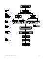
1
USER GUIDE
Freedom™ CHO-S™ Kit
For transfection of CHO-S™ Cells (cGMP-banked) and development of stable
cell lines for protein production
Catalog Number A13696-01
Publication Number MAN0003505
Revision A.0
For Research Use Only. Not for use in diagnostic procedures.
Information in this document is subject to change without notice.
DISCLAIMER
TO THE EXTENT ALLOWED BY LAW, LIFE TECHNOLOGIES AND/OR ITS AFFILIATE(S) WILL NOT BE LIABLE FOR SPECIAL, INCIDENTAL,
INDIRECT, PUNITIVE, MULTIPLE OR CONSEQUENTIAL DAMAGES IN CONNECTION WITH OR ARISING FROM THIS DOCUMENT,
INCLUDING YOUR USE OF IT.
NOTICE TO PURCHASER:
LIMITED USE LABEL LICENSE No. 335: Technology with CRO Rights
This product and its use is the subject of one or more issued and/or pending U.S. and foreign patent applications owned or controlled
by Life Technologies Corporation. The purchase of this product conveys to the buyer (whether the buyer is an academic or for-profit
entity) the right under the aforementioned patents and patent applications to use the purchased amount of the product and
components of the product in research. The buyer cannot sell or otherwise transfer (a) this product (b) its components or (c) materials
made using this product or its components to a third party or otherwise use this product or its components or materials made using
this product or its components for Commercial Purposes except that buyer may transfer this product and its components to service
providers for use by such service providers in research conducted for consideration solely for the benefit of the buyer, provided that
each such service provider agree in writing (a) not to transfer this product, its components, or materials or information made using
this product or its components to any third party, (b) to use this product, its components, and any materials or information made using
this product or its components solely for research and solely for the benefit of the buyer, and (c) to return to buyer or destroy any and
all remaining product, components, or materials made using this product or its components upon completion or termination of its
services for buyer. The buyer may also transfer information or materials made through the use of this product to a scientific
collaborator, provided that such transfer is not for any Commercial Purpose, and that such collaborator agrees in writing (a) not to
transfer such materials to any third party, and (b) to use such transferred materials and/or information solely for research and not for
Commercial Purposes. Commercial Purposes means any activity by a party for consideration and may include, but is not limited to: (1)
use of the product or its components in manufacturing; (2) use of the product or its components to provide a service, information, or
data; (3) use of the product or its components for therapeutic, diagnostic or prophylactic purposes; or (4) resale of the product or its
components, whether or not such product or its components are resold for use in research. For products that are subject to multiple
limited use label licenses, the terms of the most restrictive limited use label license shall control. Life Technologies Corporation will
not assert a claim against the buyer of infringement of patents owned or controlled by Life Technologies Corporation which cover this
product based upon the manufacture, use or sale of a therapeutic, clinical diagnostic, vaccine or prophylactic product developed in
research by the buyer in which this product or its components was employed, provided that neither this product nor any of its
components was used in the manufacture of such product. If the purchaser is not willing to accept the limitations of this limited use
statement, Life Technologies is willing to accept return of the product with a full refund. For information on obtaining additional rights,
please contact [email protected] or Out Licensing, Life Technologies Corporation, 5791 Van Allen Way, Carlsbad, California
92008. Email: [email protected].
LIMITED USE LABEL LICENSE No. 336: Transformed Host Cell Disclaimer
This product is not provided with a license to U.S. Patent No. 6,331,415, or any patents or patent applications related thereto. Each
purchaser of this product should consider whether additional rights are required for the purchaser's particular application under the
above patents.
LIMITED USE LABEL LICENSE No. 432: CHO Freedom Related Products
Notice to Purchaser: An additional license is required to use the clones derived from the Kits to produce products for use in
humans. For information on obtaining additional rights, please contact [email protected].
TRADEMARKS
All trademarks are the property of Thermo Fisher Scientific and its subsidiaries unless otherwise specified. BD is a registered
trademark of Becton, Dickinson and Company.
© 2015 Thermo Fisher Scientific Inc. All rights reserved.
Freedom™ CHO-S™ Kit User Guide
Contents
About this guide............................................................................................................. 5
Revision history ............................................................................................................ 5
Product information ...................................................................................................... 6
Product description ...................................................................................................... 6
Kit contents and storage ............................................................................................... 6
Materials required but not included ............................................................................. 7
Description of the system ............................................................................................. 7
CHO-S™ Cells (cGMP-banked)....................................................................................... 9
CD FortiCHO™ Medium ................................................................................................ 10
FreeStyle™ MAX Reagent and OptiPRO™ SFM ............................................................. 12
Selection reagents ...................................................................................................... 12
Methods ...................................................................................................................... 14
Experimental flowchart for protein expression ......................................................... 14
Create expression plasmids for the Freedom™ CHO-S™ Kit........................................ 16
Thaw and subculture CHO-S™ Cells (cGMP-banked) .................................................. 18
Freeze CHO-S™ Cells and make a research cell bank (RCB) ...................................... 20
Transfect CHO-S™ Cells for stable protein expression using the FreeStyle™ MAX
Reagent ...................................................................................................................... 23
Optional: Transfect CHO-S™ Cells for stable protein expression using the Neon™
Transfection System ................................................................................................... 27
Select stable transfectants for protein expression .................................................... 31
Assess productivity ..................................................................................................... 36
Isolate clones by limiting dilution ............................................................................... 37
Clone scale-up and screening .................................................................................... 43
Stability and fed-batch assessments of top clones .................................................... 47
Appendix A .................................................................................................................. 50
Troubleshooting ......................................................................................................... 50
Freedom™ CHO-S™ Kit User Guide
3
Appendix B .................................................................................................................. 54
Quick reference protocols .......................................................................................... 54
FreeStyle™ MAX transfection protocol and calculations ............................................. 55
Neon™ transfection protocol and calculations ............................................................ 56
Phase 1 selection ........................................................................................................ 57
Phase 2 selection ........................................................................................................ 58
Appendix C .................................................................................................................. 59
Media formulations..................................................................................................... 59
Appendix D .................................................................................................................. 60
Map and features of Freedom™ pCHO 1.0 vector ........................................................ 60
Appendix E .................................................................................................................. 63
Accessory products .................................................................................................... 63
Appendix F .................................................................................................................. 65
Safety .......................................................................................................................... 65
Chemical safety .......................................................................................................... 66
Biological hazard safety ............................................................................................. 67
Documentation and support......................................................................................... 68
Customer and technical support ................................................................................ 68
Limited product warranty ........................................................................................... 68
References .................................................................................................................. 69
4
Freedom™ CHO-S™ Kit User Guide
About this guide
Revision history
Revision
Date
A.0
July 2015
Addition of Dynamis™ fed-batch
guidance, plus minor revisions
3.0
2013
Addition of Quick Reference
protocols, plus minor revisions
2.0
2012
Addition of Neon™ protocol, plus
minor revisions
1.0
2011
New manual
Freedom™ CHO-S™ Kit User Guide
Description
5
Product information
Product description
Freedom™ CHO-S™
Kit
The Gibco™ Freedom™ CHO-S™ Kit (Cat. no. A13696-01), co-developed by Thermo
Fisher Scientific Corporation and ProBioGen AG, is designed for easy cloning and
expression of recombinant proteins in Chinese hamster ovary (CHO)-derived
suspension culture CHO-S™ Cells (cGMP-banked). The Freedom™ CHO-S™ Kit
provides reagents for:
•
cloning one or two genes that encode your protein(s) of interest
•
transfecting the DNA into CHO-S™ Cells (cGMP-banked) with high efficiency
•
generating stable cell lines that produce your protein of interest using a single,
chemically-defined animal origin-free media
Kit contents and storage
Item
CHO-S™ Cells (cGMP-banked), 1 × 10⁷ cells/mL
CD FortiCHO™ Medium
Anti-Clumping Agent
OptiPRO™ SFM
FreeStyle™ MAX Reagent
L-glutamine, 200 mM
Puromycin, 10 mg/mL
One Shot™ TOP10 Chemically Competent E. coli
(20 reactions)
Freedom™ pCHO 1.0 Expression Vector
Flash drive (contains the user manual for the
Freedom™ CHO-S™ Kit)
6
Amount
Shipping
Storage
1 mL
Dry ice
Liquid nitrogen
vapor phase
1000 mL
Ambient
temperature
2°C to 8°C,
protect from light
20 mL
Ambient
temperature
2°C to 8°C
100 mL
Ambient
temperature
2°C to 8°C,
protect from light
1 mL
Ambient
temperature
2°C to 8°C,
do not freeze
100 mL
Dry ice
–5°C to –20ºC,
protect from light
20 mL
Dry ice
–5°C to –20ºC,
protect from light
21 × 50 µL
Dry ice
–80ºC
1 kit
Dry ice
–5°C to –20ºC
1 flash drive
Ambient
temperature
Room temperature
Freedom™ CHO-S™ Kit User Guide
Materials required but not included
Unless otherwise indicated, all materials are available on our website:
www.thermofisher.com/lifescience.
•
Methotrexate (MTX), available from Sigma-Aldrich as methotrexate hydrate
(Cat. no. A6770 or M8407)
•
Disposables for molecular biology and cell culture (polypropylene conical
centrifuge tubes, microcentrifuge tubes, T-flasks, shaker flasks)
•
Cell strainer, such as BD Biosciences 40-µm nylon mesh cell strainer
(BD Biosciences, Cat. no. 352340)
•
Static and shaking incubators with humidity and CO2
•
Biosafety cabinet (i.e., laminar flow hood) for all cell manipulations
Description of the system
Components of the
Freedom™ CHO-S™
Kit
Freedom™ pCHO 1.0
vector
•
CHO-S™ Cells (cGMP-banked): cGMP-banked, CHO-derived cells adapted to
high density, serum-free suspension culture in chemically-defined medium that
are capable of producing high levels of secreted, recombinant protein. See
page 9 for more information.
•
Freedom™ pCHO 1.0 vector: A plasmid for cloning ORFs containing a
mammalian secretion signal and up to two subunits of your protein(s) of
interest. See page 60 for the map and features of the vector.
•
CD FortiCHO™ Medium: Chemically defined, animal-origin-free, serum-free
medium formulated for growth of CHO-S™ Cells (cGMP-banked) and for
expression of the recombinant protein(s) of interest. See page 10 for more
information.
•
FreeStyle™ MAX Reagent: A proprietary, AOF (animal origin-free) formulation
for high transfection efficiency of plasmid DNA into CHO-S™ Cells
(cGMP-banked). See page 12 for more information.
•
Additional components: One Shot™ TOP10 Chemically Competent E. coli cells
used for cloning your gene(s) into the Freedom™ pCHO 1.0 vector, OptiPRO™
serum-free medium (SFM) for optimal DNA:lipid complex formulation,
L-glutamine supplement for increased medium stability, Anti-Clumping Agent
to prevent cell clumping, and Puromycin for stable cell line selection.
The Freedom™ CHO-S™ Kit contains the Freedom™ pCHO 1.0 vector, designed by
ProBioGen AG, to express one or two genes of interest downstream of the vector’s
two different hybrid CMV promoters. This vector contains the dihydrofolate
reductase (DHFR) selection marker and a puromycin resistance gene, allowing
selection using MTX and Puromycin simultaneously. See page 60 for more
information on the Freedom™ pCHO 1.0 vector.
IMPORTANT! Your gene(s) of interest must be engineered with a mammalian
secretion signal for secreted protein expression into the cell culture media.
Freedom™ CHO-S™ Kit User Guide
7
Advantages of the
Freedom™ CHO-S™
Kit
FAQ eBook
8
The Freedom™ CHO-S™ Kit provides the following advantages for protein
production in mammalian cells:
•
cGMP-banked CHO-S™ Cells, derived from CHO cells (Roy et al., 1999;
Schifferli, 1999), provide stable and accurate glycosylation and are observed to
yield functional glycoproteins (Sheeley et al., 1997; Werner et al., 1998).
•
Freedom™ pCHO 1.0 is a single vector containing strong promoter elements
designed to ensure high expression levels of one or two subunits.
•
A single medium (CD FortiCHO™ Medium) is used for the entire stable cell line
development process (cell passage, transfection, selection, and limiting dilution
cloning, with easy transition into Dynamis™ production medium for
production), which eliminates the need for any adaptation of cells into other
media.
•
FreeStyle™ MAX Reagent offers high transfection efficiency of suspension CHO
cells with low cytotoxicity.
•
FreeStyle™ MAX Reagent, OptiPRO™ SFM, and CD FortiCHO™ Medium are
AOF (animal origin-free) and serum-free.
•
Robust, streamlined cell line development process with step-by-step
instructions.
We have also created an interactive FAQ eBook to supplement the
Freedom™ CHO-S™ Kit User Guide to highlight critical steps and milestones. We
highly recommend that you become familiar with both this User Guide and the
eBook http://life-technologies.uberflip.com/i/330570 prior to initiating the
workflow.
Freedom™ CHO-S™ Kit User Guide
CHO-S™ Cells (cGMP-banked)
Characteristics of
CHO-S™ Cells (cGMPbanked)
Parental cell line
The CHO-S™ cell line is a stable aneuploid cell line established from the ovary of an
adult Chinese hamster (Puck et al., 1958). The CHO-S™ cells (cGMP-banked) are:
•
prepared from low passage Master Cell Bank cultures derived from parental
CHO-S™ cells that were re-cloned by limiting dilution in CD CHO Medium,
and selected for their superior serum-free cell growth and transfection
efficiencies (Ciccarone et al., 1999; Schifferli, 1999).
•
adapted to serum-free suspension growth.
•
compatible with CD FortiCHO™ Medium for all steps of the workflow.
•
compatible with FreeStyle™ MAX Reagent for high transfection efficiency.
•
manufactured under cGMP guidelines and banked in CD CHO Medium, a
serum-free, protein-free, and chemically defined medium that is formulated
with no components of animal or human origin (Gorfien, 1998). Frozen cells are
supplied at a concentration of 1 × 10⁷ cells/mL and can be thawed directly into
CD FortiCHO™ Medium (see page 18).
•
available for commercial use with purchase of separate documentation package
and license.
Chinese Hamster Ovary (CHO) cells are among the most commonly used cell lines
for transfection, expression and large-scale production of recombinant proteins. The
CHO cell line is a stable aneuploid cell line established from the ovary of an adult
Chinese hamster (D'Anna, 1996; D'Anna et al., 1997; Deaven & Petersen, 1973; Puck
et al., 1958).
Note: While cells were banked in CD CHO Medium, cell thaw and all subsequent
workflow steps are performed directly in the kit-provided CD FortiCHO™ Medium
without any separate adaptation steps.
CAUTION! As with other mammalian cell lines, when working with CHO-S™ Cells
(cGMP-banked), handle them as potentially biohazardous material under at least
Biosafety Level 1 containment.
Note that the CHO-S™ Cells (cGMP-banked) are provided in freezing medium
containing DMSO, and the components of the product may be absorbed into the
body through the skin. In case of contact with eyes, rinse immediately with plenty
of water and seek medical advice. Always wear suitable protective clothing and
gloves when handling CHO-S™ Cells (cGMP-banked).
Freedom™ CHO-S™ Kit User Guide
9
CD FortiCHO™ Medium
Introduction
CD FortiCHO™ Medium is a chemically defined, animal-origin-free (AOF), serumfree medium for high-density suspension culture of untransfected or transfected
CHO-S™ Cells. The medium contains no human or animal origin components.
IMPORTANT! Do not use CD FortiCHO™ Medium containing either MTX and/or
Puromycin to propagate untransfected CHO-S™ Cells (cGMP-banked). Parental
CHO-S™ cells are sensitive to both Puromycin and MTX in the concentrations used
below for selection.
Only cells that have both increased DHFR enzyme and an active puromycin
resistance gene, such as cells that have been transfected with Freedom™ pCHO 1.0
vector, can be propagated in CD FortiCHO™ Medium + MTX and/or Puromycin.
Parental CHO-S™ cells, however, can be propagated in CD FortiCHO™ Medium
supplemented with L-glutamine.
If completing the Freedom™ CHO-S™ workflow (from thaw to tertiary screen) in a
single medium lot, we recommend that you pre-order or reserve at least 25 L now.
See page 63 for ordering information.
Features of
CD FortiCHO™
Medium
10
CD FortiCHO™ Medium is:
•
developed for the growth of Chinese Hamster Ovary (CHO) cells and
expression of recombinant proteins in suspension culture.
•
made with animal origin-free, chemically defined components and contains no
proteins, hydrolysates, or components of unknown composition.
•
formulated without L-glutamine for flexibility of use, and to avoid ammonia
accumulation (however, it should be supplemented as required).
•
formulated to minimize potential lactic acid accumulation under typical culture
conditions.
•
Note: Additional glucose supplementation may be required for terminal batch
cultures as determined empirically or by using our suggested simple glucose
fed-batch protocol on page 36.
Freedom™ CHO-S™ Kit User Guide
Prepare complete
CD FortiCHO™
Medium
•
CD FortiCHO™ Medium requires supplementation with L-glutamine.
Aseptically add L-glutamine to a final concentration of 8 mM to the medium
before use.
Note: We recommend that you do not use thawed L-glutamine or medium
supplemented with L-glutamine beyond one month. See page 59 for all
CD FortiCHO™ Medium formulations used in this kit.
IMPORTANT! 8 mM L-glutamine is used for all steps in this manual except
limiting dilution cloning, which requires 6 mM L-glutamine. See page 37 for
more details.
IMPORTANT! Anti-Clumping Agent will interfere with FreeStyle™ MAX
transfection. Do not use CD FortiCHO™ Medium containing Anti-Clumping
Agent during or for the two passages prior to transfection.
Growth
characteristics of
CHO-S™ Cells (cGMPbanked) in CD
FortiCHO™ Medium
•
For MTX + Puromycin selection, aseptically add drugs fresh to CD FortiCHO™
Medium. Do not store drug-containing CD FortiCHO™ Medium. See page 31
for details on selection.
•
Store complete CD FortiCHO™ Medium at 2ºC –8ºC, protected from light.
Typically, CHO-S™ Cells (cGMP-banked) cultured in CD FortiCHO™ Medium have
a doubling time in the range of 17–20 hours; however, doubling time can exceed
20 hours during the first few passages after the cells have been thawed.
Do not allow CHO-S™ Cells (cGMP-banked) to reach a cell density above
2 × 10⁶ cells/mL before transfection, because this may cause a decrease in
transfection efficiency.
Note: Individual culturing and passaging techniques of CHO-S™ Cells
(cGMP-banked) cells may result in experimental variation.
Freedom™ CHO-S™ Kit User Guide
11
FreeStyle™ MAX Reagent and OptiPRO™ SFM
FreeStyle™ MAX
Reagent
FreeStyle™ MAX Reagent is a proprietary, animal origin-free formulation for the
highly efficient transfection of plasmid DNA into eukaryotic cells. FreeStyle™ MAX
Reagent is specifically formulated to achieve the highest transfection efficiencies
and lowest cytotoxicity in CHO-S™ Cells (cGMP-banked) and other cell types.
FreeStyle™ MAX Reagent is also available separately; see page 63 for ordering
information.
Store FreeStyle™ MAX Reagent at 2–8ºC. Do not freeze.
OptiPRO™ SFM
OptiPRO™ SFM is included with the Freedom™ CHO-S™ Kit to facilitate optimal
formation of DNA-lipid complexes. OptiPRO™ SFM is a serum-free medium that is
devoid of components of animal or human origin. OptiPRO™ SFM has an ultra-low
protein concentration of 7.5 µg/mL.
OptiPRO™ SFM is also available separately; see page 63 for ordering information.
Store OptiPRO™ SFM at 2–8ºC, protected from light.
Selection reagents
Puromycin
The Freedom™ pCHO 1.0 vector contains the puromycin resistance gene, which
confers resistance to the antibiotic Puromycin. Puromycin is available separately;
see page 63 for ordering information.
Puromycin is an aminonucleoside that blocks protein synthesis in mammalian cells
by interfering with ribosomal function. Expression in mammalian cells of the
bacterial gene pac, derived from Streptomyces alboniger, results in detoxification of
Puromycin (de la Luna & Ortin, 1992; Lacalle et al., 1989; Vara et al., 1985).
CAUTION! Puromycin, while non-hazardous, should be handled with care. Avoid
contact with skin and eyes. In case of contact with skin or eyes, immediately rinse
with water thoroughly and seek medical advice. Wear suitable protective clothing
and gloves when handling Puromycin and Puromycin-containing solutions.
Store Puromycin
12
Puromycin in liquid form should be stored –5°C to –20°C. Puromycin is stable for
one year, if stored properly. Once thawed, the shelf life is only 1 month. We
recommend that, upon initial thaw of the 20 mL bottle, Puromycin be aliquoted into
smaller volumes (100 µL– 1 mL) for refreezing such that the drug can be thawed as
needed during selection. We routinely use 1 mL aliquots and store them at 4°C
post-thaw until they are used up or have been thawed for 1 month, whichever
comes first.
Freedom™ CHO-S™ Kit User Guide
Methotrexate
(MTX)
Methotrexate (MTX) is a folic acid antagonist that is actively transported into cells
by the folate transporter. In the cell, it is converted to a high molecular weight
polyglutamate metabolite by folylpolyglutamate synthase, which binds to DHFR
and inhibits its activity.
The Freedom™ pCHO 1.0 vector contains the DHFR gene. DHFR catalyzes the
reduction of 5, 6-dihydrofolate to 5, 6, 7, 8-tetrahydrofolate, which is essential for
DNA synthesis. CHO-S™ Cells (cGMP-banked) have a basal-level of DHFR activity,
which is enhanced by transfection with Freedom™ pCHO 1.0 vector. Thus, cells
carrying Freedom™ pCHO 1.0 are more resistant to MTX than parental cells.
CAUTION! CHEMICAL HAZARD. MTX is toxic to the skin, eyes, and respiratory
system. Wear suitable protective clothing, gloves, and eye and face protection when
working with MTX. Refer to the product SDS for complete precautions.
Prepare
1 mM MTX stock
solution
Note: MTX powder is insoluble in neutral pH aqueous solutions, and the below
protocol ensures complete solubilization before its addition to PBS. This volume of
MTX is sufficient for both phases of selection. We recommend performing selection
using a single lot of MTX stock solution.
1.
2.
Dissolve 10 mg MTX in 100–150 µL of 1 M NaOH. Immediately proceed to
step 2 as soon as MTX is completely dissolved as prolonged exposure to high
pH inactivates MTX.
Bring the volume up to 22 mL using PBS.
The pH of this solution should be 10–11.
Prepare
selection medium
3.
Filter-sterilize the solution through a 0.22-µm filter.
4.
Store the MTX stock solution in 100 µL–1 mL aliquots at –20ºC.
•
To prepare selection medium, use complete CD FortiCHO™ Medium
supplemented with Anti-Clumping Agent at a 1:100 dilution, and only add the
required concentration of Puromycin + MTX fresh each time (see page 59). Do
not store media containing Puromycin + MTX.
•
Alternatively, Puromycin + MTX can be added to each selection flask at each
step of selection.
•
Cells will divide once or twice in the presence of lethal doses of Puromycin, so
the effects of the drug take several days to become apparent. Complete
selection can take 3–7 weeks of growth in selective medium, depending on your
transfection, drug stocks, and protein(s) of interest.
Note: For a successful selection, Anti-Clumping Agent must be included in the
selection medium at a 1:100 dilution. Medium containing Anti-Clumping Agent can
be prepared in advance by adding 10 mL of Anti-Clumping Agent per liter of
complete medium, or by adding to each flask 100 µL of Anti-Clumping Agent for
every 10 mL of culture medium.
Development work with this kit used 10–50 μg/mL of Puromycin, and
100–1,000 nM of MTX. However, because different transfected cells may exhibit
different drug sensitivities, we recommend that you generate pools as described in
the Experimental Flowchart on page 14 with multiple concentrations of Puromycin
+ MTX to ensure that you find the optimal conditions for your protein(s).
Freedom™ CHO-S™ Kit User Guide
13
Methods
Experimental flowchart for protein expression
Introduction
The diagram on the following page schematically depicts the steps necessary to
express your one or two-subunit protein(s) of interest using the Freedom™ CHO-S™
Kit, and it shows our recommended experimental path from stable transfectants to
clone scale-up.
Note: The times shown for various experimental steps are approximations, and the
actual times depend on your protein(s) of interest and the specific workflow you
choose.
Note: For a two-subunit protein, create two expression plasmids, each containing
the subunits in a different order (SU1–SU2 or SU2–SU1). Transfect CHO-S™ Cells
(cGMP-banked) with each plasmid in parallel at least through transient transfection
to identify the optimal gene orientation. For a single-subunit protein, clone your
gene of interest behind the first promoter. For information on cloning each subunit
of your protein of interest into the Freedom™ pCHO 1.0 vector, refer to page 16. For
a map and features of the Freedom™ pCHO 1.0 vector, see page 60.
14
Freedom™ CHO-S™ Kit User Guide
Freedom™ CHO-S™ Kit User Guide
15
Create expression plasmids for the Freedom™ CHO-S™ Kit
Gene optimization
Prior to cloning your gene(s) of interest into the Freedom™ pCHO 1.0 vector, we
highly recommend that you have the nucleotide sequences optimized, not only for
codon usage, but also for cryptic splice sites, RNA secondary structure, killer
motifs, etc. GeneArt™ GeneOptimizer™ software has given us the most consistent
success from molecule to molecule, and is a service available from our website
(www.thermofisher.com/lifescience). You can assess the results of gene
optimization of your gene(s) of interest prior to any financial commitments.
Clone into
Freedom™ pCHO
1.0
The Freedom™ CHO-S™ Kit contains the Freedom™ pCHO 1.0 vector. Refer to
page 60 for map and the features of the vector.
Using the instructions in this manual, you will:
•
•
For two-subunits, create two expression plasmids by cloning two separate PCR
products corresponding to each of the two subunits (SU1 and SU2; SU: subunit)
of your protein(s) of choice separately into Freedom™ pCHO 1.0 vector. Because
protein expression may depend on the order of the different subunits of your
protein(s) of interest, clone SU1 and SU2 in two different combinations using
the following insertion sites to optimize protein expression:
-
First insertion site: AvrII/BstZ17I (blunt)
-
Second insertion site: EcoRV (blunt)/PacI
-
We recommend testing each linearized expression plasmid in parallel in
transient transfection to determine which orientation gives the best protein
expression before proceeding with stable pool generation.
For single-subunit proteins, create a single expression plasmid using the
Freedom™ pCHO 1.0 vector and the following insertion site:
•
First insertion site: AvrII and/or BstZ17I (blunt)
Optional: When creating a single expression plasmid, remove the second
expression cassette by SfiI digestion and vector re-ligation.
•
For a transfection efficiency control, create a single expression plasmid using a
GFP allele inserted into Freedom™ pCHO 1.0 vector at the first insertion site
(AvrII and/or BstZ17I (blunt)). Be sure to prepare the control plasmid using the
same reagents and procedures as used for your expression plasmid(s),
including the linearization step, before transfecting in parallel to assess
transient transfection efficiency.
•
For best ligation results, be sure your insert is in molar excess of the plasmid in
the ligation reaction. We recommend trying a few insert:vector ratios to ensure
the isolation of the desired product (for example, 3:1 and 5:1 insert:vector
ratios). You can calculate the amount of insert needed when starting with a
known amount of vector as follows:
Amount of insert (ng) =
Size of insert
Size of vector
× (insert:vector ratio) × (ng vector)
For example, to achieve a 3:1 insert:vector ratio to ligate a 2 kb insert into 20 ng of
13 kb Freedom™ pCHO 1.0, use 9.2 ng of the insert.
16
Freedom™ CHO-S™ Kit User Guide
IMPORTANT! Your inserts must contain an ATG initiation codon in the context of
a Kozak translation initiation sequence for proper initiation of translation in
mammalian cells (Kozak, 1987; Kozak, 1990; Kozak, 1991). An example of a Kozak
consensus sequence is provided below. Other sequences are possible, but the G or
A at position –3 and the G at position 4 (shown in bold) illustrates the most
commonly occurring sequence with strong consensus. Replacing one of the two
bases at these positions provides moderate consensus, while having neither results
in weak consensus. The ATG initiation codon is shown underlined.
(G/A)NNATGG
For the Freedom pCHO 1.0 vector, we have successfully used GCCACCATGG.
™
Also, if you wish to express secreted proteins, be sure to include a signal peptide
sequence for the secretion of your protein of interest.
Freedom™
pCHO 1.0 bacterial
selection marker
Types of
expression
plasmids
Freedom™ pCHO 1.0 vector contains the bacterial selection marker aph, which
allows for selection of transformed cells by generating resistance to the antibiotic
kanamycin (Oka et al., 1981; Vakulenko et al., 1987).
•
When transforming One Shot™ TOP10 Chemically E. coli with the
Freedom™ pCHO 1.0 vector, use 50 µg/mL of kanamycin.
•
If you wish to use a different strain of E. coli, ensure that it is
kanamycin-sensitive and conduct a kill-curve study to establish the ideal
concentration of kanamycin to use.
You will create two expression plasmids for two subunits, or create one expression
plasmid for a single subunit:
Vector
DNA
Freedom™ pCHO 1.0,
SU1 upstream of SU2
mammalian secretion signal and SU1
mammalian secretion signal and SU2
Freedom™ pCHO 1.0,
SU2 upstream of SU1
mammalian secretion signal and SU2
mammalian secretion signal and SU1
Freedom™ pCHO 1.0,
single SU
mammalian secretion signal and
single SU
Freedom™ CHO-S™ Kit User Guide
Selection
MTX +
Puromycin
17
Sequence
recombinant
expression
plasmids
We strongly recommend that you analyze your recombinant expression plasmid(s)
by sequencing the promoter-gene of interest junctions before transfecting
CHO-S™ Cells (cGMP-banked) and expressing your protein(s) of interest. For your
convenience, the sequences of the primers used for analyzing the promoter-gene of
interest junctions in the recombinant expression plasmid(s) are provided below.
Note: The sequencing primers are not supplied as part of the
Freedom™ CHO-S™ Kit; they need to be ordered separately and custom synthesized.
Primer
Primer sequence
Location
Comments
AB-For
5’-GTCTGAGCCTCCTTGTCTTG-3’
begins ~270 bp upstream of
AvrII/BstZ17I insertion site
forward primer for EF2/CMV
hybrid promoter ORF
AB-Rev
5’-AGAAGACACGGGAGACTTAG-3’
begins ~90 bp downstream
of AvrII/BstZ17I insertion
site
reverse primer for EF2/CMV
hybrid promoter ORF
EP-For
5’-GGTGTCGTGAGGAATTTCAG-3’
begins ~285 bp upstream of
EcoRV/PacI insertion site
forward primer for CMV/EF1
hybrid promoter ORF
EP-Rev
5’-GAGGCAGCCGGATCATAATC-3’
begins ~250 bp downstream
of EcoRV/PacI insertion site
reverse primer for CMV/EF1
hybrid promoter ORF
Recommendation: The methods for transfection into mammalian cells and the
selection process for the Freedom™ CHO-S™ Kit are the same whether you are
expressing 1 or 2 subunits. See page 14 for an overview.
Thaw and subculture CHO-S™ Cells (cGMP-banked)
Introduction
Follow the protocol below to thaw CHO-S™ Cells (cGMP-banked). The cells are
supplied in a vial that contains 1 mL of cells at 1 × 10⁷ viable cells/mL in
90% complete CD CHO Medium and 10% DMSO. Thaw the cells directly into
complete CD FortiCHO™ Medium (supplemented with 8 mM L-glutamine). No
separate adaptation is required.
For information on making a research cell bank in CD FortiCHO™ Medium, refer to
Freeze CHO-S™ Cells and make a research cell bank (RCB), page 20.
Prepare complete
CD FortiCHO™
Medium
18
•
All solutions and equipment that come in contact with the cells must be sterile.
Always use proper aseptic technique and work in a biosafety cabinet.
•
Supplement CD FortiCHO™ Medium with L glutamine to a final concentration
of 8 mM before use. See pages 10 and 59.
•
Addition of antibiotics is not recommended.
•
CD FortiCHO™ Medium is light sensitive. For optimal results, store medium at
2ºC–8ºC, protected from light.
Freedom™ CHO-S™ Kit User Guide
Required materials
You need the following reagents and materials before beginning:
•
Frozen CHO-S™ Cells (cGMP-banked) (supplied with the kit; store frozen cells
in liquid nitrogen until ready to use)
•
Complete CD FortiCHO™ Medium (prepared as above; pre-warmed to 37°C)
•
125 mL-polycarbonate, disposable, sterile Erlenmeyer flasks with vented caps
•
Orbital shaker set at 130–150 rpm in a 37°C incubator with a humidified
atmosphere of 8% CO2
Shaking speed
The 130–150 rpm shaking speed (as well as all the values used in other places in this
manual) is specific to an Infors Multitron 2 shaker incubator with an orbital throw
of 25 mm. When using a shaker with a different orbital throw, we recommend that
you modify the shaking speed to match the relative centrifugal force
(RCF = 1.118 ×10⁻⁵ × THROW × SPEED2).
Thawing procedure
1.
Remove the cryovial of cells from the liquid nitrogen and thaw quickly
(less than 2 minutes) in a 37°C water bath.
2.
Decontaminate the outside of the vial with 70% ethanol. Aseptically transfer the
entire contents of the cryovial into a disposable, sterile polycarbonate
125 mL-Erlenmeyer shaker flask containing 29 mL of pre-warmed complete
CD FortiCHO™ Medium.
Note: Removal of the DMSO from the medium is not necessary.
3.
Incubate the cells in a 37°C incubator containing a humidified atmosphere of
8% CO2 in air on an orbital shaker platform rotating at 130–150 rpm.
Note: We have thawed and subcultured CHO-S™ cells at both 130 and 150 rpm
with the only difference being a slightly faster growth rate at 150 rpm. The
speed range is provided here for your convenience.
Determine cell
density and
viability
4.
Optional: After 24 hours in culture, sample to determine the cell density and
viability using the protocol described below.
5.
Once the culture reaches 1 × 10⁶–2 × 10⁶ viable cells/mL, typically 2–3 days
post-thaw, expand the culture using the subculturing protocol, (see page 20).
Follow the procedure below to determine the viable and total cell counts.
1.
Transfer a small aliquot of the cell suspension into a microcentrifuge tube. Be
careful to avoid any cell clumps that may be present in the culture.
2.
Determine cell viability and cell density using your method of choice.
Freedom™ CHO-S™ Kit User Guide
19
Subculture cells
Passage CHO-S™ Cells (cGMP-banked) every 2–3 days into fresh medium. When
passaging the cells, use disposable, sterile polycarbonate 125-mL Erlenmeyer shaker
flasks with vented caps and pre-warmed complete CD FortiCHO™ Medium (for
instructions on preparing complete CD FortiCHO™ Medium, see page 59). We have
used a 125-mL shaker flask for 15–40 mL of suspension culture. If larger volumes
and/or cell numbers are required, increase the number of 125-ml Erlenmeyer
shaker flasks you seed or increase your shaker flask size to allow for increased
culture volume.
1.
Determine the viable and total cell counts, taking caution to avoid any cell
clumps that may be present in the culture.
2.
Using the cell density determined in step 1, calculate the volume of cell culture
suspension and fresh complete medium needed to seed each new shaker flask
by dilution. Seed the culture at a density of 2.0 × 10⁵ viable cells/mL, if a
subculture step is scheduled for 2 days. Seed the culture at a density of
1.0 × 10⁵ viable cells/mL, if a subculture step is scheduled for 3 days.
3.
Transfer the calculated volume of pre-warmed complete CD FortiCHO™
Medium into a sterile 125-mL Erlenmeyer shaker flask.
4.
Transfer the calculated volume of cell suspension into the pre-warmed
complete CD FortiCHO™ Medium to give a final cell density of 1 × 10⁵ or
2 × 10⁵ viable cells/mL, depending on the subculture schedule. Be careful to
avoid transferring any cell clumps that may be present in the culture.
5.
Incubate the flasks in a 37°C incubator with 70–80% humidified atmosphere of
8% CO2 in air on an orbital shaker platform rotating at 130–150 rpm.
6.
Repeat Steps 1–5 as necessary to maintain or expand cells.
It is normal to see a small cell ring form
on the wall of the flask and/or cell
clumps of ≤1 mm at the bottom of the
flask (see image on the right). Make sure
not to transfer this or any cell clumps
into the next passage flask. You may use
a cell strainer to remove cell clumps;
however, do not use Anti-Clumping
Agent prior to transfection.
Note: Avoid transferring cell clumps during passaging; this will minimize the
likelihood of future clumping.
•
Do not allow CHO-S™ Cells (cGMP-banked) to reach a cell density above
2 × 10⁶ cells /mL transfection to avoid a decrease of transfection efficiency.
•
A minimum of 5 passages under 2 × 10⁶ cells/mL is recommended before
proceeding with transfection.
•
After 25 passages, thaw a new vial of cells. To maintain sufficient stocks of
low-passage cells (i.e., under 25 passages), be sure to freeze aliquots of
CHO-S™ cells in liquid nitrogen vapor phase.
Freeze CHO-S™ Cells and make a research cell bank (RCB)
20
Freedom™ CHO-S™ Kit User Guide
Introduction
You may freeze CHO-S™ Cells (cGMP-banked) directly in complete CD FortiCHO™
Medium with 10% DMSO. We recommend that you freeze the cells at a density of
≥1 × 10⁷ viable cells/mL at least 3 passages post-thaw, and that you make a research
cell bank of at least 20 vials from the vial of CHO-S™ cells provided in the kit.
Guidelines for preparing freezing medium and to freeze cells are provided in this
section. These protocols can also be used to freeze cells throughout the cell line
development workflow.
Required materials
•
Complete CD FortiCHO™ Medium. See page 11.
•
Tissue culture grade DMSO
•
Reagents and equipment to determine viable and total cell counts
•
Sterile, labeled cryovials
•
Sterile, 15-mL or 50-mL conical tubes
•
Automated or manual controlled-rate freezing apparatus
Prepare freezing
medium
Prepare freezing medium immediately before use.
1.
In a sterile, conical centrifuge tube, mix together the following reagents for
every 1 mL of freezing medium needed:
Complete CD FortiCHO™ Medium
0.9 mL
DMSO
0.1 mL
Note: We recommend preparing extra freezing medium to compensate for
losses during pipetting.
2.
Place the tube on ice, or store at 2–8°C until use. Discard any remaining
freezing medium after use.
Freedom™ CHO-S™ Kit User Guide
21
Freeze cells
The following protocol describes how to prepare 1 mL of 1 × 10⁷ cells per cryovial.
Plan ahead to ensure that you have sufficient cells for the desired number of vials.
1.
Grow the desired quantity of CHO-S™ Cells in shaker flasks, harvesting when
the cells are in mid-log phase of growth with a viability >90%. Typically the
cells will be at ~1.5 × 10⁶–3 × 10⁶ cells/mL on day of freezing.
Note: Cells should be passaged at least 3 times after thaw before freezing.
2.
Transfer the cells to a sterile, conical centrifuge tube.
3.
Determine the viable and total cell counts (see page 19) and calculate the
volume of the cell suspension and the freezing medium required to yield a final
cell density of 1 × 10⁷ viable cells/mL.
4.
Centrifuge the cells at 100–200 × g for 5–10 minutes at room temperature and
carefully aspirate the medium.
5.
Resuspend the cells in the pre-determined volume of chilled freezing medium
(90% complete CD FortiCHO™ Medium and 10% DMSO; see above).
6.
Place the cryovials in a microcentrifuge rack and aliquot 1 mL of the cell
suspension into each cryovial.
7.
Freeze the cells in an automated or manual controlled-rate freezing apparatus
following standard procedures. For ideal cryopreservation, the freezing rate
should be a 1°C decrease per minute.
8.
24–72 hours after freezing the cells, transfer the frozen vials to liquid nitrogen
vapor phase for long-term storage.
Note: You can check the viability and recovery of frozen cells 24 hours after
storing the cryovials in liquid nitrogen by following the thawing procedure on
page 19.
22
Freedom™ CHO-S™ Kit User Guide
Transfect CHO-S™ Cells for stable protein expression using the
FreeStyle™ MAX Reagent
Introduction
Instructions for stably transfecting suspension CHO-S™ Cells (cGMP-banked) using
the FreeStyle™ MAX Reagent are provided below. Because individual protein
expression may depend on the order of the different subunits of your protein(s) of
interest, we recommend performing multiple transfections in parallel using your
expression plasmids (see page 17).
Prepare the
plasmid
The Freedom™ pCHO 1.0 plasmid constructs containing your gene(s) of interest
must be clean, sterile, and free from contamination with organic solvents and
sodium chloride for transfection into CHO-S™ cells. We highly recommend using
aseptic techniques and sterile vessels for both the DNA preparation and the
restriction enzyme digestion. Chemical contaminants may kill the cells, and salt
interferes with lipid complexing, decreasing transfection efficiency. We recommend
isolating plasmid DNA using an endotoxin-free or a low-endotoxin kit such as the
PureLink™ HiPure Plasmid Midiprep DNA Kit (see page 64). We have found that
the easiest and most efficient way to avoid organic solvents and salts that interfere
with transfection is to use the plasmid in the restriction digest mix without any
further processing.
Linearize the
plasmid
Prior to using the Freedom™ CHO-S™ Kit to transfect CHO-S™ Cells (cGMP-banked)
with your Freedom™ pCHO 1.0 construct, you may linearize the plasmids.
Linearizing your vectors may not improve transfection efficiency, but it does
increase the chance that the vectors will integrate into the host cell genome without
disrupting the gene(s) of interest or other elements required for expression in
mammalian cells. Follow the guidelines below to linearize your plasmids.
•
We suggest using Nru I, which cuts once in the kanamycin resistance gene on
the plasmid. Other unique restriction sites are possible. See page 62 for a list of
unique sites and non-cutting restriction enzymes. Be sure that your inserts do
not contain the restriction enzyme site you use to linearize the vector.
Note: If an appropriate linearization site is not present, you may transfect the
circular plasmid.
•
After digestion, purification of the plasmid DNA is not required. If purification
is desired, be careful to ensure complete removal of all organic solvents and
salts. Freedom™ pCHO 1.0 may precipitate during mixing with the
FreeStyle™ MAX due to carry-over of organic solvents or salts, and DNA
precipitation decreases transfection efficiency.
Freedom™ CHO-S™ Kit User Guide
23
Required materials
Optimal
transfection
conditions using
the FreeStyle™
MAX Reagent
•
CHO-S™ cells cultured in complete CD FortiCHO™ Medium at
1 × 10⁶ viable cells/mL
•
Linearized Freedom™ pCHO 1.0 plasmid DNA containing the subunit(s) of
your protein(s) of interest, see page 23.
•
FreeStyle™ MAX Reagent (supplied with the kit; store at 4°C until use)
•
OptiPRO™ SFM (supplied with the kit; pre-warmed to room temperature)
•
Disposable, sterile, 125-mL polycarbonate Erlenmeyer flasks
•
Orbital shaker in 37°C incubator with a humidified atmosphere of 8% CO2
•
Reagents and equipment to determine viable and total cell counts (e.g., Trypan
Blue, hemacytometer, or an automated cell counter)
Transfect suspension CHO-S™ cells in 30 mL, with the recommended optimized
conditions:
•
Final transfection volume: 30 mL
•
Number of cells to transfect: total of 3 × 10⁷ viable cells (cell density at time of
transfection should be 1 × 10⁶ viable cells/mL)
IMPORTANT: It is best to use CHO-S™ cells that have been passaged at least
5 times without cell densities exceeding 2 × 10⁶ viable cells/mL.
•
Amount of each plasmid DNA: 50 µg, regardless of whether the plasmid has
been engineered to express 1 or 2 subunits
•
FreeStyle™ MAX Reagent: 50 µL
Note: Further optimization of culture volume or transfection conditions is not
necessary for stable cell line production.
General guidelines
for transfection
using the
FreeStyle™ MAX
Reagent
24
•
Calculate the number of CHO-S™ Cells (cGMP-banked) that you will need for
your transfection experiment and expand cells accordingly. Make sure that the
cells are healthy and greater than 95% viable before proceeding to transfection.
•
Prepare high-quality plasmid DNA at a concentration of 1–5 µg/µL in
deionized water or TE buffer.
•
Carry-over of organic solvents and salts from DNA purification can be toxic to
the cells; therefore, DNA purification following linearization is not
recommended.
•
If possible, use an appropriate GFP plasmid transfected in parallel to determine
transfection efficiency.
•
Prepare extra volume of cell suspension to ensure that the desired cell number
is available at the time of transfection.
•
For a one-page quick reference protocol intended to be printed and used in the
laboratory, see page 55.
Freedom™ CHO-S™ Kit User Guide
Transfection
procedure using
the FreeStyle™
MAX Reagent
Follow the procedure below to transfect CHO-S™ Cells (cGMP-banked) in a
30 mL volume. Handle cells at all steps using aseptic technique in a biosafety
cabinet. We recommend including negative controls (no FreeStyle™ MAX Reagent,
no DNA) in your experiment to help you evaluate your results. We suggest that
you perform at least 2 transfections with each plasmid construct to increase the
likelihood of generating the highest-producing stable pools before initiating
limiting dilution cloning. Note that the same transfection protocol is used whether
you are expressing one or two subunits.
Note: For a one-page quick reference protocol intended to be printed and used in
the laboratory, see page 55.
IMPORTANT! Anti-Clumping Agent will interfere with FreeStyle™ MAX
transfection. We have specifically not included Anti-Clumping Agent in any cell
passage steps prior to transfection for this reason. If you choose to use
Anti-Clumping Agent in your passage medium, be sure to remove it at least
2 passages prior to transfection with the FreeStyle™ MAX Reagent.
1.
At 24 hours before transfection, pass CHO-S™ Cells (cGMP-banked) at
5 × 10⁵ to 6 × 10⁵ viable cells/mL in complete CD FortiCHO™ Medium. Place the
flask(s) on an orbital shaker platform rotating at 130–150 rpm at 37°C, 8% CO2.
2.
On the day of transfection, perform a viable cell count. To ensure optimal
transfection results, viability of cells must be over 95%.
Note: Cell clumping reduces transfection efficiency. If cell clumping is observed
in the passage flask on the day of transfection, we recommend using a cell
strainer such as the BD Biosciences 40-µm nylon mesh cell strainer
(BD Biosciences, Cat. no. 352340) to obtain a uniform single-cell suspension.
Determine the cell count and viability after using the cell strainer.
3.
For each transfection or control, seed a new 125-mL flask with
1× 10⁶ viable cells/mL in 30 mL of pre-warmed, complete CD FortiCHO™
Medium. Place the flask in shaker until ready to transfect.
Note: Do not centrifuge cells prior to transfection, because centrifugation
decreases transfection efficiency.
4.
Gently invert the tube of FreeStyle™ MAX Reagent several times to mix. Do not
vortex.
5.
Using a vessel large enough for proper mixing of the final transfection mixture
(e.g., 50 mL conical centrifuge tube), add 50 µg of plasmid DNA to a final
volume of 1.5 mL of OptiPRO™ SFM, and mix gently.
Note: While the volume of DNA added is not critical, we typically use DNA
that is ~1 µg/µL after linearization with NruI for transfection, which equates to
adding 50 µL of DNA to 1.45 mL of OptiPRO™ SFM.
6.
Add 50 µL of FreeStyle™ MAX Reagent into 1.45 mL of OptiPRO™ SFM for a 1.5
mL final volume, and mix gently.
Note: Do not vortex FreeStyle™ MAX stock solution or the FreeStyle™ MAX
reagent diluted in OptiPRO™ SFM.
Freedom™ CHO-S™ Kit User Guide
25
IMPORTANT! It is critical that step 7, below, is followed exactly as described in the
protocol to achieve the maximum transfection efficiency. Read the remaining steps
of the transfection procedure carefully before performing them exactly as described.
7.
Immediately add the diluted FreeStyle™ MAX Reagent solution to the diluted
DNA solution by first immersing the tip and expelling the solution slowly
while swirling the tip.
8.
Mix the DNA-FreeStyle™ MAX mixture gently by swirling the tube and
incubate the mixture for 10 minutes at room temperature to allow DNA-lipid
complexes to form. Do not incubate the mixture for longer than 20 minutes.
Note: If precipitation is observed upon addition of FreeStyle™ MAX Reagent to
DNA, do not proceed with transfection. Instead, prepare fresh dilutions of your
DNA and FreeStyle™ MAX reagent, and ensure that your tip is immersed in the
DNA solution when adding the diluted FreeStyle™ MAX Reagent.
9.
Drop-wise add the 3 mL of DNA:FreeStyle™ MAX Reagent complex into the
125-mL flask containing cells while slowly swirling the flask.
10. Incubate the transfected cell cultures at 37°C, 8% CO2 on an orbital shaker
platform rotating at 130–150 rpm.
11. 48 hours after transfection, proceed directly to Select stable transfectants for
protein expression, page 31.
26
Freedom™ CHO-S™ Kit User Guide
Optional: Transfect CHO-S™ Cells for stable protein expression
using the Neon™ Transfection System
Introduction
Make sure that the cells are healthy and ≥95% viable before proceeding to
transfection. Before starting, calculate the number of CHO-S™ Cells (cGMP-banked)
that you will need for your transfections and expand the cells accordingly.
Required materials
•
Suspension CHO-S™ cells cultured in complete CD FortiCHO™ Medium
(see page 11 for recipe).
•
Linearized Freedom™ pCHO 1.0 plasmid DNA containing the subunit(s) of
your protein(s) of interest, prepared as explained on page 23.
•
Neon™ Transfection System (see page 64 for ordering information)
Note: Resuspension Buffers R and E2 used in this protocol are Animal-Origin
Free (AOF); contact Technical Support (see page 68) if documentation is
required.
Optimal Neon™
transfection
conditions
•
Disposable, sterile T-75 and T-25 tissue culture flasks
•
Disposable, sterile, 125-mL polycarbonate Erlenmeyer flasks
•
Static culture incubator at 37°C with a humidified atmosphere of 8% CO2
•
Orbital shaker in incubator or a shaker incubator, set at 37°C with a humidified
atmosphere of 8% CO2
•
Reagents and equipment to determine viable and total cell counts (e.g., Trypan
Blue, hemacytometer, or an automated cell counter)
Single Neon™ transfections using the 100-µL tip generate enough cells for protocol
optimization. However, because of the scale limitations of the Neon™ Tips, four
repeat transfections of each plasmid are required to generate sufficient cells for the
subsequent selection steps. The four repeats can be pooled on the day of
transfection, or on the day of selection. Our suggested methods for this process are
detailed below.
•
Number of cells to harvest per plasmid: 4.0 × 10⁷ viable cells (this amount
allows for cell loss during washing)
•
Amount of plasmid DNA per repeat: 48 µg
•
Neon™ electroporation settings:
–Pulse voltage: 1500–1600 V
–Pulse width: 10 ms
–Pulse number: 2–3 pulses
Note: We have observed equivalent transfection efficiency (40–60%) with
Freedom™ pCHO 1.0 at 24 hours and 48 hours post-transfection using the above
Neon™ settings; the resulting stable pools produce similarly to those generated by
FreeStyle™ MAX transfection. We suggest using these settings as a starting point.
If desired, you can further optimize the instrument settings by transfecting cells
with the Freedom™ pCHO 1.0 vector expressing GFP and monitoring the
transfection efficiency (% of green cells) at 24–48 hours post-transfection.
Freedom™ CHO-S™ Kit User Guide
27
General guidelines
for Neon™
transfections
•
Use only high-quality, linearized Freedom™ pCHO 1.0 plasmid DNA
containing the subunit(s) of your protein(s) of interest, prepared as described
on page 23.
•
Do not purify the plasmid DNA following linearization because carry-over of
organic solvents and salts from DNA purification can be toxic to the cells.
•
To ensure that the amount of plasmid DNA does not exceed 10% of the total
volume used for transfection, the concentration of the plasmid DNA should be
at least 1 µg/µL. If possible, perform a control transfection in parallel using an
appropriate GFP plasmid to determine the transfection efficiency.
•
Discard the Neon™ Tips after two uses and Neon™ Tubes after ten uses.
•
Change the Neon™ Tip, Neon™ Tube, and buffer when switching to a different
plasmid DNA or cell type.
•
Prepare extra cell suspension during passage to ensure that the desired cell
number is available at the time of transfection.
Note: The calculations provided include extra cells to allow for loss during
washes and to provide sufficient volume of cells and DNA for the Neon™ tip to
be filled completely without introducing air into the tip.
Prepare cells for
Neon™ transfection
•
For a one-page quick reference protocol intended to be printed and used in the
laboratory, see page 56.
1.
24 hours before transfection, passage CHO-S™ cells at 1 × 10⁶ viable cells/mL in
complete CD FortiCHO™ Medium. Place the flask(s) on an orbital shaker
platform rotating at 130–150 rpm at 37°C, 80% relative humidity, and 8% CO2.
IMPORTANT! Cell clumping reduces transfection efficiency. If cell clumping is
observed in the passage flask on the day of transfection, we recommend using a
cell strainer such as the BD Biosciences 40-µm nylon mesh cell strainer
(BD Biosciences, Cat. no. 352340) to obtain a uniform single-cell suspension.
Determine the cell count and viability after using the cell strainer.
2.
On the day of transfection, count the cells and harvest the required amount of
cells by centrifugation at 100–200 × g for 5–10 minutes.
To ensure you have sufficient cells, harvest 4.0 × 10⁷ viable cells per plasmid to
be transfected (1.0 × 10⁷ viable cells per repeat × 4 repeats per plasmid).
3.
Wash the cells two times with at least 20 mL of phosphate-buffered saline (PBS)
without Ca2+ and Mg2+. For accuracy, recount the cells prior to centrifugation in
the second PBS wash.
Note: During the wash steps, prepare the Neon™ system and Neon™ Tube, see
page 29.
4.
28
Resuspend the cell pellet in an appropriate volume of Resuspension Buffer R
(included with Neon™ Kits) to a final density of 5.0 × 10⁷ cells/mL based on the
cell count performed in step 3. For example, if 4.0 × 10⁷ viable cells remain after
the wash in step 3, then you will need to resuspend the cells in 0.8 mL of
Resuspension Buffer R to achieve the desired 5.0 × 10⁷ viable cells/mL.
Freedom™ CHO-S™ Kit User Guide
IMPORTANT! Because the cells in Resuspension Buffer R need to be
transfected within 10 minutes of resuspension, we recommend only
resuspending enough cells for 6 repeats at a time. If more than 6 repeats are
desired, we suggest centrifuging the total desired number of cells in the first
PBS wash step, but only performing the second PBS wash and step 4 using a
maximum of 5.0 × 10⁷ viable cells at a time.
Neon™ transfection
procedure
5.
Prepare two T-75 flasks for each plasmid to be transfected by filling the flask(s)
with 10 mL of complete CD FortiCHO™ Medium without antibiotics; place
flask(s) in a humidified incubator set at 37°C and 8% CO2 while the cells are
being prepared for transfection.
1.
Fill the Neon™ Tube with 3 mL of Electrolytic Buffer E2.
Note: Change the Neon™ Tube after ten uses. Use a new Neon™ Tube for each
new plasmid DNA or cell type being transfected.
2.
Insert the Neon™ Tube in the Neon™ Pipette Station until you hear a click.
3.
Transfer the plasmid DNA (12 µg DNA per repeat) into a sterile,
1.5-mL microcentrifuge tube.
4.
Taking care to not introduce air bubbles during pipetting, add 120 µL of cells
per repeat to the tube containing the plasmid DNA and gently mix. Because of
the configuration of the Neon™ Tip, only 100 µL of cells-DNA mixture will be
transferred to the tip. As such, approximately 5 × 10⁶ cells will be transfected
per repeat.
Alternatively, for each plasmid to be transfected, create a 4X mixture by mixing
48 µg DNA and 480 µL cells. Be sure to carefully mix the cell-DNA suspension
each time before drawing it up into the Neon™ Tip.
5.
Press the push-button on the Neon™ Pipette to the second stop to open the
clamp and insert the top-head of the Neon™ Pipette into the Neon™ 100-µL Tip
until the clamp fully picks up the mounting stem of the piston. Gently release
the push-button, continuing to apply a downward pressure on the pipette,
ensuring that the tip is sealed onto the pipette without any gaps.
Note: Change the Neon™ Tip after two uses. Use a new Neon™ Tip for each new
plasmid DNA or cell type being transfected.
6.
Press the push-button on the Neon™ Pipette to the first stop and immerse the
Neon™ Tip into the cell-DNA mixture. Slowly release the push-button on the
pipette to aspirate the cell-DNA mixture into the Neon™ Tip.
IMPORTANT! Ensure that there are absolutely NO air bubbles in the
Neon™ Tip before proceeding. It may take a few attempts to properly fill the
Neon™ Tip.
Freedom™ CHO-S™ Kit User Guide
29
7.
Insert the Neon™ Pipette with the sample vertically into the Neon™ Tube placed
in the Neon™ Pipette Station until you hear a click.
8.
Ensure that you have the desired instrument settings (see page 27), and then
press Start on the touchscreen.
The Neon™ device automatically checks for the proper insertion of the Neon™
Tube and Neon™ Pipette before delivering the electric pulse. The touchscreen
displays “Complete” to indicate that electroporation is complete.
9.
Remove the Neon™ Pipette from the Neon™ Pipette Station and immediately
transfer the sample from the Neon™ Tip by pressing the push-button on the
pipette to the first stop into the prepared T-75 flask containing pre-warmed
medium without antibiotics. You can use the same Neon™ Tip and T-75 flask
for a second repeat before discarding the tip into an appropriate biological
hazardous waste container.
10. Perform steps 5–9 for the remaining repeats, seeding duplicate repeats from the
same Neon™ Tip into a single T-75 flask.
11. Gently rock the T-75 flasks for even distribution of the cells. Incubate the flasks
at 37°C in a humidified static incubator maintained at 8% CO2.
12. 48 hours after transfection, pool duplicate flasks for each plasmid transfected
and proceed directly to Select stable transfectants for protein expression,
page 31.
30
Freedom™ CHO-S™ Kit User Guide
Select stable transfectants for protein expression
Introduction
To obtain cell lines that produce high levels of your protein(s), a two-phase
selection scheme generates up to 4 pools of stably-transfected cells in which the
linearized Freedom™ pCHO 1.0 has integrated into the host cell genome. Perform
the selection using complete CD FortiCHO™ Medium containing a combination of
Puromycin and MTX. Note that only cells that have been transfected with the
Freedom™ pCHO 1.0 construct can be propagated in CD FortiCHO™ Medium
containing Puromycin and MTX, because untransfected CHO-S™ Cells
(cGMP-banked) lack pac activity and only have basal DHFR activity.
Workflow
The following flowchart depicts the major steps of the two-phase selection scheme
to generate a pool of stably-transfected cells. The two-phase selection scheme takes
approximately 3–7 weeks to complete.
Required materials
•
T-flasks (T-150, T-75)
•
Shaker flasks
•
MTX (available from Sigma, Cat. no. A6770 or M8407)
•
Anti-Clumping Agent (Cat. no. 0010057AE)
•
Static and shaking incubators.
Puromycin and
MTX
The Freedom™ pCHO 1.0 vector contains the puromycin resistance gene, which
confers resistance to the antibiotic Puromycin, and DHFR, which can be selected for
by the addition of MTX.
Complete selection can take up to seven weeks of growth in selective medium.
Freedom™ CHO-S™ Kit User Guide
31
Prepare selection
medium
•
To prepare complete CD FortiCHO™ Medium containing Puromycin and MTX,
use complete CD FortiCHO™ Medium supplemented with Anti-Clumping
Agent at a 1:100 dilution, and only add the required concentration of
Puromycin and MTX fresh each time. Do not store media containing Puromycin
and MTX.
•
Alternatively, Puromycin and MTX can be added fresh to each selection flask
during cell passage.
Note: Development work with this kit used 10–50 μg/mL of Puromycin, and
100–1,000 nM of MTX. However, because different transfected cells may exhibit
different drug sensitivities, we recommend that you generate pools with
different concentrations of Puromycin + MTX to ensure that you have at least
one set of conditions that works with your protein(s) (see Experimental
Flowchart, page 14).
Note: For the success of selection, it is critical that Anti-Clumping Agent
(Cat. no. 0010057AE, see page 64) be included at a 1:100 dilution in complete
CD FortiCHO™ Medium. Medium containing Anti-Clumping Agent can be
prepared in advance by adding 10 mL of Anti-Clumping Agent per liter of
complete medium, or by adding 300 µL of Anti-Clumping Agent to each 30 mL
culture flask.
Select stable
transfectants for
protein expression
•
The detailed protocols on pages 33–35 describe a two-phase selection scheme
that typically takes 3–7 weeks to generate stable pools. Because each phase of
selection uses two different selection pressures, each transfection yields 4 stable
pools.
•
For detailed workflow diagrams of the two-phase selection scheme, intended to
be printed and used in the laboratory, see pages 57–58.
•
48 hours after transfection, passage transfected CHO-S™ cells in selection
medium (complete CD FortiCHO™ Medium, 1:100 dilution of Anti-Clumping
Agent and varying amounts of Puromycin and MTX) as described in the
protocols below (pages 33–35).
Note: While Anti-Clumping Agent-containing complete CD FortiCHO™
Medium can be prepared in advance, we recommend that media containing
Puromycin and MTX only be prepared fresh for each passage.
32
Freedom™ CHO-S™ Kit User Guide
Selection Phase 1
(2 – 3 weeks)
Initiate the first phase of selection 48 hours post-transfection following the protocol
below.
Highly recommended: Remove a sample from each transfection to determine the
transient levels of your protein(s) of interest using an appropriate quantitation
assay. This will confirm that transfection was successful, and will provide a useful
point of reference when characterizing stable cell pools following selection.
Prepare media containing Puromycin and MTX fresh each time. Do not re-use
prepared Puromycin and MTX media. Puromycin and MTX can be added to each
flask individually, or, when multiple flasks require the same selection pressure, to a
larger volume of medium to be used to seed multiple flasks.
For a one-page workflow diagram of Phase 1 selection, intended to be printed and
used in the laboratory, see page 57.
1.
Pre-warm selection medium (complete CD FortiCHO™ Medium supplemented
with Anti-Clumping Agent at 1:100 dilution) to 37°C.
2.
Optional: If clumping is observed in the transfection flask, use a cell strainer to
obtain a uniform single-cell suspension (we recommend using BD Biosciences’
40-µm nylon mesh cell strainer, Cat. no. 352340).
3.
For each transfection, determine viable and total cell counts (see page 19).
4.
For each transfection, perform a complete media exchange (by centrifuging the
cells and resuspending them in selection medium) and seed two
150-cm2 T-flasks (T 150) at 5 × 10⁵ viable cells/mL in 40 mL of selection medium
per T-flask. If there are insufficient cells, equally reduce the volume of both
flasks as needed.
5.
To the first T-150 flask containing the transfected cells (1), add Puromycin to a
final concentration of 10 µg/mL and MTX to 100 nM (10P/100M). To the
second T-150 flask (2), add Puromycin to a final concentration of 20 µg/mL and
MTX to 200 nM (20P/200M).
IMPORTANT!: Puromycin can form a white powder at the bottom of the tube
at thaw. Ensure that the drug is in solution by inverting the tube several times
before opening.
6.
Incubate T-flasks at 37 °C, 70–80% relative humidity, and 5–8% CO2 in a static
incubator (non-shaking).
7.
Sample flasks on day 7 post-selection for a viable cell count only. This will serve
as a reference data point; if selection is occurring, you can expect a drop in
viability and cell density relative to day 0 of selection. If viability is >30%,
proceed directly to step 10. Otherwise, return the flask to incubator and
proceed to step 8.
8.
Sample flasks on day 10 or 11 post-selection for a viable cell count.
Note: If cell viability is still below the 7 day measured value, perform a
complete media exchange by centrifuging the cells and resuspending them in
fresh selection medium, adding Puromycin and MTX to maintain the selection
pressure. Reduce the volume and T-flask size as needed to keep the viable cell
density above 3 × 10⁵ viable cells/mL.
Freedom™ CHO-S™ Kit User Guide
33
Note: If viability has increased since day 7, perform a complete media exchange
as described, and transfer cells to shake flask as described in step 10. While it is
typical that cells are >30% viable when this transition occurs, for best results the
move to shake flasks should occur as soon as an increase in viability is detected
(see step 10).
9.
Sample the flasks every 3–4 days and perform a complete medium exchange
once a week until the cells show signs of recovery.
10. As soon as the cells show signs of recovery (i.e., increased viability and cell
growth from the last measured values), transfer them to a shake flask (SF) at a
seeding density of 3 × 10⁵ viable cells/mL.
11. Incubate the cells on a shaking platform at 37°C, 70–80% relative humidity,
8% CO2, and 150 rpm.
Note: While all steps preceding selection, including pre-culture and
transfection of CHO-S™ cells, have used 130–150 rpm (see Shaking speed,
page 19, we have found more consistent results in selection using 150 rpm.
Whatever rpm setting is used during selection, we recommend keeping that
setting for all subsequent shake flask pool and clone handling steps, including
productivity assessments.
12. Passage the cells in shake flasks every 3–4 days, seeding them at 3 × 10⁵ viable
cells/mL at each passage. Maintain selective pressure by adding Puromycin
and MTX appropriate for the volume of fresh media added. Centrifugation for
full media exchange is only required when the dilution factor is <2.
Note: We recommend using a cell strainer whenever clumping is observed.
13. Selection Phase 1 is complete when viability exceeds 85% and the viable cell
density exceeds 1 × 10⁶ viable cells/mL.
14. Cryopreserve at least three vials of cells from each Selection Phase 1 pool as a
back-up (see page 20) and proceed directly to Selection Phase 2 (page 35).
15. With cells remaining after cryopreservation and seeding Phase 2 selection, seed
a separate flask at 3 × 10⁵ viable cells/mL, but without adding selection
pressure, to assess productivity of your Phase 1 pools at day 5 post-seeding.
This can be compared back to transient transfection titers to confirm a titer
increase, and will serve as a reference for determining if there is an increase in
production in your Phase 2 stable pools.
Note: Do not proceed with cultures that have not recovered by day 25 of
Selection Phase 1.
Note: During the selection round, cell viability may drop dramatically
(to <10%) due to the death of untransfected and transiently-transfected cells. To
promote optimal growth of stably transfected cells, maintain cultures as
described in the protocol above.
34
Freedom™ CHO-S™ Kit User Guide
Selection Phase 2
(1– 3 weeks)
For a one-page workflow diagram of Phase 2 selection, intended to be printed and
used in the laboratory, see page 58.
1.
For each Selection Phase 1 pool, determine the viable and total cell counts
(see page 19).
2.
Seed two new 125-mL shaker flasks per Selection Phase 1 pool at
4 × 10⁵ viable cells/mL in 30 mL of selection medium by performing a complete
medium exchange and resuspending cells in fresh medium and then add the
appropriate volume of Puromycin and MTX.
3.
To the first shake flask (SF-A), add Puromycin to a final concentration of
30 µg/mL and MTX to 500 nM (30P/500M). To the second shake flask (SF-B),
add Puromycin to a final concentration of 50 µg/mL and MTX to 1,000 nM
(50P/1,000M).
Note: This protocol generates four Selection Phase 2 shake flasks per
transfection: 1A, 1B, 2A, and 2B.
4.
Incubate the cells on a shaking platform at 37°C, 80% relative humidity,
8% CO2, and 150 rpm. Sample the flasks every 3–4 days; if cells do not show
signs of recovery (i.e., cell densities above the last measured value), leave as-is
and perform a complete medium exchange once a week. Once cells show signs
of recovery, proceed to step 5.
5.
Passage the cells in shake flasks every 3–4 days, seeding them at 3 × 10⁵ viable
cells/mL at each passage. Maintain selective pressure by adding Puromycin
and MTX appropriate for the volume of fresh media added. Cell pelleting and
full media exchange is only required when the dilution factor is <2.
Note: We recommend using a cell strainer whenever clumping is observed.
6.
Selection is complete when viability meets or exceeds 90%.
Note: Do not proceed with cultures that have not recovered by day 25 of
Selection Phase 2.
7.
8.
9.
Cryopreserve at least five vials of cells from each stable pool as a back-up
(see page 20) and proceed to
Assess productivity, page 36.
Note: Maintain your stable pools by passaging the cells under final selection
pressure conditions until the pool(s) chosen for cloning are determined by
assessing pool productivity.
Freedom™ CHO-S™ Kit User Guide
35
Assess productivity
Protein production
To check for production of your protein(s) during stable cell establishment, you can
take an aliquot of spent growth medium and perform SDS-PAGE, protein-specific
ELISA, or the bioactivity assay of choice to determine that your cells are producing
your protein(s) of interest.
IMPORTANT! When you have a pool of stably-transfected cells, freeze several
aliquots of the pool using the procedure on page 20.
Assess productivity
The following protocol can be used to assess the productivity in a simple fed-batch
culture. Additional nutrition feeds can be added, if desired.
1.
Seed fully recovered cell pools (viability >90%) at 3 × 10⁵ viable cells/mL using
30 mL fresh medium (complete CD FortiCHO™ Medium supplemented with
Anti-Clumping Agent at 1:100 dilution) in 125-mL shaker flasks. Inclusion of
Puromycin and MTX is not recommended during productivity assessment.
Scale up the culture volume appropriately if you increase the sampling volume
or frequency.
2.
Incubate the cells on a shaking platform at 37°C, 80% relative humidity,
8% CO2, and 150 rpm.
Note: Be sure to use the same rpm speed used during selection.
3.
Sample cultures daily or at regular intervals (e.g., on day 0, 3, 5, 7, 10, 12, and
14) to determine the cell density, viability, and productivity until culture
viability drops below 50% or day 14 of culture is reached.
4.
After sampling, feed the cultures with glucose as follows:
•
Day 3: add 4 g/L of glucose
•
Day 5: add 4 g/L of glucose
•
Day 7: add 6 g/L of glucose
Note: To avoid prolonged culture of pools, we recommend initiating limiting
dilution cloning from the pools frozen at the end of Selection Phase 2 (see
page 35) instead of stable pool cell trains maintained during the 14-day
productivity assessment.
Alternatively, productivity of the pools can be assessed using a 5-day static or
shaking batch culture assay. With this assay, limiting dilution cloning can proceed
directly from stable pool cell trains.
Note: Batch culture (defined as no feed or supplementation during culture) beyond
5 days in CD FortiCHO™ Medium is not recommended.
Next steps
36
•
To perform clone isolation by limiting dilution to obtain single clones
expressing high levels of your protein(s), see page 37.
•
To scale up your clones for protein expression, see page 43.
Freedom™ CHO-S™ Kit User Guide
Isolate clones by limiting dilution
Introduction
Development of a CHO cell line for commercial production of a recombinant
protein requires clonality of the final cell population. This is achieved by limiting
dilution cloning (LDC), or any method to isolate single cells. We provide protocols
for LDC since it requires no special equipment.
Limiting dilution
cloning workflow
The flowchart depicts the major steps to obtain a clonal cell line (i.e., derived from a
single cell) by diluting the pool(s) of stably transfected cells to 1 cell or less per well
in a 96-well plate containing CD FortiCHO™ Medium. In most cases, a single cell
will form a distinct colony that can be scaled up using the procedures described in
this section. You may also statistically calculate the desired number of cells per well
to help ensure monoclonality, or subclone the top-producing clones.
Freedom™ CHO-S™ Kit User Guide
37
Cloning
considerations
•
When choosing a workflow, consider the amount of protein you wish to
produce, your available resources, and the amount of time it will take to obtain
your clonal, high-producing cell lines.
•
Because the growth rate and protein production of each clone varies, you may
need to optimize clone isolation conditions by adjusting the timing of clone
scale-up.
•
Based on your preferred method of statistical analysis, the number of cells per
well can be adjusted to increase the likelihood of producing monoclonal wells.
If you choose to seed below 0.5 cells/well, we recommend that you increase the
number of 96-well plates you seed to generate your desired number of
monoclones. Alternatively, proceed with cloning at 1 cell/well with the intent
of subsequently subcloning top-producing clones.
•
To achieve the highest cloning efficiency in LDC with CD FortiCHO™ Medium,
supplement only with 6 mM L-glutamine. Supplementation with conditioned
media or Anti-Clumping Agent is not necessary for LDC with CHO-S™ cells.
Note: Do not use media containing 8 mM L-glutamine for LDC.
•
Depending on how many clones you wish to screen before scale-up, you may
increase the number of LDC plates according to your anticipated cloning
efficiency. The number of clones obtained from a 96-well plate varies
depending on the experiment.
•
For greatest clone diversity, we recommend seeding multiple stable pools for
LDC. For example, if 30–40 plates are desired, seed 10–20 plates per pool if
cloning from 2–3 pools and screen an equivalent number of clones per pool.
Because the two-phase selection scheme described above will generate up to
4 stable pools per transfection, there should be multiple pools from which to
choose for LDC.
•
Due to the amount of dilution required to perform LDC and the inherent
differences between stable pools, cloning efficiency varies from experiment to
experiment and from pool to pool. One advantage of cloning from multiple
pools is that it averages out the overall pool-to-pool cloning efficiency.
IMPORTANT! When using stable pool cell trains for LDC, be sure to include
selective pressure during pool passage.
Note: In our experience, we achieve 20–45% cloning efficiency using the
conditions as described in the next section.
38
Freedom™ CHO-S™ Kit User Guide
Prepare cloning
medium
The procedure described below using one 50-mL conical tube is sufficient to seed
approximately 200 wells. For greater numbers of wells or plates, increase the
number of prepared 50-mL conical tubes accordingly.
Note: Because of the dilutions that occur during limiting dilution cloning, there is
no need to eliminate Anti-Clumping Agent in the passages prior to cloning.
1.
Thaw L-glutamine to be used in preparation of CD FortiCHO™ Medium-based
cloning medium.
Note: Cloning medium is CD FortiCHO™ Medium supplemented with
6 mM L-glutamine.
2.
For each 100 mL of cloning medium required, aseptically mix the following:
97 mL of basal CD FortiCHO™ Medium
3 mL of freshly thawed 200 mM L-glutamine
Setup plate
3.
For each every 2 × 96-well plate required, add 40 mL of cloning medium to a
50-mL centrifuge tube.
4.
Pre-warm the medium at 37°C.
1.
Label a sufficient number of 96-well plates for your procedure (we recommend
Falcon 96-well plates, Cat. no. 353072).
2.
Optional: Add 200 µL/well of sterile PBS to all peripheral wells to avoid
evaporation during incubation (as shown in the figure below).
Alternatively, peripheral wells can be used for LDC and evaporation can be
minimized by setting the incubator humidity to 95%.
Freedom™ CHO-S™ Kit User Guide
39
Cell counting and
dilution
1.
If using stable pool cell train(s) for limiting dilution cloning, ensure that the
cells are >90% viable before seeding and proceed to step 2.
If using frozen stable pool(s) for limiting dilution cloning, thaw the cells 2–5
days in advance into selection medium lacking Puromycin and MTX; no more
than one pass should be needed to reach >90% viability before seeding limiting
dilution cloning. We do not recommend extended passage of stable pools in the
absence of selection pressure.
2.
For each pool, label five 50-mL conical tubes “1” through “5”.
3.
Use a cell strainer such as BD Biosciences’ 40-µm nylon mesh cell strainer,
(Cat. no. 352340) to obtain a uniform single-cell suspension with at least 10 mL
of your transfected pool CHO-S™ cells into the 50-mL tube labeled "1".
4.
Accurately determine the viable cells/mL of the strained pool (see page 19).
5.
Serially dilute the cells to a final concentration of ~1,000 viable cells/mL using
cloning medium (as shown in figure below).
Note: Mix cells gently after each dilution by gently inverting the capped tube
5 to 6 times. Avoid foaming.
6.
Manually count the last dilution (tube 5), and adjusting the volume accordingly
in step 2 of Plate cells, page 41).
For example, pipette 8 µL of the cell suspension into 12 wells of a well plate
(96 µL total volume). Allow the cells to settle before examining them under the
microscope; no staining is required. Counting is facilitated by maintaining the
8 µL as a droplet in each well. If the cells in tube 5 are at 1,000 cells/mL, then
you will count 96 cells.
Example of serial dilution scheme of cells with initial count of 2.77 × 10⁶ viable cells/mL
40
Freedom™ CHO-S™ Kit User Guide
Plate cells
1.
Place the warmed cloning medium (page 39) in the biosafety cabinet.
2.
Pipette the necessary volume from “Tube 5” (1,000 cells/mL) into each tube
containing 40 mL of cloning medium.
This brings the final cell density to 5 cells per mL, allowing a seeding density of
1 cell per well when 200 µL of diluted cells is added into each well
(see page 40).
Note: If a different seeding density is desired or if manual counting determines
that the cells are not at 1,000 cells/mL (step 6 on page 40), adjust the volume
transferred from tube 5.
For example, if manual counting gave 72 cells in 96 µL of total volume, then
cells in tube 5 are at 72/96 × 1000 = 750 cells/mL. As such, you would need to
add the following volume from tube 5 to 40 mL of cloning medium:
0.267 mL to achieve 5 cells/mL (for a seeding density of 1 cell/well), OR
0.134 mL to achieve 2.5 cells/mL (for a seeding density of 0.5 cells/well).
Always keep the well-seeding volume at 200 µL in step 4 below.
3.
Mix the cell suspension gently by inverting the tube and transfer it into a sterile
reagent reservoir.
4.
Using a multi-channel pipettor, aseptically dispense 200 µL of the diluted cells
into each of the empty wells of each 96-well plate (marked as “CM” on the
sample plate on page 39).
5.
While stacking no more than 5 plates together, incubate the plates undisturbed
(without moving or opening the incubator) for at least 12 days at 37°C and
5–8% CO2 in humidified air in a static (non-shaking) incubator.
6.
No earlier than day 12 of incubation, examine the wells visually using a
microscope for growth of monoclonal colonies.
7.
Optional: On day 13 or 14, feed all wells with 25–50 µL of
complete CD FortiCHO™ Medium (with or without the Anti-Clumping Agent
at 1:100 dilution).
Note: The purpose of this feed is to ensure that there is sufficient volume in the
wells for both the primary screen assay and the clone scale-up procedures, and
it depends on the amount of evaporation your plates have experienced within
your static incubator. Feeding may be unnecessary if evaporation is minimized
(for example, by setting incubator humidity to 95%).
8.
Perform your primary screen (i.e., protein assay of choice) on day 17 or 18 to
identify clones of interest.
9.
Calculate the percent cloning efficiency.
Freedom™ CHO-S™ Kit User Guide
41
Calculate cloning
efficiency
Calculate the percent cloning efficiency, normalized at 1 cell per well, as follows:
Cloning Efficiency =
Number of wells showing growth
Number of wells seeded × cells per well seeded
× 100%
For example, the cloning efficiency with 120 colonies growing out of total
600 seeded wells (10 plates with 60 seeded wells/plate) at a seeding density of
0.5 cell per well is 40%.
Note: We have achieved 20–45% cloning efficiency under these conditions. The use of the cell
strainer prior to diluting cells greatly improves the frequency of monoclonality; however, the
observed frequency of monoclonality will vary from experiment to experiment, and will
decrease as seeding density increases.
Next steps
42
After testing these clones for high levels of protein production using your method
of choice, you can proceed to Clone scale-up and screening, page 43.
Freedom™ CHO-S™ Kit User Guide
Clone scale-up and screening
Introduction
After isolating your clones of interest (previous section), transfer single-cell colonies
from 96-well plates to 24 well plates in clone growth medium. Scale up the volume
of cells every 2–5 days by transferring each clone into the next larger plate or vessel
(i.e., 24-well plates to 6 well plates or T-25 flasks, and then to 125-mL shaker flasks).
We recommend performing a secondary screen in 6-well plates, and a tertiary
screen in shaker flasks.
Clone scale-up and
screening
workflow
The flowchart below depicts a suggested workflow for the major steps in clone
scale-up and screening procedure.
Freedom™ CHO-S™ Kit User Guide
43
Guidelines for
clone scale-up
Required materials
44
•
The total clone scale-up process from a 96-well plate to a 125-mL flask takes
2–3 weeks, depending on the growth rate of each clone. Assess the protein
production of each clone using your method of choice (see below or refer to
Protein production, page 36) and carry forward the top-producing clones to the
next stage in the scale-up process.
•
We have observed that clones that are ≥10% confluent on day 17 or 18 post
seeding in 96-well plates will readily scale up. However, clones <10% confluent
should also be scaled up because they can potentially score as high protein
producers. If desired, clones <10% confluent can be allowed to grow for an
additional 3–5 days and re-screened for protein production.
•
We have had success scaling clones from 96-well plates to 24-well plates to
6-well plates, followed by an assessment of clone productivity after 2 passages
in 6-well plates. This is sufficient for the vast majority of clones to be >90%
viable before the secondary screen.
•
For the secondary screen, we suggest seeding all clones at
3 × 10⁵ viable cells/mL in a 3-mL volume in 6-well plates and assessing protein
productivity at day 5 post-seeding. After assessing productivity, we
recommend counting and scaling up the top 30 clones from 6 well plates to
shake flasks. At this stage, do not let the clones grow more than 4 days without
passaging to prevent their health from becoming compromised.
•
For best results in secondary screen, we recommend that the second 6-well
plate passage as well as the secondary screen itself be performed using shaking
6-well plates. This requires the plates to be incubated in a box, which can be
done inexpensively by purchasing a plastic storage box compatible with your
6-well plate configuration and incubator space, and puncturing the lid with
multiple holes to allow for gas exchange. We generated our data using
13 × 10 × 5.5 inches plastic boxes shaking at 130 rpm. As with static plates,
shaking plates should not be stacked more than five high. If using shaking
6-well plates is not desired, we have found that both static and shaking 6-well
plates can be used to eliminate low producers during the secondary screen.
•
Single cell-derived protein-producing clones in 96-well plates
•
Clone Growth Medium (complete CD FortiCHO™ Medium and Anti-Clumping
Agent at a 1:100 dilution)
•
Sterile tissue culture dishes (24-well and 6-well) , and sterile
125-mL polycarbonate shaker flasks
•
Non-shaking static incubator set at 37°C, ≥80% humidified atmosphere, and
8% CO2
•
Shaking incubator set at 37°C, 70–80% humidified atmosphere, and 8% CO2,
shaking at 130–150 rpm
•
Assay for determining protein production
Freedom™ CHO-S™ Kit User Guide
Protocol
1.
When individual clones are 10–90% confluent in 96-well plates (day 17 or 18
post-seeding), aseptically harvest the desired clones by pipetting the cells up
and down gently and transferring the entire content of each well into a separate
well of a 24-well plate containing 0.3–0.5 mL of fresh Clone Growth Medium
(complete CD FortiCHO™ Medium and Anti-Clumping Agent at a 1:100
dilution).
Note: Until clones are safely frozen down, we recommend adding fresh
medium to the wells from which cells were scaled up at each stage as back-up.
2.
After 2–5 days, transfer the desired clones into 6-well plates using the same
procedure. The final culture volume in a 6-well plate is 2–3 mL.
3.
Perform a second passage, such as a 1:5 dilution, into new 6-well plates to
ensure that the vast majority of the clones are >90% viable before performing
the secondary screen. Ideally this passage in 6-well plate is when shaking
conditions are reintroduced, but the plates can also be incubated without
shaking; see above Guidelines for details.
Note: We recommend performing the second passage in a shaking 6-well plate
at 130 rpm as described on the preceding page; however, static 6-well plates
also work. Ideally this passage in 6-well plate is when shaking conditions are
reintroduced, but the plates can also be incubated without shaking; see above
Guidelines for details.
4.
Set up the secondary screen in static or shaking 6-well plates (consistent with
the conditions used in the preceding step). Seed the cells at 3 × 10⁵ viable
cells/mL in 3 mL of Clone Growth Medium supplemented with 3 g/L glucose
and incubate for 5 days before sampling for productivity.
Note: As with the preceding step, you may use either static or shaking 6-well
plates, but we have seen better signal-to-noise ratio and cell growth when using
shaking 6-well plates.
Note: Fast-growing clones may starve during the 5-day incubation period,
which is why we recommend supplementing the medium with additional
glucose. For example, if you are working with a 45% sterile glucose solution
and need 300 mL of fresh medium, add 2 mL glucose to 300 mL of Clone
Growth Medium before using to seed cells in 6-well plates.
5.
On day 5 post-seeding:
a.
b.
Determine the titer within each well using your protein assay of choice.
Count and passage the desired number of top-producing clones into
125-mL shaker flasks in 15-30 mL of Clone Growth Medium.
6.
Once the clones are expanded to 125-mL shaker flasks, incubate the cells at 37ºC
and 8% CO2, with shaking at 130–150 rpm and passage every 2–3 days. For best
results, use the rpm speed that was used during stable pool selection.
7.
As soon as there are sufficient cells, freeze down a mini-RCB of each
top-producing clone.
Freedom™ CHO-S™ Kit User Guide
45
8.
Perform a tertiary screen with the top-producing clones in 125-mL shaker
flasks. We recommend using a 14-day simple fed-batch in Clone Growth
Medium:
i. Sample cultures daily or at regular intervals (e.g., on days 0, 3, 5, 7, 10, 12,
and 14) to determine the cell density, viability, and productivity until culture
viability drops below 50% or day 14 of culture is reached.
ii. Feed with glucose, after sampling, as follows:
•
Day 3: add 4 g/L of glucose
•
Day 5: add 4 g/L of glucose
•
Day 7: add 6 g/L glucose
Note: We recommend passaging clones at least two times in shaker flasks
before initiating the tertiary screen to ensure that the clones' doubling times
have stabilized.
IMPORTANT! Prepare frozen cell stocks (at least 5 vials per clone) prior to
optimizing protein production. You may then optimize protein production using
different culture conditions, such as including EfficientFeed™ C + AGT™
Supplement, or continue the scale-up procedure to meet your bioproduction needs.
46
Freedom™ CHO-S™ Kit User Guide
Stability and fed-batch assessments of top clones
Guidelines for
research cell bank
and stability
assessment in
Dynamis™ Medium
After identifying your top-producing clones, we recommend transitioning your
clones for all subsequent steps (from RCB generation all the way through to final
protein production) into Dynamis™ Medium, our next-generation production
medium. Dynamis™ Medium is available in both liquid and AGT™ formats, and is
the medium of choice for large-scale protein production with Freedom™ CHO-S™
clones. Clones from the Freedom™ CHO-S™ workflow can be thawed directly and
seamlessly into Dynamis™ Medium.
Note: The starting formulation for DynamisTM Medium is the same as CD
FortiCHO™ Medium, 8 mM L-glutamine and 1:100 Anti-Clumping Agent, but the
levels of both of these supplements can be adjusted as needed to optimize for your
desired protein production and quality.
1.
Create a large cell bank of each clone for which productivity and stability will
be assessed in Dynamis™ Medium.
Note: Clones frozen down during clone screening can be directly thawed into
Dynamis™ Medium supplemented with 8 mM L-glutamine and
1:100 Anti-Clumping Agent without any negative effects.
Note: We recommend that you use the Dynamis™ Medium cell bank for
stability assessment, preliminary cell sterility testing, fed-batch and process
optimization, and subsequent cell banking needs.
Note: We recommend that at least 10 clones be assessed for production
stability.
2.
Determine stability of desired number of clones over the projected time needed
to scale up from RCB to your final protein production vessel.
For example, we typically perform stability assessment over 60 generations,
which is more than enough time to allow for clone scale-up from an RCB to a
10,000 L production tank. Adjust as needed to meet your scale-up process
needs.
3.
Optimize protein production and product quality using Dynamis™ Medium,
EfficientFeed™ C+ AGT™ Supplement, and process development.
While we provide a sample feeding schedule below, each clone could have
unique feed needs, and thus the feeding schedule provided is just a relative
starting point.
Note: For additional support, or customized medium and feed development to
optimize protein production and/or product quality, please contact
Gibco™ Bioprocess Services.
Freedom™ CHO-S™ Kit User Guide
47
Sample stability
assessment
protocol
With a 60-generation stopping point, stability assessment takes ~6 weeks for clones
with doubling times of ~17 hours. Use Dynamis™ Medium supplemented with
8 mM L-glutamine and 1:100 Anti-Clumping Agent as the medium for all
subsequent steps.
1.
Thaw each clone into a 125-mL shaker flask, and incubate as previous (37°C,
8% CO2, and 80% humidity while shaking at 150 rpm).
2.
Passage on a regular schedule, such as every 3 days or on a 3/4-day schedule,
seeding at 1 × 10⁵ –2 × 10⁵ viable cells/mL.
3.
Optional: At first passage, add 100 nM MTX to each flask for this and all
subsequent passages.
Since some customers find it acceptable to include MTX during clone scale-up
prior to production, we have provided the recommended conditions here. We
have established that most Freedom™ CHO-S™ clones will be stable (defined as
maintaining titer at ≥70% initial values) over 60 generations with a minimal
amount of MTX (100 nM), regardless of the final selection pressure that was
used to generate the pool from which the clone was isolated.
4.
Tracking clone productivity: After seeding the new passage flask, feed the cells
remaining in the old passage flask with 5 g/L glucose and incubate the flask to
day 7 before sampling for protein titer/quality.
While we have used this procedure to assess productivity once a week, the
productivity trend will be more accurate with more data points, so the glucose
feeding and sampling schedule can be used with every passage flask.
Sample feeding
schedule for
EfficientFeed™ C+
AGT™ Supplement
and Dynamis™
Medium
48
5.
Continue passage and day 7 productivity assessments as long as is needed to
simulate the time of your scale-up process.
6.
Optional: run productivity assessments as full 14-day simple fed-batch (see
page 36).
7.
Best practice: Every 20–30 generations, freeze at least 5 vials of each clone as
back-up.
We recommend using EfficientFeed™ C+ AGT™ Supplement with
Dynamis™ Medium. Compared to the original CD EfficientFeed™ C AGT™ Nutrient
Supplement, the EfficientFeed™ C+ AGT™ Supplement contains components that
are uniquely combined to allow for simple pH-neutral reconstitution, as well as the
ability to super-concentrate the supplement from 81.2 g/L (1X) to 162.4 g/L (2X) for
reduction of product dilution and increased titers with additional feeding.
1.
Thaw each clone using Dynamis™ Medium and a 125-mL shaker flask, and
incubate as previous (37°C, 8% CO2, and 80% humidity while shaking at
150 rpm).
2.
Passage at least twice before seeding a 250-mL shaker flask at
3 × 10⁵ viable cells/mL in 60 mL of Dynamis™ Medium.
3.
Sample for cell counts on day 0.
4.
Sample daily beginning on day 3 for metabolites, titer/product quality, and cell
counts.
5.
Feed glucose up to 6 g/L when levels are below 2 g/L.
Freedom™ CHO-S™ Kit User Guide
6.
Feed CD EfficientFeed™ C+ AGT™ Supplement Plus as follows:
EFC+
concentration
Culture day
0
1
2
3
4
5
6
7
8
9
10
1X
10%
10%
10%
10%
2X
5%
5%
5%
5%
Using the above protocol, viability can be readily maintained above 50% through
day 17 or longer if your protein of interest’s quantity or quality does not degrade
with extended incubation. Additional feeding schedules may also need to be tested
to find the optimal feed process for your lead clone(s), but the above protocol
provides a starting point for measuring feed responsiveness. Clone feed screening
will also generate material for Spent Media Analysis analytical service and other
media optimization and process development services available through
Gibco™ Bioprocess Services.
Freedom™ CHO-S™ Kit User Guide
49
Appendix A
Troubleshooting
In addition to a few common problems highlighted below, the most commonly asked questions regarding
the Freedom™ CHO-S™ Kit have been compiled for reference into the Freedom™ CHO-S™ FAQ eBook:
http://www.life-technologies.uberflip.com/i/330570
Thawing CHO-S™
Cells (cGMPbanked)
The table below lists some potential problems and possible solutions that may help
you troubleshoot your cell culture experiment.
Observation
No viable cells after thawing
original vial
No viable cells after thawing
stocks
Possible cause
Recommended action
Cells not stored
correctly
Order new cell stock and store in liquid nitrogen
vapor phase. Keep in liquid nitrogen vapor phase
until thawing.
Incorrect thawing
medium or method
•
Use pre-warmed CD FortiCHO™ Medium
supplemented with 8 mM L-glutamine.
•
Do not add antibiotics to medium because it may
negatively impact cell growth.
•
Incubate cultures on an orbital shaker set at
130–150 rpm in a 37°C incubator with a 70–80%
humidified atmosphere and 8% CO2.
Cells not frozen
correctly
Follow the protocol on page 20 to freeze cells.
Incorrect thawing
medium or method
•
•
•
50
Use pre-warmed CD FortiCHO™ Medium
supplemented with 8 mM L-glutamine.
Do not add antibiotics to medium because it may
negatively impact cell growth.
Incubate cultures on an orbital shaker set at
130–150 rpm in a 37°C incubator with a 70–80%
humidified atmosphere and 8% CO2.
Freedom™ CHO-S™ Kit User Guide
CulturingCHO-S™
cells (cGMPbanked
The table below lists some potential problems and possible solutions that may help
you troubleshoot your cell culture experiment.
Observation
Cells grow slowly
Possible cause
Recommended action
Incorrect growth medium
Use pre-warmed CD FortiCHO™ Medium
supplemented with 8 mM L-glutamine.
Shaker not set up properly
Shake on an orbital shaker at 130–150
rpm in 37°C incubator with a humidified
atmosphere of 8% CO2.
Medium is foamy
•
•
Cells too old
Use healthy CHO-S™ Cells (cGMP-banked)
under generation 25; do not overgrow.
Cell culture clumpy
•
Provide agitation to the culture, a
regular and frequent cell passage
schedule, and maintenance of cells at
recommended densities
(<2 × 10⁶ cells/mL).
•
Use Anti-Clumping Agent, see
page 64. Do not use Anti-Clumping
Agent during transfection; AntiClumping Agent should to be
removed 2 passages before
transfection.
•
Use a cell strainer (BD Biosciences,
40 µM nylon mesh, Cat. no. 352340)
before transfection and before
seeding for LDC
Cells overheat
Freedom™ CHO-S™ Kit User Guide
Keep shaker speed at 130–150 rpm.
At larger culture scales, you may add
FoamAway™ Irradiated AOF to prevent
foaming, see page 64.
Calibrate the incubator by comparing the
actual temperature of the medium in a
culture flask while shaking on the
platform to the temperature setting of the
incubator. Use a flask containing growth
medium, but no cells. Then adjust the
incubator setting to the defined
temperature.
51
Transfection
The table below lists some potential problems and possible solutions
that may help you troubleshoot your transfection experiments.
Observation
Very few or no stably-transfected
cells obtained
Possible cause
Improperly
cultured cells
Recommended action
•
•
•
•
Cells not passed 24
hours before
transfection
Approximately 24 hours before transfection,
pass cells at 5 × 10⁵ cells/mL.
FreeStyle™ Max
Reagent handled
incorrectly
•
•
Used poor quality
expression
construct plasmid
DNA (i.e., plasmid
DNA from a miniprep)
Do not use miniprep plasmid DNA for
transfection. Prepare midiprep plasmid DNA
with low or no endotoxin contamination.
DNA contaminated
Sterilize DNA using a 0.22-µm filter.
DNA precipitated
upon addition of
FreeStyle™ MAX
•
To ensure that it is free of salt and organic
solvents, do not purify the DNA following its
linearization with Nru I, before mixing the
DNA with FreeStyle™ MAX for transfection.
•
Be sure to add FreeStyle™ MAX to DNA, and
that the addition is made with the tip
immersed in the DNA solution with mixing,
not drop-wise to the top.
Cells did not
recover from
selection in CD
FortiCHO™ Medium
containing
Puromycin and
MTX
52
Exactly follow procedures as outlined in
Thaw and subculture CHO-S™ Cells
(cGMP-banked), page 18.
Thaw a new vial of early-passage cells.
Do not add antibiotics during transfection.
Always include Anti-Clumping Agent in your
selection medium
Store at 4°C. Do not freeze.
Mix gently by inversion. Do not vortex.
The amounts of Puromycin and MTX required for
selection of resistant cells vary with a number of
factors, including the identity of the protein(s)
being expressed. Try reducing the amount of
Puromycin and MTX used in Selection Phase 1.
Freedom™ CHO-S™ Kit User Guide
Protein expression
The table below lists some potential problems and possible solutions that may help
you troubleshoot your protein expression levels.
Observation
No or low protein detected in the
supernatant after transient or
stable transfection
Clone selection
Possible cause
Recommended action
Gene of interest does not contain
Kozak translation initiation
sequence
Add a Kozak consensus site to the
forward PCR primer, resynthesize
your gene(s) of interest.
Premature stop codons
Remove stop codons by your
method of choice.
Improper or ineffective secretion
signal
Replace secretion signal. Use
endogenous secretion signal if
possible.
Genes not optimized
Have your genes optimized, not
only for CHO codons, but also for
cryptic splice sites, RNA
secondary structure, killer motifs,
etc. We highly recommend
GeneArt™ GeneOptimizer™
Process for Multi-parameter
Gene Optimization
The table below lists some potential problems and possible solutions that may help
you troubleshoot your clone selection experiments.
Observation
Very few or no single cell clones
obtained after cloning
Possible cause
Error in cell counting and dilution
during limiting dilution cloning
Recommended action
•
•
Freedom™ CHO-S™ Kit User Guide
Follow procedures exactly as
outlined in Cell counting and
dilution, page 40.
Dilute cells no more than 1:10
with a very small amount
(<1mL) of cell suspension.
Plates moved too soon after
seeding the cells
Seeded plates should be
incubated undisturbed for at least
12 days.
Improper medium used
Prepare Cloning Medium as
described on page 39,
CD FortiCHO™ Medium
supplemented with
6 mM L-glutamine. No additional
supplements are required.
53
Appendix B
Quick reference protocols
Introduction
The following pages contain one-page quick reference protocols for transfecting
CHO-S™ Cells for stable protein expression using either the FreeStyle™ MAX
Reagent (page 55) or the Neon™ Transfection System (page 56) and detailed
workflow diagrams for Phase I and Phase II of the two-phase selection scheme to
generate a pool of stably-transfected cells (pages 57–58). Each quick reference
protocol consists of an outline of the necessary steps and space for notes that you
can then transfer to your laboratory notebook.
The quick reference protocols and the workflow diagrams are intended for
experienced users only. These protocols and workflow diagrams provide summary
instructions for transfecting CHO-S™ Cells and selecting stably-transfected cells; they
do not provide detailed instructions to successfully perform the steps necessary for
expressing your protein of interest.
Detailed protocols for transfecting CHO-S™ Cells using the FreeStyle™ MAX
Reagent are found on page 23 and using the Neon™ Transfection System on
page 27. Detailed instructions for the two-phase selection scheme are found on
pages 31–35. We recommend that you familiarize yourself with the detailed
protocols before starting your experiments.
54
Freedom™ CHO-S™ Kit User Guide
FreeStyle™ MAX transfection protocol and calculations
Cell culture before transfection: minimum 5 passages, viability ≥ 95%
Note: The numbering in this section does not match the numbering within the detailed protocol.
1.
Count cells (after cell straining if needed).
1.
a. Cell count (cells/mL):___________________
b. Viability (%):__________________________
2.
Determine volume needed for 3 × 10⁷ cells (per
transfection). Dilute cells with pre-warmed
complete medium to 1 × 10⁶ cells/mL in 30 mL;
maintain with shaking at 37°C until needed.
2.
a. mL cells (3 × 10⁷ ÷ 1a):__________________
b. mL fresh medium (30 – 2a):______________
Note: Do not centrifuge cells prior to
transfection.
3.
Dilute 50 µg of plasmid DNA in 1.5 mL final
volume of OptiPRO™ SFM.
3.
a. Plasmid ID:___________________________
b. Concentration (µg/µL):__________________
c. µL DNA for 50 µg (50 ÷ 3b):______________
d. µL OptiPRO™ SFM (1500 – 3c):____________
4.
Dilute 50 µL FreeStyle™ MAX in 1.45 mL of
OptiPRO™ SFM for each transfection; mix
without vortexing.
5.
With tip submerged, slowly add 1.5 mL diluted
FreeStyle™ MAX to diluted DNA; incubate
10–20 minutes at room temperature to allow
for DNA-FreeStyle™ MAX complex formation.
5.
DNA + FreeStyle MAX complex formation:
Incubation start time:_____________________
Incubation end time:______________________
Note: If precipitation is observed, start over
from step 3.
6.
Add all 3 mL of FreeStyle™ MAX-DNA mixture
dropwise to cells from step 2 while swirling.
7.
Incubate with shaking at 37°C in a humidified
incubator with 8% CO2 for 2 days and proceed
to Select stable transfectants for protein
expression, page 31.
Freedom™ CHO-S™ Kit User Guide
55
Neon™ transfection protocol and calculations
Cell culture before transfection: minimum 5 passages, viability ≥ 95%
Note: The numbering in this section does not match the numbering within the detailed protocol.
Prepare Neon™ Transfection System: Set desired voltage, pulse width, and pulse number (record at step 7);
fill the Neon™ Tube with 3 mL Electrolytic Buffer E2 before inserting it into the Neon™ Pipette Station.
1.
Count cells (after cell straining if needed).
Determine volume needed for harvesting
4 × 10⁷ cells by centrifugation.
1.
a. Cell count (cells/mL):___________________
2.
Centrifuge cells, discard supernatant, and
resuspend with at least 20 mL PBS to wash
(Wash 1).
2.
Wash 1 (mL):____________________________
3.
Centrifuge cells, discard supernatant, and
resuspend with at least 20 mL PBS to wash
(Wash 2).
3.
Wash 2 (mL):____________________________
4.
Recount cells prior to centrifuging Wash 2.
4.
a. Recount (cells/ mL):____________________
b. ml cells to centrifuge
(4 × 10⁷ ÷ 1a) ____________________________
b. Total cells (4a × mL Wash 2):_____________
5.
6.
Discard supernatant and resuspend cells to
5 × 10⁷ cells/mL in Resuspension Buffer R.
Mix cells with DNA: generate four tubes
containing 120 µL cells + 12 µg DNA
5.
(4b ÷ 5 × 10⁷):____________________________
6.
µL cells (120 or 480 µL):___________________
generate one tube containing 480 µL cells +
48 µg DNA.
Without introducing air, draw up 100 µL of
cell-DNA mixture into the Neon™ Tip,
electroporate, and immediately proceed to
step 8.
Plasmid ID:_____________________________
Concentration (µg/µL):____________________
OR
7.
mL Resuspension Buffer R
µL DNA (12 or 48 µg ÷ conc.):_______________
7.
Neon™ electroporation settings:
Pulse voltage (V):_________________________
Pulse width (ms):________________________
Number of pulses:_______________________
8.
Dispense cells into a T-75 flask containing
10 mL of pre-warmed complete medium (for a
total of two T-flasks).
9.
Repeat steps 7 and 8 three more times per
plasmid, with two repeats dispensed per
T-flask, for a total of four repeats and two
T-flasks.
10. Incubate T-flasks at 37°C in a humidified
incubator with 8% CO2 for 2 days, pool duplicate
T-75 flasks, and proceed to Select stable
transfectants for protein expression,
page 31.
56
Freedom™ CHO-S™ Kit User Guide
Phase 1 selection
Each time cells are handled, strain cells before counting if clumping is observed.
Freedom™ CHO-S™ Kit User Guide
57
Phase 2 selection
Each time cells are handled, strain cells before counting if clumping is observed.
Check pools every 3 – 4 days.
58
Freedom™ CHO-S™ Kit User Guide
Appendix C
Media formulations
Introduction
The table below lists the various media formulations required for the
development of stable cell lines for protein production using the
Freedom™ CHO-S™ Kit. All the media formulations use CD FortiCHO™ Medium
as the basal medium.
Note: We do not recommend using L-glutamine, or medium supplemented with
L-glutamine, beyond one month.
Medium
Basal medium
Final concentration
Anti-Clumping
L-glutamine
Agent
Additional
supplements
Complete medium*
8 mM
—
—
Selection medium
8 mM
1:100 dilution
Puromycin + MTX
6 mM
—
—
8 mM
1:100 dilution
—
8 mM
1:100 –1:100
dilution
—
8 mM
optional
10% DMSO
Cloning medium
Clone growth
medium**
CD FortiCHO
Medium
Clone productivity
medium†
Freezing medium
™
* Used for thawing and passaging parental CHO-S cells.
** Used for clone scale-up.
† Used for assessing clone productivity; inclusion of lower concentrations of Anti-Clumping Agent may be
desired for protein purification. Inclusion of higher concentrations of Anti-Clumping Agent, however, may
improve the productivity of some clones.
™
Freedom™ CHO-S™ Kit User Guide
59
Appendix D
Map and features of Freedom™ pCHO 1.0 vector
Map
60
The map below shows the elements of the Freedom™ pCHO 1.0 vector. See page
61for detailed descriptions of the elements of the Freedom™ pCHO 1.0 vector.
Freedom™ CHO-S™ Kit User Guide
Features
The Freedom™ pCHO 1.0 vector contains the following elements. Features have
been functionally tested, and the vector has been fully sequenced.
Feature
Benefit
EF2/cytomegalovirus (CMV) hybrid
promoter
Allows efficient, high-level expression of
your recombinant protein
AvrII and BstZ17I restriction
Entry points for gene insertion behind the
EF2/CMV promoter
(see page 18 for sequencing primers
used for analyzing EF2/CMV promotergene of interest junction)
CMV polyadenylation signal
Allows efficient transcription termination
and polyadenylation of mRNA
CMV/EF1 hybrid promoter
Allows efficient, high-level expression of
your recombinant protein
EcoRV and PacI restriction enzyme
Entry points for gene insertion behind the
CMV/EF1 promoter
(see page 18 for sequencing primers
used for analyzing CMV/EF1 promotergene of interest junction)
Simian virus 40 (SV40)
polyadenylation signal
Allows efficient transcription termination
and polyadenylation of mRNA
SfiI restriction enzyme sites
Allows for removal of the CMV/EF1
expression cassette when using vector
for single-subunit expression
Dihydrofolate reductase (DHFR)
gene
Allows selection of transfected CHO-S™
cells using methotrexate (MTX) (Kaufman
et al., 1985)
Puromycin resistance gene (pac)
(Puromycin N-acetyl-transferase)
Allows selection of transfected CHO-S™
cells using Puromycin (de la Luna &
Ortin, 1992; Lacalle et al., 1989; Vara et
al., 1985)
pMB1 origin of replication
Allows high-copy number replication and
growth in E. coli (Lin-Chao et al., 1992;
Yanisch-Perron et al., 1985)
enzyme sites
sites
Kanamycin resistance gene (aph)
(aminoglycoside
phosphotransferase, also known
as kanamycin kinase type I)
Freedom™ CHO-S™ Kit User Guide
Allows selection of transformants in
E. coli (Oka et al., 1981; Vakulenko et al.,
1987)
61
Unique restriction
enzyme recognition
sites
The Freedom™ pCHO 1.0 vector contains the following unique restriction
enzyme recognition sites.
Position of cut
Restriction enzyme
Recognition site
1
NdeI
CATATG
540
Sse8647I
AGGWCCT
830
ScaI
AGTACT
2,134
BplI
GAGNNNNNCTC
2,738
BaeI
ACNNNNGTAYC
3,009
BamHI
GGATCC
3,521
BclI
TGATCA
3,597
NheI
GCTAGC
4,258
SgrAI
CRCCGGYG
4,258
Sse232I
CGCCGGCG
5,737
AloI
GAACNNNNNNTCC
5,765
AvrII
CCTAGG
5,776
BstZ17I
GTATAC
6,053
EcoRI
GAATTC
6,942
FalI
AAGNNNNNCTT
7,082
FseI
GGCCGGCC
7,834
EcoRV
GATATC
7,846
PacI
TTAATTAA
8,397
FspI
TGCGCA
8,701
AscI
GGCGCGCC
8,917
BsiWI
CGTACG
8,977
RsrII
CGGWCCG
9,829
KpnI
GGTACC
10,240
PmlI
CACGTG
12,070
PvuI
CGATCG
12,070
AsiSI
GCGATCGC
12,143
SspI
AATATT
12,411
NruI
TCGCGA
The Freedom™ pCHO 1.0 vector lacks recognition sites for the following
Non-cutting
restriction enzymes restriction enzymes: AclI, FspAI, PmeI, PsrI, SbfI, SrfI, SwaI.
62
Freedom™ CHO-S™ Kit User Guide
Appendix E
Accessory products
Freedom™ CHO-S™
Kit products
Many of the components supplied with the Freedom™ CHO-S™ Kit are also
available separately. For more information, go to
www.thermofisher.com/lifescience or contact Technical Support (see page 68).
Item
Amount
Catalog no.
1 kit
A1155701
10 reactions
20 reactions
C4040-10
C4040-03
1 mL
16447-100
OptiPRO™ SFM
100 mL
1000 mL
12309-050
12309-019
L-glutamine, 200 mM, liquid
100 mL
A2916801
CD FortiCHO™ Medium
1000 mL
A1148301
Anti-Clumping Agent
20 mL
100 mL
0010057AE
0010057DG
20 mL
10 × 1 mL
A1113802
A1113803
CHO-S Cells (cGMP-banked) and Media Kit
(includes 1 × 10⁷ CHO-S™ Cells, CD CHO
Medium, and L-glutamine)
™
One Shot™ TOP10 Chemically Competent
E. coli
FreeStyle™ MAX Reagent
Puromycin, liquid
Gibco™ custom
media &
Gibco™ services
Through our Gibco™ custom media capability and Gibco™ services, we can
develop cloning or growth media formulations specifically suited to your cells.
We can provide the best nutrient media delivery scheme for your recombinant
cell line, optimizing a Gibco™ medium or one in the public domain, or your own
formulation. All final media manufacturing is performed in our ISO-9001
certified, QSR/cGMP-compliant facilities and held to the same high standards as
our own Gibco™ catalog products, ensuring scalability, robustness, and
compliance. For more information, go to or www.thermofisher.com/lifescience
contact Technical Support (see page 68).
ProBioGen AG cell
line development &
manufacturing
services
For more information of ProBioGen AG’s cell line development services,
including manufacturing, go to www.probiogen.de.
Freedom™ CHO-S™ Kit User Guide
63
Additional products
The products listed below may be used with the Freedom™ CHO-S™ Kit. For
more information, go to www.thermofisher.com/lifescience or contact
Technical Support (see page 68).
Item
Amount
Catalog no.
Trypan Blue Stain
100 mL
15250-061
PureLink HiPure Plasmid Midiprep Kit
25 preps
K2100-04
FoamAway™ Irradiated AOF
500 mL in a
1000 mL bag
A10369-02
Dynamis™ Medium (liquid)
1000 mL
A2661501
Dynamis™ AGT™ Medium
1L
10 L
100 L
10 Kg
A2617504
A2617501
A2617502
A2617503
EfficientFeed C+ AGT Supplement
1L
10 L
100 L
A25031-04
A25031-05
A25031-01
Neon™ Transfection System
1 each
MPK5000
50 reactions
192 reactions
MPK10025
MPK10096
™
™
™
Neon™ Transfection System, 100 µL Kit
64
Freedom™ CHO-S™ Kit User Guide
Appendix F
Safety
WARNING! GENERAL SAFETY. Using this product in a manner not specified
in the user documentation may result in personal injury or damage to the
instrument or device. Ensure that anyone using this product has received
instructions in general safety practices for laboratories and the safety
information provided in this document.
-
Before using an instrument or device, read and understand the safety
information provided in the user documentation provided by the
manufacturer of the instrument or device.
-
Before handling chemicals, read and understand all applicable Safety Data
Sheets (SDSs) and use appropriate personal protective equipment (gloves,
gowns, eye protection, etc). To obtain SDSs, see the Documentation and
support section in this document.
Freedom™ CHO-S™ Kit User Guide
65
Chemical safety
WARNING! GENERAL CHEMICAL HANDLING. To minimize hazards,
ensure laboratory personnel read and practice the general safety guidelines for
chemical usage, storage, and waste provided below, and consult the relevant
SDS for specific precautions and instructions:
66
-
Read and understand the Safety Data Sheets (SDSs) provided by the
chemical manufacturer before you store, handle, or work with any chemicals
or hazardous materials. To obtain SDSs, see the Documentation and
support section in this document.
-
Minimize contact with chemicals. Wear appropriate personal protective
equipment when handling chemicals (for example, safety glasses, gloves, or
protective clothing).
-
Minimize the inhalation of chemicals. Do not leave chemical containers
open. Use only with adequate ventilation (for example, fume hood).
-
Check regularly for chemical leaks or spills. If a leak or spill occurs, follow
the manufacturer's cleanup procedures as recommended in the SDS.
-
Handle chemical wastes in a fume hood.
-
Ensure use of primary and secondary waste containers. (A primary waste
container holds the immediate waste. A secondary container contains spills
or leaks from the primary container. Both containers must be compatible
with the waste material and meet federal, state, and local requirements for
container storage.)
-
After emptying a waste container, seal it with the cap provided.
-
Characterize (by analysis if necessary) the waste generated by the particular
applications, reagents, and substrates used in your laboratory.
-
Ensure that the waste is stored, transferred, transported, and disposed of
according to all local, state/provincial, and/or national regulations.
-
IMPORTANT! Radioactive or biohazardous materials may require special
handling, and disposal limitations may apply.
Freedom™ CHO-S™ Kit User Guide
Biological hazard safety
WARNING! BIOHAZARD. Biological samples such as tissues, body fluids,
infectious agents, and blood of humans and other animals have the potential to
transmit infectious diseases. All work should be conducted in properly
equipped facilities using the appropriate safety equipment (for example,
physical containment devices). Safety equipment also may include items for
personal protection, such as gloves, coats, gowns, shoe covers, boots,
respirators, face shields, safety glasses, or goggles. Individuals should be
trained according to applicable regulatory and company/ institution
requirements before working with potentially biohazardous materials. Follow
all applicable local, state/provincial, and/or national regulations. The following
references provide general guidelines when handling biological samples in
laboratory environment.
-
U.S. Department of Health and Human Services, Biosafety in
Microbiological and Biomedical Laboratories (BMBL), 5th Edition, HHS
Publication No. (CDC) 21-1112, Revised December 2009; found at:
www.cdc.gov/biosafety/publications/bmbl5/BMBL.pdf
-
World Health Organization, Laboratory Biosafety Manual, 3rd Edition,
WHO/CDS/CSR/LYO/2004.11; found at:
www.who.int/csr/resources/publications/biosafety/Biosafety7.pdf
Freedom™ CHO-S™ Kit User Guide
67
Documentation and support
Customer and technical support
Visit www.thermofisher.com/techresources for the latest in services and support,
including:
• Worldwide contact telephone numbers
• Product support, including:
– Product FAQs
– Software, patches, and updates
• Order and web support
• Product documentation, including:
– User guides, manuals, and protocols
– Certificates of Analysis
– Safety Data Sheets (SDSs; also known as MSDSs)
Note: For SDSs for reagents and chemicals from other manufacturers,
contact the manufacturer.
Limited product warranty
Life Technologies and/or its affiliate(s) warrant their products as set forth in the Life
Technologies General Terms and Conditions of Sale found on the Life Technologies
web site at www.lifetechnologies.com/termsandconditions. If you have any
questions, please contact Life Technologies at www.lifetechnologies.com/support.
68
Freedom™ CHO-S™ Kit User Guide
References
Ciccarone, V., Chu, Y., Schifferli, K., Pichet, J.-P., Hawley-Nelson, P., Evans, K., Roy, L., and Bennett, S.
(1999) LipofectamineTM 2000 Reagent for Rapid, Efficient Transfection of Eukaryotic Cells. Focus 21,
54-55.
D'Anna, J. A. (1996) Synchronization of mammalian cells in S phase by sequential use of isoleucinedeprivation G1- or serum-withdrawal G0-arrest and aphidicolin block Methods in Cell Science 18,
115-125
D'Anna, J. A., Valdez, J. G., Habbersett, R. C., and Crissman, H. A. (1997) Association of G1/S-phase and
late S-phase checkpoints with regulation of cyclin-dependent kinases in Chinese hamster ovary
cells. Radiat Res 148, 260-271.
de la Luna, S., and Ortin, J. (1992) pac gene as efficient dominant marker and reporter gene in mammalian
cells. Methods Enzymol 216, 376-385.
Deaven, L. L., and Petersen, D. F. (1973) The chromosomes of CHO, an aneuploid Chinese hamster cell line:
G-band, C-band, and autoradiographic analyses. Chromosoma 41, 129-144.
Gorfien, S. F., Dzimian, J.L., Tilkins, M.L., Godwin, G.P. and Fike, R. (1998) JAACT 9, 247
Kaufman, R., Wasley, L., Spiliotes, A., Gossels, S., Latt, S., Larsen, G., and Kay, R. (1985) Coamplification
and coexpression of human tissue-type plasminogen activator and murine dihydrofolate reductase
in Chinese hamster ovary cells. Mol Cell Biol 5, 1750-1759.
Lacalle, R. A., Pulido, D., Vara, J., Zalacain, M., and Jimenez, A. (1989) Molecular analysis of the pac gene
encoding a puromycin N-acetyl transferase from Streptomyces alboniger. Gene 79, 375-380.
Lin-Chao, S., Chen, W. T., and Wong, T. T. (1992) High copy number of the pUC plasmid results from a
Rom/Rop-suppressible point mutation in RNA II. Mol Microbiol 6, 3385-3393
Oka, A., Sugisaki, H., and Takanami, M. (1981) Nucleotide sequence of the kanamycin resistance
transposon Tn903. J Mol Biol 147, 217-226.
Puck, T. T., Cieciura, S. J., and Robinson, A. (1958) Genetics of somatic mammalian cells. III. Long-term
cultivation of euploid cells from human and animal subjects. J Exp Med 108, 945-956.
Roy, L., Cates, S., Schifferli, K., Pichet, J.-P., Ciccarone, V., Bennett, S., and Hawley-Nelson, P. (1999) High
Transfection Efficiency of Cloned Cell Lines. Focus 21, 62-63.
Schifferli, K., Jesse, J., and Ciccarone, V. (1999) LipofectamineTM 2000 Reagent for Rapid, Efficient
Transfection of Eukaryotic Cells. Focus 21, 54-55.
Sheeley, D. M., Merrill, B. M., and Taylor, L. C. (1997) Characterization of monoclonal antibody
glycosylation: comparison of expression systems and identification of terminal alpha-linked
galactose. Anal Biochem 247, 102-110.
Vakulenko, S., Kalman, M., Horvath, B., and Simoncsits, A. (1987) The nucleotide sequence of an
aminoglycoside 3'-phosphotransferase gene from E. coli. Nucleic Acids Res 15, 8111.
Vara, J., Perez-Gonzalez, J. A., and Jimenez, A. (1985) Biosynthesis of puromycin by Streptomyces
alboniger: characterization of puromycin N-acetyltransferase. Biochemistry 24, 8074-8081.
Werner, R. G., Noe, W., Kopp, K., and Schluter, M. (1998) Appropriate mammalian expression systems for
biopharmaceuticals. Arzneimittelforschung 48, 870-880.
Yanisch-Perron, C., Vieira, J., and Messing, J. (1985) Improved M13 phage cloning vectors and host strains:
nucleotide sequences of the M13mp18 and pUC19 vectors. Gene 33, 103-119.
Freedom™ CHO-S™ Kit User Guide
69
For support visit thermofisher.com/techresources or email [email protected]
thermofisher.com/lifescience
20 July 2015