1
P. methanolica
P. methanolicaExpression
Expression KitKit
A Manual of Methods for Expression of
A Manual of Methods for Expression of
Recombinant Proteins in Pichia methanolica
Recombinant Proteins in Pichia methanolica
Catalog no. K1780-01
Catalog no. K1780-01
Rev. date: 11 August 2009
Manual part no. 250288
MAN0000118
Rev. date: 25 October 2010
Manual part no. 250288
MAN0000118
Corporate Headquarters
Invitrogen Corporation
1600 Faraday Avenue
Carlsbad, CA 92008
T: 1 760 603 7200
F: 1 760 602 6500
E: [email protected]
For country-specific contact information visit our web site at www.invitrogen.com
User Manual
ii
Table of Contents
Kit Contents and Storage..................................................................................................................................... iv
Introduction ................................................................................................................... 1
Description of the System......................................................................................................................................1
Experimental Outline.............................................................................................................................................4
Methods ......................................................................................................................... 7
P. methanolica Strains..............................................................................................................................................7
General Cloning Considerations ........................................................................................................................10
Cloning into pMET...............................................................................................................................................13
Cloning into pMETα ............................................................................................................................................16
Transformation into E. coli ..................................................................................................................................19
Preparing DNA for Transformation ..................................................................................................................20
Transforming P. methanolica by Electroporation ..............................................................................................22
Small-Scale Expression ........................................................................................................................................25
Analyzing Expression ..........................................................................................................................................27
Scale-Up and Optimizing Protein Expression ..................................................................................................31
Protein Purification ..............................................................................................................................................33
Protein Glycosylation...........................................................................................................................................35
Appendix...................................................................................................................... 36
E. coli Media Recipes ............................................................................................................................................36
Pichia Media Recipes ............................................................................................................................................37
Map of pMET ........................................................................................................................................................41
Map of pMETα......................................................................................................................................................42
Features of pMET and pMETα ...........................................................................................................................43
Map of pMETα B/HSA .......................................................................................................................................44
LiCl Transformation.............................................................................................................................................45
Overlay Assay.......................................................................................................................................................47
Screening for Mut+ and MutS Transformants ...................................................................................................49
Total DNA Isolation.............................................................................................................................................52
Detecting Multiple Integration Events ..............................................................................................................54
Accessory Products ..............................................................................................................................................56
Technical Support.................................................................................................................................................57
Purchaser Notification .........................................................................................................................................58
References..............................................................................................................................................................60
iii
Kit Contents and Storage
Shipping and
Storage
Vectors and
Primers
The P. methanolica Expression Kit is shipped at room temperature. Upon receipt,
store as follows:
•
Vectors and primers: –20°C
•
BM-Y Medium: Room temperature
•
Pichia methanolica stabs: 4°C
Store at −20°C.
Reagent
Amount
pMET A, B, and C
20 μg each (40 μL at 0.5 μg/μL) in TE*, pH 8
pMETα A, B, and C
20 μg each (40 μL at 0.5 μg/μL) in TE*, pH 8
AUG1 Forward sequencing primer 2 μg, lyophilized in TE*, pH 8
AUG1 Reverse sequencing primer
2 μg, lyophilized in TE*, pH 8
2 μg, lyophilized in TE*, pH 8
α-Factor sequencing primer
*TE buffer: 10mM Tris-HCl, 1 mM EDTA, pH 8.0
Primer
Sequences
The table below provides the sequence and moles supplied of each primer
included in the kit.
Primer
Media
Sequence
pMoles
Supplied
AUG1 Forward
5´-CAATTTACATCTTTATTTATTAACG-3´
265
AUG1 Reverse
5´-GAAGAGAAAAACATTAGTTGGC-3´
293
α-Factor
5´-TACTATTGCCAGCATTGCTGC-3´
315
The following prepackaged media is included for your convenience. Instructions
for use are provided on the package. Store at room temperature.
Media
BM-Y
Amount
2 pouches
Yield
2 liters of BM-Y medium
continued on next page
iv
Kit Contents and Storage, Continued
Strains
Store P. methanolica stabs at 4°C
P. methanolica
Amount
Genotype
Phenotype
PMAD11
1 stab
ade2-11
Ade- Mut+
PMAD16
1 stab
ade2-11 pep4Δ prb1Δ
Ade- Mut+
Ade+ Mut+
ade2-11 pep4Δ
prb1Δ ADE2(S.c.) HSA(H.s.)*
*Indicates that the strain contains integrated copies of the Saccharomyces cerevisae
ADE2 gene and the human serum albumin (HSA) gene. For more information,
see page 8.
PMAD16/pMETα
B/HSA
1 stab
The Pichia stabs supplied with the kit are guaranteed until the expiration date
marked on the tube when stored at 4°C. We recommend you prepare a set of
glycerol master stocks (page 11) prior to using your Pichia cells.
Materials Supplied •
by the User
•
30°C rotary shaking incubator
30°C incubator
•
Centrifuge suitable for 50 mL conical tubes (floor or table-top)
•
Baffled cultured flasks with metal covers (50 mL, 250 mL, 500 mL, 1000 mL,
and 3000 mL)
•
50 mL sterile, conical tubes
•
6 mL and 15 mL sterile snap-top tubes
•
UV Spectrophotometer
•
Restriction enzymes and appropriate buffers
•
Agarose gel apparatus and buffers
•
Polyacrylamide gel electrophoresis apparatus and buffers
•
Media for transformation, growth, screening, and expression (see Appendix,
pages 36–40)
•
Sterile cheesecloth or gauze
•
Breaking Buffer (see page 40)
•
Acid-washed glass beads (available from Sigma-Aldrich, Cat. no. G8772)
•
Replica-plating equipment (optional)
•
Bead Beater™ (optional)
•
ProBond™ Purification System (optional)
continued on next page
v
Kit Contents and Storage, Continued
Vendors for
Baffled Flasks
Bellco Glass at www.bellcoglass.com has a wide variety of baffled flasks, ranging
from 50 to 4000 mL.
Wheaton Science Products at www.wheatonsci.com only sells side baffled flasks.
vi
Introduction
Description of the System
Before Starting
The information presented here is designed to give you a concise overview of the
Pichia methanolica expression system. This gene expression technology is not as
well-characterized as Pichia pastoris or Saccharomyces cerevisiae. For a general
review of heterologous gene expression in yeast, see Romanos, et al., 1992.
General
Characteristics
of Pichia
methanolica
Pichia methanolica is a homothallic haploid organism that offers many of the
advantages of a eukaryotic expression system such as protein processing and
protein folding, while being as easy to manipulate as E. coli or Saccharomyces
cerevisiae. It is faster, easier, and less expensive to use than other eukaryotic
expression systems such as baculovirus or mammalian tissue culture, and
generally gives higher expression levels. As a yeast, it shares the advantages of
molecular and genetic manipulations with Saccharomyces, and it has the added
advantage of 10- to 100-fold higher heterologous protein expression levels. These
features make P. methanolica very useful as a protein expression system.
Similarity to
Saccharomyces
Many techniques developed for Saccharomyces may be applied to P. methanolica,
including transformation by complementation, gene disruption, and gene
replacement. There is cross-complementation between all tested gene products in
both Saccharomyces and P. methanolica. Several wild-type genes from Saccharomyces
complement comparable mutant genes in P. methanolica. Genes such as ADE2 and
LEU2 complement their respective mutant genes in P. methanolica (Hiep et al.,
1993b; Raymond et al., 1998).
Differences from
Saccharomyces
The major difference between Saccharomyces and P. methanolica is that nonhomologous integration is strongly favored over homologous integration during
transformation. This is similar to mammalian cell transformation and is not the
case in Saccharomyces (Hiep et al., 1993b).
Pichia
methanolica as a
Methylotrophic
Yeast
P. methanolica is a methylotrophic yeast, capable of metabolizing methanol as its
sole carbon source. The first step in the metabolism of methanol is the oxidation of
methanol to formaldehyde using molecular oxygen by the enzyme alcohol
oxidase. In addition to formaldehyde, this reaction generates hydrogen peroxide.
To avoid hydrogen peroxide toxicity, methanol metabolism takes place within a
specialized cell organelle (the peroxisome) which sequesters toxic by-products
away from the rest of the cell (Kulachkovshy et al., 1997). The promoter regulating
the production of alcohol oxidase is the one used to express heterologous genes in
P. methanolica.
continued on next page
1
Description of the System, Continued
Two Alcohol
Oxidase Proteins
Two genes in P. methanolica code for alcohol oxidase-AUG1 and AUG2 (alcohol
utilizing gene) (Raymond et al., 1998). The AUG1 gene is responsible for the
majority of alcohol oxidase activity in the cell. The AUG1 gene has been isolated
and a plasmid-borne version of the AUG1 promoter is used to drive expression
of the gene of interest encoding the desired heterologous protein (Raymond et al.,
1998). The nucleotide sequence of AUG2 is ~83% identical to AUG1; strains with
an aug1Δ genotype grow slowly on methanol. Slow growth on methanol allows
isolation of MutS strains (aug1) (Raymond et al., 1998).
Expression
Expression of the AUG1 gene is repressed by dextrose (D-glucose) and induced
with methanol. Unlike Pichia pastoris, P. methanolica efficiently shifts from
utilizing dextrose as a carbon source to methanol. Grow P. methanolica cultures
on dextrose and then starve for dextrose prior to induction with methanol.
A Note About
Terminology
To differentiate media containing glucose and media containing glycerol, the
word ‘dextrose’ (D) is used to indicate D-glucose.
Phenotype of
AUG1 mutants
Loss of the AUG1 gene causes a loss of most of the cell's alcohol oxidase activity,
resulting in a strain that is phenotypically MutS (Methanol utilization slow) and
exhibits poor growth on methanol medium. Mut+ (Methanol utilization plus)
refers to the wild type ability of strains to metabolize methanol as the sole carbon
source. Identifying the Mut phenotype of your P. methanolica transformant may
help you optimize growth conditions.
Intracellular and
Secretory
Protein
Expression
Heterologous expression in P. methanolica can be intracellular or secreted.
Secretion requires an amino-terminal signal sequence for targeting to the
secretory pathway. The secretion signal sequence from the Saccharomyces
cerevisiae α-factor prepro peptide has been used with success (Raymond et al.,
1998).
continued on next page
2
Description of the System, Continued
Posttranslational
Modifications
Posttranslational modifications have not been well-characterized in
P. methanolica, but the α-factor signal sequence will be processed correctly
(Raymond et al., 1998). Other types of posttranslational modifications have not
been characterized. In general, glycosylated, recombinant proteins produced in
yeast systems may not be compatible with mammalian systems because of
differences in glycosylation patterns. In particular, proteins bearing yeast
glycosyl modifications are probably not suitable as injectable therapeutics. Use
P. methanolica to express nonglycosylated proteins or remove potential
glycosylation sites from the desired protein by site-directed mutagenesis if your
desired protein is for use in mammals. Carbohydrate moieties can also be
removed after expression by treatment with Endoglycosidase H or Peptide:Nglycosidase F (see page 35).
Proteins
Expressed in P.
methanolica
P. methanolica expression is a relatively new system and few nonproprietary
examples of recombinant protein expression exist. Intracellular expression levels
of human glutamate decarboxylase at 500 mg/liter have been documented using
P. methanolica (Raymond et al., 1998). Secreted protein expression levels have
also been documented for the extracellular domain of the human cytokine
receptor (10 mg/liter) and human leptin (500 mg/liter) (Raymond, 1999). All
expressions were performed under fermentation conditions.
3
Experimental Outline
Experimental
Process
The table below outlines the steps necessary to create and test strains for
expression of your gene of interest. In addition, each step is discussed in detail
elsewhere in the manual. Page numbers are provided so you can read about
particular steps of interest. Below the table is a general discussion of the steps
needed to express your heterologous protein in P. methanolica.
Step
Selecting the
Vector and
Cloning
Procedure
Page
1
Decide whether you want to express your protein
intracellularly or secrete it.
10
2
Clone your desired gene into pMET or pMETα.
10–18
3
Transform E. coli, select ampicillin-resistant transformants,
and confirm presence and orientation of desired gene.
19–20
4
Excise the expression cassette with the appropriate restriction
enzymes.
21
5
Transform PMAD11 or PMAD16 (P. methanolica strains) and
select Ade+ transformants.
22–24
6
Analyze several transformants for expression.
25–30
7
Select the highest expressing clones, optimize expression, and
scale-up expression for protein purification.
31–34
Two expression vectors are included in this kit. Each vector utilizes the strong,
inducible AUG1 promoter for expression of your gene of interest.
pMET is supplied as three different versions to facilitate in-frame cloning with
the C-terminal peptide encoding the V5 epitope and the 6xHis tag. It can be used
for intracellular expression or expression of a secreted protein using the protein’s
own native signal sequence.
pMETα is used for secreted expression and is also supplied as three different
versions to facilitate in-frame cloning with the N-terminal α-factor secretion
signal. pMETα also contains the C-terminal peptide encoding the V5 epitope and
the 6×His tag.
Both vectors utilize the wild-type ADE2 gene from Saccharomyces cerevisiae
(ScADE2) to select transformants.
Before cloning your insert, first:
•
Decide whether you want intracellular or secreted expression.
•
Analyze your insert for the following restriction sites: Apa I, Fse I, Pac I,
Asc I, Kpn I, and Pst I (Note: Pst I for pMET only). These sites are
recommended for excising the expression cassette prior to P. methanolica
transformation (see next page) and must not be present in your insert.
continued on next page
4
Experimental Outline, Continued
Excising the
Expression
Cassette
After cloning your gene of interest into one of the above expression vectors and
confirming the construction, digest the plasmid to release the ‘expression cassette’. The expression cassette consists of the AUG1 promoter, your gene, the
AUG1 transcription termination region, the ScADE2 gene, and downstream 3´
AUG1 sequences. Use one or a combination of the enzymes listed above to
release the expression cassette. By excising the expression cassette, you separate
the expression cassette from the plasmid backbone sequences that decrease
transformation efficiency (Raymond et al., 1998).
Transformation
After digestion of your plasmid, transform P. methanolica using electroporation or
chemical methods and isolate Ade+ transformants. The kit includes two strains,
PMAD11 and PMAD16. Both strains contain the ade2-11 allele and are
phenotypically Ade-. PMAD16 is a protease-deficient derivative of PMAD11. For
more information on these two strains, see page 7.
Integration
In P. methanolica, non-homologous recombination events (> 90%) predominate
over homologous recombination events (< 10%). Therefore, the expression
cassette integrates at a variety of locations, resulting in a Mut+ phenotype (Hiep
et al., 1993b; Raymond et al., 1998).
Phenotypes
Two different phenotypic classes of Ade+ transformants can be generated: Mut+
and MutS. MutS refers to the "Methanol utilization slow" phenotype caused by
the loss of alcohol oxidase activity encoded by the AUG1 gene. A strain with a
MutS phenotype has a mutant AUG1 locus, but is wild type for AUG2. This
results in a slow growth phenotype on methanol medium.
Multiple
Integration
Events
P. methanolica is capable of integrating multiple copies of transforming DNA.
Although the exact mechanism of multiple integration events is not fully
understood, such events are reasonably common among selected transformants
(1–5%).
Successful expression of the gene of interest to optimal levels may depend upon
the generation of a recombinant strain that contains multiple copies integrated in
the P. methanolica genome. We recommend that you directly test several
transformants for high-expressing recombinants.
continued on next page
5
Experimental Outline, Continued
Expression and
Scale-up
Purification
After isolating Ade+ transformants, test for expression of your gene of interest:
1.
Grow a small culture of each recombinant, induce with methanol, and take
time points.
•
If you are looking for intracellular expression, analyze the cell pellet
from each time point by SDS polyacrylamide gel electrophoresis (SDSPAGE).
•
If you are looking for secreted expression, analyze both the cell pellet
and medium from each time point.
2.
Analyze your SDS-PAGE gels by both Coomassie staining and western blot
and check for protein activity by assay, if available. Not all proteins express
to high levels, so it is advisable to check by western blot or activity assay,
and not just by Coomassie staining of SDS-PAGE gels for production of your
protein.
3.
Choose the recombinant strain that best expresses your protein and optimize
induction based on the suggestions on pages 31–32.
4.
Once expression is optimized, scale-up your expression protocol to produce
more protein.
In pMET and pMETα, the gene of interest can be cloned in-frame with the
sequence encoding a polyhistidine tag to facilitate purification of the fusion
protein. Metal-binding resins, such as ProBond™, can be used to purify proteins
expressed from pMET or pMETα. Use the ProBond™ Purification System (see
page 56 for ordering information) to purify C-terminal fusion proteins expressed
from pMET or pMETα. The ProBond™ kit contains precharged ProBond™ resin,
six columns, buffers for native and denaturing purification, and an instruction
manual. Preliminary preparation steps are described on pages 33–34. Note that
instructions for equilibration of and chromatography on ProBond™ resin are
contained in the ProBond™ Purification System Kit manual, which is available
at www.invitrogen.com or by contacting Technical Support (see page 57).
If you are using a metal-chelating resin other than ProBond™, follow the
manufacturer's recommendations for fusion proteins expressed in yeast.
6
Methods
P. methanolica Strains
Introduction
P. methanolica is quite similar to Saccharomyces cerevisiae in terms of general
growth conditions and handling. It is a homothallic haploid organism that can be
induced by nutritional starvation to mate and undergo a sexual cycle. Familiarize
yourself with basic microbiological and sterile techniques before attempting to
grow and manipulate any microorganism. Familiarity with basic molecular
biology and protein chemistry is also necessary. Some general references to
consult are Guide to Yeast Genetics and Molecular Biology, (Guthrie and Fink, 1991),
Current Protocols in Molecular Biology, (Ausubel et al., 1994), Current Protocols in
Protein Science (Coligan et al., 1998), Molecular Cloning: A Laboratory Manual,
(Sambrook et al., 1989), Protein Methods, (Bollag and Edelstein, 1991), and Guide to
Protein Purification, (Deutscher, 1990).
Genotype of
P. methanolica
Strain
The P. methanolica host strains PMAD11 and PMAD16 are derived from strain
CBS6515 (ATCC accession number 58372). The ade2-11 mutation in PMAD11 was
introduced using standard UV mutagenesis and nystatin enrichment (Raymond
et al., 1998). PMAD11 was further mutagenized to create PMAD16 which
contains deletions in PEP4 and PRB1 (Raymond et al., 1998). These deletions
eliminate Proteinase A and Proteinase B, respectively, and create a proteasedeficient strain for expression of protease-sensitive proteins.
All expression plasmids carry the ADE2 gene from Saccharomyces cerevisiae which
complements ade2-11 in the host, so transformants are selected for their ability to
grow on adenine-deficient medium. Spontaneous reversion of PMAD11 and
PMAD16 to Ade+ prototrophy is less than 1 out of 108 (C. Raymond, personal
communication).
Both PMAD11 and PMAD16 grow on complex medium such as YPD
supplemented with adenine (YPAD) and on minimal media supplemented with
adenine. YPD needs to be supplemented with adenine for optimal growth at
higher cell densities. Until transformed, neither PMAD11 nor PMAD16 will grow
on unsupplemented minimal medium alone as they are Ade-. Note that PMAD16
grows more slowly than PMAD11 on minimal medium.
Which Strain
Should I Use?
•
For intracellular expression of your protein we recommend PMAD16 to
prevent proteolysis. However, if your protein does not appear to be sensitive
to proteases, we recommend using PMAD11 because it grows better in
minimal medium.
•
For secretion of your protein, we recommend PMAD11. Note that it is
difficult to predict beforehand which strain will best express your protein.
Many researchers elect to test their construct in both strains.
continued on next page
7
P. methanolica Strains, Continued
Control
Expression
Strain
PMAD16/ pMETα B/HSA (Mut+) is included as a control for both intracellular
and secreted expression. The gene for human serum albumin (HSA) was
amplified by PCR without its native secretion signal and cloned in-frame with
the sequence encoding the α-factor secretion signal and the C-terminal peptide in
pMETα B, and then transformed into PMAD16. Ade+ recombinants were selected
and characterized for expression. PMAD16/pMETα B/HSA (Mut+) secretes
albumin (77 kDa) into the medium. For a map of pMETα B/HSA, see page 44.
Growth of P.
methanolica
Strains
The growth temperature of P. methanolica is 28–30°C for liquid cultures, plates,
and slants. Growth above 32°C during induction can be detrimental to protein
expression. Other important facts:
•
Doubling time of log phase P. methanolica in YPAD is ~2 hours
•
Mut+ and MutS strains do not differ in growth rates unless grown on
methanol
•
Doubling time of log phase Mut+ P. methanolica in buffered complex
methanol medium (BMMY) is ~6 hours
•
Doubling time of log phase MutS P. methanolica in BMMY is ~18 hours
•
One OD600 = ~5 × 10⁷ cells/mL
Note that growth characteristics may vary depending on the recombinant
strain.
Cells appear smaller and clump together when viewed under the microscope. This
leads to flocculation and fermentation cultures that appear to be more viscous than
similar cultures of Pichia pastoris. This is normal.
Growth on
Methanol
When plates or medium containing methanol are used as growth medium, it is
advisable to add methanol every day to compensate for loss because of
evaporation or consumption.
MEND
ION
AT
RECOM
Morphology of
P. methanolica
•
For plates, add 100 μL of 100% methanol to the lid of the inverted plate.
•
For liquid medium, add methanol to a final concentration of 0.5%.
Make frozen stocks for long-term storage of all three P. methanolica strains included
in this kit (PMAD11, PMAD16, and PMAD16/pMETα B/HSA).
continued on next page
8
P. methanolica Strains, Continued
Storage of
P. methanolica
Strains
To store cells for a few weeks, use YPAD medium or YPAD agar slants (see
page 38).
•
Streak out strain for single colonies on YPAD.
•
Transfer one colony to a YPAD stab and grow for 2 days at 30°C.
•
The cells can be stored on YPAD for several weeks at 4°C.
To store cells for months to years, store frozen at −80°C.
•
Culture a single colony overnight in YPAD.
•
Harvest the cells and suspend in YPAD containing 15% glycerol at a final
OD600 of 50–100 (approximately 2.5–5.0 × 10⁹ cells/mL).
•
Transfer cells to a cryovial and store at –80°C.
After extended storage at 4°C or –80°C, it is recommended that the Ade+
transformants be checked for correct genotype and viability by streaking on
minimal medium with and without adenine. Ade+ transformants should grow
without adenine.
9
General Cloning Considerations
E. coli Host
Many recA (recombination deficient), endA (endonuclease A deficient) E. coli
strains, such as TOP10 or equivalent (see page 56 for ordering information), are
suitable for use with the pMET and pMETα vectors.
Introduction
First decide whether you want to secrete your protein or express it
intracellularly. Then develop a cloning strategy. The vectors are supplied in three
reading frames to facilitate in-frame cloning. pMET can be used for intracellular
expression. pMETα contains the N-terminal α-factor secretion signal coding
sequence and can be used for secreted expression. The multiple cloning sites are
presented on pages 13–18 for each version of each vector to help you develop a
cloning strategy.
Selecting a
Vector
Secreting a protein is generally desirable as it facilitates downstream purification.
If your protein is cytosolic and non-glycosylated, you can express the protein
intracellularly using pMET or secrete it using pMETα. In order to secrete a
normally cytosolic protein, check your protein sequence for possible
N-glycosylation sites (Asn-X-Ser/Thr) before cloning it into pMETα (see below).
If your protein is normally secreted or directed to an intracellular organelle, try
secreting your protein. Try both the native secretion signal (clone into pMET)
and the α-factor signal sequence (clone into pMETα without the native secretion
signal) to secrete your protein.
Glycosylation
Sites
Since glycosylation is not well-characterized in P. methanolica, do not express
proteins which require specific glycosylation in P. methanolica. In addition, any
glycosylation may affect the activity of the protein, particularly for use in
mammalian cells. In general, we recommend that you minimize glycosylation
when expressing heterologous proteins in P. methanolica. If you wish to secrete
cytosolic proteins, check the sequence for glycosylation sites. You may wish to
eliminate the sites through site-directed mutagenesis or treat the purified protein
with Endoglycosidase H or Peptide:N-glycosidase F (see page 35).
Propagation and
Maintenance
To propagate and maintain the pMET or pMETα vectors, follow the guidelines
below.
•
Dilute 1 μL of each plasmid (0.5 μg/μL) to 10–100 pg/μL using sterile water
or TE buffer.
•
Transform competent E. coli with 1–2 μL of the diluted plasmid using your
method of choice and select on LB medium containing 50–100 μg/mL
ampicillin.
•
Prepare a glycerol stock for long-term storage (see next page).
continued on next page
10
General Cloning Considerations, Continued
Long-Term
Storage
Prepare a glycerol stock of your transformant for long-term storage. It is also a
good idea to keep a DNA stock of your plasmid at −20°C.
1.
Streak the original colony out on an LB agar plate containing 50–100 μg/mL
ampicillin
2.
Isolate a single colony and inoculate into 1–2 mL of LB containing
50–100 μg/mL ampicillin.
3.
Grow the culture to saturation.
4.
Mix 0.85 mL of culture with 0.15 mL of sterile glycerol and transfer to a
cryovial.
5.
Store at −80°C.
Applying Selective We recommend taking some (if not all) of the following precautions to prevent
your clone from being “overrun” by background contaminants:
Pressure
•
Use carbenicillin instead of ampicillin. Carbenicillin is more stable than
ampicillin, and allows for a longer period of selective pressure, thus
preserving your clones longer.
•
Increase the antibiotic concentration. More antibiotic means that your
clones will not be overwhelmed by β-lactamase buildup.
•
Periodically refresh plate media. If you suspect that tubes/plates may be
beginning to fail, spin them down, remove the old media, and replenish the
wells with fresh LB media plus glycerol and antibiotic.
Streak clones on selective (preferably carbenicillin) LB agar plates. After about
12 hours, isolate colonies for downstream usage. This will isolate your desired
clones from potential background contaminants.
General
Considerations
The following are some general considerations:
•
The codon usage in P. methanolica is believed to be similar to Saccharomyces
cerevisiae as many genes have proven to be cross-functional. For more
information on codon usage, see www.kazusa.or.jp/codon
•
Plasmid constructs should be maintained in a recA, endA E. coli host.
•
To secrete a protein using its native secretion signal and pMET, also clone
your gene without the native signal sequence in-frame with the α-factor
secretion signal sequence in pMETα.
•
If you do not have an antibody to your protein, be sure to clone in-frame
with the C-terminal peptide coding sequence to allow detection of your
protein with either of the Anti-V5 antibodies (see page 56).
•
If you do not wish to include the C-terminal peptide, include a stop codon.
continued on next page
11
General Cloning Considerations, Continued
Specific
Considerations
for pMET
Specific
Considerations
for pMETα
Bacterial
Transformation
The following are some specific considerations for pMET:
•
Shorter 5´ untranslated leaders reportedly work better for expression in
yeast. In pMET, make the untranslated region as short as possible (< 100 base
pairs) when cloning your gene.
•
For pMET, the fragment containing the gene of interest should have a yeast
consensus sequence for proper translation initiation, although this
requirement is not as stringent in yeast. For example, A/Y A A/U A
AUGUCU is a yeast consensus sequence (Romanos et al., 1992). Note that a
‘U’ at position -3 decreases expression 2-fold.
•
The fragment containing the gene of interest must be cloned in-frame with
the secretion signal open reading frame.
•
If the Xho I site is used for cloning, the sequence between the Xho I site and
the EcoR I site encoding the KEX2 site (Glu-Lys-Arg-X) must be recreated for
efficient cleavage of the fusion protein to occur. It is part of the α-factor
signal peptide sequence.
•
An initiating ATG is provided by the signal sequence.
•
If you wish to clone in-frame with both the N-terminal secretion signal and
the C-terminal peptide coding sequences, you may have to use PCR to
ensure that your gene will be in-frame with both coding sequences.
Once you have decided on a cloning strategy, prepare competent E. coli cells for
transformation before setting up ligation reactions. See Current Protocols in
Molecular Biology (Ausubel et al., 1994) or Molecular Biology: A Laboratory Manual
(Sambrook et al., 1989) for preparation of electrocompetent or chemically
competent E. coli.
Invitrogen provides electrocompetent and chemically competent TOP10 E. coli
strains for your convenience (see page 56 for ordering information).
12
Cloning into pMET
pMET A Multiple
Cloning Site
Below is the multiple cloning site and surrounding sequences of pMET A.
Restriction enzymes sites are labeled to indicate the cleavage sites. The boxed
nucleotides indicate the variable region. The multiple cloning site has been
sequenced and functionally tested. The sequence can be downloaded from
www.invitrogen.com or requested from Technical Support (page 57).
AUG1 Forward priming site
1081
TAGAAGTTTT ATTTAACATC AGTTTCAATT TACATCTTTA TTTATTAACG AAATCTTTAC
1141 GAAT TAA CTC AAT CAA AAC TTT TAC GAA AAA AAA ATC TTA CTA TTA AGA
*** Leu Asn Gln Asn Phe Tyr Glu Lys Lys Ile Leu Leu Leu Arg
Ecl136 II Xho I Sac I
EcoR I
Pml I
Sfi I Xma III*
Sal I
BamH I
1190 GCT CGA GAG GAA TTC ACG TGG CCC AGC CGG CCG TGG ATC CAC GCG TCG
Ala Arg Glu Glu Phe Thr Trp Pro Ser Arg Pro Trp Ile His Ala Ser
Sac II Not I/Xma III*
Avr II
V5 epitope
1238 TCG ACC CGC GGC GGC CGC CAG CTT CCT AGG GGT AAG CCT ATC CCT AAC
Ser Thr Arg Gly Gly Arg Gln Leu Pro Arg Gly Lys Pro Ile Pro Asn
Polyhistidine (6xHis) region
1286 CCT CTC CTC GGT CTC GAT TCT ACG CGT ACC GGT CAT CAT CAC CAT CAC
Pro Leu Leu Gly Leu Asp Ser Thr Arg Thr Gly His His His His His
1334 CAT TGA A CTAGTATACA ATTCTAGGGC TGCCTGTTTG GATATTTTTA TAATTTTTGA
His ***
AUG1 Reverse priming site
1391 GAGTTTGCCA ACTAATGTTT TTCTCTTCTA TGATATTTAT CATGTAGTTG
*The Xma III sites are unique to the polylinker.
continued on next page
13
Cloning into pMET, Continued
pMET B Multiple
Cloning Site
Below is the multiple cloning site and surrounding sequences of pMET B.
Restriction enzymes sites are labeled to indicate the cleavage sites. The boxed
nucleotides indicate the variable region. The multiple cloning site has been
sequenced and functionally tested. The sequence can be downloaded from
www.invitrogen.com or requested from Technical Support (page 57).
AUG1 Forward priming site
1081
TAGAAGTTTT ATTTAACATC AGTTTCAATT TACATCTTTA TTTATTAACG AAATCTTTAC
Ecl136 II Xho I Sac I
1141 GAATTAACTC AATCAAAACT TTTACGAAAA AAAAATCTTA CTAT TAA GAG CTC GAG
*** Glu Leu Glu
EcoR I
Pml I
Sfi I Xma III*
Sal I
BamH I
1197 AGG AAT TCA CGT GGC CCA GCC GGC CGT GGA TCC ACG CGT CGT CGA CCC
Arg Asn Ser Arg Gly Pro Ala Gly Arg Gly Ser Thr Arg Arg Arg Pro
Sac II Not I/Xma III*
V5 epitope
Spe I
1245 GCG GCG GCC GCC AGC TTA CTA GTA GGT AAG CCT ATC CCT AAC CCT CTC
Ala Ala Ala Ala Ser Leu Leu Val Gly Lys Pro Ile Pro Asn Pro Leu
Polyhistidine (6xHis) region
1293 CTC GGT CTC GAT TCT ACG CGT ACC GGT CAT CAT CAC CAT CAC CAT TGA
Leu Gly Leu Asp Ser Thr Arg Thr Gly His His His His His His ***
1341 TCTAGTATAC AATTCTAGGG CTGCCTGTTT GGATATTTTT ATAATTTTTG AGAGTTTGCC
AUG1 Reverse priming site
1401 AACTAATGTT TTTCTCTTCT ATGATATTTA TCATGTAGTT
*The Xma III sites are unique to the polylinker.
continued on next page
14
Cloning into pMET, Continued
Below is the multiple cloning site and surrounding sequences of pMET C.
Restriction enzymes sites are labeled to indicate the cleavage sites. The boxed
nucleotides indicate the variable region. The multiple cloning site has been
sequenced and functionally tested. The sequence can be downloaded from
www.invitrogen.com or requested from Technical Support (page 57).
pMET C Multiple
Cloning Site
AUG1 Forward priming site
1081
TAGAAGTTTT ATTTAACATC AGTTTCAATT TACATCTTTA TTTATTAACG AAATCTTTAC
Ecl136 IIXho I
Sac I
1141 GAATTAACTC AATCAAAACT TTTACGAAAA AAAAATCTTA CTATT AAG AGC TCG AGA
Lys Ser Ser Arg
EcoR I
Pml I
Sfi I Xma III*
Sal I
BamH I
1198 GGA ATT CAC GTG GCC CAG CCG GCC GTG GAT CCA CGC GTC GTC GAC CCG
Gly Ile His Val Ala Gln Pro Ala Val Asp Pro Arg Val Val Asp Pro
Sac II Not I/Xma III*
SnaB I
V5 epitope
1246 CGG CGG CCG CCA GCT TAC GTA GGT AAG CCT ATC CCT AAC CCT CTC CTC
Arg Arg Pro Pro Ala Tyr Val Gly Lys Pro Ile Pro Asn Pro Leu Leu
Polyhistidine (6xHis) region
1294 GGT CTC GAT TCT ACG CGT ACC GGT CAT CAT CAC CAT CAC CAT TGA TC
Gly Leu Asp Ser Thr Arg Thr Gly His His His His His His ***
1341 TAGTATACAA TTCTAGGGCT GCCTGTTTGG ATATTTTTAT AATTTTTGAG AGTTTGCCAA
AUG1 Reverse priming site
1401 CTAATGTTTT TCTCTTCTAT GATATTTATC ATGTAGTTGG
*The Xma III sites are unique to the polylinker.
15
Cloning into pMETα
pMETα A
Multiple Cloning
Site
Below is the multiple cloning site and surrounding sequences of pMETα A.
Restriction enzymes sites are labeled to indicate the cleavage sites. The boxed
nucleotide upstream of the EcoR I site indicates the variable region. The multiple
cloning site has been sequenced and functionally tested. The sequence can be
downloaded from www.invitrogen.com or requested from Technical Support
(page 57).
AUG1 Forward priming site
1081
TAGAAGTTTT ATTTAACATC AGTTTCAATT TACATCTTTA TTTATTAACG AAATCTTTAC
Sac I
1141 GAATTAACTC AATCAAAACT TTTACGAAAA AAAAATCTTA CTATTAAGAG CTCAAA ATG
Met
a-factor signal sequence
1200 AGA TTT CCT TCA ATT TTT ACT GCT GTT TTA TTC GCA GCA TCC TCC GCA
Arg Phe Pro Ser Ile Phe Thr Ala Val Leu Phe Ala Ala Ser Ser Ala
1248 TTA GCT GCT CCA GTC AAC ACT ACA ACA GAA GAT GAA ACG GCA CAA ATT
Leu Ala Ala Pro Val Asn Thr Thr Thr Glu Asp Glu Thr Ala Gln Ile
1296 CCG GCT GAA GCT GTC ATC GGT TAC TCA GAT TTA GAA GGG GAT TTC GAT
Pro Ala Glu Ala Val Ile Gly Tyr Ser Asp Leu Glu Gly Asp Phe Asp
1344 GTT GCT GTT TTG CCA TTT TCC AAC AGC ACA AAT AAC GGG TTA TTG TTT
Val Ala Val Leu Pro Phe Ser Asn Ser Thr Asn Asn Gly Leu Leu Phe
a-factor Forward priming site
1392 ATA AAT ACT ACT ATT GCC AGC ATT GCT GCT AAA GAA GAA GGG GTA TCT
Ile Asn Thr Thr Ile Ala Ser Ile Ala Ala Lys Glu Glu Gly Val Ser
Xho I
1440
Kex2 signal cleavage
EcoR I
Pml I
Sfi IXma III*
BamH I
CTC GAG AAA AGA GAG GCT GAA GCT GAATTCACGT GGCCCAGCCG GCCGTGGATC
Leu Glu Lys Arg Glu Ala Glu Ala
Sal I
Ste 13 signal cleavage
Sac II Not I/Xma III*
Spe I
V5 epitope
1494
CACGCGTCGT CGACCCGCGG CGGCCGCCAG CTTACTAGTA GGT AAG CCT ATC CCT
Gly Lys Pro Ile Pro
1549
AAC CCT CTC CTC GGT CTC GAT TCT ACG CGT ACC GGT CAT CAT CAC CAT
Asn Pro Leu Leu Gly Leu Asp Ser Thr Arg Thr Gly His His His His
1597
CAC CAT TGA TCTAG TATACAATTC TAGGGCTGCC TGTTTGGATA TTTTTATAAT
His His ***
Polyhistidine (6xHis) region
AUG1 Reverse priming site
1651
TTTTGAGAGT TTGCCAACTA ATGTTTTTCT CTTCTATGAT ATTTATCATG
*The Xma III sites are unique to the polylinker.
continued on next page
16
Cloning into pMETα, Continued
pMETα B
Multiple Cloning
Site
Below is the multiple cloning site and surrounding sequences of pMETα B.
Restriction enzymes sites are labeled to indicate the cleavage sites. The boxed
nucleotides indicate the variable region. The multiple cloning site has been
sequenced and functionally tested. The sequence can be downloaded from
www.invitrogen.com or requested from Technical Support (page 57).
AUG1 Forward priming site
1081
TAGAAGTTTT ATTTAACATC AGTTTCAATT TACATCTTTA TTTATTAACG AAATCTTTAC
Sac I
1141 GAATTAACTC AATCAAAACT TTTACGAAAA AAAAATCTTA CTATTAAGAG CTCAAA ATG
Met
a-factor signal sequence
1200 AGA TTT CCT TCA ATT TTT ACT GCT GTT TTA TTC GCA GCA TCC TCC GCA
Arg Phe Pro Ser Ile Phe Thr Ala Val Leu Phe Ala Ala Ser Ser Ala
1248 TTA GCT GCT CCA GTC AAC ACT ACA ACA GAA GAT GAA ACG GCA CAA ATT
Leu Ala Ala Pro Val Asn Thr Thr Thr Glu Asp Glu Thr Ala Gln Ile
1296 CCG GCT GAA GCT GTC ATC GGT TAC TCA GAT TTA GAA GGG GAT TTC GAT
Pro Ala Glu Ala Val Ile Gly Tyr Ser Asp Leu Glu Gly Asp Phe Asp
1344 GTT GCT GTT TTG CCA TTT TCC AAC AGC ACA AAT AAC GGG TTA TTG TTT
Val Ala Val Leu Pro Phe Ser Asn Ser Thr Asn Asn Gly Leu Leu Phe
a-factor Forward priming site
1392 ATA AAT ACT ACT ATT GCC AGC ATT GCT GCT AAA GAA GAA GGG GTA TCT
Ile Asn Thr Thr Ile Ala Ser Ile Ala Ala Lys Glu Glu Gly Val Ser
Xho I
1440
Kex2 signal cleavage
Sal I
1494
Pst I EcoR I
Pml I
Sfi I Xma III* BamH I
CTC GAG AAA AGA GAG GCT GAA GCT GCAGGAATTC ACGTGGCCCA GCCGGCCGTG
Leu Glu Lys Arg Glu Ala Glu Ala
Ste 13 signal cleavage
Sac II Not I/Xma III*
Spe I
V5 epitope
GATCCACGCG TCGTCGACCC GCGGCGGCCG CCAGCTTACT AGTA GGT AAG CCT ATC
Gly Lys Pro Ile
Polyhistidine (6xHis) region
1550
CCT AAC CCT CTC CTC GGT CTC GAT TCT ACG CGT ACC GGT CAT CAT CAC
Pro Asn Pro Leu Leu Gly Leu Asp Ser Thr Arg Thr Gly His His His
1598
CAT CAC CAT TGA T CTAGTATACA ATTCTAGGGC TGCCTGTTTG GATATTTTTA
His His His ***
AUG1 Reverse priming site
1651
TAATTTTTGA GAGTTTGCCA ACTAATGTTT TTCTCTTCTA TGATATTTAT
*The Xma III sites are unique to the polylinker.
continued on next page
17
Cloning into pMETα, Continued
pMETα C
Multiple Cloning
Site
Below is the multiple cloning site and surrounding sequences of pMETα C.
Restriction enzymes sites are labeled to indicate the cleavage sites. The boxed
nucleotides indicate the variable region. The multiple cloning site has been
sequenced and functionally tested. The sequence can be downloaded from
www.invitrogen.com or requested from Technical Support (page 57).
AUG1 Forward priming site
1081
TAGAAGTTTT ATTTAACATC AGTTTCAATT TACATCTTTA TTTATTAACG AAATCTTTAC
Sac I
1141 GAATTAACTC AATCAAAACT TTTACGAAAA AAAAATCTTA CTATTAAGAG CTCAAA ATG
Met
a-factor signal sequence
1200 AGA TTT CCT TCA ATT TTT ACT GCT GTT TTA TTC GCA GCA TCC TCC GCA
Arg Phe Pro Ser Ile Phe Thr Ala Val Leu Phe Ala Ala Ser Ser Ala
1248 TTA GCT GCT CCA GTC AAC ACT ACA ACA GAA GAT GAA ACG GCA CAA ATT
Leu Ala Ala Pro Val Asn Thr Thr Thr Glu Asp Glu Thr Ala Gln Ile
1296 CCG GCT GAA GCT GTC ATC GGT TAC TCA GAT TTA GAA GGG GAT TTC GAT
Pro Ala Glu Ala Val Ile Gly Tyr Ser Asp Leu Glu Gly Asp Phe Asp
1344 GTT GCT GTT TTG CCA TTT TCC AAC AGC ACA AAT AAC GGG TTA TTG TTT
Val Ala Val Leu Pro Phe Ser Asn Ser Thr Asn Asn Gly Leu Leu Phe
a-factor Forward priming site
1392 ATA AAT ACT ACT ATT GCC AGC ATT GCT GCT AAA GAA GAA GGG GTA TCT
Ile Asn Thr Thr Ile Ala Ser Ile Ala Ala Lys Glu Glu Gly Val Ser
Xho I
1440
Kex2 signal cleavage
Cla I
EcoR I
Sfi I Xma III*
Pml I
CTC GAG AAG AGA GAG GCT GAA GCA TCGATGAATT CACGTGGCCC AGCCGGCCGT
Leu Glu Lys Arg Glu Ala Glu Ala
BamH I
Sal I
Ste 13 signal cleavage
Sac II Not I/Xma III*
Spe I
V5 epitope
1494
GGATCCACGC GTCGTCGACC CGCGGCGGCC GCCAGCTTAC TAGTA GGT AAG CCT ATC
Gly Lys Pro Ile
1551
CCT AAC CCT CTC CTC GGT CTC GAT TCT ACG CGT ACC GGT CAT CAT CAC
Pro Asn Pro Leu Leu Gly Leu Asp Ser Thr Arg Thr Gly His His His
1599
CAT CAC CAT TGA TCTAGTATAC AATTCTAGGG CTGCCTGTTT GGATATTTTT
His His His ***
Polyhistidine (6xHis) region
AUG1 Reverse priming site
1651
ATAATTTTTG AGAGTTTGCC AACTAATGTT TTTCTCTTCT ATGATATTTA
*The Xma III sites are unique to the polylinker.
18
Transformation into E. coli
Bacterial
Transformation
Transform your ligation reactions into E. coli using your method of choice and
select transformants on LB agar containing 50–100 μg/mL ampicillin (see LB
agar plates, page 36).
Analyzing
Transformants
1.
Pick 10 ampicillin resistant transformants and inoculate into LB medium
with 50–100 μg/mL ampicillin. Grow overnight at 37°C with shaking.
2.
Isolate plasmid DNA and analyze for the presence and orientation of insert
by restriction analysis.
Note: We recommend isolating DNA using the PureLink™ HiPure Miniprep
Kit or the PureLink™ HiPure Midiprep Kit (see page 56 for ordering
information).
Sequencing
Recombinant
Clones
3.
We recommend that you also sequence your construct to confirm in-frame
fusions with the N-terminal secretion signal and/or the C-terminal peptide
coding sequences (see below).
4.
Make a glycerol stock of your desired clone for long-term storage by
combining 0.85 mL of a saturated bacterial culture with 0.15 mL of sterile
glycerol. Mix by vortexing and transfer to a labeled storage tube. Freeze the
tube in liquid nitrogen or a dry ice/ethanol bath and store at −80°C.
5.
Once your construct is confirmed by sequencing, proceed to Preparing DNA
for Transformation, next page.
We strongly recommend that you sequence your construct before transforming
into P. methanolica to confirm the following:
•
The correct reading frame for secretion
•
An ATG in the proper context for yeast translation initiation
•
Correct fusion to the C-terminal peptide coding sequence, if desired.
Use α-factor or AUG1 Forward primer and the AUG1 Reverse primer to sequence
your constructs. For the location of the priming sites, refer to the multiple
cloning site diagrams on pages 13–18.
For sequencing protocols, refer to Unit 7 in Current Protocols in Molecular Biology
(Ausubel et al., 1994) or Chapter 13 in Molecular Cloning: A Laboratory Manual
(Sambrook et al., 1989).
19
Preparing DNA for Transformation
Introduction
At this point, you should have a P. methanolica expression vector with your gene
of interest cloned in the correct orientation for expression. The table below
provides page numbers correlating to the next steps for protein expression.
Step
Page
1
Prepare plasmid DNA.
20
2
Excise the expression cassette by restriction digest for
transformation.
21
3
Grow either PMAD11 or PMAD16 to prepare competent cells
using the protocol for electrocompetent cells.
23
4
Transform PMAD11 or PMAD16 with your DNA.
24
5
Preparing
Plasmid DNA
Action
+
Select Ade transformants.
24
Plasmid DNA for P. methanolica transformation should be at least pure enough
for restriction digestion; however, the cleaner the DNA, the more efficient the
transformation. We recommend isolating DNA using the PureLink™ HiPure
Miniprep Kit or the PureLink™ HiPure Midiprep Kit (see page 56) for routine
P. methanolica transformations. Plan on using 1–3 μg of digested DNA for each
transformation.
continued on next page
20
Preparing DNA for Transformation, Continued
Excising the
Expression
Cassette
It is important to excise the expression cassette prior to transformation. When
1 μg of supercoiled plasmid is transformed into P. methanolica, only 1 or 2 Ade+
transformants are isolated. When the plasmid is linearized, the transformation
efficiency increases 10-fold. Finally, when sequences derived from bacterial DNA
are removed (i.e. the origin and the ampicillin resistance gene), the
transformation efficiency increases ~100-fold.
The enzymes below can be used to excise the expression cassette and separate it
from the plasmid backbone sequences. The location of each enzyme is
summarized in the table or refer to pages 41–42 for vector maps. Be sure to check
for restriction sites within your insert.
Enzyme
Recognition
Site
AUG1 promoter 3´ AUG1 sequences Vendor
Apa I
GGGCC|C
Yes
--
Invitrogen, Cat. no. 15440-019
Asc I
GG|CGCGCC
Yes
Yes
Fse I
GGCCGG|CC
Yes
Yes
New England Biolabs
New England Biolabs
Pac I
TTAAT|TAA
Yes
Yes
New England Biolabs
Kpn I
GGTAC|C
--
Yes
Invitrogen, Cat. no. 15232-010
Pst I
(pMET only)
CTGCA|G
Yes
Yes
Invitrogen, Cat. no. 15215-015
Digestion
Once you have decided on the appropriate restriction enzyme, you are ready to
digest your plasmid.
1.
Digest 10 μg each of both your construct and the parent vector. The parent
vector will be transformed into PMAD11 and/or PMAD16 and used as a
background control for expression (true negative control).
2.
Analyze a small portion of your digest by agarose gel electrophoresis to
confirm complete digestion of your fragment. The number of transformants
and frequency of targeting will be reduced if digestion is not complete.
3.
Extract the digest with phenol:chloroform:isoamyl alcohol (25:24:1) and
ethanol precipitate the digested DNA. Resuspend the DNA pellet in 5 μL of
TE buffer. It is not necessary to purify the fragment containing your gene
away from the rest of the plasmid.
4.
Determine the concentration of the DNA and store at −20°C until ready to
transform.
21
Transforming P. methanolica by Electroporation
Introduction
Electroporation provides the highest efficiency of transformation (102 stable
transformants per μg DNA) (Raymond et al., 1998). Protocols are provided below
to prepare electrocompetent P. methanolica and transform them using electroporation. If you do not have access to an electroporator, cells can be transformed
using chemical methods (see page 45). Transformation in P. methanolica is less
efficient than for Pichia pastoris.
Selecting Ade+
Transformants
Strains lacking 5-aminoimidazole ribonucleotide-carboxylase (the product of the
ADE2 gene) will become pink or red on limiting adenine. Successful
complementation of the mutant allele will result in white colonies on adeninedeficient medium (Hiep et al., 1993a; Hiep et al., 1993b). Pick the white colonies to
analyze for expression.
Before Starting
Prepare the following reagents and equipment before starting (see Pichia Media
Recipes, pages 37–40 for details).
•
YPAD medium, 1 liter
•
Fresh KD Buffer (50 mM potassium phosphate, pH 7.5; 25 mM DTT, sterile)
•
Ice-cold STM Buffer (270 mM sucrose, 10 mM Tris, 1 mM MgCl2, sterile)
•
1X YNB
•
MD plates (see page 39)
•
Sterile centrifuge bottles (large enough to hold 250 mL)
•
Sterile pipettes
•
Electroporator
•
0.2 cm electroporation cuvettes
continued on next page
22
Transforming P. methanolica by Electroporation, Continued
Preparing
Electrocompetent Cells
1.
Streak PMAD11 or PMAD16 onto a YPAD plate to isolate single colonies.
Incubate the plate at 28–30°C for 2 days.
2.
Inoculate 50 mL of YPAD in a 250 mL shake flask with a single colony of
PMAD11 or PMAD16 and grow overnight at 28–30°C with shaking
(200–250 rpm).
3.
The next morning, measure the OD600 of the culture. The OD600 can range from
1 to 20, but typically is in the 5–10 range.
4.
Inoculate 200 mL of YPAD in a one liter flask to an OD600 of 0.3 using the
overnight culture. For example, if the OD600 of the overnight culture is
5, inoculate with 12 mL of the overnight culture to obtain an OD600 of 0.3.
5.
Incubate the culture for 4 hours at 28–30°C with shaking (200–250 rpm).
6.
Check the OD600. It should be between 0.6 and 1.0. If the OD600 is below
0.6, continue to grow the culture until the OD600 is at least 0.6. If the OD600 is
over 1.0, dilute the culture to an OD600 of 0.6 with fresh YPAD and grow for at
least 1 hour. Cells must be in log-phase growth.
7.
Harvest the cells by centrifuging 5 minutes at 1,500 × g at room temperature.
8.
Decant the supernatant and resuspend the cells in 40 mL sterile, fresh KD
Buffer. Fresh, sterile DTT should be added the day of the transformation.
9.
Incubate the cell suspension for 15 minutes at 28–30°C (do not shake).
10. Centrifuge the cells 5 minutes at 1,500 × g at 4°C. Keep the cells at 0–4°C for all
subsequent steps.
11. Carefully decant the supernatant and resuspend the cell pellet in 50 mL of icecold, sterile STM buffer.
12. Centrifuge the cells 5 minutes at 1,500 × g at 4°C.
13. Repeat Steps 11 and 12 two more times for a total of 3 washes.
14. After pelleting the cells for the third time, decant the supernatant, and
resuspend the cells in 1 mL of ice-cold STM buffer. The final volume will be
greater than 1 mL because of retention of liquid in the centrifuge bottle.
15. Place the cells on ice and use immediately, or dispense in 100 μL aliquots and
freeze at −80°C for later use. Do not flash-freeze the cells. Simply place them
at −80°C and allow them to freeze slowly. Note: The transformation efficiency
will drop about 10-fold with frozen cells.
continued on next page
23
Transforming P. methanolica by Electroporation, Continued
Electroporation
For each transformation, chill a 0.2 cm electroporation cuvette on ice.
We recommend including controls to check for contamination. Include a ‘no
DNA’ and a ‘plasmid only’ control.
1.
For each transformation, transfer 100 μL of electrocompetent cells from Step
14, previous section, to a 1.5 mL microcentrifuge tube.
2.
Add 1–3 μg plasmid DNA to the tube and mix gently. Incubate the mixture
on ice for 2 minutes. Note: No significant increase in the number of
transformants has been observed with DNA concentrations > 3 μg.
3.
Transfer the cell/DNA mixture to an ice-cold 0.2 cm cuvette.
4.
Electroporate using the following parameters:
1.
Voltage: 600 V
2.
Capacitance: 50 μF
3.
Resistance: 8
5.
If you have a Bio-Rad GenePulser, try settings of 750 V (375 V/cm), 25 μF,
and a resistance setting of ∞ (Raymond et al., 1998).
6.
After electroporation, immediately add 1 mL of room temperature YPAD
and transfer to a culture tube.
7.
Incubate at 28–30°C for 1 hour without shaking.
8.
Pellet the cells by centrifuging 3 minutes at 1,500 × g at room temperature.
9.
Decant the supernatant and resuspend the cell pellet in 100 μL 1X YNB.
10. Plate 50 μL each on two minimal dextrose (MD) plates.
11. Incubate plates at 28–30°C for 3 to 4 days until colonies form.
Selecting
Transformants
In general, 2 μg of plasmid DNA yields 50–200 white, stable transformants.
Raymond et al., 1998 have reported upwards of 10,000 transformants per μg of
DNA when unstable transformants are also included in the total.
The Next Step
Once transformants are obtained, most researchers proceed directly to
expression (page 25) to identify the transformant that best expresses their protein
of interest.
Troubleshooting
Electroporation
If you do not obtain any transformants or very few transformants, review the
following points.
24
•
Make sure that you use at least 1 μg of DNA and that it is completely
digested.
•
Vary the voltage from 500 to 700 in 50 volt increments (Raymond et al., 1998).
Small-Scale Expression
Purpose
The purpose of small-scale expression is to quickly identify the highest
expressing recombinants for further characterization and optimization.
Media
We recommend BMDY/BMMY (buffered complex dextrose or methanol
medium, respectively) for small-scale expression of recombinant proteins from P.
methanolica. Specifically, high cell densities increase the likelihood that your
secreted protein will be detected in the medium. Two pouches of BM-Y medium
are included in the kit for your convenience. Follow the directions on the
package to prepare BMDY or BMMY. BMDY and BMMY recipes are provided on
page 38. Other types of media may also be useful for optimizing expression (see
page 31).
Before Starting
Run the proper controls for correct interpretation of expression results:
Important
•
Use PMAD16/pMETα B/HSA (Mut+) as a control for intracellular and
secreted expression. Note: Some unprocessed protein is retained inside the
cell and is not secreted. Be sure to assay both the cell pellet and the medium.
•
Use PMAD11 or PMAD16 containing the parent vector (no insert) as a
negative control.
Purify Ade+ transformants to ensure isolation of pure clonal isolates by streaking
for single colonies on minimal plates without adenine.
Clonal Variation
Recombination can occur in many different ways that affect expression (clonal
variation). Screen at least 6–10 recombinant clones (or more) for expression
levels. Use colonies that are less than 1 week old. Alternatively, start the cultures
with a small sample from a frozen glycerol stock that was generated from a
single colony.
Hint
Recombinant colonies may also be screened for your protein of interest using an
overlay assay. Patched colonies induced with methanol are transferred to a
nitrocellulose filter, lysed (for detection of intracellular expression only) and
probed with an antibody to the desired protein. Recombinants with the strongest
signal are selected for further characterization. The overlay assay protocol is
provided on page 47.
continued on next page
25
Small-Scale Expression, Continued
Frequency of
Expression
Greater than 50% of the transformants will express the protein of interest. Of
these, 90% will be single-copy integrants and 10% may be multi-copy. Note that
multi-copy does not necessarily mean high expression. In some cases,
particularly with secreted proteins, P. methanolica recombinants with single-copy
integrants express higher levels of recombinant protein than recombinants with
multi-copy integrants (C. Raymond, personal communication).
Procedure for
Small-Scale
Expression
Use 50 mL conical tubes or 250 mL culture flasks for small-scale expression. Be
sure that the medium is well-aerated by shaking flasks at least 250 rpm or greater
or by placing the flasks at an angle in the shaker. You can use the strain
PMAD11/pMETα B/HSA as a positive control for expression to ensure that the
induction is working properly.
Note: If you wish to normalize your samples to cell number or amount of cells,
record and use the OD600 reading for each time point or use wet cell weight.
1.
Inoculate 10–50 mL of BMDY with a single colony.
2.
Grow overnight at 28–30°C in a shaking incubator (250–300 rpm) until
culture reaches an OD600 = 2–10 (approximately 16–18 hours). Record the
OD600, if desired.
3.
Take a 500 μL sample and centrifuge at maximum speed in a microcentrifuge
for 2–3 minutes at room temperature.
•
For secreted expression, transfer the medium to a separate tube. Store the
medium and the cell pellets at −80°C until ready to assay. Freeze quickly
in liquid nitrogen or a dry ice/alcohol bath.
•
26
For intracellular expression, decant the medium and store just the cell
pellets at −80°C until ready to assay. Freeze quickly in liquid nitrogen or
a dry ice/alcohol bath.
4.
Harvest the rest of the culture by centrifuging at 1,500 × g for 5 minutes at
room temperature. Decant supernatant and resuspend cell pellet in 5 mL of
BMMY to induce expression.
5.
Grow at 28–30°C with vigorous shaking (at least 250 rpm).
6.
After 24 hours of growth, remove a 500 μL aliquot from the culture and
process the time point as described in Step 3. Supplement the culture with
500 μL 5% methanol (final concentration 0.5% methanol).
7.
Continue to grow the cells and take time points every 24 hours for 3–5 days.
At each time point, supplement the culture with additional methanol to
bring the culture to a final concentration of 0.5% methanol.
8.
Analyze the medium and cell pellets for protein expression by Coomassiestained SDS-PAGE and western blot or functional assay (see Analyzing
Expression, next page).
Analyzing Expression
Introduction
Note that any standard SDS-polyacrylamide gel apparatus and protocol will
work for protein analysis. We use Novex® 4–20% Tris- Glycine gels (see page 56
for ordering information). For other recommendations, see standard texts such
as Molecular Cloning: A Laboratory Manual (Sambrook et al., 1989), Current
Protocols in Molecular Biology (Ausubel et al., 1994), Current Protocols in Protein
Science (Coligan et al., 1998), Guide to Protein Purification (Deutscher, 1990), or
Protein Methods (Bollag and Edelstein, 1991).
Preparing
Samples
You will need PBS (137 mM NaCl, 2.7 mM KCl, 10 mM Na2HPO4, 1.8 mM
KH2PO4) and acid-washed 0.5 mm glass beads (Sigma-Aldrich, Cat. no. G8772) on
hand.
Preparing cell pellets (Intracellular and Secreted Expression):
1.
Thaw cell pellets quickly and place on ice.
2.
For each 500 μL sample, add 100–500 μL PBS to the cell pellet and resuspend.
3.
Add an equal volume of acid-washed glass beads (size 0.5 mm). Estimate
equal volume by displacement.
4.
Vortex for 1 minute and then incubate on ice for 1 minute. Repeat for a total of
4 cycles.
5.
Centrifuge at maximum speed for 10 minutes at 4°C. Transfer the clear
supernatant to a fresh microcentrifuge tube.
6.
Take 50 μL of supernatant (cell lysate) and mix with 50 μL 2X SDS-PAGE Gel
Loading buffer (Sample Buffer).
7.
Boil for 10 minutes and load 10–20 μL per well. Thickness of the gel and
number of wells will determine volume loaded. Remaining sample may be
stored at −20°C for western blots, if necessary. Cell lysates may be stored at
−80°C for further analysis.
Preparing medium samples (Secreted Expression only):
Protein
Concentration
1.
Thaw medium samples and place on ice.
2.
Mix 50 μL of the supernatant with 50 μL of SDS-PAGE Gel Loading buffer.
3.
Boil 10 minutes, then load 10–30 μL onto the gel. Remaining sample may be
stored at −20°C for western blots, if necessary. Medium samples may be
stored at −80°C for further analysis.
4.
If no protein is seen by Coomassie or by western blot, then concentrate the
medium sample 5–10 fold using a centrifugal filter device, such as a Nanosep®
or Centricon® filter, and analyze samples again by western blot.
For your convenience, Invitrogen offers Quant-iT™ Protein Assay Kits (see
www.invitrogen.com or contact Technical Support, page 57) to quantify the
amount of total protein in the cell lysates and medium supernatants.
continued on next page
27
Analyzing Expression, Continued
Controls
HSA
Analyzing
Protein
Expression
Include the following samples as controls on your SDS-PAGE:
•
Molecular weight standards appropriate for your desired protein.
•
A sample of your protein as a standard (if available).
•
A sample of PMAD11 or PMAD16 with the parent plasmid transformed into
it. This shows the background of native P. methanolica proteins that are
present. Inclusion of this sample will help you differentiate your protein
from background.
•
Analyze the PMAD16/pMETα B/HSA control also as it should indicate any
problems with the media or expression conditions.
HSA is expressed as a fusion to the α-factor signal sequence and the C-terminal
peptide encoding the V5 epitope and the 6×His tag. Since HSA is not completely
processed, cell pellets contain detectable amounts of unprocessed HSA.
Unprocessed HSA can be used as a control for intracellular expression.
Approximate molecular weights are listed below. Note that there are
glycosylation sites in the α-factor signal peptide, so molecular weights should be
considered approximate.
•
α-factor signal sequence + HSA + C-terminal peptide: ~86 kDa
•
HSA + C-terminal peptide: ~77 kDa
Inspection of your Coomassie-stained SDS-PAGE should reveal the induction of
your protein. If recombinant protein is not visible, then perform a western blot or
a functional assay if you have one (see next page).
Expression of the HSA fusion protein is detectable on a Coomassie-stained gel
24 hours post-induction, with maximal expression occurring after 48 hours. It is
also detectable with either of the Anti-V5 antibodies (see page 56).
If you do not detect expression of your recombinant protein, and you are able to
detect expression of HSA, see the next page for guidelines to optimize
expression.
Signal Sequence
Processing
P. methanolica processes the α-factor signal sequence correctly and appears to be
similar to Saccharomyces. The KEX2 gene product has been cloned from
P. methanolica (C. Raymond, personal communication). In Saccharomyces, the
processing of the α-factor mating signal sequence occurs in two steps:
1.
The preliminary cleavage of the signal sequence by the KEX2 gene product,
with the final KEX2 cleavage occurring between arginine and glutamine in
the sequence Glu-Lys-Arg * Glu-Ala-Glu-Ala, where * is the site of cleavage.
2.
The Glu-Ala repeats are further cleaved by the STE13 gene product.
continued on next page
28
Analyzing Expression, Continued
Optimizing
Signal Cleavage
In Saccharomyces cerevisiae, it has been noted that the Glu-Ala repeats are not
necessary for cleavage by KEX2, but the KEX2 cleavage after Glu-Lys-Arg may
be more efficient when followed by Glu-Ala repeats. The inclusion of Val-Pro
after Arg inhibits cleavage in both Saccharomyces and P. methanolica (Raymond,
1999). Proline also inhibits KEX2 cleavage. For more information on KEX2
cleavage, see Brake et al., 1984 or Raymond, 1999.
MEND
ION
AT
RECOM
There are some cases where STE13 cleavage of Glu-Ala repeats is not efficient,
and Glu-Ala repeats are left on the N-terminus of the expressed protein of
interest. This is generally dependent on the protein of interest.
In addition to Coomassie-stained SDS-PAGE, we strongly recommend
performing a western blot or another more sensitive assay to detect your
protein. Visualization of the expressed protein will depend on several factors,
including its expression level, its solubility, its molecular weight, and whether it
will be masked by an abundant cellular protein of the same size. Western blot
analysis, enzymatic activities, or a defined purification profile, if available, may
help identify the expressed protein among the native P. methanolica cellular
proteins.
Detecting Fusion
Proteins
Cloning your gene in-frame with the C-terminal peptide encoding the V5 epitope
and the 6xHis tag enables detection of your protein with the Anti-V5 antibodies.
Note that the HSA fusion protein is also detectable with the Anti-V5 antibodies.
Antibody ordering information may be found on page 56.
Troubleshooting
Low Expression
Levels
•
For secreted expression, check the cell pellet to see if overall expression is
low or if the protein did not secrete. If it did not secrete, try a different signal
sequence (e. g. a native or α-factor signal sequence) or express it
intracellularly.
•
Concentrate your medium by ammonium sulfate precipitation or
ultrafiltration (see page 32) and assay for expression of secreted protein.
•
Grow the cell cultures to a higher density before inducing expression.
•
Aerate the culture by shaking at 250 rpm.
•
If you are having problems with intracellular expression, try secreting your
protein. It probably will glycosylate, which may not be desirable. If
glycosylation is undesirable, oligosaccharides can be removed with
Endoglycosidase H or Peptide:N-Glycosidase F (see page 35).
continued on next page
29
Analyzing Expression, Continued
Troubleshooting
Hyperglycosylation
Multiple
Integration
Events
If your protein is hyperglycosylated:
•
Try intracellular expression as your protein will not go through the secretion
pathway and therefore, will not be modified.
•
Try deglycosylating the protein with Endoglycosidase H or Peptide:NGlycosidase F (see page 35).
As mentioned earlier, P. methanolica is capable of integrating multiple copies of
transforming DNA. Although the exact mechanism of multiple integration
events is not fully understood, such events are reasonably common among
selected transformants (< 10%).
Successful expression of the gene of interest to useful levels may depend upon
the generation of a recombinant strain that contains multiple copies integrated in
the P. methanolica genome. Estimating gene copy number is discussed on page 54.
30
Scale-Up and Optimizing Protein Expression
Guidelines for
Expression
Once you have obtained high-expressing recombinant strains, the next step is to
identify the best strain for expression of your protein. Use the following
guidelines to optimize expression.
Shake Flask or
Fermenter
You may optimize expression in shake flasks or small-scale fermenters,
depending on your needs and expertise. If you use shake flasks, use bottom or
side baffled flasks whenever possible. These are available in a variety of sizes
(see page vi for ordering information). If you are experienced with fermentation
and wish to ferment P. methanolica, refer to Raymond et al. 1998 or Raymond,
1999 for guidelines. If you have not performed a fermentation with
methylotrophic yeast, we recommend that you use shake flasks or consult with
known experts.
Media
The table below describes different media for optimizing expression of
recombinant proteins in P. methanolica. We recommend BMDY/BMMY for
general expression. The other media listed below are those that have been used
with Pichia pastoris and may be useful for optimizing expression in P. methanolica.
For recipes, see pages 37–40.
Medium
Definition
Application
BMDY/BMMY
Buffered Complex
Dextrose or Methanol
Buffered medium for high cell density
growth conditions to optimize
expression of secreted proteins.
Contains yeast extract and peptone to
allow better growth and generation of
biomass.
BMD/BMM
Buffered Minimal
Dextrose or Methanol
Buffered medium for expression of
secreted proteins.
MD/MM
Minimal Dextrose or
Methanol
Simplest medium for expression of
recombinant proteins.
Proteases
By using the protease-deficient strain, PMAD16, proteolysis can be reduced.
Aeration
The most important parameter for efficient expression in P. methanolica at high cell
densities is adequate aeration. As a general rule when inducing expression, never
allow cultures to be more than 10–30% of your total flask volume. It is strongly
recommended that baffled flasks be used. See page vi for suppliers of baffled
flasks. Cover the flasks with cheesecloth (2–3 layers) or another loose fitting cover.
Never use tight fitting covers.
continued on next page
31
Scale-Up and Optimizing Pichia Protein Expression, Continued
Kinetics of
Growth
Note that while Mut+ and MutS strains will grow at essentially the same rate in
YPAD or dextrose media, Mut+ will grow faster than MutS when both are grown
on methanol because of the presence of the AUG1 gene product.
Temperature and
Shaking
Expression is performed at 30°C in a shaking incubator. Do not allow the
temperature to exceed 30°C. If your incubator temperature fluctuates, set the
temperature at 28°C. If using a floor shaking incubator, shake at 225–250 rpm. If
using a table-top shaker that sits inside an incubator, shake at 250–300 rpm.
Concentrating
Proteins
Proteins secreted into the media usually require some additional purification. It
is optimal to concentrate the protein if the expression level is not particularly
high. General methods to concentrate proteins secreted from P. methanolica.
include:
•
Ammonium sulfate precipitation
•
Dialysis
•
Centrifuge concentrator for small volumes (e.g. Centricon® or Centriprep®
devices available from Fisher Scientific)
•
Pressurized cell concentrators for large volumes (Amicon ultrafiltration
devices)
•
Lyophilization
For a general guide to protein techniques, see Protein Methods (Bollag and
Edelstein, 1991).
Cell Lysis
A general procedure for cell lysis using glass beads is provided on page 34.
Alternative protocols may be found in Current Protocols in Molecular Biology, page
13.13.4. (Ausubel et al., 1994) and in Guide to Protein Purification (Deutscher, 1990).
Fermentation
We recommend that only those with fermentation experience or those who have
access to people with experience attempt fermentation. As a starting point,
consult Raymond, et al., 1998 or Raymond, 1999, for details.
32
Protein Purification
Purpose
For protein purification, grow and induce a 10–200 mL culture of the
P. methanolica transformant for trial purification on a metal-chelating resin such
as ProBond™. Harvest the cells and store at −80°C until you are ready to purify
the fusion protein, or you may proceed directly with protein purification. Note
that this section only describes preparation of cell lysates for use with
ProBond™. For instructions on how to prepare and use ProBond™ resin, refer to
the ProBond™ Purification manual.
ProBond™ Resin
We recommend that you use the ProBond™ Purification System (see page 56 for
ordering information) for purifying C-terminal fusion proteins expressed from
pMET or pMETα. Note that instructions for equilibration of and
chromatography on ProBond™ resin are contained in the ProBond™
Purification Kit.
If you use a metal-chelating resin other than ProBond™, follow the
manufacturer's protocol for fusion proteins expressed in bacteria or yeast.
Binding Capacity
of ProBond™
Important
One milliliter of ProBond™ resin binds at least 1 mg of recombinant protein. This
amount can vary depending on the protein.
Throughout purification, be sure to keep the cell lysate and fractions on ice.
Small-scale purifications using the 2 mL ProBond™ columns and buffers can be
done at room temperature on the bench top. For large scale purifications, all
reagents must be at 4°C.
continued on next page
33
Protein Purification, Continued
Preparing Cell
Lysates
Express the protein using a small-scale culture (10–200 mL) and the optimal
conditions for expression (if determined). Once the protein is expressed, follow
the protocol below to prepare a cell lysate for chromatography on ProBond™.
Prepare Breaking Buffer (BB) as described on page 40.
Lysing Larger
Volumes
1.
Wash cells once in BB by resuspending them and centrifuging 5–10 minutes
at 1,500 × g at 4°C.
2.
Resuspend the cells to an OD600 of 50–100 in BB.
3.
Add an equal volume of acid-washed glass beads (0.5 mm). Estimate volume
by displacement. Note: To lyse larger volumes, see the next section.
4.
Vortex the mixture for 1 minute, then incubate on ice for 1 minute. Repeat
3 more times. Alternating vortexing with cooling keeps the cell extracts cold
and reduces denaturation of your protein.
5.
Centrifuge the sample at 4°C for 5–10 minutes at 12,000 × g.
6.
Transfer the clear supernatant to a fresh container and analyze for your
protein. The total protein concentration should be around 2–3 mg/mL.
7.
Save the pellet and extract with an equal volume of 6 M urea or 1% Triton
X-100 to check for insoluble protein.
Biospec (Bartlesville, OK) makes a Bead Beater™ which can handle 5–200 mL
volumes of cell suspension.
See the ProBond™ manual for purification instructions under native and
denaturing conditions. We have observed that some contaminating proteins may
be retained on the ProBond™ column using native purification conditions.
Optimizing the purification (see the ProBond™ Purification manual) or using
denaturing purification may remove these non-specific proteins.
Analyzing
Purification
34
Save all fractions, washes, and flow-through for analysis by SDS-PAGE. You may
need to use western blot analysis to detect your protein if expression is low or
not enough protein was loaded onto the column. Refer to the ProBond™
Purification System manual for a guide to troubleshoot chromatography.
Protein Glycosylation
Analyzing
Glycoproteins
When expressing and purifying a glycosylated protein in a heterologous
expression system, it is desirable to quickly determine whether the protein is
glycosylated properly. Protocols for the carbohydrate analysis of proteins have
been published (see Unit 17 in Ausubel et al., 1994). Further information about
glycosylation in eukaryotes is available in a review (Varki and Freeze, 1994).
Enzymes for
Analysis of
Glycoproteins
Below are just a few of the enzymes available for carbohydrate analysis.
Abbreviations are as follows: Asn - Asparagine, Gal - Galactose,
GlcNAc - N-acetylglucosamine, GalNAc - N-acetylgalactosamine, and
NeuAc - N-acetylneuraminic acid.
Enzyme
Type of
enzyme
Specificity
Endoglycosidase D
Endo
Cleaves various high mannose glycans
Endoglycosidase F
Endo
Cleaves various high mannose glycans
Endoglycosidase H
Endo
Cleaves various high mannose glycans
β-galactosidase
Exo
Removes terminal galactosides from
Gal-β1,3-GlcNAc, Gal-β1,4-GlcNAc or
Gal-β1,3 GalNAc.
Peptide:N-Glycosidase F
Endo
Glycoproteins between Asn and
GlcNAc (removes oligosaccharides)
Sialidases
(Neuraminidases)
Vibrio cholerae
Clostridium perfringens
Arthobacter ureafaciens
Newcastle disease virus
Exo
NeuAc-α2,6-Gal, NeuAc-α2,6-GlcNAc
or NeuAc-α2,3-Gal
35
Appendix
E. coli Media Recipes
LB (LuriaBertani) Medium
For your convenience, Invitrogen offers prepackaged LB powder (see page 56 for
ordering information). Alternatively, follow the instructions below to prepare
your own media.
LB Medium
Recipe
1% Tryptone
0.5% Yeast Extract
1% NaCl
pH 7.0
LB agar plates
36
1.
For 1 liter, dissolve 10 g tryptone, 5 g yeast extract, and 10 g NaCl in 950 mL
deionized water
2.
Adjust the pH of the solution to 7.0 with NaOH and bring the volume up to
1 liter.
3.
Autoclave for 20 minutes at 15 lb/sq. in and 121ºC. Cool to ~55°C and add
desired antibiotics.
4.
Store at room temperature or at 4°C, if antibiotics are added.
1.
Make LB Medium above and add 15 g/liter agar before autoclaving.
2.
Autoclave for 20 minutes at 15 lb/sq. in.
3.
Cool to ~55°C and add desired antibiotics. Pour into 10 cm petri plates. Let
the plates harden, then invert, and store at 4°C.
Pichia Media Recipes
Introduction
The expression of recombinant proteins in Pichia methanolica requires the
preparation of several different media. Recipes for these media are included in
this section. In addition, Yeast Nitrogen Base is available from Invitrogen (see
page 56 for ordering information).
Stock Solutions
10X YNB (13.4% Yeast Nitrogen Base with Ammonium Sulfate without amino
acids)
Dissolve 134 g of yeast nitrogen base (YNB) with ammonium sulfate and without
amino acids in 1000 mL of water and filter sterilize. Heat the solution to dissolve
YNB completely in water. Store at 4°C. Alternatively, use 34 g of YNB without
ammonium sulfate and amino acids and 100 g of ammonium sulfate. The shelf life
of this solution is approximately one year.
Note: Pichia cells exhibit optimal growth with higher YNB concentrations;
therefore, the amount of YNB used in this kit is twice as concentrated as YNB
formulations for Saccharomyces.
500X B (0.02% Biotin)
Dissolve 20 mg biotin in 100 mL of water and filter sterilize. Store at 4°C. The shelf
life of this solution is approximately one year.
10X D (20% Dextrose)
Dissolve 200 g of dextrose (D-glucose) in 1000 mL of water. Autoclave for 15
minutes or filter sterilize. The shelf life of this solution is approximately one year.
10X M (5% Methanol)
Mix 5 mL of methanol with 95 mL of water. Filter sterilize and store at 4°C. The
shelf life of this solution is approximately two months.
1 M potassium phosphate buffer, pH 6.0:
Combine 132 mL of 1 M K2HPO4, 868 mL of 1 M KH2PO4 and confirm that the
pH = 6.0 ± 0.1 (if the pH needs to be adjusted, use phosphoric acid or KOH).
Sterilize by autoclaving and store at room temperature. The shelf life of this
solution is greater than one year.
continued on next page
37
Pichia Media Recipes, Continued
YPAD or YPD
Yeast Extract Peptone Adenine Dextrose Medium (1 liter)
1% yeast extract
2% peptone
2% dextrose (glucose)
0.01% adenine
Note: If you are using the YP Base Medium or the YP Base Agar medium
pouches, follow the directions on the pouch.
Note: Additional adenine is only required to grow ade2 mutants. Once the host is
transformed to Ade+, supplementation with adenine is no longer required.
BMDY and
BMMY
1.
Dissolve 10 g yeast extract, 20 g of peptone, and 0.1 g adenine in 900 mL of
water. Note: Add 20 g of agar if making YPAD slants or plates.
2.
Autoclave for 20 minutes on liquid cycle.
3.
Cool to ~55°C and add 100 mL of 10X D. The liquid medium is stored at
room temperature. Store YPAD slants or plates at 4°C. The shelf life is
several months.
Buffered Dextrose-complex Medium
Buffered Methanol-complex Medium (1 liter)
1% yeast extract
2% peptone
100 mM potassium phosphate, pH 6.0
1.34% YNB
4 x 10⁻⁵% biotin
2% dextrose or 0.5% methanol
Note: 2 pouches of BM-Y medium are included in the kit to help you prepare
BMDY and BMMY media. Follow the directions on the package to prepare
BMDY or BMMY. To prepare BMDY and BMMY from raw materials, see below.
1.
Dissolve 10 g of yeast extract, 20 g peptone in 700 mL water.
2.
Autoclave 20 minutes on liquid cycle.
3.
Cool to room temperature, then add the following and mix well:
100 mL 1 M potassium phosphate buffer, pH 6.0
100 mL 10X YNB
2 mL 500X B
100 mL 10X D
4.
For BMMY, add 100 mL 10X M instead of dextrose.
5.
Store media at 4°C. The shelf life of this solution is approximately two
months.
continued on next page
38
Pichia Media Recipes, Continued
BMD and BMM
Buffered Minimal Dextrose
Buffered Minimal Methanol (1 liter)
100 mM potassium phosphate, pH 6.0
1.34% YNB
4 x 10⁻⁵% biotin
2% dextrose or 0.5% methanol
1.
Autoclave 700 mL water for 20 minutes on liquid cycle.
2.
Cool to room temperature, then add the following and mix well:
100 mL 1 M potassium phosphate buffer, pH 6.0
100 mL 10X YNB
2 mL 500X B
100 mL 10X D
MD/MM
3.
For BMM, add 100 mL 10X M instead of dextrose.
4.
Store media at 4°C. The shelf life of this solution is approximately two
months.
Minimal Dextrose or Methanol Medium (1 liter)
1.34% YNB
4 x 10⁻⁵ % biotin
2% dextrose or 0.5% methanol
1.
For medium, autoclave 800 mL of water for 20 minutes on liquid cycle. For
plates, add 15 g agar to the water.
2.
Cool to about 60°C and then add:
100 mL of 10X YNB
2 mL of 500X B
100 mL of 10X D or 100 mL of 10X M
3.
Mix and store at 4°C.
4.
If preparing plates, pour the plates immediately. MD/MM stores well for
several months at 4°C.
continued on next page
39
Pichia Media Recipes, Continued
Breaking Buffer
50 mM sodium phosphate, pH 7.4
1 mM PMSF (phenylmethylsulfonyl fluoride or other protease inhibitors)
1 mM EDTA
5% glycerol
1.
Prepare a stock solution of your desired protease inhibitors and store
appropriately. Follow manufacturer's recommendations.
2.
For 1 liter, dissolve the following in 900 mL deionized water:
6 g sodium phosphate (monobasic)
372 mg EDTA
50 mL glycerol
KD Buffer
3.
Use NaOH to adjust pH and bring up the volume to 1 liter. Store at 4°C.
4.
Right before use, add the protease inhibitors.
50 mM potassium phosphate, pH 7.5
25 mM dithiothreitol (DTT)
Note: Make fresh just before use.
STM Buffer
1.
Prepare 0.2 M stock solutions of K2HPO4 (dibasic; 34.8 g/L) and KH2PO4
(monobasic; 27.2 g/L). Prepare a 1 M stock solution of DTT and filtersterilize. Dispense the 1 M DTT in 1 mL aliquots and store at –20°C.
2.
Prepare a 0.2 M solution of potassium phosphate, pH 7.5, by mixing 16 mL of
KH2PO4 and 84 mL of K2HPO4. Check the pH and adjust with KOH or HCl if
necessary.
3.
Mix 10 mL of 0.2 M potassium phosphate, pH 7.5, 1 mL of 1 M DTT, and
29 mL of sterile water.
4.
Filter sterilize and use immediately.
270 mM sucrose
10 mM Tris, pH 7.6 at 4°C
1 mM MgCl2
40
1.
To prepare 1 liter of STM buffer, combine 92.4 g of sucrose, 10 mL of
1 M Tris, and 1 mL of 1 M MgCl2 with 900 mL deionized water.
2.
Stir to dissolve and cool to 4°C.
3.
Adjust the pH to 7.6 with HCl or NaOH.
4.
Filter sterilize and store at 4°C.
Map of pMET
pMET is a 7.8 kb vector designed to express proteins either intracellularly or
target them for secretion using the protein’s native secretion signal. The
sequence can be downloaded from www.invitrogen.com or requested from
Technical Support (page 57).
Sac I
Xho I
EcoR I
Pml I
Sfi I
BamH I
Sal I
Sac II
Not I
Avr II*
Spe I*
SnaB I*
Description
V5 epitope 6xHis
MCS
P
icillin
p
Am
Comments for pMET A
7778 nucleotides
1
UG
T
pMET A,B,C
ADE2
*Frame-dependent variations.
A
Apa I
Pst I
Fse I
Pac I
Asc I
AUG
1T
stop
7.8 kb
Asc I
Pac I
Fse I
Pst I
Kpn I
5´ polylinker: bases 1-38
pU
AUG1 promoter: bases 39-1187
Co
1
ri
AUG1 Forward priming site: bases 1106-1130
UG
A
´
3
Multiple cloning site: bases 1188-1255
V5 epitope: bases 1268-1309
Polyhistidine (6xHis) tag: bases 1319-1336
AUG1 transcription termination signal: bases 1351-2165
AUG1 Reverse priming site: bases 1397-1418
ADE2 promoter: bases 2171-2819
ADE2 gene: bases 2820-4535
3´ AUG1 genomic sequences: bases 4689-5537
3´ polylinker: bases 5538-5574
pUC origin: bases 5691-6364 (complementary strand)
bla promoter: bases 7370-7468 (complementary strand)
Ampicillin (bla) resistance gene: bases 6509-7369 (complementary strand)
41
Map of pMETα
6xHis
stop
pMETa A,B,C
ADE2
Pst I*
Cla I *
EcoR I
Pml I
Sfi I
BamH I
Sal I
Sac II
Not I
Spe I
pMETα is an 8.0 kb expression vector designed to secrete expressed proteins into
the medium. The sequence can be downloaded from www.invitrogen.com or
requested from Technical Support (page 57).
Sac I
Description
a-factor
V5 epitope
MCS
P
picillin
Comments for pMETa A
8045 nucleotides
Am
*Frame-dependent variations.
A
Apa I
Fse I
Pac I
Asc I
AUG
1
UG
1T
T
8.0 kb
Asc I
Pac I
Fse I
Kpn I
5´ polylinker: bases 1-38
AUG1 promoter: bases 39-1187
AUG1 Forward priming site: bases 1106-1130
pU
Co
a-factor signal sequence: bases 1197-1463
G1
ri
U
A
Multiple cloning site: bases 1464-1532
3´
V5 epitope: bases 1534-1575
Polyhistidine (6xHis) tag: bases 1585-1602
AUG1 transcription termination signal: bases 1617-2431
AUG1 Reverse priming site: bases 1663-1684
ADE2 promoter: bases 2437-3085
ADE2 gene: bases 3086-4801
3´ AUG1 genomic sequences: bases 4956-5804
3´ polylinker: bases 5805-5841
pUC origin: bases 5958-6631 (complementary strand)
bla promoter: bases 7637-7735 (complementary strand)
Ampicillin (bla) resistance gene: bases 6776-7636 (complementary strand)
42
Features of pMET and pMETα
Features
The table below describes the general and optional features of the P. methanolica
expression vectors.
Feature
Description
Benefit
AUG1
promoter
An ~1100 bp fragment
containing the AUG1
promoter
Allows methanol-inducible high
level expression in P. methanolica
Facilitates homologous recombination at the AUG1 locus.
α-factor signal
sequence
(pMETα only)
DNA sequence coding for
an N-terminal protein
secretion signal
Targets desired protein for
secretion.
MCS
Multiple Cloning Site
Allows insertion of your gene
into the expression vector.
V5 epitope
Gly-Lys-Pro-Ile-Pro-AsnPro-Leu-Leu-Gly-Leu-AspSer-Thr
Allows detection of protein.
6xHis tag
Stretch of 6 histidines
Allows purification of expressed
protein using metal-chelating
resins (i.e. ProBond™).
AUG1 TT
Native transcription
termination and
polyadenylation signal
from AUG1 gene (~810 bp)
Permits efficient transcription
termination and
polyadenylation of the mRNA.
ADE2
Saccharomyces wild-type
gene coding for phosphoribosyl-aminoimidazole
carboxylase (~2.4 kb) and
used to complement
P. methanolica ade2 strains
Provides a selectable marker to
isolate P. methanolica
recombinant strains.
3´ AUG1
Sequences from the AUG1
gene that are further 3´ to
the TT sequences (~850 bp)
Facilitates homologous recombination at the AUG1 locus.
pUC origin
Origin of replication from
pUC
Permits replication and high
copy number in E. coli.
Ampicillin
Confers resistance to
resistance gene ampicillin in bacteria
Allows selection and
maintenance in E. coli.
Apa I
Fse I
Pac I
Asc I
Kpn I
Pst I (pMET)
Permits excision of expression
cassette for efficient integration
into the P. methanolica genome.
Rare restriction sites
43
Map of pMETα B/HSA
a-factor
HSA
Not I
Spe I
pMETα B/HSA is a 9754 bp vector designed to secrete human serum albumin
(HSA). The HSA gene was amplified from pPICZ-HSA and cloned into the Xho I
and Not I sites of pMETα B. The gene was cloned in-frame with the α-factor
prepro signal sequence for secretion. The sequence can be downloaded from
www.invitrogen.com or requested from Technical Support (page 57).
Sac I
Description
V5 epitope 6xHis
MCS
1
UG
1T
T
pMETaB/HSA
Am
9754 bp
Comments for pMETaB/HSA
9754 nucleotides
pU
Co
ri
UG
Asc I
Pac I
Fse I
Kpn I
5´ polylinker: bases 1-38
3´ A
AUG1 promoter: bases 39-1187
AUG1 Forward priming site: bases 1106-1130
a-factor signal sequence: bases 1197-1463
Human serum albumin (HSA) mature peptide: bases 1464-3221
V5 epitope: bases 3243-3284
Polyhistidine (6xHis) tag: bases 3294-3311
AUG1 transcription termination signal: bases 3326-4140
AUG1 Reverse priming site: bases 3372-3393
ADE2 promoter: bases 4146-4794
ADE2 gene: bases 4795-6510
3´ AUG1 genomic sequences: bases 6665-7513
3´ polylinker: bases 7514-7550
pUC origin: bases 7667-8340 (complementary strand)
bla promoter: bases 9346-9444 (complementary strand)
Ampicillin (bla) resistance gene: bases 8485-9345 (complementary strand)
44
1
ADE2
picillin
P
A
Apa I
Fse I
Pac I
Asc I
AUG
stop
LiCl Transformation
Introduction
P. methanolica can be transformed using a chemical method based on LiCl
(Tarutina and Tolstorukov, 1994). The transformation efficiency was reported to
be 5–20 transformants per microgram of circular YEp13. Note that YEp13
contains the 2 micron origin so it can be maintained episomally. While we have
not tested this, it is assumed that if P. methanolica is transformed with gene
fragments (i.e. the expression cassette), that transformation efficiencies will be
higher.
Before Starting
You will need the following reagents and equipment
Preparing
Competent Cells
•
YPAD
•
50 mL flasks
•
TE Buffer, pH 7.4, sterile (10 mM Tris-HCl, pH 7.4; 0.1 mM EDTA)
•
TE Buffer containing 200 mM LiCl, sterile
•
Denatured DNA (50 μg per transformation)
•
70% polyethylene glycol (PEG) 4000, sterile
•
42°C water bath
The procedure below yields enough competent cells for 5 transformations.
1.
Inoculate a single colony of P. methanolica into 10–15 mL of YPAD in a
50 mL flask and incubate with vigorous aeration (~250 rpm) at 30°C until
the culture reaches a density of 1 × 10⁶ cells/mL. Note: High culture density
leads to decreased transformation efficiency.
2.
Harvest the cells by centrifugation (1,500 × g) at room temperature.
3.
Wash the cells once with TE buffer, pH 7.4.
4.
Resuspend the cells in 10–15 mL of TE buffer containing 200 mM LiCl.
5.
Incubate the cell solution at 30°C for 1 hour.
6.
Harvest the cells by centrifugation and decant the supernatant.
7.
Resuspend the cells in 0.5 mL of TE buffer containing 200 mM LiCl. Cells
are ready to transform.
continued on next page
45
LiCl Transformation, Continued
Transformation
46
1.
Use 100 μL of the competent cell solution from Step 7 (page 45) for each
transformation.
2.
In a 1.5 mL microcentrifuge tube, add transforming DNA (2–10 μg) and
50 μg denatured DNA to 100 μL of competent cells.
3
Incubate the solution at 30°C for 30 minutes.
4.
Add 100 μL of 70% PEG 4000 and thoroughly mix.
5.
Incubate the transformation solution for 1 hour at 30°C.
6.
Heat shock the transformation solution for 5 minutes at 42°C.
7.
Incubate the transformation solution overnight (16–20 hours) at 4°C.
8.
Flick the bottom of the tube to resuspend the cells, then plate the whole
solution onto selective plates.
9.
Incubate for 5 to 7 days at 30°C until colonies appear.
Overlay Assay
Introduction
The following procedure can be used to screen recombinant colonies expressing
the protein of interest.
Before Starting
You will need the following reagents and equipment.
•
MD and YPD plates
•
MM plates containing 2% methanol (the higher concentration of methanol is
necessary for successful induction and production of secreted protein)
•
Replica plating equipment (Bio101, www.bio101.com)
•
Pre-cut nitrocellulose circles sized to fit the plate
•
100% methanol
•
PBS
•
Lysis Buffer (prepare fresh, 0.1% SDS, 0.2 M NaOH, 35 mM dithiothreitol)
•
Antibody to your protein or the Anti-V5 or Anti-V5-HRP antibody (for
ordering information, see page 56)
•
Reagents for western blot and detection
continued on next page
47
Overlay Assay, Continued
Procedure
Once you have transferred colonies to nitrocellulose and induced expression
with methanol, you can detect expression using western blot detection methods.
1.
Patch transformants onto MD and YPD plates and incubate at 30°C for
2 days. You should have 25–50 per plate. For a positive control, use
PMAD16/pMETα B/HSA.
2.
Replica plate from the MD plates onto MM plates (2% methanol).
3.
Place a dry, pre-cut nitrocellulose circle onto the plate and mark the circle
and the plate for orientation. Note: The circle will become sufficiently moist
from contact with the plate.
4.
Invert the plates and incubate for 2 to 4 days at 30°C.
5.
Add 1 mL of 100% methanol to the lid of each plate every day.
6.
To assay for expression, peel the nitrocellulose membrane off the plate with
tweezers.
7.
To detect intracellular expression, lyse the cells by incubating the filter in
Lysis Buffer for 30 minutes at room temperature.
8.
Remove lysed cells with rapidly flowing distilled water and rinse the filter
with PBS.
9.
Block the membrane with PBS containing 0.05% Tween-20 and 5% nonfat,
dry milk (PBSTM) for 1 hour.
10. Add Anti-V5-HRP (1:2000) and incubate overnight at 4°C.
11. Wash the blot two times with PBS containing 0.05% Tween-20 (PBST).
12. Detect expressing colonies using chemiluminescence (follow the
manufacturer’s instructions).
13. Circle the high expressing clones and compare with the colonies that were
patched onto the YPD plate. Select the appropriate patched colonies for
further expression experiments.
As a result of the limited ability of MutS cells to metabolize methanol, they may
show higher expression levels on a plate because of the presence of methanol.
Mut+ strains metabolize methanol resulting in depletion of methanol and
attenuation of induction. Under fermentation conditions where methanol levels
are controlled, the relative expression levels may not be different.
48
Screening for Mut+ and MutS Transformants
Introduction
Determining the Mut phenotype of P. methanolica strains may be needed to
successfully optimize growth in fermenters. Once you have obtained Ade+
transformants or a small group of high-expressing recombinants, you can
determine the Mut phenotype at any time. In addition, identification of
recombinants that have a MutS phenotype indicates that your gene has integrated
by a homologous recombination event at the AUG1 locus. A Mut+ phenotype
suggests that your gene has integrated elsewhere in the chromosomal DNA via
non-homologous recombination.
Screening for
Mut+ and MutS
Because MutS transformants are not producing Aug1 protein, they cannot
efficiently metabolize methanol as a carbon source and therefore grow poorly on
minimal methanol (MM) medium. This slow growth on methanol can be used to
distinguish Ade+ transformants in which the AUG1 gene has been disrupted
(Ade+ MutS) from Ade+ transformants with an intact AUG1 gene (Ade+ Mut+).
Before Starting
The following media (see page 39) and materials can be prepared several days in
advance and stored at 4°C :
Screening HighExpressing
Recombinant
Strains or Ade+
Transformants
•
Minimal Dextrose (MD) agar plates, 1 liter
•
Minimal Methanol (MM) agar plates, 1 liter
•
Sterile toothpicks
•
Scoring templates (page 51, optional)
•
Streak out the strain PMAD16/pMETα B/HSA on YPAD plates for use as a
Mut+ control
Take a single colony of each of your high-expressing recombinant strains or an
Ade+ transformant and test the Mut phenotype as follows. If you wish, you may
use the scoring templates on page 51.
1.
Using a sterile toothpick, streak or patch one colony onto both an MM plate
and an MD plate, making sure to patch the MM plate first.
2.
Use a new toothpick for each recombinant strain and continue until all of
your recombinant strains have been patched. Remember to patch
PMAD16/pMETα B/ HSA as a positive control for Mut+.
3.
Incubate the plates at 30°C for 2–5 days.
4.
After 2 days or longer at 30°C, score the plates. Look for patches that grow
normally on the MD plates but show little or no growth on the MM plates.
Compare the patches with the Mut+ control.
continued on next page
49
MEND
ION
AT
RECOM
Screening for Mut+ and MutS Transformants, Continued
Replica-Plating
Procedure
Purify your Ade+ transformants to ensure isolation of pure clonal isolates. This is
done by streaking for single colonies on minimal plates without adenine and
picking single colonies, either before or after testing for the Mut phenotype.
This procedure gives a lower rate of misclassifications, but it increases the overall
Mut+/MutS screening procedure by 2 days. You will need equipment to replicaplate. Note: Equipment for replica plating is available from Bio101 at
www.bio101.com
1.
Using sterile toothpicks, patch 100 Ade+ transformant on MD plates
(2–3 plates). Be sure to include PMAD16/pMETα B/HSA as a Mut+ control.
2.
Incubate the plates at 28–30°C for 2 days.
3.
After 2 days, replica-plate the patches from the MD plates onto fresh MM
and then MD plates to screen for the Mut phenotype.
4.
Incubate the replica plates at 28–30°C for 2 days.
5.
After 2 days at 28–30°C, score the replica plates. Look for patches that grow
normally on the MD replica plates but show little or no growth on the MM
replica plates. Compare the patches to the Mut+ control.
continued on next page
50
49 50 51 52
10
49 50 51 52
9
43 44 45 46 47 48
8
43 44 45 46 47 48
7
35 36 37 38 39 40 41 42
6
4
35 36 37 38 39 40 41 42
5
3
27 28 29 30 31 32 33 34
10
2
27 28 29 30 31 32 33 34
9
1
19 20 21 22 23 24 25 26
8
4
19 20 21 22 23 24 25 26
7
3
11 12 13 14 15 16 17 18
6
2
11 12 13 14 15 16 17 18
5
1
Screening for Mut+ and MutS Transformants, Continued
Scoring
Templates
51
Total DNA Isolation
Introduction
The protocol below allows you to isolate DNA from the desired Ade+
recombinant and the untransformed PMAD11 or PMAD16. The purified DNA is
suitable for Southern blot analysis, dot/slot blot analysis or genomic PCR. See
Current Protocols in Molecular Biology, pages 13.11.1 to 13.11.4 (Ausubel et al.,
1994), Guide to Yeast Genetics and Molecular Biology, pages 322–323 (Strathern and
Higgins, 1991), or (Holm et al., 1986).
Solutions
You will need to make the following solutions:
Preparing Cells
Spheroplasting
and Lysis
•
Minimal Medium
•
Sterile water
•
SCED (1 M sorbitol, 10 mM sodium citrate, pH 7.5, 10 mM EDTA, 10 mM DTT)
•
Zymolyase, 3 mg/mL stock solution in water, see Northstar BioProducts™ at
www.acciusa.com/northstar
•
1% SDS in water
•
5 M potassium acetate, pH 8.9
•
TE buffer, pH 7.4 (10 mM Tris-HCl, pH 7.4, 1 mM EDTA, pH 8.0)
•
7.5 M ammonium acetate, pH 7.5
•
Phenol:chloroform (1:1 v/v)
1.
Grow the recombinant and parental strains at 30°C to an OD600 of 5–10 in
10 mL of minimal media such as BMDY. Include adenine for growth of
PMAD11 or PMAD16.
2.
Collect the cells by centrifugation at 1,500 × g for 5–10 minutes at room
temperature.
3.
Wash the cells with 10 mL sterile water by centrifugation as in Step 2.
1.
Resuspend the cells in 2 mL of SCED buffer, pH 7.5. Make this solution
fresh.
2.
Add 0.1–0.3 mg of Zymolyase (mix well before adding to the cells). Incubate
at 37°C for 50 minutes to achieve < 80% spheroplasting.
Note: If you do not know how to check the extent of spheroplasting, see
Preparation of Spheroplasts in the Pichia Expression Kit manual, which is
available at www.invitrogen.com or from Technical Support (page 57).
3.
Add 2 mL of 1% SDS, mix gently and set on ice (0 to 4°C) for 5 minutes.
4.
Add 1.5 mL of 5 M potassium acetate, pH 8.9, and mix gently.
5.
Centrifuge at 10,000 × g for 5–10 minutes at 4°C and save the supernatant.
continued on next page
52
Total DNA Isolation, Continued
DNA
Precipitation
1.
Transfer the supernatant from Step 5 on page 52 and add 2 volumes of
ethanol to the supernatant. Incubate at room temperature for 15 minutes.
2.
Centrifuge at 10,000 × g for 20 minutes at 4°C.
3.
Resuspend the pellet gently in 0.7 mL of TE buffer, pH 7.4 and transfer to a
microcentrifuge tube.
4.
Gently extract with an equal volume of phenol:chloroform (1:1 v/v) followed
by an equal volume of chloroform:isoamyl alcohol (24:1). Split the aqueous
layer into two microcentrifuge tubes.
5.
Add 1/2 volume of 7.5 M ammonium acetate, pH 7.5, and 2 volumes of
ethanol to each tube. Place on dry ice for 10 minutes or at −20°C for 60
minutes.
6.
Centrifuge at 10,000 × g for 20 minutes at 4°C and wash the pellets once with
1 mL of 70% ethanol.
7.
Briefly air dry the pellets and resuspend in 50 μL of TE buffer, pH 7.5.
Determine the concentration of the DNA samples. The samples may be
combined or stored separately at −20°C until ready for use.
53
Detecting Multiple Integration Events
Introduction
You may wish to estimate the number of gene copies in your P. methanolica
recombinant. Note that because of non-homologous recombination, it is difficult
to determine the exact number. You may either use quantitative dot blots or
Southern hybridization to analyze gene copy number (Brierley et al., 1994; Clare
et al., 1991a; Romanos et al., 1991; Scorer et al., 1993; Scorer et al., 1994). Isolate
genomic DNA from recombinants transformed with the parent vector (0 copies
of your gene), a recombinant that contains a single copy of your gene (1 copy),
and the putative multi-copy recombinants. Note: Perform a Southern blot first to
identify a recombinant containing a single copy of your gene. Use the protocol on
page 52 to isolate genomic DNA.
Southern Blot
Analysis
Digestion of DNA from recombinants containing multiple copies will produce
bands that may either vary in intensity (depending on the number of copies of
your gene) or size (depending on the location of the gene in the genome).
Although interpretation of a Southern blot may be difficult, the band intensities
can be relatively quantified using densitometry to estimate gene copies.
For a detailed description of this technique as applied to Pichia pastoris, see Clare
et al., 1991a. It is very important to digest your DNA with the right restriction
enzyme(s) to generate a blot of digested and gel-separated genomic DNA.
General
Guidelines
Quantitative Dot
Blot Solutions
•
Follow standard procedures for Southern blotting as outlined in Molecular
Cloning: A Laboratory Manual (Sambrook, et al., 1989), pages 9.31–9.58.
•
Isolate genomic DNA and quantify. Be sure to eliminate RNA. It is very
important to load the same amount of DNA into each lane in order to
accurately determine copy number if you are using relative intensities.
•
Probe your Southern blot with a fragment complementary to your gene.
For (semi) quantitative dot blots, you will need 3MM paper and 10–15 mL of the
following solutions for each dot blot:
•
50 mM EDTA, 2.5% β-mercaptoethanol pH 9
•
1 mg/mL Zymolyase 100T in water, available from Northstar BioProducts™
at www.acciusa.com/northstar
•
0.1 N NaOH, 1.5 M NaCl, 0.015 M sodium citrate, pH 7
•
2X SSC (1X = 0.15 M NaCl, 0.015 M sodium citrate, pH 7)
continued on next page
54
Detecting Multiple Integration Events, Continued
Quantitative Dot
Blot Procedure
The following protocol is a summary of a rapid DNA dot blot technique to detect
multiple integrants (Romanos et al., 1991). It is very important to spot equivalent
numbers of cells onto filters in order to quantify copy number. Alternatively,
genomic DNA may be isolated and spotted directly onto nitrocellulose or nylon,
fixed, and analyzed.
1.
Grow recombinants in individual wells of a 96-well microtiter plate in
200 μL of YPD broth at 30°C until all wells have approximately the same
density. This may necessitate several passages. Alternatively, individual
transformants may be grown in culture tubes and the absorbance at 600 nm
normalized with the addition of medium.
2. Filter 50 μL of each sample onto a nitrocellulose or nylon filter placed into a
dot (slot) blot apparatus using a multi-channel pipettor. Air dry filters.
Mark for orientation.
3. To lyse the cells on the filter, treat the filter with four solutions as follows:
place two sheets of 3 MM paper in a tray and soak with 10–15 mL of
50 mM EDTA, 2.5% β-mercaptoethanol pH 9. Make certain that the paper is
uniformly soaked and that there are no puddles. Place the nitrocellulose
filter face down on the treated 3MM paper. Incubate for 15 minutes at room
temperature.
4. Remove the nitrocellulose filter from the 3MM paper and replace the 3MM
paper with two new sheets. Soak with 10–15 mL of 1 mg/mL Zymolyase
100T as described in Step 3. Place the nitrocellulose filter face down on the
3MM paper and incubate for 4 hours at 37°C.
5. Remove the nitrocellulose filter from the paper and replace the paper with
two new sheets. Soak with 10–15 mL of 0.1 N NaOH, 1.5 M NaCl, 0.015 M
sodium citrate, pH 7. Place the nitrocellulose filter face down on the paper
and incubate for 5 minutes at room temperature.
6. Remove the nitrocellulose filter and replace with two new 3MM sheets.
Soak with 10–15 mL of 2X SSC. Place the nitrocellulose filter face down on
the 3MM paper and incubate for 5 minutes at room temperature. Repeat.
7. Bake nitrocellulose filters at 80°C or UV-crosslink DNA to nylon. The filters
may be probed with a nonradioactive-labeled or a random-primed,
32
P-labeled probe complementary to your gene.
Multi-copy integrants can be identified by a strong hybridization signal relative
to the single copy control. Dot blots can then be quantified for copy number by
densitometry of the film or blot, or by using a β-scanner (if radiolabeled).
55
Accessory Products
Additional
Products
The following additional products may be used with the P. methanolica
Expression Kit. For more information, visit www.invitrogen.com or contact
Technical Support (see page 57).
Item
®
20 reactions
C4040-03
One Shot® TOP10 Electrocomp™
10 reactions
C4040-50
20 reactions
C664-55
25 westerns
R960-25
Electrocomp™
Anti-V5
Anti-V5-HRP
25 westerns
R961-25
™
6 purifications
K850-01
™
50 mL
R801-01
150 mL
R801-15
Purification Columns
(10 mL polypropylene columns)
50 columns
R640-50
Ampicillin
200 mg
11593-027
Carbenicillin
ProBond Purification System
ProBond Resin
5g
10177-012
™
100 preps
K2100-03
™
PureLink HiPure Plasmid Midiprep Kit
25 preps
K2100-04
Apa I
6,000 U
15440-019
Kpn I
2,000 U
15232-010
Pst I
3,000 U
15215-015
(Miller’s LB Broth Base)® Luria Broth Base,
powder
500 g
12795-027
LB Broth
500 mL
10855-021
Yeast nitrogen base (YNB)
1 pouch
Q300-07
500 g
Q300-09
PureLink HiPure Plasmid Miniprep Kit
56
Catalog no.
One Shot TOP10 Chemically Competent
TOP10
Gels,
Electrophoresis,
and Blotting
Reagents
Amount
A wide variety of NuPAGE® Novex® precast gels, premade buffers, transfer
membranes and protein standards are available from Invitrogen. For details, visit
www.invitrogen.com or contact Technical Support (see page 57).
Technical Support
Web Resources
Contact Us
Visit the Invitrogen website at www.invitrogen.com for:
• Technical resources, including manuals, vector maps and sequences, application
notes, MSDSs, FAQs, formulations, citations, handbooks, etc.
•
Complete technical support contact information
•
Access to the Invitrogen Online Catalog
•
Additional product information and special offers
For more information or technical assistance, call, write, fax, or email. Additional
international offices are listed on our website (www.invitrogen.com).
Corporate Headquarters:
5791 Van Allen Way
Carlsbad, CA 92008 USA
Tel: 1 760 603 7200
Tel (Toll Free): 1 800 955 6288
Fax: 1 760 602 6500
E-mail: [email protected]
Japanese Headquarters:
LOOP-X Bldg. 6F
3-9-15, Kaigan
Minato-ku, Tokyo 108-0022
Tel: 81 3 5730 6509
Fax: 81 3 5730 6519
E-mail: [email protected]
European Headquarters:
Inchinnan Business Park
3 Fountain Drive
Paisley PA4 9RF, UK
Tel: +44 (0) 141 814 6100
Tech Fax: +44 (0) 141 814 6117
E-mail: [email protected]
MSDS
Material Safety Data Sheets (MSDSs) are available on our website at
www.invitrogen.com/msds.
Certificate of
Analysis
The Certificate of Analysis provides detailed quality control and product qualification
information for each product. Certificates of Analysis are available on our website. Go to
www.invitrogen.com/support and search for the Certificate of Analysis by product lot
number, which is printed on the box.
Limited Warranty
Invitrogen (a part of Life Technologies Corporation) is committed to providing our customers
with high-quality goods and services. Our goal is to ensure that every customer is 100%
satisfied with our products and our service. If you should have any questions or concerns
about an Invitrogen product or service, contact our Technical Support Representatives.
All Invitrogen products are warranted to perform according to specifications stated on the
certificate of analysis. The Company will replace, free of charge, any product that does not
meet those specifications. This warranty limits the Company’s liability to only the price of the
product. No warranty is granted for products beyond their listed expiration date. No
warranty is applicable unless all product components are stored in accordance with
instructions. The Company reserves the right to select the method(s) used to analyze a
product unless the Company agrees to a specified method in writing prior to acceptance of
the order.
Invitrogen makes every effort to ensure the accuracy of its publications, but realizes that the
occasional typographical or other error is inevitable. Therefore the Company makes no
warranty of any kind regarding the contents of any publications or documentation. If you
discover an error in any of our publications, please report it to our Technical Support
Representatives.
Life Technologies Corporation shall have no responsibility or liability for any special,
incidental, indirect or consequential loss or damage whatsoever. The above limited
warranty is sole and exclusive. No other warranty is made, whether expressed or implied,
including any warranty of merchantability or fitness for a particular purpose.
57
Purchaser Notification
Limited Use Label
License No. 5:
Invitrogen
Technology
The purchase of this product conveys to the buyer the non-transferable right to use
the purchased amount of the product and components of the product in research
conducted by the buyer (whether the buyer is an academic or for-profit entity). The
buyer cannot sell or otherwise transfer (a) this product (b) its components or (c)
materials made using this product or its components to a third party or otherwise
use this product or its components or materials made using this product or its components for Commercial Purposes. The buyer may transfer information or materials
made through the use of this product to a scientific collaborator, provided that such
transfer is not for any Commercial Purpose, and that such collaborator agrees in
writing (a) not to transfer such materials to any third party, and (b) to use such
transferred materials and/or information solely for research and not for
Commercial Purposes. Commercial Purposes means any activity by a party for
consideration and may include, but is not limited to: (1) use of the product or its
components in manufacturing; (2) use of the product or its components to provide a
service, information, or data; (3) use of the product or its components for
therapeutic, diagnostic or prophylactic purposes; or (4) resale of the product or its
components, whether or not such product or its components are resold for use in
research. For products that are subject to multiple limited use label licenses, the
terms of the most restrictive limited use label license shall control. Life Technologies
Corporation will not assert a claim against the buyer of infringement of patents
owned or controlled by Life Technologies Corporation which cover this product
based upon the manufacture, use or sale of a therapeutic, clinical diagnostic, vaccine
or prophylactic product developed in research by the buyer in which this product
or its components was employed, provided that neither this product nor any of its
components was used in the manufacture of such product. If the purchaser is not
willing to accept the limitations of this limited use statement, Life Technologies is
willing to accept return of the product with a full refund. For information about
purchasing a license to use this product or the technology embedded in it for any
use other than for research use please contact Out Licensing, Life Technologies, 5791
Van Allen Way, Carlsbad, California 92008; Phone (760) 603-7200 or e-mail:
[email protected].
Limited Use Label
License No. 22:
Vectors and
Clones containing
sequences coding
for Histidine
Hexamer
This product is licensed under U.S. Patent Nos. 5,284,933 and 5,310,663 and foreign
equivalents from Hoffmann-LaRoche, Inc., Nutley, NJ and/or Hoffmann-LaRoche
Ltd., Basel, Switzerland and is provided only for use in research. Information
about licenses for commercial use is available from QIAGEN GmbH, Max-VolmerStr. 4, D-40724 Hilden, Germany.
continued on next page
58
Purchaser Notification, Continued
Limited Use Label
License No. 73:
Pichia
Methanolica
Expression
System
Pichia methanolica is covered by one or more of U.S. Patent Nos. 5,716,808; 5,
854,039; 5,955,349; 6,001,597, and 6,153,424 patents pending, and corresponding
foreign patents owned by ZymoGenetics Inc. and licensed to Life Technologies
Corporation (“Life Technologies”).
59
References
Ausubel, F. M., Brent, R., Kingston, R. E., Moore, D. D., Seidman, J. G., Smith, J. A., and Struhl, K. (1994).
Current Protocols in Molecular Biology (New York: Greene Publishing Associates and WileyInterscience).
Bollag, D. M., and Edelstein, S. J. (1991). Protein Methods (New York: Wiley-Liss).
Brake, A. J., Merryweather, J. P., Coit, D. G., Heberlein, U. A., Masiarz, G. R., Mullenbach, G. T., Urdea, M.
S., Valenzuela, P., and Barr, P. J. (1984). a-Factor-Directed Synthesis and Secretion of Mature Foreign
Proteins in Saccharomyces cerevisiae. Proc. Natl. Acad. Sci. USA 81, 4642-4646.
Brierley, R. A., Davis, G. R., and Holtz, G. C. (1994). Production of Insulin-Like Growth Factor-1 in
Methylotrophic Yeast Cells. United States Patent 5,324,639.
Clare, J. J., Rayment, F. B., Ballantine, S. P., Sreekrishna, K., and Romanos, M. A. (1991a). High-level
Expression of Tetanus Toxin Fragment c in Pichia pastoris Strains Containing Multiple Tandem
Integrations of the Gene. Bio/Technology 9, 455-460.
Coligan, J. E., Dunn, B. M., Ploegh, H. L., Speicher, D. W., and Wingfield, P. T. (1998). Current Protocols in
Protein Science, V. B. Chanda, ed. (New York: John Wiley and Sons, Inc.).
Deutscher, M. P. (1990) Guide to Protein Purification. In Methods in Enzymology, Vol. 182. (J. N. Abelson and
M. I. Simon, eds.) Academic Press, San Diego, CA.
Guthrie, C., and Fink, G. R. (1991) Guide to Yeast Genetics and Molecular Biology. In Methods in
Enzymology, Vol. 194. (J. N. Abelson and M. I. Simon, eds.) Academic Press, San Diego, CA.
Hiep, T. T., Kulikov, V. N., Noskov, V. N., Sizonenko, G. I., Chernoff, Y. O., and Pavlov, Y. I. (1993a). The 5Aminoimidazole Ribonucleotide-Carboxylase Structural Gene of the Methylotrophic Yeast Pichia
methanolica: Cloning, Sequencing, and Homology Analysis. Yeast 9, 1251-1258.
Hiep, T. T., Noskov, V. N., and Pavlov, Y. I. (1993b). Transformation in the Methylotyrophic Yeast Pichia
methanolica Utilizing Homologous ADE1 and Heterologous Saccharomyces cerevisiae ADE2 and LEU2
Genes as Genetic Markers. Yeast 9, 1189-1197.
Holm, C., Meeks-Wagner, D. W., Fangman, W. L., and Botstein, D. (1986). A Rapid, Efficient Method for
Isolating DNA from Yeast. Gene 42, 169-173.
Kulachkovshy, A. R., Moroz, O. M., and Sibirny, A. A. (1997). Impairment of Peroxisome Degradation in
Pichia methanolica Mutants Defective in Acetyl-CoA Synthetase or Isocitrate Lyase. Yeast 13, 1043-1052.
Raymond, C. K. (1999) Recombinant Protein Expression in Pichia methanolica. In Gene Expression Systems:
Using Nature of the Art of Expression, J. M. Fernandez and J. P. Hoeffler, eds. (San Diego, CA: Academic
Press), pp. 193-209.
Raymond, C. K., Bukowski, T., Holderman, S. D., Ching, A. F. T., Vanaja, E., and Stamm, M. R. (1998).
Development of the Methylotrophic Yeast Pichia methanolica for the Expression of the 65 Kilodalton
Isoform of Human Glutamate Decarboxylase. Yeast 14, 11-23.
Romanos, M. A., Clare, J. J., Beesley, K. M., Rayment, F. B., Ballantine, S. P., Makoff, A. J., Dougan, G.,
Fairweather, N. F., and Charles, I. G. (1991). Recombinant Bordetella pertussis Pertactin p69 from the
Yeast Pichia pastoris High Level Production and Immunological Properties. Vaccine 9, 901-906.
Romanos, M. A., Scorer, C. A., and Clare, J. J. (1992). Foreign Gene Expression in Yeast: A Review. Yeast 8,
423-488.
continued on next page
60
References, Continued
Sambrook, J., Fritsch, E. F., and Maniatis, T. (1989). Molecular Cloning: A Laboratory Manual, Second
Edition (Plainview, New York: Cold Spring Harbor Laboratory Press).
Scorer, C. A., Buckholz, R. G., Clare, J. J., and Romanos, M. A. (1993). The Intracellular Production and
Secretion of HIV-1 Envelope Protein in the Methylotrophic Yeast Pichia pastoris. Gene 136, 111-119.
Scorer, C. A., Clare, J. J., McCombie, W. R., Romanos, M. A., and Sreekrishna, K. (1994). Rapid Selection
Using G418 of High Copy Number Transformants of Pichia pastoris for High-level Foreign Gene
Expression. Bio/Technology 12, 181-184.
Southern, J. A., Young, D. F., Heaney, F., Baumgartner, W., and Randall, R. E. (1991). Identification of an
Epitope on the P and V Proteins of Simian Virus 5 That Distinguishes Between Two Isolates with
Different Biological Characteristics. J. Gen. Virol. 72, 1551-1557.
Strathern, J. N., and Higgins, D. R. (1991) Recovery of Plasmids from Yeast into Escherichia coli: Shuttle
Vectors. In Guide to Yeast Genetics and Molecular Biology, C. Guthrie and G. R. Fink, eds. (San Diego, CA:
Academic Press).
Tarutina, M. G., and Tolstorukov, I. I. (1994). Development of a Method for the Vector Transformation of
the Methylotrophic Yeast Pichia methanolica. Russian Journal of Genetics 30, 689-695.
Varki, A., and Freeze, H. H. (1994). The Major Glycosylation Pathways of Mammalian Membranes. A
Summary. Subcell. Biochem. 22, 71-100.
©2009, 2010 Life Technologies Corporation. All rights reserved.
For research use only. Not intended for any animal or human therapeutic or diagnostic use.
61
Corporate Headquarters
5791 Van Allen Way
Carlsbad, CA 92008
T: 1 760 603 7200
F: 1 760 602 6500
E: [email protected]
For country-specific contact information, visit our web site at www.invitrogen.com
User Manual

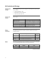
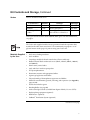

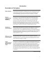
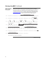
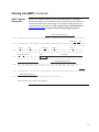
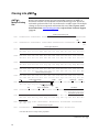
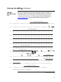
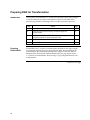
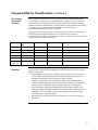

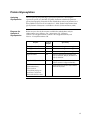
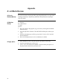
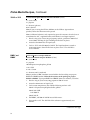

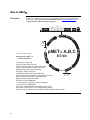
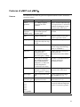
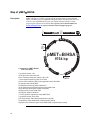

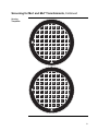
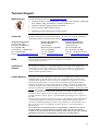


![[Phytophthora infestans (Mont) de Bary] EN CHAPINGO, MÉXICO](http://vs1.manualzilla.com/store/data/006207786_1-5a8924e6b4674eefa2af9dcbcb4dc0ac-150x150.png)