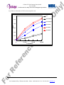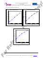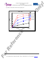
1
On
Cell-Based ELISA Kit for Measuring cyclobutane pyrimidine dimers in situ
ly
!
Cellular UV DNA-Damage Detection Kit
User’s Manual
For Research Use Only, Not for use in diagnostic procedures
CycLex Cellular UV DNA-Damage
Detection Kit
Cat# CY-1141
en
ce
Pu
rp
Intended Use................................................ 1
Storage......................................................... 1
Introduction.................................................. 2
Principle of the Assay.................................. 2-4
Materials Provided....................................... 5
Materials Required but not Provided........... 5
Precautions and Recommendations.............. 6
Detailed Protocol.......................................... 7-10
Troubleshooting........................................... 10
Reagent Stability...........................................10
Example of Test Results.............................. 11-14
References.................................................... 15
Related Products........................................... 15
os
e
For 200 Assays
Intended Use
The CycLex Research Product CycLex Cellular UV DNA-Damage Detection Kit is a non-isotopic
immunoassay used for the semi-quantitative measurement of CPDs (cyclobutane pyrimidine dimers) in
genomic DNA formed by UV-irradiation to cells.
er
Applications for this kit include:
1) Detection and semi-quantification of cyclobutane pyrimidine dimers in cells.
2) Monitoring the effects of UV on formation of cyclobutane pyrimidine dimers in cells.
3) Monitoring the effects of UV protection reagent on formation of cyclobutane pyrimidine dimers in
cells.
4) Study on the repair mechanisms of UV-induced DNA damage by the nucleotide excision repair in
cells.
ef
This assay kit is for research use only and not for use in diagnostic or therapeutic procedures.
Storage
rR
• Upon receipt store all components at 4°C.
• Don’t expose reagents to excessive light.
Fo
Cat#: CY-1141
1
Version#: 120420
15A Constitution Way · Woburn, MA 01801 · Phone: 1.800.200.5459 · Fax: 781-939-6963 · mblintl.com
On
Introduction
ly
!
Cellular UV DNA-Damage Detection Kit
User’s Manual
For Research Use Only, Not for use in diagnostic procedures
rp
os
e
DNA damage in cells exposed to ultraviolet (UV) radiation plays significant roles in cell-cycle arrest,
activation of DNA repair, cell killing, mutation, and neoplastic transformation. The major types of DNA
damage induced by UVB (280-315 nm, component of sunlight) and by UVC (200-280 nm) are
cyclobutane pyrimidine dimers (CPDs) and (6-4) photoproducts (6-4PPs), which are formed between
adjacent pyrimidine nucleotides on the same strand of DNA. Approximately 70-80% of UV-induced
DNA damage is CPDs and the remaining is 6-4PPs and Dewar isomer of 6-4PPs. These types of DNA
lesions are repaired by nucleotide excision repair (NER) system in normal human cells. Mori et al (10)
have established monoclonal antibodies specific for CPDs or 6-4PPs. These antibodies enable one to
quantitate photoproducts in DNA purified from cultured cells or from the skin epidermis using an
enzyme-linked immunosorbent assay (ELISA) and to visualize and measure photoproducts in DNA in
cultured cells or the skin using indirect immunofluorescence (IIF). This technology would contribute to
understanding of molecular mechanisms of cellular responses to UV and DNA damage in the
photobiology and the pigment cell biology.
en
ce
Pu
A rapid and convenient method for evaluating the formation of cyclobutane pyrimidine dimers
(CPDs) in a cell after UV-irradiation was developed which detects CPDs adducts on the genomic DNA
by means of anti-CPDs antibody in a cellular ELISA format. It has been shown that a precise evaluation
of CPDs formation, could be performed by the measurement of CPDs adducts in purified genomic DNA
by ELISA. In addition, there is a good reverse proportion between the cellular CPDs ELISA value and
ability of nucleotide excision repair (NER) when the cell are incubate for several hours after
UV-irradiation for repair as shown for a variety of NER deficient cell lines, such as xeroderma
patient-derived cell lines.
The CycLex Cellular UV DNA-Damage Detection Kit can be used in many different in vitro cell
systems without any purification step of genomic DNA.
Principle of the Assay
rR
ef
er
The CycLex Cellular UV DNA-Damage Detection Kit based on the formation of CPDs on the
genomic DNA after UV-irradiation in the cells that are cultured in microtiter plates. After formation of
CPDs on DNA, CPDs in the cell is detected by anti-CPDs antibody.
During UV-irradiation is carried out on wells of the microtiter plate, CPDs will be formed on the
DNA of living cells. To enable antibody binding to the CPDs on the genomic DNA, cells must be fixed,
permeabilized and the DNA denatured. Detector anti-CPDs antibody is pipetted into the wells and
allowed to incubate for one hour, during which time it binds to any CPDs. Unbound antibody is washed
away and horseradish peroxidase-conjugated secondary antibody is added, which binds to the detector
antibody. The horseradish peroxidase catalyzes the conversion of the chromogenic substrate
tetra-methylbenzidine (TMB) from a colorless solution to a blue solution (or yellow after the addition of
stopping reagent). The color is quantified by spectrophotometry and reflects the relative amount of CPDs
in the cells.
Fo
Cat#: CY-1141
2
Version#: 120420
15A Constitution Way · Woburn, MA 01801 · Phone: 1.800.200.5459 · Fax: 781-939-6963 · mblintl.com
ly
!
Cellular UV DNA-Damage Detection Kit
User’s Manual
For Research Use Only, Not for use in diagnostic procedures
I. Summary of Procedure for adherent cells
Culture adherent cells in microplate at 1-4 x 10⁴ cells/well.
On
The CycLex Cellular UV DNA-Damage Detection Kit is designed to measure the relative levels of
cyclobutane pyrimidine dimers on genomic DNA in situ. The summary of the assay is shown in below.
os
e
Incubate O/N at 37°C in CO2 incubator
UV-irradiate the cells.
Incubate an appropriate time at 37°C in CO2 incubator
Discard the culture medium.
rp
Add 200 µL of Fixing/Denaturing solution.
Stand for 30 min at room temp.
Pu
Discard the Fixing/Denaturing solution.
Add 200 µL of Blocking Solution.
Incubate 30 min at room temp.
en
ce
Discard the Blocking Solution
Add 50 µL of Primary Antibody (Anti-CPDs antibody)
Incubate 1 hr at room temp.
Wash the wells
Add 50 µL of HRP conjugated Secondary Antibody
Incubate 1 hr at room temp.
Wash the wells
er
Add 50 µL of Substrate Reagent
ef
Add 50 µL of Stop Solution
rR
Measure absorbance at 450 nm
Fo
Cat#: CY-1141
3
Version#: 120420
15A Constitution Way · Woburn, MA 01801 · Phone: 1.800.200.5459 · Fax: 781-939-6963 · mblintl.com
On
II. Summary of Procedure for suspension cells
ly
!
Cellular UV DNA-Damage Detection Kit
User’s Manual
For Research Use Only, Not for use in diagnostic procedures
Culture suspension cells in microplate at 1-2 x 10⁴ cells/well.
Incubate O/N at 37°C in CO2 incubator
os
e
UV-irradiate the cells.
Incubate an appropriate time at 37°C in CO2 incubator
c.f.g. the microplate at 3,500 rpm for 10 min.
rp
Discard the culture medium
Air dry the cells by hair-dryer for 15 min
Pu
Add 200 µL of Fixing/Denaturing Solution.
Stand for 30 min at room temp.
Discard the Fixing/Denaturing Solution
en
ce
Add 200 µL of Blocking Solution.
Incubate 30 min at room temp.
Discard the Blocking Solution
Add 50 µL of Primary Antibody (Anti-CPDs antibody)
Incubate 1 hr at room temp.
Wash the wells
er
Add 50 µL of HRP conjugated Secondary Antibody
Incubate 1 hr at room temp.
Wash the wells
ef
Add 50 µL of Substrate Reagent
rR
Add 50 µL of Stop Solution
Fo
Cat#: CY-1141
Measure absorbance at 450 nm
4
Version#: 120420
15A Constitution Way · Woburn, MA 01801 · Phone: 1.800.200.5459 · Fax: 781-939-6963 · mblintl.com
On
Materials Provided
ly
!
Cellular UV DNA-Damage Detection Kit
User’s Manual
For Research Use Only, Not for use in diagnostic procedures
All compounds treatment should be assayed in duplicate. The following components are supplied and
are sufficient for 200 assays.
os
e
Fixing/Denaturing Solution: One bottle containing 50 mL of Fixing/Denaturing Solution. Ready to use.
The solution is a strong alkaline. Wear disposable gloves and eye protection when handling.
Blocking Solution: One bottle containing 50 mL of Blocking Solution. Ready to use.
10X Wash Buffer: One bottle containing 100 mL of 10X Wash Buffer containing 2%Tween®-20.
20X Primary Antibody Solution: One vial containing 600 µL of anti-cyclobutane pyrimidine dimers
antibody.
rp
Primary Antibody Dilution Buffer: One bottle containing 12 mL of Primary Antibody Dilution Buffer.
Ready to use.
Pu
20X Secondary Antibody Solution: One vial containing 600 µL of HRP (horseradish peroxidase)
conjugated Secondary Antibody.
Secondary Antibody Dilution Buffer: One bottle containing 12 mL of Secondary Antibody Dilution
Buffer. Ready to use.
en
ce
Substrate Reagent: One bottle containing 20 mL of the chromogenic substrate, tetra-methylbenzidine
(TMB). Ready to use.
Stop Solution: One bottle containing 20 mL of 1 N H2SO4. Ready to use. Sulfuric Acid is a strong acid.
Wear disposable gloves and eye protection when handling.
Materials Required but not Provided
rR
ef
er
• 96-well microplate (tissue culture grade, flat bottom)
• UV crosslinker or irradiator: Stratalinker 1800 (Stratagene) etc.
• Centrifuge with rotor for microplate (for suspension cell only).
• Cell culture media
• 1X PBS pH 7.2
• Pipettors: 2-20 µL, 20-200 µL and 200-1000 µL precision pipettors with disposable tips.
• Precision repeating pipettor
• Orbital microplate shaker
• Microcentrifuge and tubes for sample preparation.
• Vortex mixer
• Microplate washer: optional (Manual washing is possible but not preferable)
• Software package facilitating data generation and analysis :optional
• Reagent reservoirs
• Deionized water of the highest quality
• Absorbent paper: disposable paper towels
• Plate reader capable of measuring absorbance in 96-well plates at dual wavelengths of 450 nm/540
nm. Dual wavelengths of 450/550 or 450/595 nm can also be used. The plate can also be read at a
single wavelength of 450 nm, which will give a somewhat higher reading.
Fo
Cat#: CY-1141
5
Version#: 120420
15A Constitution Way · Woburn, MA 01801 · Phone: 1.800.200.5459 · Fax: 781-939-6963 · mblintl.com
On
Precautions and Recommendations
ly
!
Cellular UV DNA-Damage Detection Kit
User’s Manual
For Research Use Only, Not for use in diagnostic procedures
Safety Warnings and Precautions: The CycLex Cellular UV DNA-Damage Detection Kit is designed
for research use only and not recommended for internal use in humans or animals. All chemicals should
be considered potentially hazardous and principles of good laboratory practice should be followed.
os
e
Technical Notes
Pu
rp
1. When performing washes manually, avoid introducing bubbles when dispensing liquids into the wells,
and ensure each well is filled with buffer, but not overflowing to avoid cross-contamination between
wells. Empty wells with a wrist flick motion over an appropriate receptacle, and while still inverted,
blot any remaining moisture onto clean absorbent paper.
2. Agitation of wells during incubation of Blocking Buffer and Antibody steps is recommended to reduce
non-specific background. If microtiter plate agitator is not available, a platform vortex at a low setting
can be used (e.g. level 1 of Fisher’s Genie II platform vortex). If background problems occur, simply
increase the number and/or duration of washes.
3. A brief 1X PBS rinse is recommended prior to the addition of the HRP substrate to remove any traces
of the Tween®-20 with can interfere with the HRP activity.
4. Do not allow the wells to dry out during the protocol.
5. Incubation temperatures for Primary Antibody and Detection Antibody can be varied and should be
empirically determined.
General Notes
• Allow all the components to come to room temperature before use.
en
ce
• Do not use kit components beyond the indicated kit expiration date.
• Rinse all detergent residue from glassware.
• Use deionized water of the highest quality.
• Do not mix reagents from different kits.
er
• The buffers and reagents used in this kit contain NaN3 as preservatives. Care should be taken to avoid
direct contact with these reagents.
• Dispose of tetra-methylbenzidine (TMB) containing solutions in compliance with local regulations.
ef
• Avoid contact with the alkaline Fixing/Denaturing Solution, the acidic Stop Solution and Substrate
Solution which contains hydrogen peroxide.
rR
• Wear gloves and eye protection when handling immunodiagnostic materials and samples of human
origin, and these reagents. In case of contact with the Stop Solution and the Substrate Solution, wash
skin thoroughly with water and seek medical attention, when necessary.
• CAUTION: Sulfuric Acid is a strong acid. Fixing/Denaturing Solution is a strong alkaline. Wear
disposable gloves and eye protection when handling Fixing/Denaturing Solution and
Stop Solution.
Fo
Cat#: CY-1141
6
Version#: 120420
15A Constitution Way · Woburn, MA 01801 · Phone: 1.800.200.5459 · Fax: 781-939-6963 · mblintl.com
On
Detailed Protocol
ly
!
Cellular UV DNA-Damage Detection Kit
User’s Manual
For Research Use Only, Not for use in diagnostic procedures
os
e
The CycLex Cellular UV DNA-Damage Detection Kit includes all reagents except cell culture
microplate, for detection of CPDs formed in genomic DNA in cultured cells. Since experimental
conditions may vary, treatment cells with test compound should be assayed in duplicate. Disposable
pipette tips and reagent troughs should be used for all liquid transfers to avoid cross-contamination of
reagents or samples.
Preparation of Reagents
All reagents need to be brought to room temperature prior to the assay. Assay reagents are supplied
ready-to-use, with the exception of 10X Wash Buffer, 20X Primary Antibody Solution and 20X
Secondary Antibody Solution.
rp
1. Prepare a working solution of Wash Buffer by adding 100 mL of the 10X Wash Buffer (provided) to
900 mL of deionized (distilled) water. Mix well. Store at 4°C for two weeks or -20°C for long-term
storage.
Pu
2. Prepare a working solution of Primary Antibody Solution: Dilute 20X Primary Antibody Solution
1:20 with Primary Antibody Dilution Buffer. Primary Antibody Solution can be stored for up to
one week protected from light at 4°C.
Prepare appropriate volume for your assay. You will need 50-60 µL of Primary Antibody Solution
per assay well. Discard any unused Primary Antibody Solution after diluted.
en
ce
3. Prepare a working solution of Secondary Antibody Solution: Dilute 20X Secondary Antibody
Solution 1:20 with Secondary Antibody Dilution Buffer. Secondary Antibody Solution can be
stored for up to one week protected from light at 4°C.
rR
ef
er
Prepare appropriate volume for your assay. You will need 50-60 µL of Secondary Antibody Solution
per assay well. Discard any unused Secondary Antibody Solution after diluted.
Fo
Cat#: CY-1141
7
Version#: 120420
15A Constitution Way · Woburn, MA 01801 · Phone: 1.800.200.5459 · Fax: 781-939-6963 · mblintl.com
On
Assay Procedure
ly
!
Cellular UV DNA-Damage Detection Kit
User’s Manual
For Research Use Only, Not for use in diagnostic procedures
A. Culture cells in 96-well microplate and UV irradiation
1. Plate adherent cells into a 96-well microplate at 1-4 x 10⁴ cells/well* in a final volume of 100
µL/well.
2. Incubate the microplate at 37°C over night in CO2 incubator.
3. Remove the culture media. Add 50 µL of sterile PBS in well.
os
e
* For most experiments, 2.5 x 10⁴ cells/well are sufficient and adequate for most adherent cells. 1-2 x
104 cells/well are sufficient and adequate for most suspension cells (See Fig. 1-4).
rp
4. Perform UV irradiation** to each well. No irradiation control should be run in duplicate as a
negative control.
** 0, 0.1, 0.2, 0.5, 1, 5, 10, 40 J/m2.
Pu
5. Remove the PBS. Add 100 µL of the culture medium in well.
6. Incubate the microplate at 37°C for appropriate time in CO2 incubator (in case of repair experiment).
en
ce
Note: Although we suggest to conduct experiments as outlined above, the optimal experimental
conditions will vary depending on the parameters being investigated, and must be
determined by the individual user. Especially, an appropriate cell number must be
optimized for your experiment
B. Fixing and denaturing the cells to 96-well plate
Fixing and denaturing the cells in the 96-well plates should be done as soon as the desired incubation
has completed.
er
For adherent cells:
1. Remove media from wells with a wrist flick. Avoid touching the bottom of the well and removing
cells.
ef
For suspension cells:
1’. Remove media from wells by centrifugation the 96 well microplate at 2,100 x g for 10 min and then
suction using a canulla. Avoid touching the bottom of the well and removing cells. Dry the cells
using a hair-dryer for about 15 minutes.
After this step, the dried cells can be stored for up to one week at 4°C.
rR
2. Add 200 µL/well of Fixing/Denaturing Solution slowly to insure cells are not detached from the
plastic. Let stand for 30 minutes at room temperature (ca.25°C).
3. Remove Fixing/Denaturing Solution from wells with a wrist flick. While still inverted, tap the plate
gently onto absorbent paper to remove any excess fixing agent still within the wells.
Fo
Cat#: CY-1141
8
Version#: 120420
15A Constitution Way · Woburn, MA 01801 · Phone: 1.800.200.5459 · Fax: 781-939-6963 · mblintl.com
ly
!
Cellular UV DNA-Damage Detection Kit
User’s Manual
For Research Use Only, Not for use in diagnostic procedures
On
C. Detection of Signals (Addition of Primary and Secondary Antibodies and Substrate Reagent)
1. Add 200 µL/well of Blocking Solution and incubate for at least 30 minutes at room temperature
(ca.25°C).
2. Remove Blocking Solution with a wrist flick.
os
e
3. Add 50 µL/well of Primary Antibody Solution and incubate for 1 hour at room temperature
(ca.25°C).
Alternatively, this incubation period can be varied between 30–120 minutes, depending on
individual requirements.
4. Remove Primary Antibody Solution with a wrist flick.
rp
5. Rinse the wells once with 200µl/well of Wash Buffer.
6. Remove Wash Buffer with a wrist flick. While still inverted, tap the plate onto absorbent paper.
Pu
7. Wash wells 4 times with 200 µL/well of Wash Buffer for 2 minutes each with shaking at ca. 200 rpm
on an orbital microplate shaker. Remove Wash Buffer in-between each wash with a wrist flick.
8. Add 50 µL/well of Secondary Antibody Solution and incubate for 1 hour at room temperature
(ca.25°C).
en
ce
9. Remove Secondary Antibody Solution with a wrist flick.
10. Rinse wells once with 200 µL/well of Wash Buffer.
11. Remove Wash Buffer with wrist flick and tap plate onto absorbent paper.
12. Wash wells 4 times with 200 µL/well of Wash Buffer for 2 minutes each with shaking at ca. 200
rpm on an orbital microplate shaker. Remove Wash Buffer in-between each wash with a wrist flick.
13. After last wash with Wash Buffer, rinse wells once with 300 µl/well of 1X PBS. Remove with a
wrist flick and tap onto absorbent paper. Ensure that that no liquid remains in the well.
er
14. Add 50 µL/well of Substrate Reagent. (Avoid exposing the microplate to direct sunlight covering
the plate with e.g. aluminum foil is recommended). Return Substrate Reagent to 4°C immediately
after the necessary volume is removed.
ef
15. Incubate the plate for 15-25 minutes at room temperature (ca.25°C). (The incubation time may be
extended up to 30 minutes if the reaction temperature is below than 20°C).
rR
16. Add 50 µL of Stop Solution to each well in the same order as the previously added Substrate
Reagent.
17. Measure absorbance in each well using a spectrophotometric microplate reader at dual wavelengths
of 450/540 nm. Dual wavelengths of 450/550 or 450/595 nm can also be used. Read the microplate
at 450 nm if only a single wavelength can be used. Wells must be read within 30 minutes of adding
the Stop Solution*.
Fo
Cat#: CY-1141
9
Version#: 120420
15A Constitution Way · Woburn, MA 01801 · Phone: 1.800.200.5459 · Fax: 781-939-6963 · mblintl.com
ly
!
On
Cellular UV DNA-Damage Detection Kit
User’s Manual
For Research Use Only, Not for use in diagnostic procedures
Note-1: Complete removal of liquid at each step is essential to good performance. After the last wash,
remove any remaining Wash Buffer by aspirating or decanting. Invert the plate and blot it
against clean paper towels.
os
e
Note-2: Although we suggest to conduct experiments as outlined above, the optimal experimental
conditions will vary depending on the parameters being investigated, and must be determined
by the individual user. Especially, an appropriate cell number must be optimized for your
experiment
Troubleshooting
rp
1. The signals and backgrounds are influenced a great deal by cell line and cell number that you
plated, please ensure the appropriate cell number for your experiment. Please conduct the
experiment to optimize cell number for this assay. See “Example of Test Results Fig.2 and
Fig.4”.
Pu
2. Unavoidable background (no UV irradiation) is observed even if an appropriate cells number is
used. It is usually around 0.3-0.4 See “Example of Test Results Fig.1 and 3”.
3. With some cell lines, higher cell concentrations (more than 1 × 10⁴ cells/well in case of adherent cells)
may lead to increasing absorbance values in no UV irradiation (background) rather than those in UV
irradiation.
en
ce
4. All UV irradiation including no UV irradiation control should be run in duplicate, using the protocol
described in the Detailed Protocol. Incubation times or temperatures significantly different from
those specified may give erroneous results.
5. Poor duplicates, accompanied by elevated values for wells containing no UV irradiation control,
indicate insufficient washing or vigorous washing. Wash the plate thoroughly and gently.
6. Overall low signal may indicate that desiccation of the plate has occurred between the final wash and
addition of Substrate Reagent. Do not allow the plate to dry out. Add Substrate Reagent immediately
after wash.
er
Reagent Stability
ef
All of the reagents included in the CycLex Cellular UV DNA-Damage Detection Kit have been
tested for stability. Reagents should not be used beyond the stated expiration date. Upon receipt, kit
reagents should be stored at 4°C. For long-term storage of 20X Primary Antibody Solution and 20X
Secondary Antibody Solution, it is recommended to store at below -70°C.
rR
For research use only, not for use in diagnostic or therapeutic procedures
Fo
Cat#: CY-1141
10
Version#: 120420
15A Constitution Way · Woburn, MA 01801 · Phone: 1.800.200.5459 · Fax: 781-939-6963 · mblintl.com
On
Example of Test Results
Fig.1 UV dose dependency using HeLa cells
CPDs 12,500 cells/well HeLa
CPDs 25,000 cells/well HeLa
1.2
1.2
os
e
1.0
1.0
0.8
A450
0.8
A450
ly
!
Cellular UV DNA-Damage Detection Kit
User’s Manual
For Research Use Only, Not for use in diagnostic procedures
0.6
0.6
0.4
rp
0.4
0.2
0.2
0.0
1
10
J/m2
Pu
0.0
0
100
0
1
J/m2
10
100
CPDs 50,000 cells/well HeLa
en
ce
1.2
1.0
A450
0.8
0.6
er
0.4
0.2
rR
ef
0.0
Fo
Cat#: CY-1141
0
1
J/m2
11
10
100
Version#: 120420
15A Constitution Way · Woburn, MA 01801 · Phone: 1.800.200.5459 · Fax: 781-939-6963 · mblintl.com
On
Fig.2 Effect of cell number on ELISA value using HeLa cells
CPDs HeLa
1.2
ly
!
Cellular UV DNA-Damage Detection Kit
User’s Manual
For Research Use Only, Not for use in diagnostic procedures
UV 0
UV 0.156J/m2
os
e
1.0
UV 0.625J/m2
UV 2.5J/m2
UV 10J/m2
0.6
rp
A450
0.8
0.2
0.0
12,500
25,000
Cell number
50,000
rR
ef
er
en
ce
0
Pu
0.4
UV 40J/m2
Fo
Cat#: CY-1141
12
Version#: 120420
15A Constitution Way · Woburn, MA 01801 · Phone: 1.800.200.5459 · Fax: 781-939-6963 · mblintl.com
On
Fig.3 UV dose dependency using Raji cells
CPDs 12,500 cells/well Raji
1.4
1.2
1.2
1.0
1.0
0.8
0.8
0.6
0.6
0.4
rp
0.4
os
e
CPDs 25,000 cells/well Raji
1.4
A450
A450
ly
!
Cellular UV DNA-Damage Detection Kit
User’s Manual
For Research Use Only, Not for use in diagnostic procedures
0.2
0.2
0.0
0.0
1
J/m2
10
100
0
1
Pu
0
J/m2
10
CPDs 50,000 cells/well Raji
en
ce
1.4
1.2
A450
1.0
0.8
0.6
er
0.4
0.2
rR
ef
0.0
Fo
Cat#: CY-1141
0
1
J/m2
13
10
100
Version#: 120420
15A Constitution Way · Woburn, MA 01801 · Phone: 1.800.200.5459 · Fax: 781-939-6963 · mblintl.com
100
On
Fig.4 Effect of cell number on ELISA value using Raji cells
ly
!
Cellular UV DNA-Damage Detection Kit
User’s Manual
For Research Use Only, Not for use in diagnostic procedures
CPDs Raji
UV 0
1.4
os
e
UV 0.156J/m2
1.2
UV 0.625J/m2
UV 2.5J/m2
1.0
UV 40J/m2
0.6
rp
A450
UV 10J/m2
0.8
0.2
0.0
12,500
25,000
Cell number
50,000
rR
ef
er
en
ce
0
Pu
0.4
Fo
Cat#: CY-1141
14
Version#: 120420
15A Constitution Way · Woburn, MA 01801 · Phone: 1.800.200.5459 · Fax: 781-939-6963 · mblintl.com
rp
Related Products
os
e
1. Nishiwaki, Y., et al., J. Invest. Dermatol. 122, 526-532 (2004)
2. Imoto, K., et al., J. Invest. Dermatol. 119, 1177-7782 (2002)
3. Kobayashi, N., et al., Pigment Cell Res. 14, 94-102 (2001)
4. Katsumi, S., et al., J. Invest. Dermatol. 117, 1156-1161 (2001)
5. Otoshi, E., et al., Cancer Res. 60, 1729-1735 (2000)
6. Nakagawa, A., et al., J. Invest. Dermatol. 110, 143-148 (1998)
7. Kobayashi, N., et al., J. Invest. Dermatol. 110, 806-810 (1998)
8. Komatsu, Y., et al., Nucleic Acids Res. 25, 3889-3894 (1997)
9. Kobayashi, N., et al., J. Invest. Dermatol. 101, 685-689 (1993)
10. Mori, T., et al., Photochem. Photobiol. 54, 225-232 (1991)
On
References
ly
!
Cellular UV DNA-Damage Detection Kit
User’s Manual
For Research Use Only, Not for use in diagnostic procedures
en
ce
Pu
* CycLex Cellular Histone Acetylation Assay Kit: Cat# CY-1140
* CycLex Cellular UV DNA-Damage Detection Kit: Cat# CY-1141
* CycLex BrdU Cellular ELISA Kit: Cat# CY-1142
* CycLex Histone H2A.X Phosphorylation Cellular ELISA Kit: Cat# CY-1143
PRODUCED BY
er
CycLex Co., Ltd.
1063-103 Terasawaoka
Ina, Nagano 396-0002
Japan
Fax: +81-265-76-7618
e-mail: [email protected]
URL: http://www.cyclex.co.jp
rR
ef
CycLex/CircuLex products are supplied for research use only. CycLex/CircuLex products and
components thereof may not be resold, modified for resale, or used to manufacture commercial
products without prior written approval from CycLex Co., Ltd.. To inquire about licensing for
such commercial use, please contact us via email.
Fo
Cat#: CY-1141
15
Version#: 120420
15A Constitution Way · Woburn, MA 01801 · Phone: 1.800.200.5459 · Fax: 781-939-6963 · mblintl.com