1
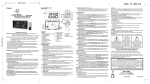
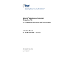
Nuclear-ID™ Green Chromatin Condensation Detection Kit for fluorescence microscopy and flow cytometry Instruction Manual Cat. No. ENZ-51021-K200 For research use only. Rev. 1.1.0 October 2009 200 assays Notice to Purchaser The Nuclear-ID Green Chromatin Condensation Detection Kit is a member of the CELLestial™ product line, reagents and assay kits comprising fluorescent molecular probes that have been extensively benchmarked for live cell analysis applications. CELLestial™ reagents and kits are optimal for use in demanding cell analysis applications involving confocal microscopy, flow cytometry, microplate readers and HCS/HTS, where consistency and reproducibility are required. This product is manufactured and sold by ENZO LIFE SCIENCES, INC. for research use only by the end-user in the research market and is not intended for diagnostic or therapeutic use. Purchase does not include any right or license to use, develop or otherwise exploit this product commercially. Any commercial use, development or exploitation of this product or development using this product without the express prior written authorization of ENZO LIFE SCIENCES, INC. is strictly prohibited. Limited Warranty These products are offered under a limited warranty. The products are guaranteed to meet appropriate specifications described in the package insert at the time of shipment. Enzo Life Sciences’ sole obligation is to replace the product to the extent of the purchase price. All claims must be made to Enzo Life Sciences, Inc. within five (5) days of receipt of order. Trademarks and Patents Enzo and Nuclear-ID are trademarks of Enzo Life Sciences, Inc. Several of Enzo’s products and product applications are covered by US and foreign patents and patents pending. Contents I. Introduction ............................................................... 1 II. Reagents Provided and Storage.............................. 1 III. Additional Materials Required ................................. 2 IV. Safety Warnings and Precautions........................... 2 V. Methods and Procedures ......................................... 3 A. REAGENT PREPARATION ................................................... 3 B. CELL PREPARATIONS ........................................................ 4 C. STAINING LIVE CELLS FOR FLOW CYTOMETRY ................... 4 D. STAINING PERMEABILIZED CELLS FOR SUB-G0 ANALYSIS BY FLOW CYTOMETRY ....................................... 5 E. WIDE FIELD FLUORESCENCE / CONFOCAL MICROSCOPY .... 5 F. MICROPLATE ASSAY ......................................................... 6 VI. Appendices ............................................................... 7 A. FLUORESCENCE CHANNEL SELECTION FOR DATA COLLECTION ..................................................... 7 B. EXPECTED RESULTS......................................................... 7 VII. References ................................................................ 8 VIII. Troubleshooting Guide ......................................... 10 I. Introduction During the apoptotic process, chromatin undergoes a phase change from a heterogeneous, genetically active network to an inert highly condensed form that is subsequently fragmented and packaged into apoptotic bodies. Recently, three stages of apoptotic chromatin condensation have been defined based upon morphological and biochemical criteria using a cellfree system: stage 1 “ring” condensation; stage 2 “necklace” condensation; and stage 3 “nuclear collapse/disassembly”.2 Cells appear to possess an apoptosis-specific system for the induction of chromatin condensation, with phase 2 of the process requiring DNAse(s) and phase 3 requiring hydrolysable ATP. A number of other factors have been implicated in nuclear condensation, including AIF, acinus and phospholipase A2 and research efforts continue in order to better define the key physiological factors that drive chromatin condensation during apoptosis in vivo. Enzo Life Sciences’ Nuclear-ID™ Green Chromatin Condensation Detection Kit provides a convenient approach for analysis of late stage apoptosis by flow cytometry and microscopy. It can also be used in microplate assays. The basis of the assay is that the compacted chromatin of apoptotic cells binds higher amounts of nuclear dye compared to the healthy cells. The kit is suitable for differentiating between healthy and apoptotic cells with condensed nuclei. A control apoptosis inducing agent, Staurosporine, is provided for monitoring apoptotic changes in nuclear organization. Potential applications for live-cell studies include quickly testing cell cultures for overall health, as well as rapid screening for compounds that induce apoptosis and chromatin condensation. II. Reagents Provided and Storage All reagents are shipped on dry ice. Upon receipt, the kit should be stored at ≤-20°C, protected from light. When stored properly, these reagents are stable for at least twelve months. Avoid repeated freezing and thawing. Reagents provided in the kit are sufficient for approximately 200 assays using either live or permeabilized cells. Reagent Quantity Nuclear-ID™ Green Detection Reagent Apoptosis Inducer (Staurosporine) 10X Assay Buffer 100 µL 50 nmoles 30 mL 1 III. Additional Materials Required Flow cytometer equipped with 488 nm laser source Microscope equipped with standard FITC filters Tubes appropriate for holding cells for the flow cytometer Calibrated, adjustable precision pipetters, preferably with disposable plastic tips Adjustable speed centrifuge with swinging buckets (for suspension cultures) Deionized water Anhydrous DMSO Total growth medium suitable for cell type 10% Triton X-100 in water (for Sub G0 analysis of permeabilized cells, optional) IV. Safety Warnings and Precautions This product is for research use only and is not intended for diagnostic purposes. The Nuclear-ID™ Green Detection Reagent contains DMSO which is readily absorbed through the skin. DMSO is harmful if ingested or absorbed through the skin and may cause irritation to the eyes. Observe appropriate precautions when handling these reagents. Reagents should be treated as possible mutagens and should be handled with care and disposed of properly. Observe good laboratory practices. Gloves, lab coat, and protective eyewear should always be worn. Never pipet by mouth. Do not eat, drink or smoke in the laboratory areas. All blood components and biological materials should be treated as potentially hazardous and handled as such. They should be disposed of in accordance with established safety procedures. To avoid photobleaching, perform all manipulations in low light environments or protected from light by other means. 2 V. Methods and Procedures The procedures described in this manual assume that the user is familiar with the basic principles and practices of flow cytometry and is able to run samples according to the operator’s manual pertaining to the instrument being used. NOTE: Allow all reagents to thaw at room temperature before starting with the procedures. Upon thawing, gently hand-mix or vortex the reagents prior to use to ensure a homogenous solution. Briefly centrifuge the vials at the time of first use, as well as for all subsequent uses, to gather the contents at the bottom of the tube. A. REAGENT PREPARATION 1. Positive Control The Apoptosis Inducer (Staurosporine) is supplied as a lyophilized powder (50 nmoles) and should be reconstituted in 50 μL DMSO for a 1 mM stock solution. It is recommended that with the apoptosis inducer of choice, induction occurs at the effective concentration (EC50) and that the final percent DMSO in the assay not exceed 0.2%. 2. 1X Assay Buffer Allow the 10X Assay Buffer to warm to room temperature. Make sure that the reagent is free of any crystallization before dilution. Prepare enough 1X Assay Buffer for the number of samples to be assayed by diluting each milliliter (mL) of the 10X Assay Buffer with 9 mL of deionized water. 3. DNA Staining Solutions The concentration of Nuclear-ID™ Green Detection Reagent for optimal staining will vary depending upon the application. Suggestions are provided to use as guidelines, though some modifications may be required depending upon the particular cell type employed and other factors such as the permeability of the dye to the cells or tissues. To reduce potential artifacts from overloading the cells, the concentration of the dye should be kept as low as possible. Refer to sections C, D and E for details on the preparation of the staining solution for specific applications. Prepare sufficient amount of the staining solution for the number of samples to be assayed. 3 B. CELL PREPARATIONS Positive control cells should be pretreated with Apoptosis inducer for 1-4 hours. Response to Staurosporine is time and concentration dependent and may also vary significantly depending upon cell type and cell line. Negative control cells should be treated with a vehicle (DMSO, media or other solvent used to reconstitute or dilute an inducer or inhibitor) for an equal length of time under similar conditions. C. STAINING LIVE CELLS FOR FLOW CYTOMETRY 1. Cells should be maintained via standard tissue culture practice. Grow cells overnight to log phase in a humidified incubator at 37°C, 5% CO2. 2. Treat cells with compound of interest and Negative Control cells with vehicle. 3. Prepare positive control cells by incubating with Apoptosis inducer (0.5-2 µM, see section A-1, page 3) for 1-4 hours under normal tissue culture conditions. 4. At the end of treatment, trypsinize (adherent cells), or collect cells (suspension cells). Samples may contain 1 x 10⁵ to 1 x 10⁶ cells. 5. Centrifuge at 400 x g for 5 minutes to pellet the cells. Re-suspend in media, 1X Assay Buffer, or other buffer of choice and centrifuge as before. 6. Immediately prior to staining the live cell samples, prepare fresh DNA Staining Solution as follows: NOTE: A concentration of 1-5 µM Nuclear-ID™ Green Detection Reagent is recommended for Chromatin Condensation analysis. The procedure described below is for preparation of 5 µM dye solution. For each sample to be stained, dilute 1 µL Nuclear-ID™ Green Detection Reagent to a final volume of 1 mL with cold media or buffer of choice. Mix well and keep on ice. 7. Resuspend each live cell sample in 0.5 mL of freshly prepared cold DNA Staining Solution (from step 6). Incubate for 30 minutes at 4°C in the dark. It is important to achieve a monodisperse cell suspension at this step by gently pipetting up and down repeatedly. 8. Analyze the samples in the FL1 channel of a flow cytometer. 4 D. STAINING PERMABILIZED CELLS FOR SUB-G0 ANALYSIS BY FLOW CYTOMETRY Sub-G0 analysis of apoptotic cells, a measure of DNA fragmentation, may be performed using the Nuclear-ID™ Green Chromatin Condensation Detection Kit simply by permeabilizing the cells. The NuclearID™ Green dye intercalates with the chromosomal DNA and a population of lower fluorescence fragmented DNA can be analyzed as follows. 1. Grow cells overnight to log phase in a humidified incubator at 37°C, 5% CO2. 2. Treat cells with or without experimental compounds. 3. Prepare positive control cells by incubating with pre-diluted Staurosporine (0.5-2 µM, see section A-1, page 3) for 1-4 hours under normal tissue culture conditions. 4. At the end of treatment, trypsinize (adherent cells), or collect cells (suspension cells). Samples may contain 1 x 10⁵ to 1 x 10⁶ cells. 5. Centrifuge at 400 x g for 5 min. to pellet the cells. Re-suspend in media, 1X Assay Buffer, or other buffer of choice and centrifuge as before. 6. Immediately prior to staining the cell samples, prepare fresh DNA Staining/Permeabilizing Solution as follows: NOTE: A concentration of 1-5 µM Nuclear-ID™ Green Detection Reagent is recommended for permeabilized cells. The procedure described below is for preparation of 5 µM dye solution. For each sample to be stained, dilute 1 µL Nuclear-ID™ Green Detection Reagent and 10 µL of 10% Triton X-100 to a final volume of 1 mL with media or buffer of choice. 7. Re-suspend each fixed cell sample to be fixed in 0.5 mL freshly prepared DNA Staining/Permeabilizing Solution (from step 6) to a final concentration of 1 x 10⁵ to 1 x10⁶ cells/mL. 8. Incubate for 30 min. at 37°C in the dark. 9. Analyze the samples in the FL1 channel of a flow cytometer with a 488 nm excitation laser. E. WIDE FIELD FLUORESCENCE / CONFOCAL MICROSCOPY 1. Grow cells directly onto glass slides. 2. Treat cells with compound of interest and Negative Control cells with vehicle. 3. Carefully wash cells twice with ice-cold 1X Assay Buffer in a volume sufficient for covering the cell monolayer. 5 4. Carefully remove supernatant and dispense the cold DNA staining solution (e.g., 5 µM Nuclear-ID™ Green dye in 1X Assay Buffer) in a volume sufficient for covering the cell monolayer. 5. Protect samples from light and incubate for 30 minutes at 4˚C. 6. Flick the staining solution onto a paper towel and if necessary add a few drops of 1X Assay Buffer to prevent the cells from drying out. 7. Cover cells and observe under a fluorescence/confocal microscope with a filter set for GFP/FITC (Ex/Em: 488/514nm). 8. Positive Control Samples: It is recommended that positive control samples be pretreated with the Apoptosis Inducer at a final concentration of 2 μM for 4 hours, though optimal conditions may vary significantly among cell types and cell lines. Follow steps 3-7 post treatment. F. MICROPLATE ASSAY FOR CHROMATIN CONDENSATION The Nuclear-ID™ Green Chromatin Condensation Detection Kit can be employed to analyze chromatin condensation using a fluorescence microplate reader. A 96-well black wall, clear bottom microplate is recommended for this method. Optimal conditions will depend upon the cell type and apoptosis inducer of choice. Fluorescence signals are generally in Relative Fluorescence Units (RFUs) and linearity should be verified with appropriate filter sets according to instrument specifications. 1. Cells should be maintained via standard tissue culture practices. 2. Plate 40,000-50,000 cells/100 µL/well in a 96-well black wall, clear bottom plate. Grow overnight. 3. Replace the media with the apoptosis inducer of choice at required concentration and incubate for 4-6 hours at 37˚C. 4. Remove the media, then wash with 1X Assay Buffer. 5. Add DNA staining solution (1-5 µM Nuclear-ID™ Green dye in 1X Assay Buffer). 6. Incubate at 4˚C for 30 minutes. 7. Read the plate in the microplate reader with excitation wavelength ~488 nm and emission wavelength ~520 nm. 6 VI. APPENDICES A. FLUORESCENCE CHANNEL SELECTION FOR DATA COLLECTION The selection of optimal filter sets for a fluorescence microscopy application requires matching the optical filter specifications to the spectral characteristics of the dyes employed in the analysis. Consult the microscope or filter set manufacturer for assistance in selecting optimal filter sets for your microscope. Absorbance Fluorescence emission Fluorescence channel FL1 is recommended for imaging Nuclear-ID™ Green Detection Reagent with 488 nm excitation. 400 450 500 550 Wavelength (nm) 600 Figure 1. Absorption and fluorescence emission spectra for Nuclear-ID™ Green dye. Spectra were determined in 1X Assay Buffer. B. EXPECTED RESULTS Apoptosis is characterized by a diverse repertoire of biochemical and cellular signals that alter cellular morphology. Some of the downstream effects observed via fluorescence microscopy include cell shrinkage, loss of membrane symmetry, membrane blebbing and cytoplasmic condensation. Changes at the nuclear level are also evident, including the condensation and fragmentation of nuclear and chromosomal DNA. 1. Microscopy When observed under the fluorescence microscope, cells treated with Staurosporine or other apoptosis inducers should demonstrate stages of chromatin condensation. Nuclei of the healthy cells demonstrate even nuclear staining with distinctly stained nucleoli. Whereas the cells undergoing apoptosis lose the nucleolar staining and the nuclei take up more stain to demonstrate more dense and compact staining pattern. 7 2. Flow Cytometry a. Positive Control. Flow cytometry resolves the healthy cell population from the apoptotic cell population undergoing chromatin condensation based upon an approximately 40-fold increased staining of the condensed chromosomes. Control uninduced Jurkat (T-Cell leukemia) cells are stained as well but mostly display low fluorescence with the exception of a small percentage of cells (up to 15%) with condensed chromatin, as expected in routine cultures of untreated cells (Figure 2, panels A and C). In the samples treated with 2 μM Staurosporine for 4 hours more than 50% of the cells become highly fluorescent, indicating late-stage apoptosis (Figure 2, panels B and D). Panels A and B display the raw, ungated data. The rectangles in panels A and B are suggested gating settings for flow cytometry. Panels C and D show the data after the suggested gated has been applied, demonstrating clear separation of healthy cells from and apoptotic cells with condensed chromatin (panel D). b. Appearance of Additional Populations. In addition to the two major populations of weakly (healthy) and strongly (apoptotic) fluorescent cells, additional populations may be resolved based upon the overall granularity and shape size of the cells (as observed by side scatter and/or fluorescence FL1), depending on the nature of the apoptosis inducer. These populations may represent cells undergoing apoptosis at different stages or cells compromised in any other way. c. Sub-G0 Analysis. Sub-G0 analysis of apoptotic cells, a measure of DNA fragmentation may be performed using the Nuclear-ID™ Green Chromatin Condensation Detection Kit, simply by permeabilizing the cells. The Nuclear-ID™ Green dye intercalates with the chromosomal DNA and a population of lower fluorescence fragmented DNA can be analyzed as shown in Figure 3. VII. References 1. Telford, Komoriya and Packard. Multiparametric analysis of apoptosis by flow and image cytometry. Methods Mol Biol. 2004; 263:141-60. 2. Tonéa, Sugimotoc, Tandaa, Sudab, Uehirab, Kanouchia, Samejimad, Minatogawaa, and Earnshawd (2007) Three distinct stages of apoptotic nuclear condensation revealed by time-lapse imaging, biochemical and electron microscopy analysis of cell-free apoptosis. Exp. Cell Res. 313: 3635-3644. 3. Tawar, Bansal, Shrimal, Singh, Tandon (2007) Nuclear condensation and free radical scavenging: a dual mechanism of bisbenzimidazoles to modulate radiation damage to DNA. Mol Cell Biochem. 305(1-2): 221-33. 8 B C D After Gating Raw Data A Control Staurosporine Figure 2 Flow cytometry analysis of Control and Staurosporine treated Jurkat cells. Raw data is presented in panels A and B where suggested gating is indicated by rectangles drawn around the cell populations. Panels C and D represent the separation of healthy and apoptotic nuclei based on their characteristic fluorescence. Control Staurosporine Figure 3. Detection of the Sub-G0 phase in permeabilized cells using Nuclear ID™ Green dye. In the permeabilized cells, the dye intercalates with the chromosomal DNA, resulting in a highly fluorescent healthy cell population. Upon Staurosporine treatment, a population with lower fluorescence, corresponding to fragmented DNA, can be detected via flow cytometry. 9 VIII. Troubleshooting Guide Problem Potential Cause Suggestions Make sure the cell preparation protocol generates single cells with minimal clumping. Filtration or trituration of cell suspensions may be required. Systematically vary the dye to cell ratio. Chromatin condensation is not observed. Incorrect drug concentration or time of incubation for cell type Optimize for cell type, medium or buffer used, time of incubation, temperature of incubation. Verify that the flow cytometer is correctly aligned, with stable fluidics, using calibrated fluorescent beads of known CV. Verify that the cell concentration is 1 x 10⁵ to 1 x10⁶ cells/mL. Lower PMT setting. The healthy and apoptotic populations are not well separated. More than two populations appear upon apoptosis induction Healthy cells show very high fluorescence intensity Incorrect dye concentration Optimize for cell type, medium or buffer used, time of incubation, temperature of incubation. Unhealthy cells Use healthy cell cultures. Property of the dye Apoptosis is a multi-stage process and Nuclear-ID™ Green dye can distinguish different levels of condensation. PMT setting is too high Lower the PMT setting. Temperature of incubation Incubation with the dye should be at 4˚C. Permeablized cells In permeabilized cells the dye gets intercalated in the DNA giving higher fluorescence. 10 www.enzolifesciences.com