1
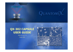
QX-302
User Manual
UQX009 Issue 2.0
© QuantomiX Ltd. 2005. All rights reserved.
April 2005
1
QX-302 User Manual
QuantomiX Ltd. 2005. All rights reserved.
This publication is the copyright of QuantomiX ltd. and contains information
that may not be used or reproduced unless agreed in writing. QuantomiX ltd.
reserves the right to alter without notice the specifications, design or supply
of any product or service.
The information provided in this User Manual is believed to be accurate. It is
the user’s responsibility to confirm the technical aspects and the suitability of
the technology for any particular application.
QuantomiX Ltd., 12 Hamada St., Tamar Science Park, Rehovot
POB 4037, Nes-Ziona 70400, Israel
Tel: +972-8-946-2244, Fax: +972-8-946-5874
http://www.quantomix.com
2
Table of Contents
1
Safety...................................................................................7
2
Technical Data .....................................................................9
2-1
2-2
2-3
2-4
3
Introduction ........................................................................15
3-1
3-2
3-3
4
Getting Started ..................................................23
Specimen Preparation.......................................24
Inserting Specimens into the QX-302 Capsule .27
Storing Specimens in the QX-302 Capsule .......30
Imaging ..............................................................................31
5-1
5-2
6
Manual Scope and Contents .............................15
References........................................................16
Technology and Components............................17
Using the QX-302 Capsules ..............................................23
4-1
4-2
4-3
4-4
5
QX-302 Capsules ................................................9
Capsule Plates (MP-12) ....................................12
Imaging Buffer (IB-74) .......................................13
Spacers .............................................................13
Optimal Imaging Conditions ..............................31
Recommendations for SEM Imaging with
QX- 302 Capsules.............................................34
Appendices ........................................................................35
6-1
6-2
6-3
6-4
6-5
Appendix A: Protocols for Specific Applications 35
Appendix B: Glossary........................................36
Appendix C: Troubleshooting ............................37
Appendix D: Ordering Information.....................38
Appendix E: Legal Notices ................................39
3
4
List of Figures
Figure 1. Sealed Capsule - Dimensions.........................................10
Figure 2. The Capsule Plate...........................................................12
Figure 3. QX-302 Capsule Parts....................................................18
Figure 4. QX-302 Capsule, Positioned in the Capsule Plate .........19
Figure 5. Opening the QX-302 Capsule ........................................28
Figure 6. Placing a Sample in the Specimen Dish.........................29
List of Tables
TABLE 1: Material Compatibility Table .....................................11
TABLE 2: Chapters and Appendices in the User Manual ............16
TABLE 3: Features Enhanced by Different Staining Agents .......26
TABLE 4: Recommendations for SEM Imaging with QX-302
..................Capsules ......................................................................34
TABLE 5: Troubleshooting ..........................................................37
Cited registered trademarks:
Whatman®
Tween®
Auto/Zyme®
VibratomeTM
Triton®
All trademarks are the properties of their respective owners.
5
6
Safety
Chapter 1: Safety
Cautions
Please observe the following cautions while using the
QuantomiX QX-302 capsules:
1. Correct sealing of the capsule is essential for its
proper functioning. To ensure that correct sealing is
achieved, please note that the wings of the middle
section of the capsule should be aligned with the
wings of the upper and lower sections.
2. The QX-302 sealing stub includes an O-ring, a circular rubber ring found in the lower section of the sealing stub. If the O-ring accidentally detaches from the
sealing stub, it should be re-positioned inside the
sealing stub (see Figure 3. on page 18).
3. Use powder-free gloves to maintain cleanness and
sterility of the QX-302 capsules. Powdered gloves
should be avoided.
4. Do not place the liquid dish or the capsule with the
capsule membrane facing down, except in the capsule plate. To prevent rupture, avoid touching the capsule membrane at all stages.
7
QX-302 User Manual
8
Technical Data
Chapter 2: Technical Data
2.1 QX-302 Capsules
Storage
The capsules should be stored in a dry,
dark environment at room temperature.
Shelf life
12 months from specified production
date (printed on the box).
Application
The capsules are intended for single use
and are not reusable.
They are intended for research purposes only.
The QX-302 capsule is suitable for
imaging thick, non-adherent samples
such as tissue biopsies, plants, and
material specimens, in a wet environment. The capsule is suitable for various
sample sizes, with a maximum diameter
of 3 mm. Sample thickness can range
between 300-1000 microns.
Dimensions
Specimen dish - diameter 3 mm,
Sealed capsule - see Figure 1 on
page 10. In cases where the QX-302
capsule does not fit your SEM stage,
inquire for available adaptors.
Sterility
Gamma-sterilized.
When sterility is required, opening of the
packaging should be done in a sterile
environment (laminar flow). Opened
packages should be stored under sterile
conditions.
Operation
Temperature
4° to 40° C
9
QX-302 User Manual
For compatibility of reagents with the
QX-302 capsule materials, see Table 1
on page 11. For other materials or specific concentrations, please contact
[email protected]. For materials not
compatible with the QX-302 capsule,
inquire about other capsule types.
Reagents containing DMSO should not
be used during SEM imaging (DMSO
can be present in the sample preparation process).
Material
Compatibility
Figure 1. Sealed Capsule - Dimensions
16
28
8
3.10
12.5
Dimensions are given in mm.
10
Technical Data
TABLE 1: Material Compatibility Table
Material
Compatible
Not Compatible
Acetone
x
DMSO
x
Ethanol
x
x
Ethyl Acetate
Formalin
x
2% Glutaraldehyde
x
Isopropanol
x
Methanol
x
4% Paraformaldehyde
x
1% Tannic Acid
x
0.5% Triton®X-100
x
0.5% Tween® 20
x
Toluene
x
Xylene
x
11
QX-302 User Manual
2.2 Capsule Plates (MP-12)
Storage
Should be stored in a dry, dark environment at room
temperature.
Shelf Life
Three years from specified production date.
Application
The plates should be used for all stages in which capsules are being handled.
Dimensions
85 x 128 x 33 mm
Sterility
ETO sterilized
Operation
Temperature
4° to 40°C
Figure 2. The Capsule Plate
Capsule plate lid
Capsule plate, as seen
from above
12
Capsule plate
Technical Data
2.3 Imaging Buffer (IB-74)
Storage
The lyophilized product is stored at room temperature.
After reconstitution, store in the dark at 4°C. The
reconstituted product is stable for 1 month at 4°C.
Shelf life
12 months. See expiry date printed on the bottle.
Application
Imaging buffer should be added to the specimen dish
prior to SEM imaging in order to protect specimen
from beam damage.
2.4 Spacers
Paper type
Whatman® 3 mm CHR
Diameter
3 mm
Thickness
0.30 mm
Packaging
Spacers are supplied inside the Imaging Buffer container.
Application
Spacers should be used to enhance the attachment of
the specimen to the QX-302 capsule’s membrane.
13
QX-302 User Manual
14
Introduction
Chapter 3: Introduction
Imaging fully hydrated samples has been a long-needed capability in electron microscopy. The WETSEMTM technology developed by QuantomiX combines the resolution that characterizes Scanning Electron Microscopes
(SEM) with the easy sample preparation that characterizes light microscopy.
The technology is based on a thin, electron-transparent membrane that completely isolates the sample from the vacuum in the microscope chamber.
Fully hydrated specimens are placed in the capsule. The capsule is sealed,
and imaging is done through the electron-transparent membrane.
3.1 Manual Scope and Contents
This manual provides a detailed description of the components required for
using the QX-302 capsules. It also provides guidelines for inserting samples
into the capsules and imaging.
References for specific protocols for preparing the samples are provided in
Appendix A of this manual.
This User Manual consists of the following chapters:
15
QX-302 User Manual
TABLE 2: Chapters and Appendices in the User Manual
Chapter
Heading
Provides
1
Safety
Cautions for safe use
of the QX-302 capsules
2
Technical Data
Technical data about
the QX-302 capsules
and accessories
3
Introduction
User Manual description, References, and
Components descriptions
4
Using the QX-302
Capsules
Instructions for preparing samples for use
with the QX-302 capsule, inserting samples into the capsule,
and storing samples in
the capsules
5
Imaging
Instructions for imaging with the QX-302
capsules
6
Appendices
Protocols for Specific
Applications, Glossary, Troubleshooting,
Ordering Information,
and Legal Notices
3.2 References
3.2.1 Sites
http://www.quantomix.com
16
Introduction
3.2.2 Technical Support
For technical support please contact your local distributor or
[email protected].
3.3 Technology and Components
The WETSEMTM technology is a proprietary technology developed by QuantomiX Ltd., which enables direct imaging of fully hydrated samples in scanning electron microscopes. The patented technology is based on a thin,
electron-transparent membrane, which seals the sample from the vacuum in
the microscope chamber. No coating or embedding of the sample are
required, enabling electron microscopy imaging with easy sample preparation comparable with light microscopy.
The QX-302 capsules can be used for imaging various materials and biological samples. The samples can be visualized either directly or by following
the appropriate contrast enhancement staining or labeling procedures, for
which some typical protocols are provided on the QuantomiX website at
http://www.quantomix.com.
The QuantomiX components required for using the technology are listed
below.
• The QX-302 capsule
• Capsule plate
• Spacers
• Imaging Buffer
3.3.1 The QX-302 Capsule
The QX-302 capsule shown in Figure 3 on page 18 is a sterile single use
specimen enclosure consisting of a specimen dish and a sealing stub.
The base of the capsule is the specimen dish, which is designed as a small
dish in which specimens can be placed. The top of the capsule (the sealing
17
QX-302 User Manual
stub) is designed to seal the specimen chamber and to ensure the attachment of the specimen to the capsule’s membrane.
1. Prior to sealing the capsule, ensure the O-ring is properly positioned in
the sealing stub (as seen in Figure 3 on page 18).
2. The specimen dish should be positioned in the capsule plate (as seen in
Figure 4 on page 19).
3. Place the sealing stub on top of the specimen dish.
4. Hold the sealing stub from the middle and turn clockwise until full closure
is obtained. Check that the wings of the sealing stub are aligned with the
wings of the specimen dish.
Figure 3. QX-302 Capsule Parts
Sealing
stub
O-ring
Plunger
18
Specimen
dish
(includes a
membrane
with a supporting grid)
Introduction
Figure 4. QX-302 Capsule, Positioned in the Capsule Plate
Note
Prior to closing the capsule, ensure that the O-ring is positioned appropriately in the sealing stub.
Caution
a. Correct sealing of the capsule is essential for its proper functioning.
b. Capsule sealing is achieved when the wings of the specimen
dish and the sealing stub are aligned.
c. Do not use sharp objects, such as sharp-ended forceps, to
hold the rubber seal, in order to prevent damage to the sealing.
3.3.2 Capsule Plate (MP-12)
The QX capsule plate is a sterile disposable plastic holder for the capsules. It
is designed to enable parallel handling of a number of individual QX-302
capsules. It serves for holding the capsules during specimen preparation,
and for storage.
19
QX-302 User Manual
The QX capsule plates are supplied sterile and intended for single use. They
are not compatible with autoclave sterilization.
Note
Placing the QX-302 capsule in the QX capsule plate protects the membrane from rupture.
3.3.3 Imaging Buffer (IB-74)
QX-302 Imaging Buffer is a solution optimized for imaging samples in an
SEM with QX-302 capsules and is formulated to minimize damage to the
samples by the electron beam. Imaging of samples should be done in QX
Imaging Buffer whenever possible, especially for biological samples.
QX Imaging Buffer should be used unless a specific experimental requirement dictates a different medium.
QX-302 Imaging Buffer is applied on samples after sample preparation steps
and prior to imaging, as described in Section 4.3 on page 27.
3.3.4 Spacers
The spacer is a piece of absorbent paper that should be placed in the specimen dish above the sample. The spacer is used to protect the sample from
damage that could be incurred by the plunger of the sealing stub. In addition,
in case of a thin sample, multiple spacers are placed above the sample,
enhancing the attachment of the sample to the membrane. Sample attachment to the membrane is critical for SEM imaging.
The spacers are included in the Imaging Buffer vial (Cat. No. IB-74).
20
Introduction
Note
For additional product details, please refer to instructions
accompanying the product.
3.3.5 Calibration Capsule (RT-58)
The Calibration Capsule (Cat. No. RT-58) is a QX-302 capsule designed for
finding the optimal imaging conditions for wet samples with WETSEMTM
technology.
It is strongly recommended that new users initially use the Calibration Capsule to find the optimal working conditions in the SEM.
Imaging of samples with a QX-302 capsule in an SEM differs from standard
SEM imaging in some aspects. The factors that affect imaging vary among
applications and differ from one SEM model to another. The Calibration Capsule contains nanoparticles (40 and 500 nm in size) stably attached to the
capsule membrane. The particles are easily imaged in an SEM and provide
a convenient means to calibrate the parameters for optimal wet imaging conditions.
Note
Always use the Calibration Capsule with QX Imaging
Buffer.
For details of use and calibration, please refer to the
instruction page accompanying the product.
21
QX-302 User Manual
22
Using the QX-302 Capsules
Chapter 4: Using the QX-302 Capsules
The following procedures are general and appropriate for many different QX302 applications. For specific applications protocols, please see the QuantomiX website, at http://www.quantomix.com.
4.1 Getting Started
The QX-302 capsules and the QX multi-capsule plates (MP-12) are supplied
sterile in a sealed container. If maintaining sterility is desired, the opening
and further handling of capsules should be performed in a sterile environment.
Note
It is recommended to open the capsules only after specimen preparation (section 4.2 on page 24) has been com-
Caution
To prevent rupture of the membrane, avoid touching the
membrane at all stages. Keep the specimen dish properly
positioned in the capsule plate at all times.
•
•
•
Unwrap the QX-302 capsule container by peeling back the paper cover.
Position the individual capsules in the QX capsule plate.
Open the positioned capsules by turning the middle part of the sealing
stub counter-clockwise (see Figure 5 on page 28). Do not open the top
of the sealing stub.
23
QX-302 User Manual
•
•
Put aside the sealing stub until all preparations are completed and the
sample is ready for imaging. It is recommended to use the original QX302 capsule package for storing the sealing stubs.
Cover the QX-302 capsule plate with the lid to keep the specimen dish
undamaged.
To prepare a specimen for SEM imaging, follow the steps described in the
following section.
4.2 Specimen Preparation
Any fresh or freshly-fixed soft specimens can be used and imaged in the QX302 capsule. Experience shows that older biological specimens, even when
fixed, tend to disintegrate and loose their internal structure. For best results,
these samples should be avoided.
Note
Hard or sharp specimens can damage the capsule membrane and tear it. It is not recommended to use the QX302 capsule for imaging hard or sharp specimens.
4.2.1 Fixation
Most biological specimens require fixation prior to further treatments. The
purposes of the fixation are:
• To preserve the biological structures as close to the living state as possible.
• To protect the specimen from morphological alteration and damage during subsequent treatments.
The most common fixation reagent is formalin. However, other fixatives can
be applied to the specimens. Since no fixative preserves all biological struc-
24
Using the QX-302 Capsules
tures, the choice of a fixation reagent and conditions is dependent on the
specimen itself and the features of interest.
For specific applications please consult your local distributor or
[email protected].
4.2.2 Slicing and Cutting
Using the QX-capsules, the electron beam can penetrate a specimen to a
depth of up to 3 microns. Thickness of specimens beyond 3 microns does
not have an effect on the quality of the imaging.
Specimens prepared for use with the QX-302 capsule should be sliced to a
thickness of 300-1000 microns. Samples can be sliced manually using a
scalpel knife or with slicing equipment such as VibratomeTM
(www.vibratome.com).
Manually slicing is a simple and fast process. Its disadvantages are the lack
of accuracy of final sample thickness and the roughness of the sliced surface.
New, sharp blades must be used. The quality of the final slices will depend
on the skill and experience of the operator.
The use of slicing equipment will provide slices of a defined, uniform thickness. The process will usually require additional steps in order to stabilize
the specimen in agarose prior to slicing. For best results, please consult the
user manual of the instrument to be used.
Once a specimen is sliced to the required thickness, use a knife to reduce its
size. The final section should be small enough to fit into the QX-capsule
specimen dish, which is 3mm in diameter.
25
QX-302 User Manual
Note
The final specimen should be less than 1000 microns
thick and smaller than 3 mm in diameter.
4.2.3 Contrast Enhancement
The imaging contrast in QX-302 capsules is created from variations in the
average atomic number of the sample constituents. Thus, heavy metal
stains, such as Uranium and Osmium compounds, are best suited for
improving the general contrast of biological samples. Table 3 on page 26
describes some common staining reagents and the features highlighted by
them.
TABLE 3: Features Enhanced by Different Staining Agents
Reagent
Features Enhanced
Uranyl Acetate
General contrast agent. Binds to nucleic acids,
proteins and membranous structures
Osmium Tetroxide
Lipids, membranous structures, vesicles
PTA
(Phosphotungstic
acid)
Positively charged structures such as basic proteins associated with nuclear DNA and nucleoli.
General staining protocols are provided on the QuantomiX website at
http://www.quantomix.com. These should be used as guidelines and be
modified to suit the particular specimen.
26
Using the QX-302 Capsules
4.2.4 Immunogold Labeling
Immunogold labeling techniques are common in electron microscopy and
are suitable for imaging with the QX-302 capsule. These techniques allow
the visualization of a single gold colloid attached to a single molecule, thus
enabling the localization and quantification of specific receptors.
General guidelines for immunogold labeling are provided on the QuantomiX
website at http://www.quantomix.com. Since there is no standard labeling
protocol which is suitable for all labeling reactions, optimal labeling conditions should be established on the basis of prior experience with the particular antibody and antigen or on preliminary experiments.
4.3 Inserting Specimens into the QX-302 Capsule
4.3.1 Preparing the capsule
Place a new QX-302 capsule in the capsule plate (see Figure 4 on page 19).
Turn the middle part of the capsule (the sealing stub) counter-clockwise (see
Figure 5 on page 28) and place it inside the capsule plate lid. The specimen
dish should be resting in the capsule plate during the entire procedure.
Note
The specimen dish contains the membrane that enables
viewing of the samples with an SEM. The membrane is
very delicate and must not be touched at any time. Special care should be taken when using sharp instruments.
The sealing stub contains a plunger that, once the capsule is closed, pushes
the sample towards the membrane and mechanically attaches it to the membrane.
27
QX-302 User Manual
This action ensures that the sample is within the electron beam penetration
range and enables SEM imaging.
Figure 5. Opening the QX-302 Capsule
Sealing stub
Specimen dish
Membrane
4.3.2 Placing the sample
Carefully place your sample into the specimen dish. The sample should
have a diameter of 3 mm or less in order to fit into the dish. Use a spacer as
an aid in this procedure, as described in Section 4.3.3 on page 29.
Caution
Placing a sample often requires the use of a tool such as
tweezers. To avoid damage to the QX capsule membrane,
the tool must not touch the membrane at any time.
A damaged membrane is not suitable for use in an SEM.
28
Using the QX-302 Capsules
4.3.3 Using spacers
Reconstitute the QX-302 Imaging Buffer with 1 ml of double-distilled water.
The spacers will absorb the Imaging Buffer. Remove a spacer from the vial
and use it as described below:
1. Place a spacer on the working surface and place the sample gently on
the spacer. Holding the spacer with tweezers, turn it over and place it in
the specimen dish. (See Figure 6. on page 29.) Remember that the sample itself must be touching the membrane, and the spacer is to be placed
above the sample, further from the membrane.
2. If your sample is thinner than 300 microns, place an additional spacer, in
order to make sure that the sample is held in close contact with the capsule membrane.
Figure 6. Placing a Sample in the Specimen Dish
Sample on top
of spacer
Specimen dish
Spacer
To place sample, turn over the spacer with the
sample and position in the specimen dish.
4.3.4 Adding Imaging Buffer
Carefully add 2 to 5 microliters of Imaging Buffer into the specimen dish.
29
QX-302 User Manual
4.3.5 Sealing the capsule
Now that the sample is placed in the specimen dish, the capsule may be
sealed. While keeping the specimen dish in the capsule plate, gently place
the sealing stub on it. Turn the sealing stub clockwise until the “wings” are
aligned. At this point, the capsule is tightly closed and ready for imaging in
the SEM (see Chapter 5 on page 31). To ensure it remains intact, leave the
capsule in the capsule plate until it is placed in the SEM. For further protection, the capsule plate itself should be covered.
4.4 Storing Specimens in the QX-302 Capsule
Samples can be stored in sealed capsules for a limited period. Sample quality will deteriorate over time. The rate of deterioration is a function of the
nature of the sample itself and the quality of the storage conditions. Over a
period of approximately one week samples could gradually dry out and
therefore lose their original features. It is recommended to store samples at
4°C.
Note
The sealed capsules are conveniently stored in the capsule plate.
30
Imaging
Chapter 5: Imaging
The factors that affect imaging vary with different applications and different
SEMs. This chapter provides guidelines for achieving the best imaging
results for wet samples with WETSEMTM technology.
The imaging conditions are best optimized using the Calibration Capsule.
The Calibration Capsule contains nanoparticles, which are easily visualized
in the SEM and provide a convenient means to calibrate the parameters for
optimal imaging conditions. The Calibration Capsule is provided with an
image to evaluate the optimization process. Please read the instructions
accompanying the product prior to use.
5.1 Optimal Imaging Conditions
Follow the guidelines below to find the optimal imaging conditions.
Note
Recommendations for SEM parameters are summarized
in Table 4 on page 34. Optimize the conditions first with
the Calibration Capsule, and then move on to imaging
your sample.
Caution
The QX-302 capsule is taller than the average specimen.
Bring the stage to a low position before placing the capsule in the SEM.
31
QX-302 User Manual
1. Place the closed QX-302 capsule on the microscope stage as a conventional ‘stub’ with the capsule membrane facing up. In case the QX-302
capsule does not fit your SEM stage, inquire for available adaptors.
2. The QX-302 capsules are suitable for use with high vacuum or low vacuum modes.
3. Ensure sufficient working distance. Note that the QX-302 capsules may
be taller than conventional SEM stubs. Adjust the height of the stage as
necessary.
4. Perform the imaging with a Back Scattered Electron Detector (BSED).
The capsule also enables X-ray microanalysis using suitable detectors.
Secondary Electron (SE) imaging can also be performed, but with a
lower signal. For systems without a BSED, consult QuantomiX representatives regarding the use of secondary electron detectors in conjunction
with the QX-302 capsule.
5. For best imaging, adjust the working distance (WD) to maximize the
sample BSE signal. See Table 4 on page 34 for recommended WD
range.
6. To obtain best imaging conditions, start with an acceleration voltage of
30 kV and a mid-range spot size.
7. Increase the contrast until the desired signal from the sample is obtained
(usually maximum contrast). It is recommended to start imaging with a
low scan speed (a few seconds per frame). Note that since the signal
from the sample is generally weaker and of lower contrast than the signal from the support grid, the optimization of contrast and brightness
should be carried out with respect to the sample and not to the grid.
8. Focus on the sample. If you have difficulties in focusing on your sample
at this stage, focus first on the supporting grid of the capsule using SE
(secondary electron) detector, then switch back to BSE detector.
9. The range of recommended acceleration voltage is 10 kV to 30 kV. Different acceleration voltages correspond to different penetration depths.
32
Imaging
10. Probe current/spot size is determined empirically by the optimal signal
obtained. A higher probe current generates a larger signal. A higher
probe current may also damage the sample. The optimal probe current/
spot size is determined by optimizing the signal and minimizing damage
to the sample. For maximal allowed probe current see Table 4 on
page 34.
11. Scan speed should be adjusted according to the signal from the sample.
For low contrast samples, it is recommended to work with lower scan
speeds. For samples sensitive to higher beam damage, it is recommended to integrate several frames instead of scanning a single frame
at a lower scan speed.
33
QX-302 User Manual
5.2 Recommendations for SEM Imaging with QX302 Capsules
TABLE 4: Recommendations for SEM Imaging with QX-302
Capsules
Parameter
Recommended
range
Comments
Acceleration
voltage
15-30 kV
Not lower than 10 kV
Probe current (based on source type)
Tungsten
filament
0.4-1.0 nA
Not higher than 1.0 nA
FEG
0.1-0.5 nA
Not higher than 0.5 nA
Working distance (based on detector type)
Semiconductor
(BSE)
6-10 mm
Acceptable: 5-15 mm
Robinson
(BSE)
10-20 mm
Better efficiency at higher kV
Scintillator
(BSE)
6-10 mm
Acceptable: 6-10 mm
EverhartThornley (SE)
8-12 mm
Acceptable: 6-15 mm
In-lens /
Through the
lens (all
detectors)
2-4 mm
Manufacturer dependent
34
Appendices
Chapter 6: Appendices
6.1 Appendix A: Protocols for Specific Applications
For the latest applications protocols, please see the list of published protocols at www.quantomix.com, on the Technology tab.
Each of the protocols provided on our web-site includes a list of the required
reagents, a step-by-step procedure and Notes highlighting issues important
for the successful completion of the protocol.
It should be noted that the protocols are based on work conducted at QuantomiX laboratories. They are given as a starting point which will facilitate the
user’s first steps in acquiring the desired imaging results. It is the user’s
responsibility to determine the suitability of any protocol published by QuantomiX to their applications. Users may find it necessary to modify protocols in
order to obtain the information required for their study.
Note
All fixing, staining and labeling protocols of samples for
QX-302 capsules can be done in small test-tubes. During
incubation steps, samples should be gently agitated in the
appropriate reagent to ensure a homogeneous reaction.
35
QX-302 User Manual
6.2 Appendix B: Glossary
BSED
Back-scattered electrons detector
BSE
Back-scattered electrons
Calibration
Capsule
QX-capsule with reference sample used for optimization of imaging conditions
ETO
Ethylene Oxide Sterilization
MP-12
Capsule plate; a sterile, transparent holder for parallel
handling of up to 12 individual QX-302 capsules, serving to hold and store the capsules
QX-302
Capsule used for SEM-imaging of various wet, thick
non-adherent samples.
QX Imaging
Buffer
Buffer optimized for imaging samples in an SEM with
QX-302 capsules
SE
Secondary electrons
SED
Secondary electron detector
SEM
Scanning electron microscope
Sealing Stub
Part of the QX-302 capsule used for sealing the capsule and for holding the capsule in the SEM
Spacer
A round piece of absorbent paper designed to
enhance specimen attachment to the QX-302 capsule
membrane.
Specimen Dish
QX-302 capsule base designed as a dish for placing
specimens
36
Appendices
6.3 Appendix C: Troubleshooting
TABLE 5: Troubleshooting
Phase
Problem
Possible
Cause
Solution
Immediately
after sample
insertion
There is a droplet on the multicapsule plate,
underneath the
membrane.
Membrane
was ruptured during
sample
insertion.
A new sample must
be prepared. A capsule with a torn
membrane should
not be placed in the
SEM.
Imaging
No signal is
observed.
The sample
is not in contact with the
membrane.
Another spacer
should be added
above the sample.
The image is
not clear.
There is
insufficient
contrast
between the
constituents
of the sample.
The sample may
require contrast
enhancement, such
as heavy metal
staining. See Section 6.1 on page 35.
High signal artifacts appear
during imaging.
No Imaging
Buffer was
added.
Add 2 to 5 microliters of Imaging
Buffer prior to imaging.
High signal
imaging (charging effect)
Sample is
old and has
dried out.
Prepare a new
sample.
Sample has
not been
kept at 4°C
and has
dried out.
Prepare a new
sample.
37
QX-302 User Manual
6.4 Appendix D: Ordering Information
Please see www.quantomix.com for your local distributor or QuantomiX
Sales Representative or contact [email protected].
38
Appendices
6.5 Appendix E: Legal Notices
6.5.1 Product Warranty, Liability and License for Use
1. QuantomiX guarantees the performance of all Products in the manner
described in our product literature. The Purchaser must determine the
suitability of the product for its particular use or application. Should any
product fail to perform satisfactorily, within a period of twelve months following the date of delivery, due to any reason other than misuse or
unsuitable application, QuantomiX will replace it free of charge or refund
the purchase price. Please contact [email protected] or your local
distributor or sales representative.
2. QuantomiX QX capsules have been validated for single-use in Scanning
Electron Microscopes of the major manufacturers. Reuse of capsules is
prohibited, is deemed misuse of the Products and releases QuantomiX
from all warranty obligations.
3. QuantomiX products are covered by patents owned by QuantomiX Ltd.
as well as patents owned by Yeda Research and Development Co. Ltd
and licensed to QuantomiX. Upon delivery of the Products, QuantomiX
shall be deemed to have granted the Purchaser a non-transferable, nonexclusive license to use the Products for the sole purpose of performing
research and development applications. The Purchaser will not have the
right to market, sub-license, or otherwise grant any right in, or to, the
Products or make any commercial use or any disposition whatsoever in
the Products.
4. The limited warranties set forth in these terms and conditions are given
to the Purchaser only, are not enforceable by any other entity or person,
and are the sole and exclusive warranties given by QuantomiX with
respect to the Products. QuantomiX expressly disclaims any and all
implied warranties, including but not limited to, implied warranties of
merchantability and fitness for a particular purpose, title and noninfringement.
5. In no event shall QuantomiX bear any liability, obligation or responsibility
for any indirect, incidental or consequential damages in connection with
the Products, regardless of the form of action, including but not limited
39
QX-302 User Manual
to, loss of revenue or anticipated profits arising in any way in connection
with the use of the Products. In no event shall QuantomiX be liable for
any amount greater than the amount paid to it in respect to the specific
product giving rise to the liability.
40