1
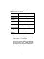
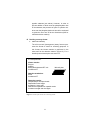
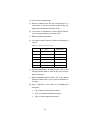
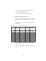
PERKINELMER LIFE AND ANALYTICAL SCIENCES
USING THE ALPHASCREENTM PHOSPHOSENSOR KIT
CATALOG NUMBERS:
6760307D, 6760307M, 6760307R
For Laboratory Use Only
Research Chemicals for Research Purposes Only
Precautions
•
AlphaScreen™ beads are light-sensitive. All assays using
the AlphaScreen beads should be performed under subdued laboratory lighting of less than 100 lux. Alternatively,
green filters (Roscolux filters #389 from Rosco, or equivalent) can be applied to light fixtures. Any incubation of
AlphaScreen™ beads should be performed in the dark.
Plates can be covered by an opaque microplate to minimize the effect of light.
•
Due to the small volumes used in the assay, it is recommended that the plates be covered with TopSeal-ATM adhesive sealing film to reduce evaporation during incubation
periods (PerkinElmer® Inc., Cat. No. 6005185). The assay
can be read with the TopSeal-A film in place.
•
The PhosphoSensor Acceptor beads contained in this kit
may slightly aggregate with time. This is normal. It is advised to vortex the beads prior to use.
•
Beads should be stored in the dark at 4oC.
2
TABLE OF CONTENTS
I.
BEFORE STARTING
5
II.
INTENDED USE
8
III.
PRINCIPLE OF THE ASSAY
8
IV.
ASSAY DEVELOPMENT
9
A. Deciding on substrate configuration
B. Deciding on assay format
C. Titration of biotinylated phosphopeptide and/or
non-phosphorylated peptide substrate
D. Evaluating optimal enzymatic assay conditions
E. Termination of the kinase reaction
F. Kinetics of the detection reaction
G. Titration of PhosphoSensor Acceptor beads
10
11
13
14
18
20
21
V.
SUMMARY OF ASSAY DEVELOPMENT
22
VI.
DETAILED PROTOCOLS
24
A. Titration of phosphorylated versus
non-phosphorylated substrate
B. All-in-one-well and transfer kinase assays
C. AlphaScreen beads quality control
24
26
29
VII.
30
TROUBLESHOOTING GUIDE
3
4
I.
BEFORE STARTING
Receiving the AlphaScreen PhosphoSensor Kit
Upon receiving the AlphaScreen PhosphoSensor Kit, ensure
that it is on blue ice and that the ice packs are not completely
melted. Verify that all components are present in the kit using
the table below.
Provided Reagents and Materials
The following kit sizes are available*:
1,000 assay points (catalog number 6760307D)
10,000 assay points (catalog number 6760307M)
50,000 assay points (catalog number 6760307R)
*The number of assay points is based on the use of 500 ng
of each bead per well.
The reagents and materials provided in the AlphaScreen PhosphoSensor Kit are listed in the Table I:
5
Table I. Reagents and materials supplied
1,000
assay
points
6760307D
10,000
assay
points
6760307M
50,000
assay
points
6760307R
PhosphoSensor
Acceptor beads
0.1 mL
Stored in 100 mM Tris-HCl (5 mg/mL)
pH 7.0, 0.05% ProclinTM-300
1.0 mL
(5 mg/mL)
5.0 mL
(5 mg/mL)
Streptavidin-Donor beads
0.1 mL
Stored in 100 mM Tris-HCl
(5 mg/mL)
pH 7.4, 0.05% Proclin-300
1.0 mL
(5 mg/mL)
5.0 mL
(5 mg/mL)
KIT
COMPONENTS
Positive control bio-LCK-P
Stored in 25 mM Hepes pH
7.4, 0.05% Proclin-300
0.05 mL
(5 mM)
0.05 mL
(5 mM)
0.05 mL
(5 mM)
10X Control buffer
100 mM MES pH 6.0, 1M
NaCl, 0.05% Proclin-300
1.5 mL
(10X)
1.5 mL
(10X)
1.5 mL
(10X)
Note before use:
•
For maximum recovery of content, briefly centrifuge
the vials prior to removing the caps and resuspend
the beads by vortexing.
•
Reagents should be stored at +2 - 8°C.
•
Acceptor and Donor beads should not be frozen
and should be stored protected from light.
•
10X buffer may not be suitable as a kinase assay
or as a detection buffer.
6
Recommended Additional Reagents and Materials
Table II. Recommended reagents and materials
Item
Suggested source
Catalog #
Kinase of choice
N/A
N/A
Biotinylated
substrate of choice
HPLC water or
equivalent
ATP
N/A
N/A
Fisher Scientific
W5-4
Sigma-AldrichTM Co.
A-3377
TM
MgCl2
Sigma-Aldrich
EDTA
GIBCO®
15575-038
Staurosporine
Sigma-AldrichTM Co.
S-4400
OptiPlate™-384
(white opaque
384-well microplate)
TopSeal-A Adhesive
Sealing Film
Tween® 20
PerkinElmer® Inc.
6007290 (pack of 50)
6007299 (pack of 200)
PerkinElmer® Inc.
6005185
Pierce
Biotechnologies
N/A
28320
Single-channel
Pipettors§
§
Co.
M-9272
N/A
For lower volume additions (2-10 µL), we recommend a pipet-
tor precision ≤ 2%. For higher volume additions (25-1000 µL), a
pipettor precision of ≤ 1% is recommended.
Assay must be read using an AlphaScreen compatible reader
such as all PerkinElmer EnVisionTM multilabel plate readers
with AlphaScreen module, Fusion-AlphaTM multilabel readers or
AlphaQuest® original AlphaScreen readers.
7
II.
INTENDED USE
The AlphaScreen PhosphoSensor Kit is intended to perform
antibody-free detection of phosphorylated protein or peptide.
III.
PRINCIPLE OF THE ASSAY
In cells, protein kinases mediate the phosphorylation of a variety of different protein substrates in the presence of ATP.
Kinases catalyze the reversible addition of phosphate molecules to tyrosine, serine and threonine residues. There are several commercially available antibodies that recognize phosphotyrosine residues with high affinity. However, such generic antibodies are not currently available for phosphoserine and phosphothreonine. The AlphaScreen PhosphoSensor Kit allows detection of the phosphorylation of tyrosine, serine, and threonine
residues without the need for such sequence specific antibody.
The principle of the assay is illustrated in Figure 1. In this assay, the kinase driven addition of a phosphate group to a biotinylated substrate will result in the simultaneous capture of the
phosphorylated substrate by the PhosphoSensor Acceptor
(coated with a Lewis Metal Chelate) and the streptavidin (SA)
Donor beads. Upon laser excitation of the Donor beads, the
proximity of the Donor and PhosphoSensor Acceptor beads will
generate an AlphaScreen signal between 520 and 620 nm. In
the absence of phosphorylation, no signal should be observed.
Since the Acceptor beads allow for the detection of phosphates, the activity of phosphatases can also be monitored using these beads.
8
Biotinylated substrate
(Peptide or protein)
ATP
kinase
ADP
1O
Emission
520-620 nm
2
LM3+
LM
3
LM
Excitation
680 nm
+
3+
LM3+
LM3+
Streptavidin
Donor Beads
Biotinylated
Phospho-substrate
LM
LM3+
LM
3
+
3+
P
PhosphoSensor
Acceptor Beads
Figure 1. Illustration of the detection of a phosphorylated peptide using the AlphaScreen PhosphoSensor Acceptor beads. Legend: LM3+ = Lewis Metal Chelate.
IV.
ASSAY DEVELOPMENT
The AlphaScreen technology has been widely used for the development of kinase assays using specific antibodies. It is important to stress that the optimal detection buffer as well as
ATP, substrate, and enzyme concentrations, which have been
determined for an antibody based AlphaScreen assay, will not
necessarily apply to an antibody-free assay using the PhosphoSensor beads. Consequently, it is strongly advised to follow
the assay development steps presented in this section.
9
A. Deciding on substrate configuration
The following guidelines should be followed when preparing
a novel biotinylated substrate to be included in a kinase
assay reaction monitored by the AlphaScreen technology.
The same substrate configuration applies for both an antibody-based and an antibody-free kinase assay development using the AlphaScreen technology.
1.
Peptide substrate
Peptide substrates should be designed such that they
possess at least 20 carbons between the biotin label
and the amino acid targeted for phosphorylation
(tyrosine, serine or threonine). For small peptides, this
can be achieved by including either a glycine stretch, or
a LC (long chain) spacer.
N-hydroxysuccinimidyl ester (NHS) or maleimide driven
chemical reaction can be used for the addition of biotin
to amino acid sequences. The NHS driven reaction will
target the secondary amine present on lysine residues
and at the N-terminal of the peptide. On the other hand,
the maleimide driven reaction will target the sulphydryl
group present in cysteine residues.
If the peptide contains many internal lysine residues,
which will be targeted by NHS, it is recommended to
have biotin integrated during peptide synthesis with the
required spacer.
2.
Protein substrate
Protein substrates can be biotinylated using either NHS
or maleimide driven reactions, in the same way as a
10
peptide substrate (see above). However, in order to
prevent addition of biotin near the phosphorylation site
of the substrate, the presence of lysine or cysteine residues near the phosphorylation site should be evaluated
to guide the use of one of the two chemistries (NHS or
maleimide driven reaction).
B. Deciding on assay format
1.
All-in-one-well assay
The all-in-one-well (homogenous) assay format represents the format of choice for screening purposes. In
this format, the kinase reaction is performed in the
same well as the detection reaction (Figure 2). For a
detailed protocol description see section VI.
In a microplate add:
Kinase reaction:
Enzyme
Inhibitor
Biotinylated substrate/ATP mix
Incubate at RT
384-well plate
Reaction termination:
EDTA
Incubate at RT
Detection reaction:
PhosphoSensor Acceptor beads
Streptavidin Donor beads
Incubate at 1 hour at RT
Read with an AlphaScreen capable reader
Incubate overnight and read again
Figure 2. Scheme of the all-in-one-well assay format.
11
2.
Transfer assay
The advantage of the transfer assay format is to dilute
potential interfering reagents that are present in the
kinase reaction before adding the detection beads. This
format can also be used for screening purposes. It is
especially useful when:
•
the enzyme is intrinsically highly phosphorylated
and therefore interferes with the detection of the
phosphorylated peptide by the PhosphoSensor
Acceptor beads;
•
the signal generated is low and high ATP concentrations are required (e.g., over 100 µM).
A scheme of the transfer assay protocol is illustrated in
Figure 3 (for a detailed protocol description see section
VI).
Kinase reaction:
In a 96-well microplate add:
• Biotinylated-substrate
• Enzyme
• ATP
• Incubate at RT
• Add EDTA to stop the reaction
96-well plate
Dilution of reaction mix:
Dilute the reaction mix 1/20
Detection reaction:
In a microplate add:
• Diluted mix
• PhosphoSensor Acceptor beads
• Streptavidin Donor beads
Incubate 1 hour at RT
Read with AlphaScreen capable reader
Figure 3. Scheme of the transfer assay format.
12
384-well plate
C. Titration of biotinylated phosphopeptide and/or nonphosphorylated peptide substrate
To ease the optimization of an antibody-free kinase assay
using the AlphaScreen PhosphoSensor Acceptor beads,
we recommend obtaining the phosphorylated version of the
peptide substrate. Titration of this positive control in parallel
with the biotinylated substrate will allow evaluation of the
expected signal window. It should be noted, however, that
the availability of a phosphorylated peptide is not essential
to develop a kinase assay using the AlphaScreen PhosphoSensor Kit.
For peptide titration assays, the biotinylated peptide (as
well as the phosphopeptide, if available) should be added
to the plate diluted in the kinase reaction buffer, whereas
the beads (both Acceptor and Donor) should be added to
the plate in the detection buffer.
For the majority of biotinylated peptides tested, the standard detection buffer composition is:
10 mM Tris‑HCl pH 7.0, 100 mM NaCl, 0.1% Tween-20.
However, optimization of the detection buffer must be performed when observing high non-specific binding of the
biotinylated substrate. Ionic strength, nature of the buffer,
and pH of the detection reaction can influence the nonspecific binding of some peptide sequence.
Note 1: Do not use PBS since it contains phosphate,
which will bind to the PhosphoSensor Acceptor
beads
and
displace
the
phosphorylated
substrate.
Note 2: Be aware that the presence of ATP in the detection reaction may alleviate the non-specific bind-
13
ing of the substrate (see section IV-D). Thus, we do
not use plus or minus ATP as an indication of the
assay window.
D. Evaluating optimal enzymatic assay conditions
The following section presents the steps necessary to determine the optimal ATP, substrate, and enzyme concentrations in order to obtain an optimal signal window. The optimal signal window is defined here as the optimum S/B ratio
measured between the background signal (basal signal
obtained in the presence of staurosporine; see note below)
and the maximal enzymatic activity.
Note 3: The non-specific binding of some biotinylated
peptides will be strongly diminished by the
presence, in the detection reaction, of ATP and/
or some enzymes that are themselves phosphorylated for activation. In these cases, control incubations lacking either ATP or enzyme
as a reference for background could be misleading when evaluating assay window (S/B
values). For such “sticky” peptides, it is advisable to determine assay window using control
incubations containing all the reaction components (i.e., enzyme, substrate and ATP) in the
presence or absence of a generic protein
kinase inhibitor such as staurosporine.
Note 4: We do not recommend terminating the kinase
reaction using EDTA at this stage of assay development. Since EDTA interferes to some extent with the detection by the PhosphoSensor
Acceptor beads, it should be integrated later
during assay development (see section IV-E).
14
The following example shows preliminary assay development using the commercially available protein kinase A
(PKA) and the biotinylated substrate kemptide. All assays
were performed at room temperature (RT), in white,
opaque 384‑well microplates, in a final volume of 26 µL
using 2 µL of enzyme, 2 µL of biotinylated peptide, 2 µL of
ATP, 10 µL of PhosphoSensor Acceptor beads and 10 µL
of Streptavidin Donor beads (both acceptor and donor
beads were used at a final concentration of 20 µg/mL).
1.
ATP/ substrate titration
The first optimization step consists of titrating both the
substrate and ATP concentrations. We recommend the
matrix depicted in Table III, which uses both fixed enzyme (3 nM) and beads concentrations (20 µg/mL).
The assay window should be determined by performing
the assay in the absence or presence of staurosporine.
Table III. Scheme of preliminary kinase assay development. For preliminary assay
conditions, perform the titration of ATP and substrate using 3 nM of enzyme. The assay
should be performed in the absence and presence of 10 µM of a generic inhibitor (such
as staurosporine) with at least one concentration of substrate to evaluate assay background.
With 10 µM
staurosporine)
Substrate (µM)
ATP
(µM)
0
0.3
1
3
3
3
(3,0)
(3, 0.3)
(3, 1)
(3, 3)
(3, 3)
10
(10, 0)
(10, 0.3)
(10, 1)
(10, 3)
(10, 3)
30
(30, 0)
(30, 0.3)
(30, 1)
(30, 3)
(30, 3)
Microplates were read after a detection time of either
1 hour (Figure 4A) or 17 hours (overnight incubation;
15
Figure 4B). After 1 hour of incubation, it was observed
that substrate concentrations higher than 0.3 µM did
not improve the signal window. A decrease of the signal window was observed for ATP concentrations
higher than 3 µM, due to ATP interference. Although
similar results were obtained following an incubation
period of 17 hours, a greatly improved signal window
was observed. Thus, for both incubation periods tested,
the optimal signal window was obtained when using
0.3 µM of biotinylated substrate and 3 µM of ATP. Under these conditions, S/B values of approximately 6
and 25 were observed following 1 hour and 17 hours
detection time, respectively
A
AlphaScreen Signal
(counts)
30000
[biotin-kemptide] (µM)
0
0.3
1
3
25000
20000
15000
10000
5000
0
0
3
10
30
[ATP] (µM)
B
[biotin-kemptide] (µM)
AlphaScreen Signal
(counts)
200000
0
0.3
1
3
175000
150000
125000
100000
75000
50000
25000
0
0
3
10
30
[ATP] (µM)
Figure 4. Scheme of the preliminary kinase assay optimization. Kinase reaction was
performed in kinase reaction buffer (25 mM Hepes pH 7.4, 100 mM NaCl, 2.5 mM
MgCl2, 1 mM DTT, and 0.01% Tween-20) and beads were added in detection buffer
(100 mM Tris pH 7.0, 100 mM NaCl, and 0.1% Tween-20). Incubations (25 µL) were
conducted in 384-well microplates. Detection time was A) 1hour and B) 17 hours
16
2.
Enzyme titration
The second optimization step consists of enzyme titration, using the optimal substrate and ATP concentrations determined previously.
As observed in Figure 5, increasing the concentration
of enzyme up to approximately 0.3 nM led to a proportional signal increase. A S/B ratio of approximately 31
was obtained when using 0.3 nM of enzyme. Above
this enzyme concentration, a signal decrease was observed, which may reflect either 1) saturation of both
beads (Acceptor and Donor) by an excess of phosphorylated product or 2) competition of biotinylated
product binding to the PhosphoSensor Acceptor beads
by the kinase itself. As a matter of fact, some kinases
are phosphorylated for activation.
AlphaScreen Signal
(counts)
70000
without staurosporine
with staurosporine
60000
50000
40000
30000
20000
10000
0
∞ -11
-10
-9
-8
-7
-6
log [PKA] (M)
Figure 5. Enzyme titration using optimal concentrations of ATP
and substrate. PKA was titrated using 3 µM of ATP and 0.3 µM of
biotinylated substrate; a detection time of 1 hour was used.
3.
Transfer assay format
In the transfer assay format, a bulk kinase reaction is
performed; subsequently, the reaction mix is diluted
before adding the AlphaScreen beads. In this format,
the following concentrations of ATP, enzyme and bioti-
17
nylated substrate are suggested as a starting point:
100 µM, 30 nM, and 10 µM, respectively.
After incubation of the kinase reaction, the mix is diluted in order to obtain final concentrations of biotinylated substrate between 0 and 1 µM in the detection
reaction. The detection reaction using the Acceptor and
Donor beads is then conducted as for the all-in-onewell assay format (see section VI-B).
E. Termination of the kinase reaction
EDTA is a commonly used chelator for termination of
kinase reactions. However, excessive concentrations of
EDTA should be avoided when performing a detection using the PhosphoSensor Acceptor beads.
It is recommended to perform all kinase assay development
using 2.5 mM MgCl2 and then to determine the optimal concentration of EDTA required to terminate the enzymatic
reaction. If necessary, titration of MgCl2 can also be performed before EDTA titration.
Figure 6 shows the effect of increasing concentrations of
EDTA on the PKA kinase reaction. In this assay EDTA was
added either before starting the kinase reaction or 2 hours
following the initiation of the reaction. Detection times of 1
and 17 hours were compared (Figure 6A-B). To confirm the
specificity of the reaction, each incubation was performed
in the presence or absence of a generic kinase inhibitor
(10 µM staurosporine) (data not shown). It was determined
that 5 mM EDTA is sufficient to completely stop the kinase
reaction, while leaving an acceptable signal window (S/B
ratio of 13). Overnight incubation of the detection reaction
clearly results in an improved S/B ratio (Figure 6B).
18
AlphaScreen Signal
(counts)
A
50000
EDTA added at the beginning
40000
EDTA added at the end
30000
20000
10000
0
0.0
2.5
5.0
12.5
25.0
[EDTA] (mM)
AlphaScreen Signal
(counts)
B
1400000
EDTA added at the beginning
1200000
EDTA added at the end
1000000
800000
600000
400000
200000
0
0.0
2.5
5.0
12.5
25.0
[EDTA] (mM)
Figure 6. Titration of EDTA. Following the addition of EDTA, a detection time of 1 hour (A) or 17 hours (B) was used.
If no signal window is obtainable when using EDTA, it is
recommended to either use the transfer assay to dilute
EDTA before reading, or to add staurosporine at 1-10 µM to
terminate the enzymatic reaction.
19
F. Kinetics of the detection reaction
Phosphopeptide detection using the PhosphoSensor Acceptor beads involves lower binding affinities than an antibody-based detection method. Thus, following termination
of the kinase activity, it is recommended to incubate the
detection reaction overnight to obtain the optimal signal
window.
Figure 7 depicts the kinetics of the detection reaction following termination of PKA activity with 5 mM of EDTA. A
proportional increase in the signal window was observed as
a function of time.
AlphaScreen Signal
(counts)
1500000
with staurosporine
without staurosporine
1250000
1000000
750000
500000
250000
0
0
10
20
30
40
50
60
time (h)
Figure 7. Kinetics of the detection reaction using the PhosphoSensor
Acceptor beads to perform antibody-free kinase activity detection. The
signal to background ratio is defined with or without 10 µM staurosporine.
Note 5: The time course of the detection will vary for
other peptide or protein substrates.
20
G. Titration of PhosphoSensor Acceptor beads
In general, it is recommended to use a final concentration
20 µg/mL of AlphaScreen beads per reaction. However, for
kinases which have affinities for ATP in the high micromolar
range, tolerance of the PhosphoSensor Acceptor beads to
ATP can be improved by increasing their concentration in
the detection reaction. The following table shows an example using phosphorylation of the biotinylated–crosstide peptide substrate by MSK-1 (Table III). When the concentration
of beads was increased to 50 µg/mL, more than two-fold
increase in S/B was observed.
Table III. Effect of Acceptor beads concentration on tolerance to ATP. In this reaction, 0.3 µM of biotinylated crosstide was phosphoryated by 3 nM of MSK-1 in the
presence of different concentration of ATP. Enzymatic reactions were allowed to proceed for 2 hours in the absence (max) or presence (min) of staurosporine. Detection was
performed using different bead concentrations, as indicated.
[PhosphoSensor Acceptor beads]
20 µg/mL
[ATP]
(µM)
0
10
30
100
min
21395
7897
6348
6315
max
43965
110645
54727
23108
50 µg/mL
S/B
2.1
14
9
4
21
min
262727
26400
19214
15102
max
227959
970873
447904
113449
S/B
1
37
23
8
V.
SUMMARY OF ASSAY DEVELOPMENT
Figure 9 illustrates the different steps that should be undertaken in the development of a kinase assay using the
AlphaScreen PhosphoSensor Kit.
In summary, the development of a kinase assay involves the
following steps:
A. Identify substrate and perform the appropriate biotinylation
(see section IV-A).
B. Perform titration of the biotinylated substrate together with
the phosphorylated version of the substrate (if available) to
evaluate the expected signal window (see section IV-C).
C1. If a high background signal is observed due to non-specific
binding of the biotinylated substrate, optimize the detection
buffer.
Note 6: Be aware that the presence of ATP in the detection
reaction may alleviate the non-specific binding of
the substrate (see section IV-D). Thus, we do not
use plus or minus ATP as an indication of the assay window.
C2. If the peptide titration assay generates a specific signal
window, continue with the kinase assay development using an
all-in-one-well assay format (see sections IV-D, E, and F).
D. Titrate substrate, ATP and enzyme using an all-in-one-well
kinase assay format. If a specific signal is observed in the
kinase assay, proceed with assay optimization (see sections
IV-D, E, and F).
22
E. If no specific signal is observed in the kinase assay, perform
the assay in a transfer format to eliminate possible interferences with the detection reaction.
A. Choose substrate
biotinylation strategy
Perform substrate
biotinylation
B. Titrate of substrate
and phoshorylated
substrate (if available)
C1. Specific window with
high
non-specific background
C2. Specific window
observed
with low non-specific
background
Determine optimal buffer
to reduce non-specific
binding
D. Titrate kinase
reagents using all-in-onewell format
Signal generated
No signal generated
Determine MgCl2
requirements
E. Perform transfer assay
to eliminate possible
interferences
Include EDTA to terminate
kinase activity
Determine optimal
detection time
Figure 9. Scheme of assay development using the AlphaScreen PhosphoSensor Kit.
23
VI.
DETAILED PROTOCOLS
A. Titration of phosphorylated versus nonphosphorylated substrate
Protocol suggested for optimizing buffer conditions using
phosphorylated and non-phosphorylated substrates (see
section III-C): the following protocol will allow the titration of
both the phosphorylated and non-phosphorylated peptides.
For performing more than two titration curves, increase the
volume of each reagent.
1. Prepare reaction buffer
2. Prepare detection buffer: 10 mM Tris pH 7.0, 100 mM
NaCl, 0.1% Tween-20.
3.
Prepare biotin-non-phospho and biotin-phospho sub-
strate by making serial dilutions in kinase reaction buffer
supplemented with ATP and EDTA.
Note 7: Supplementation with ATP and EDTA is necessary to mimic closely the conditions that will be
observed in an actual kinase assay.
Table IV shows an example of peptide dilution using a peptide stock at 50 µM.
24
Table IV. Example of peptide dilution using a peptide stock at 50 µmol/L
Dilution
[final in
assay]
(mol/L)
[intermediate ]
(mol/L)
1
1 X 10⁻⁶
5.0 X 10⁻⁶
-7
-6
2
3
4
5
6
7
8
9
10
11
12
3 X 10
1 X 10⁻⁷
3 X 10⁻⁸
1 X 10⁻⁸
3 X 10⁻⁹
1 X 10⁻⁹
3 X 10⁻¹⁰
1 X 10⁻¹⁰
3 X 10-11
1 X 10-11
-
1.5 X 10
5.0 X 10⁻⁷
1.5 X 10⁻⁷
5.0 X 10⁻⁸
1.5 X 10⁻⁸
5.0 X 10⁻⁹
1.5 X 10⁻⁹
5.0 X 10⁻¹⁰
1.5 X 10⁻¹⁰
5.0 X 10⁻¹¹
-
Volume of
dilution
(µL)
20 of 50
µmol/L
60 of dil 1
60 of dil 2
60 of dil 3
60 of dil 4
60 of dil 5
60 of dil 6
60 of dil 7
60 of dil 8
60 of dil 9
60 of dil 10
-
Kinase reaction buffer
(µL)
180
140
120
140
120
140
120
140
120
140
120
100
4. Dilute the PhosphoSensor Acceptor beads 1/100 in the
detection buffer
5. Dilute the Streptavidin Donor beads 1/100 in the detection buffer
6. Protocol of addition
The assay is performed in triplicates in a white OptiPlate-384 in a total volume of 25 µL:
a) Add 5 µL of the biotinylated peptide dilution
b) Add 10 µL of PhosphoSensor Acceptor beads dilution
(20 µg/mL final concentration)
c) Add 10 µL of Streptavidin Donor beads dilution
(20 µg/mL final concentration)
d) Incubate in the dark for 1 hour at room temperature
e) Read on an AlphaScreen reader
25
B. All-in-one-well and transfer kinase assays
These assays are divided into three major steps:
Kinase reaction
It is recommended but not always necessary to perform the
kinase reaction in the smallest volume possible (e.g. 6 µL)
to allow for dilution of potential interferences during the detection reaction.
Termination of kinase activity
Before adding the detection beads, it is recommended to
terminate the kinase reaction by the addition of EDTA
(diluted in the optimal detection buffer). For the concentration to use in the assay, see section IV-E.
If the signal window is too low using EDTA, it is recommended to use a generic inhibitor such as staurosporine to
terminate the enzymatic reaction. A titration should be performed to determine the optimal staurosporine concentration.
Detection reaction
Beads are added following the inactivation of the kinase. It
is recommended to use an overnight detection to increase
the signal window. This will not affect the pharmacological
parameters of the assay since the enzyme has been inactivated before the final detection step. For detection buffer
composition, see section IV-C.
Note 8: Acceptor beads can be titrated to increase tolerance to ATP (See Section IV-G).
26
1. Prepare reaction buffer as suggested by the provider of
the enzyme.
2. Prepare detection buffer: 10 mM Tris pH 7.0, 100 mM
NaCl, 0.1% Tween-20.
3. Protocol of addition for the all-in-one-well assay
The assay is performed in triplicates in a white OptiPlate-384 in a total volume of 28 µL:
a) Add 2 µL of enzyme diluted in the kinase reaction
buffer
b) Add 2 µL of inhibitor or buffer diluted in the kinase reaction buffer
c) Add 2 µL of biotinylated substrate/ATP dilution mix diluted in the kinase reaction buffer
d) Incubate 2 hours (kinase dependent) at room temperature
e) Add 2 µL of EDTA diluted in the optimal detection
buffer
f)
Add 10 µL of PhosphoSensor Acceptor beads diluted
1/90 in the optimal detection buffer
g) Add 10 µL of Streptavidin Donor beads diluted 1/90 in
the optimal detection buffer
h) Incubate in the dark for 1 hr at room temperature
i)
Read on an AlphaScreen reader
j)
Incubate overnight in the dark and read again.
Note 9: Detection reaction should read after an overnight incubation for optimal results.
27
4. Protocol for the transfer assay
a) Enzyme, substrate and ATP are mixed together in a
total volume of 100 µL of kinase reaction buffer (for
reagents concentrations see section IV-D)
b) The mixture is incubated for 2 hours (kinase dependent) at room temperature (see section IV-E)
c) EDTA is added to the mixture
d) The kinase reaction mixture is diluted as described in
Table V
Table V. Dilution of the kinase mixture
1
Volume of dilution
(µL)
reaction mix
Kinase reaction
buffer (µL)
-
2
30 of reaction mix
70
3
30 of dilution 2
60
4
30 of dilution 3
70
5
30 of dilution 4
60
6
-
100
Dilution
e) Dilute PhosphoSensor Acceptor beads 1/100 in the
optimal detection buffer in order to get a final concentration of 20 ug/mL
f)
Dilute Streptavidin Donor beads 1/100 in the optimal
detection bufferin order to get a final concentration of
20 ug/mL
g) Add in triplicates to the wells of an Optiplate-384
microplate:
•
5 µL of kinase reaction dilution (step d)
•
10 µL of Acceptor beads dilution (step e)
•
10 µL of Donor beads dilution (step f)
28
h) Incubate in the dark for 1 hour at room temperature
i)
Read on an AlphaScreen reader
j)
Incubate overnight in the dark and read again
C. AlphaScreen beads quality control
1. Prepare 1X control buffer: dilute 1.5 mL of 10X control
buffer with 13.35 mL of H2O and add 0.15 mL Tween-20
10%.
2. Prepare serial dilutions of the kit’s positive control at
5µM (Table VI):
Table VI: Kit’s positive control probe dilution using a peptide stock at 5 µmol/L
Dilution
[final in
assay]
(mol/L)
[intermediate ]
(mol/L)
1
1 X 10⁻⁷
5.0 X 10⁻⁷
2
3
4
5
6
7
8
9
10
3 X 10⁻⁸
1 X 10⁻⁸
3 X 10⁻⁹
1 X 10⁻⁹
3 X 10⁻¹⁰
1 X 10⁻¹⁰
3 X 10-11
1 X 10-11
-
1.5 X 10⁻⁷
5.0 X 10⁻⁸
1.5 X 10⁻⁸
5.0 X 10⁻⁹
1.5 X 10⁻⁹
5.0 X 10⁻¹⁰
1.5 X 10⁻¹⁰
5.0 X 10⁻¹¹
0.00
Volume of
dilution
(µL)
20 of 5
µmol/L
60 of dil 1
60 of dil 2
60 of dil 3
60 of dil 4
60 of dil 5
60 of dil 6
60 of dil 7
60 of dil 8
0
1X control
buffer
(µL)
180
140
120
140
120
140
120
140
120
200
3. Dilute the PhosphoSensor Acceptor beads 1/100 in the
1X control buffer
4. Dilute the Streptavidin Donor beads 1/100 in 1X control
buffer
29
5. Protocol of addition:
The assay is performed in triplicates in a white OptiPlate-384 in a total volume of 25 µL
a) Add 5 µL of control phosphopeptide
b) Add 10 µL of PhosphoSensor Acceptor beads dilution
(20 µg/mL final concentration)
c) Add 10 µL of Streptavidin Donor beads dilution
(20 µg/mL final concentration)
d) Incubate in the dark for 1 hour at room temperature
e) Read on an AlphaScreen reader
h) Incubate overnight in the dark and read again.
Note 10: The 10X detection buffer included in the kit
may not be suitable for the detection of all
phosphorylated peptides and should only be
used for quality control of the beads.
Expected results: maximum signal should be reached at
30 nM of control probe with an EC50 between 1.5 to 15 nM.
The absolute maximum counts generated will be dependent on the instrument used for the readout.
VII.
TROUBLESHOOTING GUIDE
The following section describes the possible problems which
could be encountered when developing an antibody-free kinase
assay using the AlphaScreen Technology. If more information
is required, please consult your local PerkinElmer technical
support division (see page 34 for customer support information).
30
Problem
No signal
Possible Cause
Detection
conditions
Effect/Remedy
Interference of EDTA with the
assay / since EDTA is used to
chelate the Mg ions, titrate MgCl2 to
limit the concentration of EDTA
added for chelation. Add a small
volume of EDTA before adding the
larger volumes of beads.
Stop the kinase reaction using a
generic inhibitor such as
staurosporine (1-10 µM)
ATP interferes with the detection
1) Increase the concentration of
Acceptor beads to increase the
tolerance of the assay to ATP.
2) Perform the assay in a transfer
assay.
3) Perform kinase assay in smaller
volume and/or detection assay in
large volume to increase the dilution
of ATP before the detection.
Kinase assay
conditions
Perform the assay in a transfer
format to evaluate if it is due to
interference of ATP and/or enzyme.
Instrument/plates
Incompatible microplate choice / use
solid opaque white plates such as
PerkinElmer Optiplates.
Ensure that your reader contains an
AlphaScreen reading mode.
Kinase assay
reagents
No or improper biotinylation of
substrate peptide or protein / check
extent of biotinylation using the
AlphaScreen TruHitTM kit (Cat. No
6760627).
31
Problem
No signal
Possible Cause
Kinase assay
reagents
Effect/Remedy
Verify that the distance between the
phosphorylated amino acid and the
biotin is at least 20 carbons.
Protein contains lysine residues near
the phosphorylation site / target
cysteines for biotinylation using
maleimide driven coupling reaction.
Cofactor required for optimal
enzymatic activity / add the cofactor in
the kinase reaction buffer.
Peptide substrate not sufficient for
efficient phosphorylation by the
enzyme / use the full length or a
longer domain as substrate.
Reagents degradation / perform the
assay with fresh enzyme and/or
substrate.
High
background
signal
Detection
conditions
Non-phosphorylated peptide is binding
non-specifically to the PhosphoSensor
Acceptor beads / test different buffer
conditions to reduce non-specific
binding by varying:
• pH (6 to 8)
• NaCl from 0 to 400 mM
• Tween-20 from 0 to 0.1%
and by evaluating Tris-HCl, HEPES or
MES as potential buffers.
Kinase assay
reagents
Ensure that the signal to background
ratio is established in the presence
and absence of staurosporine, since
the presence of ATP and/or enzyme in
the reaction could alleviate the
substrate non-specific binding to the
PhosphoSensor Acceptor beads.
32
Problem
High
background
signal
Possible Cause
Kinase assay
reagents
High degree Microplates
of signal
variability
Effect/Remedy
When using a full-length kinase as a
substrate, ensure that it is not
activated by phosphorylation. Only
use non-activated kinase as a
substrate.
Warped or distorted microplates /
avoid storage of microplate under
heavy objects or next to sources of
heat.
Uneven plate molding.
Light penetrating edges of
microplate / ensure use of black
cover plate during bead incubation.
Incubate microplate in dark
environment such as inside a drawer
or cover microplate entirely with foil
or material impenetrable to light.
Poorly fitted plate seals inducing
evaporation of reaction mixture.
Instrument
Temperature control problem within
the instrument / for the EnVision
readers using the 1.07 software
version, adjust the internal
temperature of the instrument. For
other readers, consult the technical
service department.
Assay conditions
Beads are interacting together /
Avoid premixing the Acceptor and
Donor beads since signal will
decrease substantially following 15
minutes of pre-incubation.
Day-to-day
variability
Inappropriate standard operation
procedures / ensure that
experimental procedures are the
same from day to day: 1) prepare
the beads in the same area, 2)
ensure that incubation times are
constant and temperature does not
fluctuate in the room.
33
MANUFACTURED BY:
PerkinElmer BioSignal, Inc.
1744, William Street
Montreal, Quebec Canada H3J 1R4
For further technical information or to place an order, call:
PerkinElmer LAS, Inc.
710 Bridgeport Avenue
Shelton, CT 06484
USA
800-762-4000 or 203-925-4600
[email protected]
European Headquarters:
PerkinElmer LAS, Inc.
Imperiastraat 8
BE-1930 Zaventem
Belgium
[email protected]
Country
Austria
Belgium
Denmark
France
Germany
Italy
Netherlands
Norway
Spain
Sweden
Switzerland
United Kingdom
Telephone
0800 293 515
0800 94 540
80 88 3477
0800 90 77 62
0800 1 81 00 32
0800 79 03 10
0800 02 23 042
800 11 947
900 973 255
020 79 07 35
0800 55 50 27
0800 89 60 46
34
35
PerkinElmer Life and Analytical Sciences, Inc.
710 Bridgeport Avenue
Shelton, CT 06484-4794 USA
(800) 762-4000 or (+1) 203-925-4602
www.perkinelmer.com
For a complete listing of our global offices, visit www.perkinelmer.com/lasoffices
M-6760307-01
36