1
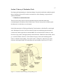
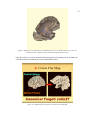
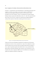
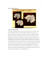
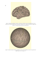
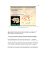
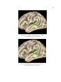
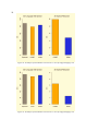
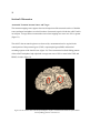
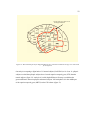
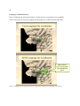
20
2.2 Surface flattening tools
The complex geometry of the human brain contains many folds and fissures making it impossible
to view the entire surface at once. Since most of the cortical activity occurs on these folds, it is
desirable to view the entire surface of the brain in a single view. This can be achieved using flat
maps of the cortical surface, which are essentially unwrapped cortical surfaces in a 2D plane (Van
Essen et al., 2001). Cortical flat maps also make it easier to see the depths and complete shape of
the sulci. Algorithms for creating flat maps do require cutting, compression and stretching of the
surface, causing some distortion. All cortical flattening methods aim to minimize geometric
distortion. We considered the following tools for creating cortical flat maps in this case study:
•
Computerized Anatomical Reconstruction and Editing Tool Kit (CARET)
(http://brainmap.wustl.edu/caret)
•
mrUnfold-5.0 (http://white.stanford.edu/~brian/mri/segmentUnfold.htm)
•
BrainVoyager (http://www.brainvoyager.de)
•
FreeSurfer (http://surfer.nmr.mgh.harvard.edu)
We selected Caret to flatten surfaces for two reasons. First, SureFit was selected for image
segmentation and is distributed and supported by the same lab as Caret. Thus, SureFit is designed
to interface seamlessly. In fact, work is underway to incorporate SureFit into the Caret software
suite. Secondly, since we selected Caret as one of the spatial normalization methods, using the
same software suite for flattening made for a streamlined evaluation protocol.
2.3 Target Atlases
Ideally a target atlas will not bias the final solution. An ideal template would consist of the
average of a large number of MR images that have been registered to within the accuracy of the
spatial normalization technique (Ashburner and Friston, 2000).
Talairach
Jean Talairach and Pierre Tournoux created the now famous book, Co-Planar Stereotaxic Atlas
of the Human Brain, in 1988. Talairach and Tournoux dissected and photographed a post
mortem brain of a 60 year-old female subject, creating a proportional coordinate system often
referred to as “Talairach space” for neurosurgical studies. This atlas was widely used in
international brain imaging studies and continues today to be the most widely used human brain