1
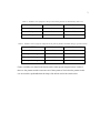
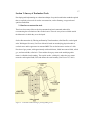
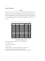
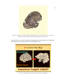
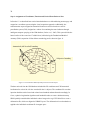
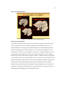
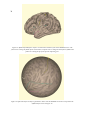
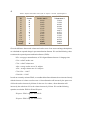
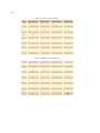
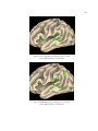
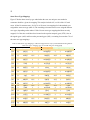
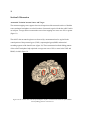
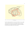
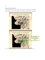
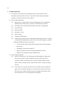
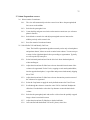
28
Section 3: Methods
Subjects
The subjects were 11 patients (5 female, 6 male, age range 23-52 years) undergoing resection
treatment at the University of Washington Medical Center for chronic epilepsy (n = 11). Seven
patients were right handed. All cortical stimulation occurred in the subject’s left hemisphere,
which was identified as the subject’s language-dominant hemisphere in all subjects determined by
pre-surgery WADA testing (Corina et al., 2005). Subject demographics are summarized in
table 3.
Table 3. Subjects’ gender, age, handedness and verbal IQ (VIQ)
BrainID
54
55
58
60
61
62
63
117
164
170
176
Gender
M
M
M
M
F
F
M
F
M
F
F
Age
25
30
23
38
35
24
42
41
42
52
41
Handed
ness
R
R
R
R
R
R
R
VIQ
107
83
86
72
91
92
125
97
94
75
82
Evaluation technique protocol
To test our hypothesis we developed a six-step evaluation protocol:
1: select MRI volumes
2: create surface reconstruction
3: create flat map
4: assign coordinates, function and cortical parcellation to each CSM site
5: apply spatial normalization to anatomical and functional data
6: evaluate methods using spread reduction and anatomical localization measures