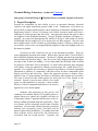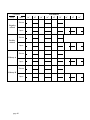
1
Manual for Chemical Biology Laboratory
Chemistry 427b
Spring 2005
Lab Manual Contents
1.
2.
3.
4.
5.
6.
7.
8.
Course Description (page 2)
Semester Schedule (page 2)
Course Materials (page 3)
Policies (page 4)
Lab Faculty, Staff, and Hours (page 5)
Lab Safety and Waste Management (page 6)
Material Safety Data Sheets (page 9)
Writing in your Lab Notebook (page 9)
General Guidelines (page 10)
Before Lab: Experimental Plan (page 10)
During Lab: Recording experiments and data (page 11)
9. Grading Information (page 12)
10. Guidelines for Final Report (page 13)
11. The Research Projects (page 14)
Sub-group 1: The mini protein specificity project (page 16)
Sub-group 2: The b-peptide library project (page 19)
Sub-group 3: The transcription cell culture project (page 22)
12. Keeping up with the Literature (page 26)
13. Protocols for Chemical Biology Laboratory Techniques (page 27)
Affinity chromatography of GST fusion proteins (page 28)
Agarose gel electrophoresis (page 34)
Bacterial expression of miniature proteins (page 39)
Centrifuge usage (page 44)
Circular dichroism (page 47)
Cloning basics (page 51)
DNA technical information (page 62)
Fluorescence background and techniques (page 64)
Fluorophore labeling of miniature proteins (page 67)
HPLC purification (page 71)
Mass spectrometry (department MALDI-TOF instrument) (page 77)
Sample preparation for amino acid analysis (page 94)
Using the lyophilizer (page 97)
Using the Phastsystem (page 99)
Using the speed-vac (page 104)
Ultra-violet visible spectroscopy (page 106)
Western blots (page 114)
Cell Culture Protocols (page 119)
14. Useful Internet Links (page 130)
page 1
1. Course Description (return to Contents)
The goal of Chemical Biology Laboratory is to involve undergraduates enrolled in
Chemical Biology I in the challenge and excitement of independent discovery at an early
stage of their Yale experience, long before such research opportunities would usually be
available to them. Each student will have an individual project that is a sub-project of a
major funded investigation currently underway in graduate research laboratories at Yale.
Since the projects in this course represent novel research, they differ from those in a
traditional laboratory course which tend to have pre-determined outcomes. Students in
Chemical Biology I will share the excitement—and perhaps the frustration—of hands-on
experience with original research.
Sub-groups of four students working on related projects will be guided by a TA who is
working on related research in his or her own lab. This feature fosters independence and
communication, since the students within a particular sub-group will be working on
individual problems but may encounter similar challenges. There will be frequent minimeetings of the research sub-groups for troubleshooting and discussion of results. The
schedule is designed so that significant milestones can be reached by the end of each
semester.
As students progress through the semester, they will gain experience performing a variety
of indispensable laboratory techniques while they gain exposure to research methods. By
the end of the semester, students will be expected to analyze their results and propose
logically related future experiments.
2. Semester Schedule (return to Contents)
The laboratory classroom is SCL 168, with sub-groups meeting from 1 – 5:00 p.m. (124:00 pm on Wednesday) on two days of the week. A 30-40-minute lecture will also be
given by the course instructor once a week in a classroom in SCL. All Chemical Biology
Laboratory students are encouraged (though not required) to attend the Schepartz
laboratory group seminars, which are held at 3 p.m. in SCL 201 (the Faculty Lounge).
Note: Each sub-group will have distinct schedule guidelines for experimental progress.
Because of the inherently open-ended, results-driven nature of research, it is impossible
to provide an exact script of the semester! This is part of the excitement of research, but it
also requires students to maintain focus and look to their TA for direction as necessary.
Other than how experimental time is organized, the rest of the schedule is identical for all
sub-groups. Every student is responsible for attending all scheduled laboratory and
lecture periods. If an illness, injury, or family emergency prevents you from fulfilling this
obligation, please email the instructor and your TA before the absence.
page 2
3. Course Materials (return to Contents)
Lab text:
This lab manual serves as the guiding text for the course. Each sub-group description will
contain references to relevant techniques and background journal articles from the
literature. There are also several recommended textbooks to provide background
information for many of the techniques and biological processes with which you will
become acquainted during this course.
Recommended Textbooks:
Nucleic Acids in Chemistry and Biology
by G. Michael Blackburn (Editor), Michael G. Gait, Michael J. Gait (Editor); Irl Pr;
2nd edition (December 1996), ISBN: 0199635331
Introduction to Protein Structure
by Carl-Ivan Branden, John Tooze; Garland Publishing; 2nd edition (January 15,
1999), ISBN: 0815323050.
Proteins: Structures and Molecular Properties
by Thomas E. Creighton; W. H. Freeman & Co.; 2nd edition (1993), ISBN:
071677030X.
Structure and Mechanism in Protein Science: A Guide to Enzyme Catalysis and
Protein Folding
by Alan Fersht; W H Freeman & Co.; (January 1999), ISBN: 0716732688.
Recommended Journal Articles:
In addition to the textbooks listed above, each sub-group has a list of relevant journal
articles for background and supplementary reading. Most of these are available online
for free to Yale IP addresses.
Click on the link below for the list of literature specific to your sub-group project:
Sub-group 1 literature references
Sub-group 2 literature references
Sub-group 3 literature references
*Section 13 below contains links to information about the protocols and laboratory
techniques you will be using.
Laboratory notebook: Available at the Yale bookstore, or you may use a lab notebook
begun in a previous course. The notebook cannot be spiral or loose-leaf! A copy of your
page 3
entire notebook must be left with your TA at the end of the semester either by using a
notebook that contains carbon copies, or by making photocopies at the end of the
semester
Calculator: You will need a reliable scientific calculator for calculation of yields,
concentrations, molarity conversions, etc. To prevent loss, please label it with your name.
Lab safety glasses or goggles: Eyewear will be provided. Students who wear glasses
should wear goggles or safety glasses over them.
Lab coat: This is suggested but optional for all subgroups except for subgroup 3 who
will be working in a cell culture facility. The other two subgroups may instead opt to
wear durable, inexpensive clothing that you don’t mind staining.
4. Policies (return to Contents)
Attendance and Tardiness
It is critical that you attend all scheduled lab periods. If you must miss a
scheduled lab period due to illness, injury, family emergency, religious
observance, or sports team travel, you are required to notify the course
instructor and your lab TA in advance of the absence. You are responsible to
make arrangements for making up missed work and submitting your
assignments.
Punctuality is a virtue. Late arrivals are disruptive to others and may
compromise your safety if you miss instructions from the TA. Be on time,
unless tardiness is unavoidable – in which case you must contact the course
instructor and your TA in advance to make arrangements.
Exams and Grading
For more information on grading policies, refer to Section 9.
Students will be graded on their understanding and performance of the techniques that are
entailed in the class, the quality of their experimental design, laboratory notebooks,
experimental data, and their ability to draw conclusions from results and place them in a
scientific context. There will be four quizzes on material presented in the Friday lectures,
a written proposal for future experiments, and a final report in the format of a short
Journal of the American Chemical Society article for the final exam.
Breakdown:
20% 4 quizzes on lecture material
10% Pre-lab assignments
40% Laboratory notebooks
10% Proposal for future experiments
page 4
20% Final report (format of short JACS article)
Academic Honesty
Honor Code
As an enrollee in this chemical biology laboratory course, I agree to work independently unless I
am specifically instructed to work with a partner. I will not copy another student’s work on any of
the assignments or quizzes. I will not allow another student to copy my work on any of the
assignments or quizzes. In my laboratory notebook, I will record all data with honesty and submit
only my own work, unless I have clearly credited another student for contributing data in
collaboration.
5. Lab Faculty, Staff, and Hours (return to Contents)
Abby Maranda: course instructor and administrator
Phone: 432-3984, email [email protected]
Office: KCL 110
Office Hours: Monday 10-12
Teaching Assistants
Sub-group 1: Kamil Woroniwicz
Phone: 432-3984, email [email protected]
Laboratory: KCL 100
Sub-group 2: Dr. Olen Stephens
Phone: 432-3984, email [email protected]
Laboratory: KCL 110
Sub-group 3: Rachel Dexter
Phone: 432-3984, email [email protected]
Laboratory: KCL 112
Hours for Experimentation
Monday, Tuesday, Thursday from 1 PM to 5 PM
Wednesday from 12 Noon to 4 PM
The lab doors are LOCKED outside of scheduled lab periods. With your TA’s
permission, you are welcome to additional lab time during another scheduled lab period,
or at another time that you arrange with your TA. There may be occasions when your
experiments require attention outside of your scheduled lab period, and in that eventuality
you will be invited (but not required) to participate.
page 5
6. Lab Safety and Waste Management (return to Contents)
Safety
Your safety is of primary importance. You should always arrive prepared to work
effectively, with an experimental plan for the day and basic knowledge of the
instruments, materials, and techniques you will be using. In addition to thorough
preparation, know the cardinal rules of lab safety listed below. Safety Rules are also
posted at each TA blackboard and at the bulletin board by the organic lab (145 SCL).
Safe Housekeeping Rules are posted at each chemical ventilation hood.
"Guidelines for Safe Laboratory Practices in the Department of Chemistry, Yale
University," is an online laboratory safety manual composed by Department of Chemistry
faculty and graduate students (http://www.chem.yale.edu/resources.html). A hard copy of
this safety manual is available at each TA bench. Pay particular attention to Section III Cardinal Rules of Chemical Safety, Section V - Laboratory Accidents, and Section
VB - Personal Contamination and Injury. Students who plan to pursue laboratory
internships or work-study assignments would benefit additionally from taking the
chemical safety training course offered by the campus Office of Environmental Health
and Safety (http://www.yale.edu/oehs/trainreq.htm).
Although you will not be working with any live pathogens or infectious materials, you
should familiarize yourself with the basic guidelines for biological safety and waste
management (http://www.yale.edu/oehs/LabIssues/Bio/bioreqmain.htm; see Section X,
Biological Waste Disposal). Additional information on biological safety can be found at
http://www.cdc.gov/od/ohs/biosfty/biosfty.htm.
Additionally, subgroup 3 will be extensively using a tissue culture facility and human cell
lines for their experiments. The experiments that subgroup 3 will be doing are classified
as Biosafety Level 2 (BL2). BL2 labs have special regulations for equipment used,
protocols followed, and disposal of waste. The Office of Environmental Health and
Safety will be preparing an afternoon safety course with the specific needs of this course
– including BL2 procedures – as the focus and all members of the lab will be required to
attend.
Finally, your lab TA will provide information about lab safety and waste management for
specific experiments and techniques not covered in the safety training course. If you
don’t know, ASK.
You are expected to know and follow the safety rules listed below. In addition, Chemical
Biology Laboratory students are responsible for following the guidelines for safe
housekeeping, also listed below. This course gives you more independence than other
undergraduate laboratories, and maintaining a clean and organized work environment is
important for conducting experiments safely and efficiently.
Emergency Telephone Numbers
Chemical or Biological Spills or Exposure
page 6
435-3555
Yale Campus Police
Yale Health Services
111 (2-4400)
432-0123
Safety Rules:
Come to lab on time to avoid missing important information and rushing
through experiments.
Notify your TA/staff immediately in case of accident, injury, fire, leak, or
chemical/water spill.
Wear safety goggles or safety glasses with side shields at all times in the lab.
Know the location and operation of the shower and eyewash in your lab.
Know the location of the fire extinguisher and all exits in the lab.
Tie back long hair.
Do not wear open-toe shoes (sandals) in the lab.
Wear gloves when performing experiments.
Read the label of every chemical before you use it.
Use chemical reagents under the chemical ventilation hoods, and take care to
recap the bottles after use.
LABEL your tubes, beakers, buffers, and other solutions to avoid mix-ups.
Dispose of solid and liquid waste products in the correct waste container. If you
are not sure how to deal with waste, ask your TA.
Clean up any spilled chemicals immediately.
Place broken glass in the appropriate waste container.
Shut off gas, water, steam and electrical devices before leaving the lab.
Wash your hands well before leaving the lab.
Never perform any unauthorized experiments. Always ask your TA before you
try something novel.
Never eat or drink in the lab.
page 7
Never heat a closed system.
Safe Housekeeping Rules:
Return everything you use to its proper place.
When using the balances, clean up spills after each use.
If you break it or use it up, replace it or notify your TA/staff.
Do your part to keep the hoods, benches, reagent containers, and equipment
clean by wiping up spills immediately.
Throw away your trash/discard waste as you create it.
Avoid risk of flooding by keeping all trash and debris out of the sinks.
Clean your glassware and bench space at the end of the lab day.
Waste
Proper waste management is an important part of safe laboratory practice. Guidelines for
handling chemical and biological waste are posted in the lab, and individual waste
containers are clearly labeled to prevent inappropriate mixing. Learn to distinguish
between containers for non-hazardous waste (trash), glass waste (gray buckets at each
TA bench), biological waste level 1 (green buckets lined with an autoclave bag, located
throughout the lab), biological waste level 2 (red buckets located only in the cell culture
lab), needles and syringes (beige ‘sharps’ containers at each TA bench), solid chemical
waste, including filter paper (cardboard box under the waste bench), flammable
solvents, and acid, base, or oxidizer waste (containers located in the hoods). If you are
not sure about how to dispose of something, ASK.
Waste Management Rules:
Check that each chemical waste container is labeled as “Hazardous Waste” and
has a tag that lists its contents.
Replace the cap or cover on the waste container as soon as you have finished
depositing your waste into the container.
Dispose of solid chemical waste and filter paper into the solid waste container.
Dispose of solid biological waste in the autoclave bags in green buckets located
throughout the lab.
page 8
Dispose of liquid chemical waste into the properly labeled waste bottle and recap
the bottle tightly as soon as you are done.
Dispose of broken glass into the gray buckets.
Dispose of non-hazardous trash into the trash barrels.
Dispose of needles and syringes into the “sharps” biohazard containers under
the TA benches.
Report full, smoking or foul smelling waste containers immediately.
Report all violations in waste management to the lab staff or TAs.
7. Material Safety Data Sheets (MSDS) (return to Contents)
Material Safety Data Sheets (MSDS) are available in the lab in accordance with Federal
“Right to Know” laws. They provide information about the physical and chemical
properties of chemicals used in the laboratory such as toxicity, flammability, and special
precautions to be aware of in case of spills, exposure, and incompatibilities with other
substances.
Yale’s Office of Environmental Health and Safety provides a description of MSDS
sheets, which you can find on their website (http://www.yale.edu/oehs/safelnks.htm). The
most useful sections for students in Chemical Biology Laboratory are Section 3
(describing physical properties of the compound of interest), Section 5 (outlines health
hazards and emergency procedures in case of exposure), and Section 9 (describing
handling and storage procedures). This website also lists additional safety-related links of
interest.
8. Writing in Your Laboratory Notebook (return to Contents)
The goal of chemical biology research is to collect data from which new insight and
theoretical understanding can be developed. Very little insight is likely to arise from
messy, incomplete, or incoherent records, so you must take care to make your notebook
as detailed and accurate as possible. Not only will you facilitate your own analysis of
your results with a thorough, organized notebook, but you will also be recording
information that might be of use to future investigators.
"The guiding principle for note-keeping is to write with enough detail and clarity that
another scientist could pick up the notebook some time in the future, repeat the work
based on the written descriptions, and make the same observations that were originally
recorded. If this guideline is followed, even the original author will be able to understand
the notes when looking back on them after considerable time has passed." (From Kanare,
H. M. Writing the Laboratory Notebook; American Chemical Society: 1985, p. 1.)
page 9
Additionally, the lab notebook for this class will be used as a diary of sorts. Primary
scientific research is rarely simply a product, but is instead a process in which the end
and the means are continually being revised. Frequently scientific lab notebooks serve as
a record of the thoughts, analysis, and intentions of the scientist. Your notebook will
actually be an expanded lab diary. The notebook should contain preparation notes for the
lab including calculations needed for the upcoming experiments as well as any pre-lab
assignment from your TA. Each experiment initiated should have a short summary of the
“why” and “how” as well as a demonstration that you understand how the experiments
results contribute to the larger question being asked by the project. Additionally, the lab
notebook will serve as a place for your TA to track the growth of your understanding of
scientific research as a whole. As you read primary literature for both your subgroup’s
project as well as for the weekly lectures, keep a running log of questions you are unable
to resolve. Also, your TA will frequently ask you to complete short writing assignments
that will also be included in your notebook. The final notebook will be a collection of
snapshots of your progress through the semester in the goal of becoming a good research
scientist.
General Guidelines (return to Contents)
•
•
•
•
•
•
•
•
•
•
Use a bound laboratory notebook
Label the cover with your name, the course title, your TA’s name, and your lab
day and time.
Leave a few pages in the front blank for a Table of Contents, which you should
keep up-to-date with a brief description of the experiments performed, the page
numbers, and the dates.
Label pages with your name, the date, and a title that refers to what you are doing.
Use a blue or black ballpoint pen and write firmly and legibly.
Use past tense.
Strike through mistakes with a single line. Your notebook does not need to be a
polished work of art, and you should get in the habit of writing directly in the
notebook as you work.
Clearly label experiments and procedures within them with descriptive headings.
When continuing from one page to another, make sure you write “continued on
page __” at the top of the page and “continued from page __” at the bottom of the
next page.
Sketches or diagrams to illustrate procedures and equipment may be appropriate.
Before Lab: Experimental Plan (return to Contents)
The pre-lab assignments for each week will be determined by the individual TAs for each
of the subgroups depending on the needs of each group. However, all pre-lab
assignments will be centered around the need to think about the details of the week as
well as the role of the upcoming week in the over all goal of the research project.
page 10
Preparing a plan for the upcoming lab time is very important to enable you to work
efficiently, having thought through your experiments and the necessary preparation for
each step. Always start by reading relevant sections of the lab manual, textbook or
literature references, and the appropriate protocols. However, more importantly than
thinking through the logistical details of the upcoming experimental, is your critical
thinking regarding the progress of your project. Think about what you accomplished
during the last lab period and what you aim to do in the lab period for which you are
preparing the plan. Because of the day-to-day unpredictability of laboratory research (part
of its charm), it is very important that you spend some time after each lab period, and
before the next one, reflecting on what happened and what you learned. Think about the
following questions with respect to your progress in the lab: what did I learn? What new
questions can I ask? What do I need to do to answer them? What problems arose and how
can I attempt to solve them?
During Lab: Recording experiments and data (return to Contents)
What you write down while you are in lab is the most important part of your lab
notebook. Learn to keep detailed notes as you go. Memory is not sufficient, and jotting
notes on a paper towel or auxiliary sheet of paper to transpose into the notebook later is
not acceptable. Remember that you need to include enough detail so that someone could
repeat your experiment exactly by referring to your notebook. Please observe the
following guidelines for notebook writing as you perform your laboratory research.
•
The in-lab section of your notebook should contain details of all the procedures
you perform and all of your observations and data. Try to write with brevity and
legibility. Tables, sketches, and diagrams can be useful.
•
You only need to record a technique in excruciating detail the first time you
perform it. After that, make note of its specific application and any modifications
from the procedure you recorded initially.
•
Record the actions you take and the observations you make in the order in which
they occur.
•
Pay attention to details such as chemical names or formulas, concentrations and
amounts, and use proper units.
•
Also important are experimental conditions: time, temperature, solvent or buffer,
whether you add something drop-wise or all at once, stirring, and any adjustments
that you make along the way.
•
Carefully record your observations, noting color, odor, transparency, viscosity,
and any physical changes that occur.
•
Record calculations in your notebook, clearly showing the formula used and
taking care to include units.
page 11
•
For data generated on an instrument (the HPLC, the UV-Vis, Mass Spectrometer,
CD, etc.), keep copies of all spectra and other printouts taped in your notebook.
•
Label all spectra with your name, the date, and what is being analyzed. Attach
spectra, chromatograms, photographs of gels, and other data sheets into your
notebook. All figures, spectra, tables, etc. should be given an identification
number by the following convention: your initials, the notebook number, and the
page number. For example, an HPLC trace that Joe P. Student attaches to page 38
of his first notebook would be coded JPS-I-38.
Your notebook will be graded on thoroughness, the quality of your data, clarity of
experimental details, your ability to plan and execute research experiments, and how well
you demonstrate an ability to interpret data and draw conclusions from it. Organization
and legibility will be taken into account.
9. Grading Information (return to Contents)
In general, students will be graded on their understanding of the techniques they are
learning to perform, the quality of their experimental design, laboratory notebooks,
experimental data, and their ability to draw conclusions from results and place them in a
scientific context. There will be four quizzes on material presented in the formal lectures,
a written proposal for future experiments, and a final report in the format of a short
Journal of the American Chemical Society article for the final exam.
Grades will be determined based on the following breakdown:
20% 4 quizzes on lecture material
10% Pre-lab assignments
40% Laboratory notebooks/Laboratory technique
10% Proposal for future experiments
20% Final report (format of short JACS article)
Quizzes are given at the beginning of the lecture period according to the schedule. They
will cover material presented in the previous lecture periods. If you miss a quiz, you must
arrange with the course instructor in advance to make it up. Make-ups for cases without
advance notice require a Dean’s excuse.
Laboratory notebooks will be graded periodically throughout the semester at the TAs
discretion. The notebooks will be subjectively assessed by your TA, who will take into
account evidence of preparation, understanding of techniques and concepts, efficiency
and safety. Additionally, the TA will be looking for evidence of intellectual engagement
in the research project through primary literature reading assignments, short writing
assignments, and ability to manipulate and analyze data obtained.
page 12
Each student will develop a proposal for future experiments at the end of the semester.
It should be no longer than five pages in length and must include a summary of the
semester’s results and a detailed description of experiments designed to build upon, or
further refine, those results. Include sequences, structures, and an experimental plan as
appropriate.
In lieu of a final exam, students will write a final report presenting results from the
semester’s research in the format of a short J. Am. Chem. Soc. article. See Section 10
below for detailed information about how to write this report.
Letter Grades
Letter grades are based on the total percentage of points earned. If necessary, your TA
can scale the grades at the end of the semester. Always keep all of your graded work in
case there is a mistake made in your grade. Letter grades are assigned at the end of the
semester. You can get your grade from the Registrar as soon as it is posted online.
10. Guidelines for Final Report (return to Contents)
You are required to submit a final report by (date) in the style of a short article from J.
Am. Chem. Soc. All the information you need to help you prepare your article can be
found in this section of the manual, but you may also wish to view the instructions for
authors posted on the JACS website, which is accessible through Yale IP addresses
(https://paragon.acs.org/paragon/application?pageid=content&parentid=authorchecklist&
mid=ag_ja.html&headername=Author%20Information%20%20Journal+of+the+American+Chemical+Society).
This journal requires that manuscripts be presented “with the utmost conciseness
consistent with clarity.” Your report should be as brief as possible while allowing
adequate treatment of your results and conclusions. Each paper should contain the
following:
•
•
•
•
•
•
A descriptive title and list of authors (those who contributed intellectually to the
work).
A paragraph or two to provide background that will orient the research into a
larger scientific context (what experimental findings informed and inspired your
particular research questions?).
A description of the question you are asking and the methods you are using to
address it.
An outline of the experiments performed and the results obtained therein. For
each experiment, clearly state what was being investigated and how the results
provide relevant information.
To present your results, use figures as appropriate: molecular structures, amino
acid sequences, representative spectra, etc.
For results that don’t make sense, suggest possible explanations (an excellent
thinking exercise) and ways to test them.
page 13
•
•
Summarize your results and present your conclusions. What did you learn?
Provide an opening for future experiments, and suggest a direction for
continuation of the research.
Include references as numbered footnotes.
Submit your paper in duplicate along with a CD containing your report, all figures, and
all of your data files from the semester.
11. The Research Projects (return to Contents)
Teams of four students will be participating in research projects organized by sub-group.
While each student is responsible for a different aspect of the project, his or her
individual project will be quite similar in design to that of others within the sub-group.
Therefore, everyone will encounter similar challenges and will benefit significantly from
an atmosphere of collegiality and teamwork. You will be encouraged to work with others
when appropriate, and to work for others when something you do can benefit the entire
sub-group. If you feel comfortable with a new technique, take time to help another
student who feels less proficient. If you are making more of a buffer or solution than you
need, advertise this so that others can share the extra. If you notice a reagent or supply
running low, notify the TA so that it can be quickly restocked. Depending on your
particular project, there may occasionally be times outside of scheduled lab hours when a
student needs to stop into the lab to stop a reaction or execute a minor experimental step
(put something in the freezer, for example). Coordinate with your lab-mates to take turns
to spread out the workload.
Each team of four students will be led by one TA who is an expert in the research project
for that sub-group. Think of your TA as the most valuable resource you have for your
research. He or she will not only supervise your progress, provide experienced technical
assistance, and help you troubleshoot when necessary, but will also serve as a mentor to
your research. Your TA will help you analyze your results and determine what to do next,
thereby directly training you in how to approach and execute research. In addition, your
TA can help you keep up-to-date with the latest developments in your specific research
area by alerting the sub-group to relevant newly published articles.
Sub-group 1: The mini protein specificity project: EVH1 specificity of miniature
proteins
Sub-group 1 project description
Sub-group 1 background reading
Sub-group 1 semester schedule
Sub-group 2: The b -peptide library project: Rational Design of b 3-Peptide
Library to Identify Ligands for Proteins
Sub-group 2 project description
Sub-group 2 background reading
page 14
Sub-group 2 semester schedule
Sub-group 3: The transcription cell culture project: Correlating CBP-KIX
Binding Affinity to Transcriptional Activity through Designed PPKID4 Mutants
Sub-group 3 project description
Sub-group 3 background reading
Sub-group 3 semester schedule
page 15
Chemical Biology Laboratory (return to Contents)
Sub-group 1: EVH1 specificity of miniature proteins
1. Project Description
By studying the in vitro and in vivo interactions between rationally designed peptides and
biological macromolecules, we can increase our understanding of the structural and
energetic features of vital cellular events. Many biological events are governed by
interactions between proteins, a large number of which have a-helices located at the
protein-protein interface. Isolating the a-helices from the context of a native protein fold
typically destabilizes the a-helix and destroys folding and recognition. Protein grafting is
an approach we designed to circumvent this problem by effectively minimizing almost
any a-helical epitope. This strategy grafts recognition residues from a helical protein onto
the avian pancreatic polypeptide (aPP) protein scaffold. aPP is a 36 amino acid peptide
whose structure contains an a-helix joined by a type I b-turn to a type II polyproline
helix. Because it is small and exceptionally stable, aPP provides a versatile scaffold for
the miniaturization of proteins employing an a-helix in macromolecular recognition.
Recently, protein grafting has been extended to minimize polyproline type II helices.
L.J. Ball et al., 2000
In this sub-group, we will explore the
specificity of miniature proteins based on ActA
towards EVH1 domains of Mena, Vasp and
Evl. These proteins regulate actin dynamics,
and are involved in processes like cellular
motility, formation of neural netwoks, and
virulence. ActA is a protein from Listeria
monocytogenes that enables the bacterium to
use actin filaments to propel itself inside of the
mammalian cell. However, several questions
remain unanswered. What are the roles of
Mena, Vasp, and Evl? Do pGolemi and its
variants bind in the same fashion to EVH1
domains as ActA? To address these questions,
students in this group will generate variants of
Mena, Vasp, and Evl EVH1 domains that vary
in the binding pocket. They will then characterize them for binding with Mena, Vasp,
and Evl EVH1 domains. They will use fluorescence polarization analysis to determine
the in vitro equilibrium dissociation constants of EVH1-miniature protein complexes. By
the end of the semester, students will be able to analyze the results individually and
collectively to orient their findings within the larger scientific context. Based on these
results and the conclusions they draw from them, students will propose logical future
experiments for the project.
page 16
Techniques to be learned during this project:
1.
2.
3.
4.
5.
6.
7.
8.
HPLC purification
Amino acid analysis*
MALDI Mass Spectrometry of peptides
Automated solid phase peptide synthesis
FPLC purification of proteins
Fluorescence Polarization Binding Experiments
Overexpression of recombinant proteins
Molecular cloning
*out-sourced
1. Background Reading
High affinity, paralog-specific recognition of the Mena EVH1 domain by a
miniature protein. D. Golemi-Kotra, T. Mahaffy, M. J. Footer, J. H. Holtzman, T. D.
Pollard, J. A. Theriot, A. Schepartz
J Am Chem Soc. 2004, 126, 4-5
Highly Specific DNA Recognition by a Designed Miniature Protein N. J. Zondlo, A.
Schepartz, J. Am. Chem. Soc., 1999, 121, 6938-6939.
Cellular Motility Driven by Assembly and Disassembly of Actin Filaments. T. D.
Pollard and G. G. Borisy. Cell, 2003, 112, 453-465.
Dual Epitope recognition by the VASP EVH1 domain modulates polyproline ligand
specificity and binding affinity. L. J. Ball, R. Kühne, B. Hoffman, A. Häfner, P.
Schmieder, R. Volkmer-Engert, M. Hof, M. Wahl, J. Schneider-Mergener, U. Walter, H.
Oschkinat and T. Jarchau. The EMBO Journal, 2000, 19, 4903-4914.
Interaction of Human Arp2/3 Complex and the Listeria monocytogenes ActA Protein
in Actin Filament Nucleation. M. D. Welch, J. Rosenblatt, J. Skoble, D. A. Portnoy, T.
J. Mitchinson, Science, 1998, 281, 105-108.
Methodology for Optimizing Functional Miniature Proteins Based on Avian
Pancreatic Polypeptide Using Phage Display J. W. Chin, R. M. Grotzfeld, M. A.
Fabian, A. Schepartz, Bioorg. & Med. Chem. Lett., 2001, 11, 1501-1505.
Concerted Evolution of Structure and Function in a Miniature Protein J. W. Chin,
A. Schepartz, J. Am. Chem. Soc., 2001, 123, 2929-2930.
page 17
Semester Schedule for Sub-group 1: EVH1 specificity of miniature proteins
Day 1
Week 1
January 17th
Week 2
January 24th
Week 3
January 31st
Week 4
February 7th
Week 5
February 14th
Week 6
February 21st
Week 7
February 28th
March 7th
March 14th
Week 8
March 21st
Week 9
March 28th
Week 10
April 4th
Week 11
April 11th
Week 12
April 18th
page 18
Cloning
Peptide Synthesis
Day 2
Oligo Design
Peptide Synthesis
Cloning
Peptide Synthesis
Transformation
Expression
Protein Purification
Protein Purification
Tag cleavage
Oligo Design
Protein Purification
Protein Analysis
Peptide Purification
Peptide Purification
Peptide Labeling
Peptide Labeling
Spring Break
Spring Break
Peptide
Characterization
Direct Binding
Experiments
Direct Binding
Experiments
Peptide
Characterization
Direct Binding
Experiments
Direct Binding
Experiments
Flexible
Flexible
Write Paper
Edit Paper
Chemical Biology Laboratory (return to Contents)
Sub-group 2: Rational Design of b3-Peptide Library to Identify Ligands for Proteins
2. Project Description
Proteins are remarkable for their ability to serve as structural elements, chemical
catalysts, and signal transducing agents within a cell. Furthermore, all proteins are
derived from a simple peptide polymer with a limited set of monomeric building blocks.
Replicating Nature’s success in forming well folded structural motifs has been a
challenge for which progress has been slow. One approach towards this goal is under
investigation in the Schepartz laboratory. Using b3-peptides, analogues of Nature’s apeptides, our group has demonstrated the ability to design a water-stable 14-helical
scaffold that can use roughly 66% of its surface area for other purposes. We have used
this available surface on the 14-helix to graft binding surfaces of known a-helices onto
our scaffold. In two cases, our designed ligand competes for the same binding surface as
the natural a-peptide.
14-helices are still a relatively new toy in the biochemical toolbox. There are
some fundamental questions that remain. What is their general utility? How flexible is
this scaffold for targeting other proteins? Can these b3-peptides generate lead compounds
that could develop into new drugs? Thus far we have only designed ligands that utilize
one edge of the 14-helix for binding. Can we fully exploit the flexibility of this scaffold
by using two of the three faces as designed? To answer these questions, theis subgroup
will employ a shot-gun approach to identify protein targets for a small library of b 3peptides. It is common for researchers to generate large libraries of related compounds
(thousands to millions of compounds!) and then screen for those compounds which
display specificity and selectivity. Often, this approach involves a complex, laborintensive step of ‘deconvoluting’ the library to determine what ligands are successful.
This step is non-trivial and would require too much time for a class limited to one
semester. Instead, it may be more effective for a small
group of undergraduates to generate a much smaller
library of known compounds and then use an iterative
approach to evolve better ligands.
Students who participate in this project will
synthesize a 96-member library of b3-peptides and an apeptide library as a control; the a-peptide will allow us
to determine which b3-peptides bind because of the 14helical scaffold and which bind non-specifically. These
two libraries will be robotically arrayed onto a flexible
membrane. Fluorescently labeled proteins (which the
students will overexpress, purify, and label) will be
panned against these membranes. b3-peptides that bind
these proteins will be detected by the fluorescence
associated with the bound protein. Together, the
page 19
students and I will examine the set of b3-peptides that bind a particular protein and
determine what attributes made these good ligands. The students will use this
information to design a second generation of the original library. This library will be
chemically synthesized by the students and arrayed onto a new membrane. The panning
process will be repeated to evaluate our design strategy. Students will also be trained in
solution phase binding experiments where they will determine quantitative binding
affinities of individual b3-peptides for their protein targets.
Techniques to be learned during this project:
1.
2.
3.
4.
5.
6.
7.
8.
HPLC purification
Amino acid analysis*
MALDI Mass Spectrometry of peptides
Solid phase peptide synthesis
FPLC purification of proteins
Fluorescence Polarization Binding Experiments
Overexpression of recombinant proteins
Panning combinatorial libraries
*out-sourced
2. Background Reading
Can We Infer Peptide Recognition Specificity Mediated by SH3 Domains Gianni
Cesareni, Simona Panni, Giuliano Nardelli, Luisa Castagnoli Fed. of Eur. Biochemical
Soc. Letters 2002, 513, 38-44
Helical b-Peptide Inhibitors of the p53-hDM2 Interaction Joshua A. Kritzer, James D.
Lear, Michael E. Hodsdon, and Alanna Schepartz J. Am. Chem. Soc., 2004, 126, 94689469.
Minimal Activators that Bind to the KIX domain of p300/CBP Identified by Phage
Display Screening. John V. Frangioni, Leah M. LaRiccia, Lewis C. Cantley, and Marc
R. Montminy. Nature Biotechnology, 2000, 18, 1080-1085.
page 20
Semester Schedule for Sub-group 2: Rational Design of b3-Peptide Library to
Identify Ligands for Proteins
Week 1
January 17th
Week 2
January 24th
Week 3
January 31st
Week 4
February 7th
Week 5
February 14th
Week 6
February 21st
Week 7
February 28th
March 7th
March 14th
Week 8
March 21st
Week 9
March 28th
Week 10
April 4th
Week 11
April 11th
Week 12
April 18th
page 21
Day 1
Overexpression
Protein and Making
Buffers
Labeling and
Purifying Proteins
Purification of
Protein Targets
Panning Against
beta-peptide plates
Purification of betapeptides
Day 2
Overexpression
Protein and Making
Buffers
Labeling and
Purifying Proteins
Purification of
Protein Targets
Purification of betapeptides
Direct Binding of
beta-peptides
Design of Next
Direct Binding of
Generation of
beta-peptides
Ligands
Combinatorial
Combinatorial
Synthesis of 2nd
Synthesis of 2nd
generation Library
generation Library
Spring Break
Spring Break
Panning of protein Purification of betatargets
peptides
Purification of betaDirect Binding of
peptides
beta-peptides
Direct Binding of
Direct Binding of
beta-peptides
beta-peptides
Flexible
Flexible
Write Paper
Edit Paper
Chemical Biology Laboratory (return to Contents)
Sub-group 3: Correlating CBP-KIX Binding Affinity to Transcriptional Activity
through Designed PPKID4 Mutants
3. Project Description
Mimicking protein-protein interactions has long been a goal of synthetic chemists
and biologists. Small molecules have been successful in targeting proteins with small,
deep clefts, usually containing reactive functional groups. However, proteins usually
recognize each other using large, shallow complementary surfaces. Lacking a cleft and
active side chain residues to target, these large surface interactions have been elusive as
drug targets. Using protein grafting, a method developed in the Schepartz Laboratory,
miniature proteins have been designed that selectively target the surface of a protein.
Protein grafting takes advantage of a miniature, yet
stable protein, the avian pancreatic polypeptide (aPP)
to serve as a scaffold. The functional epitope of the
protein to be mimicked, is grafted onto the solvent
exposed face of the _-helix in aPP (Fig 1). These
miniature proteins are better suited than small
molecules to target the large surfaces proteins use for
recognition.
Transcription is a viable target in treating genetic diseases, since the effects of
genetic diseases are shown through the over- or under-expression of proteins. The KID
domain of CREB recruits the transcription coactivator CBP through interaction with the
KIX domain of CBP. In order for transcription to occur, the transcriptional machinery
must be brought into close proximity with the DNA to be transcribed. CREB KID binds
to CBP which leads to recruitment of the transcriptional machinery. Thus, through the
interaction of KID and KIX, the transcriptional machinery is recruited and transcriptional
activity begins. While there is a small cleft present in the KIX domain of CBP, Helix B
of CREB KID (red) only inserts one residue into this cleft to recognize KIX. Rather than
exploiting the tight interactions of the cleft, Helix B instead lays across the long, shallow
surface of KIX (light blue) providing a wonderful model for mimicing large, surface
interactions. Through combinatorial libraries using phage display and a protein grafting
approach, the Schepartz laboratory has designed a miniature protein, PPKID4, that
successfully mimics the B helix of CREB KID and
selectively binds the KIX surface of CBP. Luciferase
assays show that the expression of PPKID4 attached to a
DNA binding domain (DBD) can activate transcription
via the CBP pathway, or alternatively free floating
PPKID4 not attached to a DBD can sequester CBP and
effectively inhibit transcription.
It is expected that PPKID4’s ability to influence
transcription depends on its ability to bind to KIX.
Binding studies have been done on PPKID4 and various
page 22
mutants of PPKID4 (Fig 1 yellow proline residues and blue residues) with all mutants
having a decreased or insignificant (DDG ≤ 0.2 kcal/mol) binding affinity for CBP KIX.
While it is thought that those mutants with decreased binding will also show decreased
transcription activity, no studies have been done to correlate these two factors. Students
on this project will make mutants and perform transcriptional assays to determine the
correlation between binding affinity and transcriptional activity.
Starting at the DNA level, students will generate specific mutants of PPKID4
through site-directed mutagenesis. In order to perform transcriptional assays, students
will be certified to work in cell culture facilities at Yale. Since the minature protein
contains only natural amino acids, it can be expressed inside the cell allowing in vivo
studies to be performed. Each mutant will be attached to the Gal4 DBD and expressed in
human HEK23 cells, using a luciferase assay to monitor transcriptional activity.
Imperative to these studies is the ability of the cell to express the desired mutants. To
ensure this, western blots will be performed. Since students are using the same mutants
as those for the binding studies, direct correlations between binding affinity and
transcriptional activity can be made. Students will analyze these results and make direct
conclusions correlating binding affinity and transcriptional activity. Using their
conclusions, students will be able to generate future projects for this field.
Techniques to be learned during this project:
1.
2.
3.
4.
5.
Tissue cell culture manipulations
DNA sequencing*
Luciferase assays
Western Blot
Molecular cloning
*out-sourced
3. Background Reading
Molecular Recognition of Protein Surfaces: High Affinity Ligands for the CBP KIX
Domain. Rutledge, S. E., Volkman, H. M., Schepartz, A., J Am Chem Soc. 2003, 125,
14336-14347.
Binding Mode and Transcriptional Activation Potential of High Affinity Ligands for
the CBP KIX Domain. Volkman, H. M., Rutledge, S. E., Schepartz, A., in preparation
Solution Structure of the KIX Domain of CBP bound to the Transactivation
Domain of CREB: A Model for Activator:Coactivator Interactions. Radhakrishnan I.,
Pérez-Alvarado, G, C, Parker, D., Dyson, H. J., Montminy, M. R., Wright, P. E. Cell,
1997, 91, 741-752.
page 23
Magnitude of the CREB-dependent Transcriptional Response Is Determined by the
Strength of the Interaction Between the Kinase-Inducible Domain of CREB and the
KIX domain of CREB-binding protein . Shaywita, A. J., Dove, S. L. , Kornhauser, J.
M., Hochschild, A., Greenberg, M. E., Molecular and Cellular Biology, 2000, 20, 94099422.
Minimal Activators that Bind to the KIX domain of p300/CBP Identified by Phage
Display Screening. John V. Frangioni, Leah M. LaRiccia, Lewis C. Cantley, and Marc
R. Montminy. Nature Biotechnology, 2000, 18, 1080-1085.
page 24
Semester Schedule for Sub-group 3: Correlating CBP-KIX Binding Affinity to
Transcriptional Activity through Designed PPKID4 Mutants
Day 1
Week 1
January 17th
Week 2
January 24th
Week 3
January 31st
Week 4
February 7th
Week 5
February 14th
Week 6
February 21st
Week 7
February 28th
March 7th
March 14th
Week 8
March 21st
page 25
Safety Training
Quick-change: PCR
Miniprep DNA
Day 2
Miniprep/desalt
DNA
Quick-change:
Digests
Run gels,
Sequencing
Streak plates
Maxi Prep
Maxiprep: Gels and
UV detemination
Flexible
Transfect Cells
Harvest Cells,
Lucifease Assay
Luciferase Assays
Luciferase Assays
Spring Break
Spring Break
Buffer Making Day
Split/Feed Cells
Week 9
March 28th
Western Blot:
Harvest Cells
Western Blot:
Harvest Cells,
Running Gel,
Transfer Gel
Week 10
April 4th
Week 11
April 11th
Week 12
April 18th
Western Blot:
Visualization, BSA
Flexible
Flexible
Flexible
Write Paper
Edit Paper
12. Keeping up with the Literature (return to Contents)
Conducting research will be unlike any other laboratory experiments you have performed
so far in several ways. First, the results are unknown and await your discovery. Second,
you are participating in an active research field where many other scientists at Yale and
other institutions play a role. The scientific community values collaboration and strives
for broad dissemination of new findings. Just as you will draw upon the published
experimental results of others to guide and inspire your research, others may also benefit
from learning about your results. Reading articles written by others who are working on
scientific questions related to your own can alert you to new techniques, new approaches,
and maybe even lead you to propose a new approach of your own. Whether or not the
work you do this semester develops into a publishable article, a poster presentation, a
research talk that you can share with others in the department, or all of these things, your
contributions to the field are interesting beyond the scope of the class.
Because research is ongoing and dynamic, there are always discoveries being made and
new things being reported to the scientific public. How can you tap into this constantly
evolving body of knowledge? Since keeping up with the voluminous amount of research
published in the scientific literature is impossible, you’ll want to start to develop an
efficient strategy for keeping up with the more focused area of science that is relevant to
your research interests. Below are some suggested starting points and links for literature
searches. Along with the recommended background reading, each sub-group project
description has a list of key words, which are useful for searches.
Try to set aside some time each week to explore the literature via these gateways. At first,
you might be overwhelmed or unable to easily pinpoint relevant materials. Over time and
with diligent practice, you will become familiar with searching the literature and learn
how to find relevant material in a time-efficient manner.
Recommended starting points for literature searches
http://www.ncbi.nlm.nih.gov/entrez/query.fcgi?db=PubMed
One of a collection of government-supported databases, PubMed is one of the best
starting points for literature searches on topics of interest. You can enter keywords,
specify limits for the search, and collect your results in an organized fashion.
http://www.library.yale.edu/science/subject/chemistry.html or
http://www.library.yale.edu/science/subject/biology.html
These Yale library links provide a set of discipline-specific resources. A particularly
useful site, accessed via the chemistry page, is the Web of Science, which contains online
journals available to Yale IP addresses. There are also other lists of electronic journals
and links to campus libraries, departments, and reference help.
page 26
13. Protocols for Chemical Biology Laboratory Techniques (return to
Contents)
Affinity chromatography of a GST fusion protein
Agarose gel electrophoresis
Bacterial expression of miniature proteins
Centrifuge usage
Circular dichroism
Cloning basics
DNA technical information
Fluorescence background and techniques
Fluorophore labeling of miniature proteins
HPLC purification
Mass spectrometry (department MALDI-TOF instrument)
Sample preparation for amino acid analysis
Using the lyophilizer
Using the Phastsystem
Using the speed-vac
Ultra-violet visible spectroscopy
Western blots
Cell Culture Protocols
page 27
Affinity Chromatography: Purification of GST Fusion Proteins
by Heather Volkman
adapted for Chemical Biology Laboratory by J. Frederick
(return to Contents)
I.
Introduction
Affinity chromatography is one of the most selective types of
chromatography, and it can be a very useful technique for
protein purification. It employs a specific interaction that takes
place between one kind of molecule in the solute and a second
molecule that is immobilized to the stationary phase. The high
affinity binding that occurs between protein molecules and
their specific ligands can be exploited by this technique.
Examples are histidine binding to metal ions, and glutathioneS-transferase binding to glutathione, as will be further
discussed in this protocol.
A convenient method of protein expression and subsequent purification is to fuse
a protein with a glutathione-S-transferase (GST) domain. The DNA encoding for this
25 kDa protein domain is ligated in-frame with the gene for the desired protein so
that, upon expression, your desired protein is fused to the GST domain. This is an
incredible help in protein purification, since GST binds glutathione extremely
strongly. The general purification strategy is thus to bind the GST fusion protein on a
column of immobilized glutathione, wash away all the other stuff, and then elute the
protein. The protein can then be used directly in experiments, with the GST domain
still attached, although in many cases one must then control the experiments with
GST to rule out interactions between GST and other molecules. Alternatively, the
GST fusion protein is often constructed with a protease cleavage site between the
GST domain and the protein, so that digestion with a protease such as thrombin or
blood coagulation Factor Xa and subsequent separation will remove the GST domain
altogether.
II.
Purification of a GST Fusion Protein
Starting with the supernatant of the cell lysis, there are two steps to GST fusion
protein purification. First, the GST fusion protein is separated from all other proteins by
running the supernatant over a glutathione column; the GST fusion protein binds to the
glutathione column and all other proteins are washed away. The GST protein is then
eluted from the column with glutathione. Second, the eluted GST protein is run over a
Nap10 column to remove the glutathione, resulting in a very pure sample containing only
the GST fusion protein.
page 28
Solutions to prepare
Buffer A (for glutathione column)
This buffer is specific to your protein and is usually specified in the literature describing
the fusion protein's purification. Examples include:
For GST-KIX (100 mL):
1x PBS
1 mM DTT
0.01% Tween
1 protease inhibitor tablet
fill with H2O to 100 mL
For GST-MDM2 (100 mL):
10 mL 5 M NaCl
270 µL 1 M KCl
142 mg Na2HPO4
24.5 mp KH2PO4
200 µL EDTA
70 µL b-mercaptoethanol
fill with H2O to 100 mL
adjust to pH 8.0
Glutathione Elution Buffer
10 mM glutathione
50 mM Tris-HCl, pH 8.0
dH20
(for 15 mL)
(0.046 g glutathione)
(0.75 mL 1M Tris-HCl)
(14.25 mL dH20)
Column Regeneration Buffer 1 (pH = 8.5)
(for 50 mL)
0.1M Tris
(5mL 1M Tris)
0.5M NaCl
(5mL 5M NaCl)
dH20
(~40 mL dH20)
pH sample to 8.5 with NaOH.
Column Regeneration Buffer 2 (pH = 4.5)
(for 50 mL)
0.1 M sodium acetate
(0.41 g sodium acetate)
0.5 M NaCl
(5 mL 5M NaCl)
Fill with dH20
(~45 mL dH20)
pH sample to 4.5 with HCl.
Storage Buffer (for Nap-10 column)
This buffer is also specific to your protein and should be what you want to store
your protein in for the long run.
Glutathione column purification
Before starting, remove and save an aliquot of lysis supernatant for later analysis
on Phast Gel. The column used in the following protocol is the Bulk GST Purification
Module from Amersham Pharmacia.
Column Preparation - Day 1
page 29
1. Add 1.33 mL 75% glutathione sepharose slurry to column (both 75% slurry and
column are provided in kit).
2. Drain the column of its storage buffer.
3. Wash column 3 to 5 times with 3 mL Buffer A.
4. Add 1 mL of Buffer A to the sepharose. Mix so that beads are suspended and then
add the slurry to the lysis supernatant in 50 mL orange-cap tube.
5. Wash column with an addition 1mL Buffer to remove any remaining sepharose and
add this to the tube.
6. Incubate sample overnight at 4ºC with shaking or rotation.
Pack Column/First Elution - Day 2
All centrifugation and incubation is to be done at 4ºC!!
1. Pour some of the sample back into the column.
2. Place the column in an orange cap tube and centrifuge at 20,000rpm for one minute at
4ºC.
3. Pour flow-through into another tube and save.
4. Add more sample to the column and centrifuge again. Repeat until all of the sample
has passed through the column. As the column becomes more packed it will be
necessary to centrifuge for longer amounts of time.
5. Wash column 3x with 5 mL Buffer A and save the flow-through from each wash
separately.
6. Add 2 mL of Elution Buffer and incubate for one hour.
7. Collect the eluent (this is eluent 1) by centrifugation.
8. Add 2mL of Elution Buffer to column and incubate overnight.
Second Elution / Column Regeneration- Day 3
Collect eluent 2 by centrifugation. If you still have a lot of sample coming off in
eluent 2, you might want to do additional elutions. If this is the first time you're doing the
purification, you should probably collect additional elutions just in case.
Regenerate column:
1. Wash column with 2 to 3 volumes of alternating high pH and low pH buffers.
2. Repeat cycle 3x.
3. Wash column 2x with 1X PBS.
4. Store column in 1X PBS at 4ºC ( column can be stored this way for ~1 month).
Evaluation of purification
At this point you will want to run a Phast Gel on the following fractions:
• starting material (lysis supernatant)
• flow through
• wash1
• wash 2
• wash 3
• eluent 1
• eluent 2
• other eluents
page 30
Removal of glutathione on a Nap-10 column
1. Equilibrate column with 3 volumes of storage buffer.
2. Add 1 mL of eluent from glutathione column, collect flow-through and save it for
step 4. This is the void volume and should not have any protein in it.
3. Add 1.5 mL of storage buffer to column and collect flow-through.
4. Do "dot blot" test (blot filter paper with void and protein fractions, then stain with
Coomassie blue) to ensure that your protein is in the 1.5 mL fraction.
5. Use a new Nap-10 column for each 1mL of eluent. When finished, combine fractions
with protein and run a Phast Gel to check for purity.
III.
Thrombin Cleavage
If the protein is desired without GST attached to it, and there is a cleavage site
built in to the fusion between GST and your protein, you can use a protease to remove the
GST. The following protocol describes cleavage with thrombin using the Novagen
Thrombin Kit.
Thrombin is an endoprotease that cleaves at the sequence Leu-Val-Pro-Arg-ØGly-Ser. There are two ways to accomplish cleavage. The first (and most common)
method involves carrying out cleavage while the GST fusion protein is still bound to the
glutathione column. This method is excellent if you are only interested in recovering your
protein, because after cleavage the GST is still bound to the glutathione and the protein
elutes by itself.
If you need to recover pure GST as well, purify the sample as described above,
then carry out the thrombin reaction to completion in a tube. Run the completed reaction
back through a glutathione column as described above using 1x Thrombin Buffer as
Buffer A. Flow-through will contain your protein plus thrombin, and then you can
remove thrombin as described below. Finally, you can elute GST from column as
described above. Time and amount of thrombin required for cleavage reaction is
dependent on the protein. You may want to optimize the reaction conditions on a small
scale first, starting with a general estimate of 1 unit of thrombin per mg of target protein.
Solutions to prepare
3x Thrombin Cleavage Buffer
60 mM Tris pH 8.5
300 mM NaCl
1 mM CaCl2
1X Thrombin Cleavage Buffer with 1mM DTT
(to make ~50 mL)
16 mL 3x Thrombin Cleavage Buffer
32 mL dH20
Filter, then add:
7.7 mg DTT
50 µL Tween (if necessary for your protein)
page 31
Cleavage on glutathione column
1.
2.
3.
4.
Add 1.33 mL of 75% glutathione slurry to column and allow to settle.
Drain column.
Wash 3x with 5 mL 1x Thrombin Cleavage Buffer.
Add sample in 1x Thrombin Cleavage Buffer 2-3 mL at a time. Load and incubate
column in batches if sample volume is bigger than 3 mL.
5. Incubate and rotate/shake for 1 hour at 4ºC.
6. Centrifuge to pack column and save flow-through.
7. Wash column 3x with 1x Thrombin Cleavage Buffer and save washes.
8. Add biotinylated thrombin in 2 mL 1x Thrombin Cleavage Buffer (~1 unit/mg
protein). Incubate and rotate/shake for 2 hours at room temperature or 4ºC, depending
on robustness of target protein.
9. Remove a 20 µL aliquot from the slurry.
10. Spin down aliquot and use supernatant to run Phast Gel to determine extent of
cleavage. At this time you can also run flow-through and washes on the gel.
11. If necessary, incubate overnight and/or add more thrombin
12. When complete cleavage is verified, collect protein by centrifugation. Protein will be
in flow-through.
13. Elute GST as per GST purification protocol (if desired).
Collected protein will also contain thrombin. To remove thrombin, add 32 µL
50% streptavidin agarose (provided in the kit) per unit of thrombin used in the reaction.
Incubate for 30 minutes at room temperature on a shaker; streptavidin will bind the
biotinylated thrombin. Transfer the reaction to a spin filter and centrifuge at 2300 rpm in
a microcentrifuge for 5 minutes. Filtrate should contain only your protein. Check purity
by Phast Gel.
Small scale optimization (for cleavage off column)
1. Make 1:25, 1:50, 1:100 and 1:200 serial dilutions of thrombin in thrombin dilution
buffer (provided in the kit).
2. To each of five tubes, add:
5 µL 10x Thrombin Cleavage Buffer (provided in kit)
10 µg target protein
1 µL diluted thrombin (each tube receives a different dilution, to the fifth tube add
1µL of Dilution Buffer)
dH2O to 50 µL final volume
3. Incubate the reactions at room temperature.
4. Remove 10 µL aliquots from the reactions after 2, 4, 8 and 16 hours and put into 10
µL 2x SDS buffer.
5. Determine extent of cleavage by SDS-PAGE (or test aliquots by Phast Gel).
It may also be necessary to test cleavage at various temperatures between 4ºC and
37ºC. Once the appropriate conditions are found, scale up the reaction. For more
information on factors affecting cleavage, refer to the instructions provided with the kit.
page 32
Additional information (products, handbooks and instructions as pdf files, etc.)
can be found on the Amersham website at the following address:
http://www1.amershambiosciences.com/aptrix/upp01077.nsf/Content/Products?OpenDoc
ument&parentid=366157&moduleid=38861.
page 33
Agarose Gel Electrophoresis
by Kamil Woronowicz
adapted for Chemical Biology Laboratory by J. Frederick
(return to Contents)
I.
Theory
In theory, electrophoresis should be a wondrously simple technique that allows us
to determine the charges and molecular weights of all sorts of macromolecules. The basic
tenet is a simple one: more negatively charged molecules will migrate in an electric field,
over time, toward the positively charged cathode. In practice, however, it is not that
simple. A matrix (such as agarose or polyacrylamide) must be used to conduct heat
evenly and provide an extra sieving effect. Frictional forces that act on the molecules are
difficult to estimate because few molecules can be approximated as a simple sphere (most
are ellipsoid). Coulombic forces are even more difficult to calculate because counterions
in solution will interfere with the field, and also partially shield the charges of the
macromolecules being separated. These complications and others mean that
electrophoresis is a very poor quantitative tool. Electrophoresis is still somewhat useful
as a qualitative tool for estimation of molecular weights, but its real power is in
separation of complex mixtures of macromolecules into their components.
In particular, agarose gel electrophoresis is generally used to separate DNA
(single-stranded, double-stranded, and supercoiled) and RNA. Since DNA is negatively
charged, it migrates in an electric field toward the positively charged cathode. The
agarose matrix retards DNA migration roughly proportionally to DNA length when the
DNA being separated is small. Longer oligonucleotides have a harder time traveling
through the matrix, while shorter oligonucleotides (and small molecules such as ATP)
breeze right through it.
Gel concentration
The concentration of agarose in the gel can be fine-tuned to achieve optimal
separation for a specific range of sizes. The general equation for the relationship between
electrophoretic mobility (µ) and the gel concentration (C) is:
log µ = log µo – Kr C
where µo is the “free” (matrix-free) electrophoretic mobility, and Kr is the retardation
coefficient (a scaling factor) which is related to the properties of the gel and the size and
shape of the migrating molecules (see Maniatis, p. 6.5). A plot of log µ versus C is called
a Ferguson plot and can be used to optimize gel concentration for difficult separations. A
steep slope on such a plot (large Kr) usually indicates a larger molecule, so that sieving
effects become more pronounced at higher gel concentrations. A higher intercept (large
µo) usually indicates a more charged molecule.
For the typical DNA separation experiment, however, this simple chart is
sufficient for selecting a gel concentration:
Agarose Gel Concentration
(%w/v)
page 34
DNA Size Range for
Optimal Separation
0.3
0.6
0.7
0.9
1.2
1.5
2.0
5,000 – 60,000
1,000 – 20,000
800 – 10,000
500 – 7,000
400 – 6,000
200 – 3,000
100 – 2,000
Usually 1 to 2% gels are used for detecting plasmids (several kb long) or their
fragments (ie. from digestions). For resolving much shorter DNAs, use polyacrylamide
gel electrophoresis (PAGE, see separate section). Gels with a lower percentage of agarose
tend to be flimsy, so if you do use them run them at low temperature (4ºC).
Agaroses
There are a few different types of agarose available. For analytical purposes, such
as running digested plasmids to see whether a ligation was successful, you can usually
use agarose from USB. However, if you want to recover your DNA and/or perform some
in-gel reactions, you should use the low melting agaroses (the NuSieve GTG, etc). These
specific agarose protocols are usually provided with the reagent and are available online.
1. Agarose Gel Electrophoresis Protocol
Equipment
To run a gel you will need the following:
1. Two 1L orange cap bottles.
2. 250 mL flask
3. Volumetric cylinders
4. Spatula
5. Gel casting tray
6. Gel combs
7. Tape
8. Electrophoresis tank
9. Power supply and cables
The first six items are used to pour the gel, and the last three are required for running
the gel.
Buffers
There are several buffers that can be used. TAE is typically used, but TBE and
others can be used also (again, see Maniatis). Making a stock of 50x TAE for yourself
saves time and prevents variations in salt concentration from gel to gel. Also, make or get
0.5 M EDTA ahead of time and adjust pH to 8.0 (it can be somewhat time consuming).
50x TAE
242 g Tris-base
57.1 mL Acetic Acid, glacial
page 35
100 mL 0.5 M EDTA
Filter
6x Loading Dye
0.25% Bromophenol blue – BB— (or tiny amount on the spatula tip)
0.25% Xylene cyanol FF –XC— (or same as BB)
15% Ficoll
120 mM EDTA (240 µL of 0.5 M EDTA in 1 mL total 6x loading dye)
Note: Very little loading dye is used; 1 mL of 6x dye should last a long time!!
Pouring the gel
1. Tape both sides of the casting tray so that it will hold the liquid gel. It is good to do
this step first when you’re pouring the gel the first time, as it may require some
practice. Also, select a comb. It should have enough teeth to make wells for all your
samples, but keep in mind that the more wells, the less sample each will hold.
2. Make 1 L of 1x TAE (20 mL into 1 L).
3. Pour 100 mL of 1x TAE into a clean 250 mL flask (or any other container that is
>>100 mL and fits well into the microwave) and save the other 900 mL for running
buffer. Add 1 g of Agarose to make 1% gel (w/v), 2g for a 2% gel, etc.
4. Microwave until solution is clear, and just starting to bubble. **IT WILL BE HOT**.
Use insulated mitts, a paper towel folded several times over, or the like to transport
the flask. Be careful as it may bubble or spill over. If you prefer, you can eschew the
microwave method and instead stir the agarose on a hot plate until it is liquid.
5. Check for undissolved agarose. It will appear as floating “lenses”.
6. If the solution is clear and fully liquefied, bring it to your bench and let it cool to
about 60ºC – it should feel quite warm to your hand, but not hot. If you are impatient,
you can cool it under running water or in the deli case, but if you cool it too much it
will solidify, and you will need to re-heat it. It is better if the gel is warm during
pouring, but if it is too hot, it may melt the glue holding the casting tray together.
With practice you will be able to recognize the proper pouring temperature.
7. Add Ethidium Bromide (EtBr) to a final concentration of 0.05 µg/mL. So for a 100
mL gel add 5 µL of 10 mg/mL stock (you can also stain later). Note that EtBr is a
carcinogen (it intercalates DNA) and so must be handled with care and gloves at all
times!
8. Pour the warm liquid agarose. Place the comb into the casting tray by placing the
sides into the notches.
9. Wait until the gel polymerizes. It usually takes about an hour. The gel should look
opaque and uniform. Use this time to do other things, finish preparing samples, or get
the power supply if you haven’t done so already.
10. Carefully remove the comb to expose the sample wells.
Running the gel
1. Place the gel into the tank.
2. Add EtBr to the ~900 mL 1x TAE to make it 0.05 µg/mL (45 µL of a 10 mg/mL
stock solution). Pour the buffer into the tank high enough to cover the gel. Be careful
since the buffer now has EtBr in it.
page 36
3. Add 1 µL loading dye per 5 µL sample (because the dye is 6x).
4. Add samples:
a) Loading 100-500 ng of DNA per lane is usually sufficient.
b) Total sample volume should be from 10-35 µL (depends on the gel thickness
of the gel and well size used).
c) One of the samples should be a marker that contains DNA fragments of
known lengths that are in the range of your samples.
5. Connect the tank to the power supply:
a) Set the voltage at ~150 V. The passage of current will produce bubbles at the
electrodes. Also, flipping the display switch to mA should show you a value
(usually 2 or 3 digits). If you have no current, check the connections. The
samples will migrate towards the “+” electrode.
b) Watch the gel carefully in first couple of minutes to ensure that the dyes are
migrating in the correct direction. If they are not, turn off the power, switch
the electrodes and turn the power back on. The gel should still come out
reasonably well.
6. Run for about an hour or until the faster dye (BB) migrates most of the way through
the gel. You can monitor the progress of the DNA directly (if the EtBr was added) by
shining UV light on the gel as it is running. Just be careful with the UV lamp.
Staining a gel
If you did not add the ethidium bromide earlier, you will need to do so before you
can visualize it. The advantage of staining it after running is reduced probability of DNA
damage and perturbed migration. The disadvantage, however is that you can’t visualize
the DNA directly during the run. If you didn't add EtBr, put the unstained gel in a
container and pour some TAE buffer (you can reuse the one from the tank you just used
to run the gel) just enough to cover the gel, and add ~50µL EtBr (from 10 mg/mL stock
solution). Incubate for about an hour with mild shaking.
Imaging the gel
To visualize the DNA bands, you must look at them under UV light. Use the UV
transilluminator next to the STORM in the Schepartz hot room. **WEAR GLOVES**.
You can carry your gel in the casting tray or transfer it onto saran wrap first. Be very
careful with gels, as they can be very fragile.
• Open the plastic cover.
• Transfer gel from the casting tray by sliding onto the transilluminator (if you’re using
saran wrap, you can leave it on the saran wrap).
• Close the plastic cover. This should protect you from UV, but you can use a face
mask, a plastic shield, or other protection as well.
• Turn on UV to 100%. You should see bands at this point.
• TURN OFF UV. You want to minimize the UV exposure time as it may damage
DNA. Turn off UV before you open the plastic cover.
If you want to take a picture of your gel and save the image:
page 37
1.
2.
3.
4.
5.
6.
7.
8.
9.
10.
11.
Make sure you have a directory on the E: drive on Pompeii (that’s the computer
next to the STORM). Your TA will create one designated for Chemical Biology
students.
Place the digital camera (with its black “hood”) onto the gel so that the four
corners of the hood align with the marks on the transilluminator.
Open Adobe Photoshop on Pompeii.
Go to File‡ Import and click on TWAIN_32. You’ll get a window called “Kodak
DC120 Digital Access (Twain Acquire)." Click “Camera Functions” and set the
following:
• Single Spot (Auto Focus)
• Best (Quality)
• Flash OFF
• Shutter speed to 1/2 second (in the Manual Exposure box); this is a good
starting point as it works for most gels; you can optimize it for your gel, but
keep in mind that you can bring out a lot of details in image processing later.
• Click “Update Camera”. **VERY IMPORTANT** Settings will not take
effect unless you update camera. Update after you have made all desired
changes.
• Close the Camera Functions window.
Turn on UV. Click on “Take a picture!” and wait; you should see a small picture
of your gel. If it is all black, you forgot to turn on the UV. If it is faint, you can
change the shutter speed, or you can try to see whether you’ll be able to recover it
in Photoshop (usually you can). If that doesn’t work, double check all previous
steps and try again.
Now click on “Transfer picture” and wait for the picture to transfer to Photoshop.
When it does, close the “Kodak DC120” window.
Go to Image ‡ Mode ‡ and click on Grayscale; click OK (discard color
information). You don't have to make it black-and-white, but it will make the file
smaller, and only need to determine the positions of the bands.
Go to Image ‡ Adjust ‡ and click on Auto Levels. This will work great most of
the time. If you still don’t like the way your gel looks, you can change contrast
and brightness manually (under the Image ‡ Adjust menu).
Also, invert the image so that the bands are black and the gel is white/gray. This
will save the ink when you print.
Save the image in your directory on the E: drive.
Print the image to include in your lab notebook. Remember to always label each
gel image with the contents of each lane so you know what you are looking at.
page 38
Recombinant Protein Expression and Purification
by Alexis Kays Leonard
adapted for Chemical Biology Laboratory by J. Frederick
(return to Contents)
This protocol describes how to isolate recombinant protein from an E. coli
expression system. The protocol below is optimized for expression of yeast TBP mutant
K97C. At the end of the expression and purification protocol, there is a list of possible
factors to alter to optimize expression of a different recombinant protein. It is important
to note that, for expression of K97C, it is highly common to have to try three or four
expression attempts before one works successfully.
For your particular over-expression, you will be supplied with a set of optimal (or
near optimal) expression conditions for your protein. Depending on the molecule you
wish to isolate, you may use a different purification protocol (such as HPLC, affinity
chromatography, etc.).
Overexpression of K97C in BL21 (DE3) pARG electrocompetent cells
(p.31, notebook VI, ARK)
K97C is transformed into BL21(DE3) pARG electrocompetent cells. These cells
contain a plasmid encoding the eukaryotic arginine tRNA, which is not present in
prokaryotic cells. Plate 50 µL and 500 µL on LB agar containing 200 µg/mL amp (to
select for pK97C) & 50 µg/mL kanamycin (to select for pARG) and incubate 12 hours at
37ºC. Directly proceed to the small growth – putting the colonies at RT or at 4ºC slows
the growth of the cells considerably, and I have found this affects the expression of
K97C. Colonies should be visible. Inoculate a 60 mL growth in LB containing 200
µg/mL amp and 50 µg/mL kanamycin with one colony of K97C. Shake at 37ºC until the
small growth becomes slightly turbid (cloudy)- again, allowing this step to go overnight
slows the growth of the cells and effects the expression of K97C.
(For a time schedule suggestion: plate cells at 4 or 5 pm, incubate 12 hours, start
small growth at 4 or 5 am. You should be able to start the large growths in another 5-7
hours (around noon). The large growths can take 3-7 hours to reach the appropriate stage
for induction of K97C expression, after which expression continues for 3 hours (yes, it's a
long day).
Inoculate five 4 L flasks containing 1 L TB with:
1 mL 100 mg/mL ampicillin
1 mL 50 mg/mL kanamycin
10 mL small growth
Shake at 37ºC for 2 hours. Monitor growth by UV absorbence until OD600 = ~1.0 (~4 or 5
hours on a good day). Collect 1 mL sample when OD600 = 1.0 to set aside for phast gel.
Induce each flask with:
500 µL 1M IPTG (sterile filtered)
Shake at 37ºC for 3 hours, collect a 1 mL sample after 3 hours to set aside for phast gel.
Set flasks at 4 ºC. Harvest cells by pelleting them in Sorvall centrifuge, GS-3 rotor,
8,000rpm, 10 minutes, 4ºC.
page 39
Phast gel sample preparation for 1 mL samples from growth: First pellet cells in a
microcentrifuge and carefully decant the supernatant. Then resuspend pellet in 200 µL
2xSDS buffer and heat shock the cells for 2-5 minutes at 95ºC. Finally, run samples on
Homo-20 or Homo-12 phast gel to verify that K97C was not expressed before addition of
IPTG and was expressed after addition of IPTG. You may have to dilute the phast gel
samples to be able to see bands clearly.
Harvesting protein from cells
Done at 4 ºC. After harvesting over-expressed protein, (p.32, notebook VI, ARK),
resuspend cells in 1xRanish Buffer (65 mL total) on ice.
1x Ranish Buffer
30 mM Tris-HCl, pH 7.5 at 25ºC
10% glycerol
50 mM KCl
1mM EDTA
2mM DTT (Boehringer Manheim), always added immediately before buffer is used!
In general, make a 2L stock of 5x Ranish, which is then diluted as needed
throughout the purification process. Make sure to make 1x Ranish enough in advance of
needing to use it that it can be sterile filtered, degassed and chilled to 4ºC. Add DTT
about 30 min before using the buffer.
Once the cells have been resuspended, lyse the cells by first freezing them to -70
ºC on dry ice (can leave them at –70 ºC until ready for next step), then thawing them on
ice (takes about 2.5 hours). Sonicate the cell resuspension on Cycle 8, 80% duty, using
the macrotip, and keeping the cells on ice. Sonicate for four 35 second bursts, with 2
minutes between each sonication. Collect a 100 µL sample to set aside for phast gel.
Centrifuge to separate soluble and insoluble fractions by pelleting the insoluble
fraction in Sorvall centrifuge, SA-600 rotor, 16,350 rpm, for one hour, at 4ºC. Collect
supernatant (yellow and translucent) and set aside a 100 µL sample from supernatant for
phast gel.
Run phast gel (Homo-12.5 or Homo-20 SDS PAGE) to check for expression of
plasmid. To each collected sample, add 200 µL 2xSDS buffer and denature at 95ºC for 2
minutes.
Q and SP sepharose column purification (FPLC)
The Q column is an anion exchange column that will bind negatively charged
proteins. The SP column is a cation exchange column that will bind positively charge
proteins and TBP.
Load soluble fraction on a Q column that is hooked in tandem to an SP sepharose
FPLC column at 4ºC, both equilibrated in 3 column volumes of 1x Ranish Buffer
containing fresh DTT and hooked to a peristaltic pump at a rate of <2 ml/min. Run 1x
Ranish buffer over the columns to remove excess protein until OD280 < 0.1 (~300 mL).
Disconnect the SP column from the Q column and connect the SP column to the
FPLC at 4ºC. Separate fractions using a salt gradient from 50 mM to 600 mM KCl over
120 mL. Collect 4 mL fractions, run column at 2 mL/min, with backpressure on FPLC set
page 40
to 2. yTBP elutes around 360 mM KCl (around fractions 17-20). Monitor fractions for
protein concentrations with the UV monitor on the FPLC. Check fractions showing
protein concentration on phast gel (or check all fractions on a Coomassie-stained filter
paper "dot blot"). For the phast gel, mix 3 µL of fraction with an equal amount of 2x SDS
buffer, run a HOMO-20 phast gel. Select TBP-containing fractions.
Concentrate/buffer exchange fractions in Centriprep-10 concentrators at 4ºC.
Reduce KCl concentration to < 100 mM by buffer exchanging with 1x Ranish. Reduce
total volume to less than 5 mL.
Heparin Hi-Trap column purification
Load concentrated TBP-containing fractions on a Heparin Hi-Trap column (1 mL)
that has been equilibrated in 1x Ranish (DTT freshly added). Load protein using a 5 mL
syringe that is connected to the column with a luerlock/screw piece. Collect flow-through
and load again, to ensure that everything is on column. Connect Heparin-HiTrap column
to FLPC.
Purify TBP using a salt gradient from 50 mM to 600 mM over 80 mL. Collect 2
mL fractions, run column at 1 mL/min, backpressure on FPLC set at 2. yTBP elutes
around 375 mM KCl (around fractions 24-26). Monitor fractions for protein
concentrations with UV monitor on FPLC. Check fractions showing protein
concentration on phast gel by mixing 3 µL of fraction with equal amount of 2x SDS
buffer and running a HOMO-20 phast gel. Select TBP-containing fractions
Concentrate/ Buffer exchange fractions in Centriprep-10 concentrators at 4ºC.
Reduce KCl concentration to < 100 mM as above. Reduce total volume to ~3 mL.
Estimate final concentration using by checking absorbence at 280 nm and using the
following equation:
A280 – 0.0051
[conc] ≈
0.0177
Store at -70ºC until ready to use.
Expression optimization
The following is a list of different factors in recombinant protein expression that
can be altered to achieve higher expression of the protein.
1. Transformation
Different cell lines can be used for protein expression. The general cell line used
in our lab is Bl21 (DE3) cells, which can be purchased from Novagen in a competent
form. We also currently have in the lab BL21 (DE3) pARG cells which contain a plasmid
encoding the eukaryotic arginine tRNA that is not naturally present in E. coli cells. There
are many other variations on expression systems as well.
2. Small Growth
A small growth (5-10 mL per 1 L culture) is typically started 24 hours or more
after the transformation of the plasmid into the cells. (Often a transformation is done one
afternoon, and plates are incubated overnight, then the small growth is started the next
afternoon and incubated overnight.)
page 41
I have found greater success in starting the small growth 12 hours after the
transformed cells have been plated. The small growth often takes 3-5 hours to become
cloudy when the colony used to start it is fresher, and the expression of the recombinant
protein is more robust as a result of fresher cells.
3. Large Culture
Try inoculating the large growth (usually 1 L growths in 4 L flasks) when the
small growth first becomes cloudy. As I stated above, keeping the cells multiplying at a
healthy rate seems to result in the best expression.
4. Temperature of Growth
Some proteins may be less stable and therefore will need to be expressed at a
lower temperature. Try expression at 30 ºC rather than 37 ºC. This will greatly increase
the time required for growth, but it may be a more stable environment for the
recombinant eukaryotic protein in a prokaryotic environment.
5. Media for growth
Sometimes minimal media encourages cell growth, and sometimes a rich media
does better. Maniatis lists several different broths that may be used for growing cultures.
6. IPTG concentration
With IPTG, sometimes less is more and other times, brute force (lots of IPTG) is
necessary to get acceptable expression of recombinant proteins. Usually, 0.4 to 1 mM
final concentration of IPTG in your large growth is a good range to test.
7. OD600
If aggregation is a problem (i.e., you get expression but see little or nothing when
you purify your protein), perhaps inducing the expression of your protein earlier may
help reduce the aggregation. If you see no expression at all, perhaps your cells need to
grow longer before recombinant protein expression can be induced. A good range to test
is OD600 = 0.6 – 1.0 (log phase growth).
8. Duration of protein expression
If a protein is fragile or has problems folding, it may degrade during a long
expression period. After induction of the recombinant protein, expression can be as short
as 0.5 hours and last up to 3 hours.
9. Troubleshooting:
1. Do you need to use a protease inhibitor?
2. Are you truly meticulous about keeping cell cultures / cell pellets / protein
solutions on ice when they are not growing? You should be!!!
3. Do you need to increase the volume of the growth to increase expression (500 mL
– 5L)?
4. Are you achieving complete cell lysis?
5. Perhaps a protein cannot survive a PEI or ammonium sulfate precipitation –
perhaps it needs to stay in solution?
page 42
When having trouble, ask yourself:
Do you see overexpression in pre-induction? In post-induction?
Do you see protein in significant concentration in any discarded fractions during the
purification process?
page 43
Centrifuges
by Scott Hart and Kamil Woronowicz
adapted for Chemical Biology Laboratory by J. Frederick
(return to Contents)
The following protocol describes the use of the centrifuges in the Schepartz laboratory.
You can apply this information to guide your use of the centrifuges in the Chemical
Biology Laboratory as well.
I.
Centrifuges in the Schepartz Lab
Each lab within the Schepartz Lab Complex has a microcentrifuge for use with
1.5 mL or 0.5 mL Eppendorf tubes. These microcentrifuges are also used for the
QIAGEN Miniprep kits, etc. – anything that requires a tabletop centrifuge. Use is
amazingly straightforward: place your tubes in the centrifuge in a balanced arrangement,
close the top, and do one of two things: either set the timer for a long run, or press the
button on the front for a moment to simply 'pulse' the tubes. For work that must be done
at 4ºC, there is a microcentrifuge in the deli case in the hot room.
The deli case in the hot room and the deli case near Kamil's bench (room
KCL100) also contain centrifuges appropriate for 50 mL conical tubes. Make sure your
tubes are balanced, cap included, before spinning.
A swinging rotor centrifuge is in Joshua's hood (K102). The rotor for this
centrifuge is appropriate for 15 mL Falcon tubes.
Keep in mind that Speed-Vacs are not typical centrifuges; they are specifically for
drying small samples, and procedures for speed-vacs are outlined elsewhere in this
manual.
For proper use of any centrifuge, keep a few simple things in mind: balance your
sample tubes, clean up any messes you happen to make, and inform your TA of any
problems with the centrifuges.
II.
Other (Bigger) Centrifuges Available to the Schepartz Lab
This is a general overview of the preparative centrifuges available to our lab in the
Kline Chemistry Building. Remember, none of these centrifuges belong to our group, so
be conscientious when using them. Betty Freeborn in the Moore Lab is the person in
charge of them. Always clean up after yourself, and log usage in the appropriate logbook.
Always ask for help from someone who has used the centrifuge you need before starting
for the first time.
For information on appropriate rotors and conversion information from RPG to
RPM, see the Schepartz lab website directory of 'Cool Science Links'
(http://www.paris.chem.yale.edu/links.html), where you will find links to Sorvall and
Beckman rotor caclulators.
Sorvall Superspeed RC2-B
page 44
This centrifuge belongs to the Crothers group and is not an untracentrifuge. It is
useful for spinning bacterial broths and working up the cells. The common rotor is the
SGA rotor that accepts bottles with volumes of 250 mL each. This one is ideal if you
have up to three 500 mL broths because a single spin will be sufficient to pellet the cells.
There is also a GS-3 rotor that takes 6 500 ml tubes. It is great for large volumes (as is the
Beckman J2-21 discussed below). When using this centrifuge, remember that there is no
vacuum in the chamber, so you cannot perform very high-speed spins. For the SGA rotor,
and for the GS-3 rotor, you will need to get above 6000 rpm to do your work. To display
the speed in "g" you can flip a switch from rpm to rcf.
Procedure:
1. Place the rotor you wish to use in the centrifuge. Set the temperature and wait for at
least 30 minutes to allow the rotor to cool before you do your run.
2. Pour your sample into at least two bottles that have screw top lids. The O-ring lids
work best to prevent leakage during the spin.
3. Balance the samples to within 0.1 g (including the lid).
4. Place the bottles in the cooled rotor.
5. Attach the lid with the two attachment screws in the direction shown.
6. Secure the top on the chamber.
7. Set the speed.
8. Set the time and begin your run.
9. Remove your samples when the run is complete.
10. Check carefully that the bottles have not ruptured or leaked into the rotor.
11. Remove the rotor from the centrifuge. Turn off the power when finished and leave the
lid open.
12. Clean and dry the rotor thoroughly when finished. Do not use harsh chemicals to
clean the rotors or damage will result. Use soap and water and a teflon brush (in the
Moore's lab across the hall; ask Betty if you can't find it). Place rotor upside down on
the paper towels when finished.
13)
Clean all the condensed water that formed in the open centrifuge.
Beckman J2-21
This centrifuge belongs to the Moore Lab and is useful for the same types of runs
as the Sorvall. It will accept several different rotors, including one with a maximum
bottle size of 500 mL, so it is useful for large bacterial growths.
Procedure:
The procedure is the same as for the Sorvall. There is also a vacuum that must
register in the green region of the gauge prior to starting your run. For the rotor that
accepts the 500 ml tubes, use a special crowbar to insert a rotor into the centrifuge; the
rotor is quite heavy and the inside of the centrifuge is narrow. You will find the crowbar
next to the rotor (usually). The crowbar screws into the center of the rotor with the arm
that has threads.
Beckman L-70K (and L8-70M)
page 45
This is an ultracentrifuge useful for cesium chloride gradients of plasmid
preparations or ammonium sulfate fractionations in protein preparations.
Procedure:
1)
Choose your rotor. Only use rotors designed for use in this centrifuge!
2)
Cool the rotor by storing at the desired temperature for at least 1 hour.
3)
Once the sample/rotor is in the centrifuge, turn on the vacuum and wait until the
chamber is <100 microns. This can take awhile so you do not need to monitor it
constantly.
4)
Set the desired speed, time, and brake. Do not exceed the speed ratings of the
rotor!
5)
Start the run.
6)
When run is complete, follow the above instructions for cleaning and storage of
the rotor.
7)
Turn off the instrument.
page 46
Circular Dichroism
by Neal Zondlo (1994), edited by Scott Hart (2001)
adapted for Chemical Biology Laboratory by J. Frederick (2003)
(return to Contents)
I.
Applications of CD in the Schepartz Lab
Background
The most important physical/optical concept in CD is the idea of circularly
polarized light. In CD, the polarized beam of light can be considered to be composed of
right- and left-handed circularly polarized components. The CD instrument itself uses a
double monochromator to take a beam of light (UV range) and eliminate stray light. The
two monochromators are oriented in different axial directions, which serves to produce
linearly polarized light. That's the simple part. The fancy part is called the CD Modulator.
The linearly polarized light is passed through a quartz crystal that has been subjected to
mechanical stress, producing circular polarization within the crystal. Polarized light that
has passed through this crystal is thus modulated to circular polarization.
Now imagine this light passing through an optically active substance. When the
light passes through an optically active material, its two components (left- and righthanded circularly polarized light) are absorbed to different degrees. This difference in
absorbance of the two forms of light is called circular dichroism.
The light that has passed through the optically active substance shows a net effect
of being elliptically polarized. Much math is involved in truly understanding this, but if
you consider the result of equal portions of left- and right-circularly polarized light as
resulting in a circle (no circular dichroism), consider differential amounts of these types
of light as producing an ellipse. At the cartoon level, this hopefully makes sense. The
molecular ellipticity resulting from this phenomenon is represented by the symbol Q
(theta).
Applications of CD and why we use it
Various protein and peptide secondary structures interact in different and
predictable ways with circularly polarized light, and give signature CD spectra. a-Helical,
b-sheet, and random coil structures all give signature CD spectra (Figure 1). The
absorbances at various minima and maxima of these spectra can be used as diagnostic
tools to determine the amount of the pertinent secondary structure present. For instance,
as seen in Figure 1, an a-helix has minima at 222 nm and 208 nm. The absorbance, or
ellipticity (usually meaning residue ellipticity), at 222 nm is often used as a quantitative
measure of helical content when the number is compared to the theoretical ellipticity of a
100% helical peptide.
page 47
Figure 1. Representation of circular dichroism spectra for various secondary structures.
Top curve is helix, middle curve is sheet, bottom curve is coil. Taken from Greenfield
and Fasman. For more information of diagnostic spectra for various secondary structures,
see: Greenfield & Fasman, Biochemistry, 8(10), 4108-4116, 1969.
II.
Use of a Circular Dichroism Spectrophotometer
There are instrument-specific procedures for the CD spectrometer that is available to
Chemical Biology Laboratory students. Your TA will provide training on the instrument
you will be using. No matter what instrument you use, there are some general procedural
things to keep in mind. Before you attempt to collect circular dichroism data, be sure you
carefully review the sections below and get answers to any questions you have about use
of the CD.
Where is the CD?
The location of the instrument designated for use by Chemical Biology students will be
provided by your TA.
Are there any sign-up procedures?
Your TA will provide information if any advance scheduling is required.
Where can I find general information on CD usage?
The manual can provide more detailed information on the use of the instrument
and its software. Additionally, general information about CD, analysis and interpretation
of spectra, sample handling, and the limitations of CD (many) can usually be found with
the manual.
Start-up procedures
Spectrometers often require up to an hour to warm up and get to the proper temperature.
Make sure that this takes place before you prepare your samples and materials to do the
page 48
experiment so you can avoid wasting time. Your TA will sometimes take care of this in
advance, but it is your responsibility to think about it.
Checklist of things to bring to the CD room
Collect everything you need before heading out. Things you might need include: CD cell
and holder (0.1 or 1.0 cm), tubes with sample(s), DOS-formatted high density disk, lens
paper, parafilm (for 0.1 cm cells), ethanol squirt bottle, water squirt bottle, 1% SDS, P200
pipetteman, P200 tips, extra tubes, beaker for tips, buffers, etc., gloves, a pen, and
Kimwipes.
Sample handling
The specific details of sample handling will vary depending on the exact instrument used.
In general, you first need to decide which size cell you want to use, 0.1 or 1.0 cm. This
decision is influenced by the concentration of your sample (which affects the absorbance:
you need a strong signal, but not too strong to overwhelm the instrument) and the
strength of the CD signal. If either of these gets too high, you will see the dynode voltage
(dynV) rise, and data acquisition may stop. When handling CD cells, you should wear
gloves and use only lens paper to dry or wrap the cells. Use only plastic tips to remove
solutions from the cell. For 0.1 and 1.0 cm cells you will use 150-200 µL and 300 µL of
sample, respectively. Be sure to mark down the size of the cell you used!
During a run you should close the cells with parafilm or the teflon stopper to
guard against evaporation. After the run, remove the sample from the cell, wash
repeatedly with 1% SDS, water and ethanol solutions, and finally rinse with ethanol and
dry using N2, wiping off excess ethanol with lens paper. A useful diagnostic experiment
to try is to run a spectrum, remove the sample and wash the cell, and then return the
sample to the cell to assure that the spectrum remains the same. At the end of the
experiment, clean the cell thoroughly, wrap it well in lens paper, and immediately return
it to the CD supplies drawer.
Use of the software to run experiments
Your TA will train you on the specific software for the CD instrument you’ll be using.
The software is generally quite user-friendly, but you must think through the details of
your experiment in advance. Many CD spectrometers allow you to run experiments in
several modes, including CD signal vs. wavelength (W), temperature (T) for thermal
melts, or time (K) for kinetics.
Depending on your experiment, you will be able to adjust parameters such as wavelength,
temperature, step size, and number of scans. Your TA will help you determine the
appropriate settings for your experiment. Examples of parameters that have worked well
for b-peptide experiments are as follows:
Temperature
25°C
Path length
2 mm
Averaging time
2 seconds
Bandwidth
2 nm
Peptide Concentration80, 40, 20, and 10 mM
page 49
Collect the relevant data and obtain printouts of the spectra to include in your lab
notebook. Be sure to save your data!
Shut-down procedures
Just as there are steps to go through when you are starting up the CD, there are also shutdown steps to put the CD into a safe stand-by configuration. You will also need to turn
off the power, allow the lamp to cool, and turn off the gas and water supplies. Follow the
specific instructions for the CD spectrometer you are using.
page 50
Cloning, Version 2.0
by Stacey E. Rutledge
adapted for Chemical Biology Laboratory by J. Frederick
(return to Contents)
1. General Considerations
Cloning is mysterious. What works well for one person will, for no apparent
reason, not work well for another person. You have to find things that work well for you,
and stick with them. That said, this protocol is meant to provide you with some general
guidelines.
When you prepare your insert, you inevitably lose quite a bit of DNA at each step.
Therefore, do everything on a fairly large scale (especially when working with libraries).
It may take an extra half hour to set up ten extra PCR reactions or even an extra day to
gel purify more oligos, but if you play it safe, you won’t ever get to the end of your insert
preparation and realize you do not have enough DNA to do ligations, thus necessitating
weeks of more work to prepare more insert.
General preparation strategies
There are a few main strategies that can be used to prepare inserts to be cloned
into pCANTAB_5E (or any other vector). In both strategies, a double-stranded DNA is
created and then digested on either end with appropriate restriction enzymes. The length
of the sequence you wish to insert will dictate what strategy you use to create the doublestranded insert: if the size of the insert is less than 100 bp (including restriction sites and
random ends, as discussed below), the double-stranded insert can be created by primer
extension of a single synthetic oligo. A longer DNA insert (100-200 bp) can be prepared
by mutually primed synthesis.
In addition, short (less than 100 bp) inserts (but not libraries!) can be prepared by
annealing of two complementary synthetic oligos. Single sequence inserts can also be
created by PCR, using two short primers (usually with overhangs which introduce
restriction sites) to amplify a sequence from another vector.
General design considerations
Protein-coding inserts must either contain a methionine initiation codon (ATG) or
be ligated in-frame to a vector-encoded initiation codon. Your insert must also contain a
stop codon or be ligated in-frame to a vector-encoded stop codon.
Ideally, the codons in your insert should be optimized for expression in E.Coli
(see any phage display book or manual). Note that you will not have control over this if
you are creating your insert by PCR from another vector. You can, however, use sitedirected mutagenesis (Stratagene's Quikchange kit is particularly easy and effective) to
make small insertions, deletions, or mutations in your vector, which may be used to
optimize codons for expression.
page 51
It is useful to design your insert so that ligation of your insert into your vector
creates a restriction site which can be used as a positive screen for the presence of insert.
If you are lucky, the sequence you are inserting will contain a single restriction site which
is not contained in your vector. If not, you can use silent mutagenesis to create a unique
restriction site. I have found the program Webcutter (available from our links page) to be
useful in this regard.
Because most DNA inserts are very small (50-200 bp), it is often difficult to
distinguish fully cut insert from uncut or singly cut insert. For this reason, it is useful to
design your predigested insert such that restriction digest on either end will cut off 15-20
base pairs or more.
Preparing an Insert from a Single Synthetic Oligo via Primer Extension
Oligos can be ordered on an 0.2 or 1.0 µmole scale. They can be ordered from the
Keck center at Yale Medical School (which has always worked well for me) or from
Operon (see Kevin), or other commercial vendors. If you are ordering long oligos for
library construction, Lori (the resident oligo synthesis expert) has the particulars of how
they should be ordered to ensure efficient synthesis.
Purification
Time estimate: One day per gel + overnight for elution + a few hours for drying.
Primers do not need to be gel purified, and can be used directly after desalting.
Longer oligos can be ordered purified (Beware! They are not always very pure!) or they
can be easily purified by denaturing PAGE followed by desalting. I generally resuspend
oligos on a 1.0 µmole scale in 500 µL dH20, add 500 µL formamide loading buffer (For 1
mL: 980 µL deionized formamide, 20 µL 0.5 M EDTA, spatula tip of xylene cyanol and
bromophenol blue). The oligos are heated to >95 °C for 10 minutes, then quick cooled on
dry ice before being loading on an appropriate percentage denaturing acrylamide gel (see
table).
I load 200-300 µL of each oligo in each well (3 wells/comb). The oligos are
excised from the gel (take care to avoid n–1 contaminants!), crushed through a 5 mL
syringe into a 15 mL orange-cap tube, and eluted in 3 volumes TE (10 mM Tris, pH 8.0;
1 mM EDTA) overnight with shaking. The acrylamide is pelleted by centrifugation and
the supernatant carefully transferred to eppendorf tubes. The oligos can be dried in the
speed-vac and resuspended in 1 mL dH2O for desalting.
Dye Migration in Polyacrylamide Denaturing Gels
Gel percentage
Bromophenol blue
Xylene cyanol
5%
35 nt
140 nt
6%
26 nt
106 nt
8%
19 nt
75 nt
10%
12 nt
55 nt
20%
8 nt
28 nt
page 52
Recommended Polyacrylamide Gel Percentages
for Resolution of DNA
Gel percentage
DNA size range
3.5%
100-1000 bp
5%
75-500 bp
8%
50-400 bp
12%
35-250 bp
15%
20-150 bp
20%
5-100 bp
Desalting
Time estimate: 1 hour + drying time
NAP10 columns (Pharmacia) are used for desalting. The columns are equilibrated
with three column volumes of water or TE. The load volume for the columns is 1 mL.
Oligos can be eluted with 1.5 mL dH20. The concentration of desalted oligos can be
determined by measuring UV absorbance (A260) and converting this to concentration via
the Schepartz Lab biopolymer calculator. The oligos are dried in the speed-vac and
resuspended to a concentration of 50 µM.
Annealing
Time estimate: 1.5 hours + 1.5 hours for agarose gel
To anneal, equimolar amounts of each oligo (either two long oligos, or a long oligo
and a primer) are mixed, heated to >95 °C for 10 minutes, then slowly cooled to
room temperature. To monitor the success of the annealing reaction, run a 3%
agarose gel with your single-stranded DNA #1 in one lane, DNA #2 in another lane,
and then the annealed DNA in a third lane. Run a DNA ladder (100 bp (NEB) or
fX174 HinfI (Promega)) in another lane for comparison. Note that the DNA ladders
are double-stranded, so your single-stranded oligos will not have the same mobility
as markers of the same length.
Preparation of double-stranded inserts via primer extension
Time estimate: 3 hours + 1.5 hours for agarose gel + 0.5–2 hours for optional
cleanup
The primer for primer extension reactions should be 20-30 bp in sequence,
complementary to either the 3´ or 5´ end of the template oligo, and GC rich. Both ends of
the primer should be a G or C base (preferably two in a row). Primer extension reactions
are performed as follows:
Step 1. Annealing
page 53
400 pmol long DNA
400 pmol primer
40 µL 5x sequenase buffer (USB)
200 µL total volume
The reaction should be heated to >95 °C for 10 minutes, and then slow cooled to room
temperature by removing the hot block from the heating apparatus.
Step 2. Extension
To each annealing reaction, add:
2 µL 25 mM dNTP's
2 µL 10 µg/µL BSA
2 µL 100 mM DTT
4 µL 13U/µL sequenase (USB)
The reaction is incubated at 37 °C for 30 minutes, and then incubated at 65 °C for 1 hour
to heat inactivate the sequenase.
One test reaction should be attempted first, and the success of the primer
extension reaction determined by running 5 µL of the reaction on a 3% agarose gel
(single-stranded DNA should be run also, as a comparison.) If the reaction is successful,
multiple primer extensions can be performed.
After primer extension, digests can be performed immediately on the primer
extension mixture (this is the way I have always done it). However, the high salt and/or
high protein concentration in the primer extension reaction may interfere with restriction
digests. If you find this is a problem, you can clean up your DNA in a number of ways:
EtOH precipitation – add 2.5 vol ice-cold EtOH, 1/10 vol 3 M NaOAc, incubate on dry
ice for 30 minutes, spin for 15 minutes, remove supernatant, wash pellet carefully with 1
vol 70% EtOH (room temperature), remove supernatant, dry pellet, resuspend in
appropriate volume of dH2O or TE.
Phenol/Chloroform extraction – add 1 vol 25:24:1 Tris-buffered
phenol:chloroform:isoamyl alcohol, vortex, centrifuge for 10 minutes, transfer aqueous
layer (top, contains DNA!) to different eppendorf tube.
Nucleotide removal kit (Qiagen) or PCR Purification Kit (Qiagen).
Preparing an Insert from Multiple Synthetic Oligos using Mutually Primed Synthesis
(MPS)
Time estimate: 3 hours + 1.5 hours for agarose gel + 0.5–2 hours for cleanup
(optional)
MPS is just glorified primer extension! The oligos to be used in the MPS reaction
should contain a 20-25 bp overlap, should be fairly GC-rich in the overlap region and
should contain G or C bases at either end of the overlap region.
page 54
The oligos can be annealed and extended under the same conditions as those
described above for primer extension (substitute 400 pmol long oligo #2 for primer). The
success of the MPS reaction should be monitored by running 5 µL of the MPS reaction
and each ssDNA on a 3% agarose gel. Multiple reactions should then be performed. If the
MPS reaction is not clean, reaction conditions can be varied. Some things to try are
changing the MgSO4 concentration, eliminating BSA, changing the annealing
temperature, or changing the extension time.
As with primer extension, these reactions can be cleaned up by one of the
methods discussed previously, or can be used directly.
Preparing an Insert from a Different Vector using PCR
Time estimate: 3 hours + 1.5 hours for agarose gel + 0.5–2 hours for cleanup
To PCR an insert out of a different vector, you need to order two primers (0.2
µmole scale), one for each end. These PCR primers must contain a 20-25 bp region
identical to the 5´ region of the gene to be amplified (5´ primer) or a 20-25 bp region
antiparallel to the 3´ region of the gene to be amplified (3´ primer). The hybridization
portions of the primer should be GC rich and should contain GC clamps at the ends. In
addition, you may want your primers to contain an overhang region coding for restriction
sites, an initiation codon (5´ primer) and/or a stop codon (3´ primer).
The success of the PCR reaction can be affected by a number of things (especially
primer concentration and Mg2SO4 concentration), and thus a number of PCR conditions
should be screened. Note that you need only a very small amount of template DNA (1 µL
of a 1:100 dilution of miniprepped plasmid is sufficient). A good place to start in testing
PCR conditions:
Primer 1 (10 µM)
Primer 2 (10 µM)
100 µM Mg2SO4
25 mM dNTP’s
Template DNA
Thermo Pol Buffer
Vent (exo-) Polymerase
PCR Program:
page 55
Volume (µL)
4
4
6
6
4
4
6
6
2
4
2
4
1
1
1
1
1
1
1
1
5
5
5
5
2
2
2
2
50 µl total reaction volume
8
8
2
1
1
5
2
8
8
4
1
1
5
2
94 °C, 1 min, 40 sec
20 x (94 °C, 30 sec, 55 °C, 1 min, 72 °C, 1 min)
72 °C, 6 min
5 to 10 µL of each PCR reaction should be run on an agarose gel to determine the
success of the reactions. If the reactions look clean (only your desired product is visible)
the reactions can be cleaned up as discussed above. I find the PCR purification kit
(Qiagen) works very well for this (use 1 column/ PCR reaction). If the PCR does not look
very good, some other things to try are varying the concentrations of primer or Mg2SO4
further, adding BSA, using a different polymerase or changing the annealing temperature.
PCR can also be used to amplify small amounts of any double-stranded DNA
made by any of the previous methods. To do this, you need primers identical to the 5´
region of the insert to be amplified (5´ primer) and antiparallel to the 3´ region of the
insert to be amplified (3´ primer).
Restriction Digests of Double-Stranded Inserts
Time estimate: As long as it takes! (1–3 days, generally) including gels to monitor
reactions + cleanup of digested oligos (2 hours – 2 days)
Digestion
The NEB catalog should be consulted for buffer requirements and enzyme
compatibility. Some enzyme combinations are compatible, others are not. Note that many
restriction enzymes cut poorly close to the ends of oligos. Thus, for library inserts, it is
essential to prepare A LOT of double-stranded insert, because you will most likely need
to purify doubly cut insert away from uncut or singly cut insert.
If the required enzymes are compatible (BglII/NotI, for example), the DNA can
be cut with both enzymes at the same time. (For library inserts, I generally do
MULTIPLE (5-7) digests in a volume of 50 µL, cutting 10 µg of DNA in each reaction,
with an appropriate volume of enzyme). In general, you want to keep the volume of the
reaction as small as possible while still cutting as close to completion, so that you can
load more DNA across a smaller number of lanes when you purify it, whether on
acrylamide or agarose.
If the enzymes are not compatible (SfiI/NotI, for example), the DNA should be
cut sequentially. I do this by cutting first with one enzyme (again, 10 mg DNA in 50 µL
reaction), EtOH precipitating the DNA after the first digest is complete, resuspending the
DNA in an appropriate volume dH2O (generally 30 µL), and digesting with the second
enzyme (again in 50 µL reactions). Alternatively, you could cut with the enzyme
requiring less salt first, then add salt to the required concentration and cut with the second
enzyme.
Digestion should be monitored along the way. If a digest has not progressed
sufficiently, it is a good idea to add more enzyme to the reaction and continue the
incubation. For sequential digests, I always run an agarose gel to check that the first
digest has gone to completion BEFORE I go on to EtOH precipitate the DNA and cut
with the second enzyme.
page 56
In the end, if the digest appears to have gone to completion, it may not be
necessary to gel purify. In this case, the digested DNA could be cleaned up using the
PCR purification kit, the nucleotide removal kit, or just by EtOH precipitation, or
phenol/chloroform extraction. If this is not the case, gel purification is necessary to purify
your digested insert.
Agarose gel purification
After the second digest, I gel purify my inserts on 3% agarose gel. I load as much of the
digests as I can in each well, excise the appropriate doubly cut bands, and use the
QIAquick gel extraction kit to purify the DNA. Even at its best, the kit only recovers 20%
of the DNA. Thus, you need LOTS of doubly digested material to recover enough to do
ligations, especially in the case of your libraries. As far as the kit goes, a couple of things
help recovery: First, the columns can each accept 400 mg of agarose. I find that I get
more DNA out if I do not overload the columns with agarose, but similarly do not
UNDERLOAD the columns (in other words, use almost exactly 400 mg of
agarose/column). Second, always do the optional isopropanol step. Third, elute with 30
µL elution buffer (comes with kit) and wait at least 1 minute before centrifuging. The
DNA that comes out of this kit is VERY salty. I find a single EtOH precipitation is
enough to clean up the DNA.
Acrylamide gel purification
One drawback to using the agarose gel purification columns is that if your insert
is less than 100 base pairs you lose even more DNA. For this reason, other people have
found other methods for purifying their doubly digested DNA to be more successful. For
example, the DNA can be gel purified on native acrylamide (15%, 29:1 acryl:bisacryl)
(use the fat wells). The DNA needs to be very very clean (i.e., free from contaminating
proteins) before loading on the gel or the lanes will be smeary, so it is advisable to doubly
phenol/chloroform extract the DNA before loading. First, a test gel should be run, in
which various amounts of the DNA (in glycerol loading dye) are loaded in each lane, to
determine the best amount of DNA to load (generally, about 1/15 to 1/10 of your 400
pmol scale primer extension reaction). To gauge how far to run the gel, consult the
Pharmacia catalog (useful info in the back). Stain the gels in TBE/EtBr. Once you have
determined the appropriate amount of DNA to load, run enough gels to purify all of your
digests. The appropriate bands should be excised, and eluted in TE as with denaturing
purification. The DNA should then be cleaned up by EtOH precipitation (multiple times
if necessary).
Quantification
The DNA should be quantified by UV prior to ligation. The final concentration of
the DNA insert should be around 10 ng/µL for it to be useful in ligations. For cloning of
libraries, you need at least 1 mg of insert, but more is better (I usually aim for 3 µg total).
For single sequences, 300 ng is probably enough (I usually aim for 1 µg total).
Preparing a Vector for Ligation
page 57
Time estimate: Overnight + 4-5 hours for maxiprep of vector, 1-2 days for digests
and cleanup
As starting material, a small maxiprep (100-250 mL) of your vector should be
way more than enough to digest for use in ligations. It's useful to have extra uncut vector
to use as controls for later restriction digest screens.
Digests are performed on the vector in a manner analogous to the insert
(sequentially if necessary, etc.). However, the restriction enzymes will cut your vector
much more efficiently than your insert, so vector digestion is generally much more
quickly than insert digestion and generally does not require as much cleanup. For
libraries, I usually cut 5 µg of the vector in a 50 µL reaction (do four or so reactions). For
single sequence clones, one digest should be sufficient. Care should be taken to ensure
that the vector is indeed doubly digested. If it is only singly digested, the vector will religate to itself with fairly high efficiency, and you will see a lot of background. The
double digest can be monitored by agarose gel, but this has two disadvantages: First, you
often cannot clearly gauge the success of the second cut without performing additional
restriction digests to reduce your vector to two or more pieces of smaller size. Second,
very small amounts of uncut vector will not be detected on agarose gels, yet will re-ligate
very efficiently.
A better way to ensure that your vector is doubly digested is to digest your insert
with a third enzyme that cuts at a site between those of the two enzymes of interest. This
will cut the DNA you are attempting to remove in half, thus making it significantly less
likely that the "old" insert will be re-ligated to the vector. It will also decrease the chance
of any vector remaining singly cut (and thus able to easily re-ligate to itself).
After the digests are complete (monitor on a 1% agarose gel), I combine the
digests into 100 µL aliquots, and use the Chromaspin 1000 columns to clean up my
doubly digested vector. The protocol is simple: spin the column for 5 minutes to remove
the buffer, load your sample (100 µL) and spin column 5 minutes more. The vector is in
the eluent. (FYI, I generally lose about 1/3 of my DNA on the column.) The DNA may
also be cleaned up by EtOH precipitation or phenol/chloroform extraction.
The final concentration of the doubly digested vector should be around 70ng/µL
(for libraries) or 25 ng/µL (for single sequences) for it to be useful in ligations.
Ligation
The old-fashioned way
Time estimate: 1 hour to set up + 16 hour incubation
A number of vector:insert molar ratios should be used to investigate ligation
efficiency. A good starting point is 1:1, 1:5 and 1:10. You may also do a vector-only
ligation reaction with no insert added. Note however that the number of colonies you get
after transforming a vector-only ligation does not necessarily accurately represent the
background in your insert-containing reactions.
10x ligation buffer (NEB)
5 µL 1 M MgCl2
page 58
5 µL 2.5 mg/mL BSA
5 µL 100 mM ATP
25 µL 1 M Tris, pH 8
10 µL 0.5 M DTT
50 µL total volume
Ligations are generally performed on a 150-500 ng scale, although some people
have successfully scaled ligation reactions up (for libraries). The reactions should be
performed in a total volume of 10 to 15 µL (for 150-500 ng scale), with 1 µL of T4 DNA
ligase (NEB). The reactions are incubated at 16 °C (in the hot block in the deli case) for
16 hours. The reactions can be spiked with ligase and ATP, if necessary. These reactions
can be transformed directly into cells as described below.
FastLink ligation kit
Time estimate: 1 hour
Ligations can be performed with the Epicentre Fast-Link ligation kit as described
in the manufacturer's protocol. If cloning libraries, a number of vector:insert molar ratios
should be tried, to determine which ratio gives the highest ligation efficiency, and hence,
the largest number of transformants. The ligation reaction should be assembled as
described in a total reaction volume of 15 µL, with 1 µL of Fast-Link DNA Ligase. The
reaction is incubated at room temperature for 5 to 15 minutes and then incubated at 70 °C
for 15 minutes to heat-inactivate the ligase. The reactions can be transformed directly into
cells as described below.
Transformation and Plating
Electroporation
Time estimate: 2–2.5 hours including plating + overnight for plates
Generally, 1 µL of each ligation reaction is transformed into 40 µL store-bought
XL1Blue cells (or any other electrocompetent cells). The cells are thawed on ice, the
DNA is added, and the mixture is stirred gently with a pipette tip. Cells and DNA are
incubated together for ~15 minutes before transformation. Electroporation cuvettes
should be chilled on ice. The cells are added to the electroporation cuvette, and the
cuvette is placed in the electroporation safety stand, with the shield down. XL1 Blue cells
should be pulsed at 1.38 kV. Push the charge button, and wait until the green light stops
flashing. Proceed immediately to recovery (below). Be sure to press the reset button
between transformations. The display should show a value around 4 while charging.
However, sometimes our electroporator shows a negative number and cannot be
reset…when this happens, the transformation should still work (it's a display problem,
not a pulsing problem). If the electroporator sparks while pulsing, this means that your
mixture of DNA and cells is too salty. The DNA may need to be cleaned up more before
you attempt another transformation. Note that any healthy cells will be killed when it
sparks, so those cells should be discarded. It is always a good idea to do control
transformations along with transformations of your DNA. Water can be transformed in
page 59
place of DNA for a negative control, and another plasmid (such as pUC) can be
transformed as a positive control.
Cells are recovered by immediately adding 960 µL of any rich media (+ glucose
for phagemids) lacking antibiotics to the cuvette. The cells can be transferred to 5 mL
falcon tubes and are incubated for 1 hour with shaking at 37 °C. Plate 50-100 µL of the
recovered cells (neat) and multiple dilutions made in media without antibiotics (10-1 to
10-5) on appropriate agar plates (2xYT-AG for phagemid vectors, LB Amp for most other
plasmids) to ensure that that you will be able to pick single colonies and/or determine the
number of transformants. Plates are incubated at 37 °C for 12-16 hours (not longer!). The
remaining cells are mixed with 500 µL of 50% glycerol and frozen on dry ice.
Heat Shock
Time estimate: 1.5-2 hours including plating + overnight for plates
Heat shock competent cells, including BL21(DE3) and many other strains, are
first thawed on ice. 1 µL of DNA is added to 20 µL cells and mixed by gentle stirring
with a pipette tip. The cells and DNA are incubated for 5 minutes on ice, and then heated
to 42 °C for 30 seconds exactly. The cells are incubated for 2 additional minutes
(exactly) and 80 µL of any rich media (without antibiotics) is added to the tube. The cells
are recovered at 37 °C for one hour, with shaking. Plating and controls are performed as
described above for electroporation.
Screening and sequencing
Time estimate: Overnight + 1 hour for minipreps + 1 hour for digests + 1.5 hours
for agarose gel
Screen around 10 colonies for single sequences, or at least 20 for libraries, off of
the plate with the highest ligation efficiency. Grow 5 mL overnight cultures in
appropriate media, each inoculated with a single colony, for 15-16 hours. Minipreps of 2
mL of each overnight culture should be sufficient for screening and sequencing. Two
miniprep kits popular in the lab are the Promega Wizard kit and the Qiagen spin kit. Both
are easy to use, and the manufacturer's directions can be followed exactly. Two digests of
each clone should be performed: one with an enzyme whose site is found in the new
insert but not in the vector (this enzyme should cut your clones!), and another with an
enzyme whose site is found in the region of the vector cut out, but not in the new insert
(this enzyme should not cut your clones!). The digests are performed in a 10 µL volume,
with 1 µL enzyme and 5 µL miniprepped DNA for one hour, and then loaded on a 1%
agarose gel. Clones that show the correct pattern of digestion can be sent to Keck for
sequencing. For some people, sequencing is more successful when they follow the Keck
guidelines for sample preparation exactly. Other people find that, in their hands, other
amounts of DNA are more likely to be sequenced cleanly. I use 15 µL miniprepped
DNA, 7 µL dH2O, 2 µL µM primer.
Cloned Libraries
The number of transformants needs to exceed the theoretical diversity of your
library. To calculate number of transformants, determine the best plate for counting
colonies and multiply:
page 60
(# of colonies) ¥ (dilution) ¥ (1000/how much you plated).
For example, if you have 46 colonies on the 10-4 dilution plate (and you plated 100 µl),
you have 4.6 x 10⁶ transformants / 1 µl of ligation. If your diversity is 3.2 x 10⁷ (5
residues randomized), you will have to transform multiple aliquots of your ligations and
possibly do multiple ligations. To guarantee 90% completion of your library, you need a
number of transformants that is at least 2.3 times the theoretical diversity.
Preparation of cells for phage display experiments
Time estimate: Overnight + ~5 hours for maxiprep + 0.5 hours for aliquoting
The recovered cells from the required number of transformations are combined and
grown overnight in a large volume of 2XYT-AG (the volume required will depend on
how many transformations you need to combine). You want to have a healthy culture
of cells in the morning. From this culture, you can maxiprep your library. In addition,
you should make multiple (~30) glycerol stocks (1 mL cells + 500 µL 50% glycerol;
freeze on dry ice). These aliquots should each contain multiple copies of every
transformant and can be used directly in phage display experiments.
page 61
DNA Technical Information
Adapted by Lori Yang from www.biosyn.com/t_dna.htm
Adapted for Chemical Biology Laboratory by J. Frederick
(return to Contents)
Calculations
Using Absorbance at 260 nm to Measure DNA Concentration
A useful estimation of DNA concentration relates to the amount of
oligonucleotide which, when dissolved in 1 mL of water, results in an absorbance of 1
when measured at 260 nm in a 1 cm path length cuvette. This is often simply called the
A260 of a sample. The actual concentration can range from 39 µg/mL (for a homopolymer
of C) to 20 µg/mL (for a homopolymer of A). For most practical experiments, an A260 of
1.0 represents approximately 33 mg of oligo with an equal mixture of the four bases.
A260 conversion factors
A260 = 1.0 ‘ 33 µg/mL ssDNA
‘ 40 µg/mL ssRNA
‘ 50 µg/mL dsDNA
Molecular Weight of an Oligonucleotide
MWoligo = ((A x 312.2)+(G x 328.2)+(C x 288.2)+(T x 303.2)-61)
where A,C,G,T represent the number of A's, C's, G's and T's in an oligo.
Molar conversions
1 µg of 1,000 bp DNA = 1.52 pmol (3.03 pmoles of ends)
1 µg of pBR322 DNA = 0.36 pmol DNA
1pmol of 1,000 bp DNA = 0.66 µg
1pmol of pBR322 DNA = 2.78 µg
Melting Temperature (Nucleic Acid Hybridization)
Up to 25 bp:
Tm = 4ºC (G+C) + 2ºC (A+T)
More than 25 bp:
Tm = 81.5ºC + 16.6 log M + 0.41 %(G+C) – 500 / n – 0.61 (% formamide)
M = [Na+] in moles/liter ; n = length of shortest chain in duplex
Other Information
Resuspension Buffers
1. Sterile Water (dd H2O)
2. TE Buffer (10 mM Tris-HCI, 1mM EDTA) pH 7.5
DNA Storage Conditions and Stability
Lyophilized (-20˚C) = 6 months to several years
Lyophilized (25˚C) = 2 months to 1 year
page 62
Dissolved (-20˚C) = 1 month to 6 months
Dissolved (25˚C) = 1 week to 3 months
DNA Conformations
Helix type
A
B
Z
Direction of
rotation
Right
Right
Left
Residues
per turn
11
10
12
Rotation per
residue
33º
36º
–30º
Helix rise per
residue
2.55 Å
3.4 Å
3.7 Å
Helix pitch
PAGE Purification Information
% Gel
5.0
8.0
12.0
15.0
20.0
Dye migration on a denaturing gel (7 M UREA), 1x TBE
Separation range (bases)
Bromophenol Blue
Xylene Cyanol
100-200
35
140
80-150
20
75
40-100
12
53
12-80
10
44
8-60
8
24
References
1. Suggs, S., et. al. Proc. Natl. Acad. Sci 78:6613('81)
2. George H. Keller, Mark M. Manak, DNA probes; p 15; M Stockton Press, '89.
page 63
28 Å
34 Å
45 Å
Fluorescence Techniques
by Tanya Schneider
adapted for Chemical Biology Laboratory by J. Frederick
(return to Contents)
In the Schepartz lab, fluorescence techniques are useful in evaluating
protein•protein interactions, protein•DNA interactions, and protein folding. Both kinetic
and thermodynamic measurements can be made using fluorescence. Three common
fluorescence methods used to evaluate the above processes are fluorescence quenching,
fluorescence resonance energy transfer (FRET), and fluorescence polarization
(anisotropy). A brief discussion of each method follows.
I. Fluorescence Quenching
It is possible to use fluorescence quenching to evaluate protein binding or folding
if a unique fluorophore undergoes a change in environment in the binding or folding
process that is reflected by a change in fluorescence signal intensity. For example, the
fluorophore fluorescein quenches itself. If two protein monomers were covalently
modified with fluorescein, one might be able to measure dimerization as a function of
fluorescein self-quenching as the two fluorescein molecules were brought into close
proximity. Similar changes in fluorescence can be seen during protein folding if a
fluorophore moves from a hydrophobic to hydrophilic environment or vice versa. To
execute these experiments, one would perform wavelength scans of the protein of interest
over a range of desired conditions (ie, different concentrations for dimerization
experiments, varying concentrations with DNA for DNA binding experiments) and
measure the change in fluorescence of the fluorophore under investigation. These
changes can then be plotted to give values for the equilibrium binding constant Kd.
Fluorescence quenching can be very convenient if your protein of interest has one
intrinsic fluorophore (tryptophan is most useful) located in a useful position, thus
removing the need to fluorescently label your protein. However, this technique has
limitations. It may be difficult to ensure that fluorescence quenching is due only to the
changes that you make in the system – and it may be hard to prove your case. Many other
factors can lead to quenching, including photobleaching of the fluorophore, buffers, etc.
A related, but perhaps more satisfying, technique is fluorescence resonance energy
transfer.
For reference, measurements of dimerization and DNA binding using intrinsic
tryptophan quenching were performed in:
DNA specificity enhanced by sequential binding of protein monomers. J.J. Kohler, S.J.
Metallo, T.L. Schneider & A. Schepartz. Proc. Natl. Acad. Sci. USA (1999) 96: 1173511739.
II. Fluorescence Resonance Energy Transfer (FRET)
page 64
FRET can also be used to measure protein•protein and protein•DNA binding. In
contrast to a quenching experiment, FRET requires a pair of fluorophores. Here, a donor
fluorophore is excited and, when in close proximity with an appropriate acceptor
fluorophore, transfers energy to the acceptor fluorophore. In this case, increased
fluorescence is detected for the acceptor fluorophore and decreased fluorescence is
detected for the donor fluorophore. One is able to detect binding by measuring the energy
transfer that occurs when the molecules are proximal (usually donor quenching is
quantified for Kd measurements). A successful donor-acceptor pair must have overlap
between the emission wavelength of donor and the absorbance wavelength of the
acceptor. The pair must also be able to transfer energy over the distance that you estimate
to be relevant for your system. The characteristic transfer distance (R0) is known for
common donor-acceptor pairs. Fluorescein and rhodamine are commonly used as a
donor-acceptor pair due in part to the strong signal of fluorescein.
Unlike fluorescence quenching as described above, FRET gives an indication that
the quenched signal of the donor fluorophore is related to an interaction with the acceptor
molecule based on the acceptor signal. However, there are limitations here as well. If the
donor and acceptor fluorophores are not positioned correctly, no transfer will be seen, so
some thought and molecular modeling may be necessary prior to covalent modification.
Also, it is important to make sure that donor quenching is not due to any factors except
for the presence of the acceptor. One can perform control wavelength scans without the
acceptor fluorophore to check this.
Useful references:
1. Molecular Probes catalog or website (http://www.probes.com/).
2. Fluorescence resonance energy transfer. P. R. Selvin. Methods in Enzymology (1995)
246: 300-334.
3. Kinetic studies of Fos-Jun-DNA complex formation: DNA binding prior to
dimerization. J.J. Kohler & A. Schepartz. Biochemistry, (2001) 40, 130-142.
4. Hepatitis B Virus protein pX enhances monomer assembly pathway of bZIP-DNA
complexes. T. L. Schneider & A. Schepartz. Biochemistry, (2001) 40, 2835-2843.
5. Kinetic preference for oriented DNA binding by the yeast TATA-binding protein
TBP. Y. Liu & A. Schepartz. Biochemistry, (2001) 40, 6257-6266.
III. Fluorescence Polarization
Fluorescence polarization measurements allow one to identify changes in the size
of a complex based on the way the complex moves in solution. For example, when a
fluorescently tagged DNA sequence is bound by a protein, the DNA likely will tumble
more slowly in solution because the bound protein adds considerable size to the complex.
This change is measured using a fluorimeter outfitted with polarizers. Polarized light is
used to excite the sample, and emitted light is read in two dimensions, also through a
polarizer. Polarization (P) is related to the difference between the parallel and
perpendicular components of emitted light (I), when parallel excitation is used.
Anisotropy is a similar measurement, and polarization and anisotropy are mathematically
related.
page 65
P=
Ipara –
Anisotropy =
Ipara –
Anisotropy =
Iperp
Ipara
Iperp
Ipara
+ Iperp
+ 2Iperp
2P
3–P
The positioning of the fluorophore is also important for polarization
measurements, but in a different way from FRET. Here, it is necessary that the
fluorophore be linked to the peptide/DNA of interest through a tether which is not very
flexible. It is important that the hydrodynamic properties of the fluorophore mirror those
of the labeled peptide/DNA; otherwise, your measurements will not reflect the behavior
of the peptide/DNA, only the floppy fluorophore. Ideally, the fluorophore is attached to
the smaller peptide/DNA sequence in the complex as complexation will then show a
greater difference in polarization. It is not considered possible to extract a precise
measure of the size of a complex from a polarization measurement; instead, one merely
judges relative changes that suggest complexation.
Useful references (in addition to Schepartz papers):
1. Jameson, D. M. and Sawyer, W. H. Fluorescence anisotropy applied to biomolecular
interactions. Methods in Enzymology, (1995) 246: 283-300.
2. Hill, J. J. and Royer, C. A. Fluorescence approaches to study of protein-nucleic acid
complexation. Methods in Enzymology, (1997) 278: 390-416.
Useful curve fits
1. Used for dimerization curve fit in several Schepartz papers, adopted from CD
Kd fit in:
Zitzwitz, J. A, Bilsel, O., Luo, J., Jones, B. E., and Matthews, C. R. Biochemistry,
(1995) 34: 12812-12819.
F app =
K 2dim + (8* K dim *[A tot ]) - K dim
4 *[A tot ]
where Fapp = apparent fraction of unfolded protein at any concentration and Atot is
the total protein concentration, expressed in terms of the monomer.
2. Used for binding curves in several papers, often for polarization data, taken
from:
Heyduk, T. and Lee, J. C. PNAS, (1990) 87: 1744-1748.
Nice because it can be derived from first principles with no assumptions.
(K a * AT + Ka * BT + 1)2 - 4K2a * AT * BT
1
page 66 P = 0.5(AT + BT + K ) ±
2Ka
a
where P = polarization, AT and BT are total protein/DNA concentration, and Ka is
equilibrium association constant. Typically, either A or B would be fluorescently labeled,
and the concentration of the labeled molecule would be kept constant while the other
species varied over a range of concentrations.
IV. Covalent Modification of Peptides with Fluorophores
(return to Contents)
It is generally necessary to tag peptides with a fluorophore unless an intrinsic tryptophan
is conveniently located for your experiment. The signal from tryptophan is typically too
weak to use in polarization experiments where the polarizers reduce the intensity of
signal. Fluorescein is an ideal fluorophore as it has a strong signal and is cooperative in
labeling reactions. While other successful protocols exist for fluorescein labeling, the
following protocol works well for me. I find that purifying a crude synthetic peptide
before labeling works best – less mess on the HPLC trace. Crude labeled peptides made
at Keck have been, in my experience, painful to purify.
The Molecular Probes website contains useful information about fluorophores, including
a helpful (though general) protocol providing guidelines for labeling peptides with thiolreactive probes (see http://www.probes.com/media/pis/mp00003.pdf).
Modification of cysteine with fluorescein
Combine approximately 50 mM peptide with 500 mM 5iodoacetamidofluorescein (Molecular Probes) in 20 mM sodium phosphate, pH 7.4.
Fluorescein should be dissolved initially in DMF at a high concentration (1-10 mg/mL) to
minimize the amount of DMF in the final reaction. Stir reactants at room temperature for
one hour in the dark. Modified peptide can be separated crudely from free fluorescein
through the use of a Nap-10 column (Pharmacia) in most cases with reasonably soluble
peptides. Modified peptide can be separated from unmodified peptide or peptide dimer
using HPLC. Usually the HPLC protocol used to purify the peptide originally can be used
as a starting point. Modification with fluorescein often shifts the peptide to a later
retention time, but be prepared for anything. Collect any HPLC peaks that have a signal
at 280 nm (peptide) and 490 nm (fluorescein). Purified fractions can be run on a Phast gel
to give an initial idea of whether the peptide is labeled – you can scan the unstained Phast
gel on the STORM using the blue fluorescence option to detect fluorescein, and then
stain the gel to detect peptides. Further confirmation through mass spectrometry is also
necessary.
Modification with other fluorophores
Rhodamine labeling is similar to fluorescein but more challenging. I have had
success following the protocol above for fluorescein with a few modifications, mainly
due to the low solubility of rhodamine in aqueous buffers (Tetramethylrhodamine-5iodoacetamide is available from Molecular Probes.):
page 67
1. Use a lower concentration of both peptide and rhodamine in the reaction (closer to 25
mM) to avoid precipitating modified peptide, rhodamine, or both.
2. Do not use a Nap-10 column - material will get stuck in the column.
3. Use higher concentrations of DMF or other organic solvent (acetonitrile may also
help) to keep rhodamine in solution if necessary.
4. If everything crashes out of solution, try separating precipitate from supernatant – the
precipitate likely contains modified peptide which you may be able to re-dissolve in
DMF and purify on HPLC.
All other fluorophores have proven more challenging in my experience than fluorescein,
so, if possible, use that.
Words of caution regarding potential effects of fluorophores on peptide
dimerization can be found in:
Daugherty, D. L. and Gellman, S. H. A fluorescence assay for leucine zipper
dimerization: avoiding unintended consequences of fluorophore attachment.
JACS, (1999) 121: 4325-4333.
Other useful references:
Waggoner, A. Covalent labeling of proteins and nucleic acids with fluorophores.
Methods in Enzymology, (1995) 246: 362-373.
Molecular Probes has some protocols supplied with their products.
V. Factors to Consider in Setting Up a Binding Experiment
Buffer: 1X PBS may be fine, or your peptide may demand detergents or other
components to stay in solution. Check to make sure that additions to your buffer do not
greatly change the polarization of your labeled molecule alone if you’re doing
polarization – glycerol has a large effect which masks many polarization changes due to
binding.
Equilibration time and temperature: You can check to see when binding has
reached equilibrium by watching polarization or FRET or quenching with time in the
fluorimeter. This will give you an idea of how long you need to wait before taking
measurements. The fluorimeter currently does not have temperature control. Some people
have done 4 ºC measurements by incubating their binding reaction on ice prior to
measurement, then adding the sample to the cuvette and measuring immediately.
Concentration of fluorophore: Having at least 25 nM fluorescein-labeled peptide
or DNA seems to be optimal for polarization experiments, though less may be possible. 5
nM fluorescein was sufficient in FRET assays performed in the PTI. Rhodamine has a
somewhat weaker signal which photobleaches more easily, and tryptophan is weaker yet.
Trial and error may be necessary to see how much peptide is necessary to give a good
signal with limited noise. Some change in signal can be brought about by varying the
slitwidth of the opening from the lamp to the sample and/or to the emission
photomultiplier tube (PMT).
page 68
VI. Using the PTI Fluorimeter
The following is a guide to aid you in use of the instrument. This does not replace
personal instruction from a lab member well acquainted with the instrument.
Start-up
5. Sign in and record start time in order to keep track of lamp hours.
6. Make sure that all other components (computer, motor) are turned off, and turn
on the lamp power. The lamp will ignite automatically after a few seconds. Allow the
lamp to warm up for at least 15 minutes at ~60 watts. Set lamp to 70-75 watts after
warm up. **Igniting the lamp with the computer on can cause damage to the
computer. Also, it is better to leave the lamp on if you’re only leaving the system for
an hour or so - ignition is what really wears on the lamp.**
7. Turn on Motor Drive Box, which powers most of the system. Check to see that the
PMT digital readout is set at 1000V (max = 1100V).
12.
Turn on computer. Operating software for the PTI system is Felix. In the Felix
program, first choose Configure. Under Hardware, choose Initialize to set the
monochromators. Check to see that the monochromators are actually set to the
values that the computer gives after initialization.
Taking measurements
Wavelength scans: useful for FRET or quenching experiments. Choose Emission
scan under Acquire to bring up a relevant window. Input the desired excitation
wavelength for your sample and the emission wavelengths you wish to scan. I find that
the default settings for step size (1 nm) and integration time (1 sec) are generally
reasonable, but can be changed as needed. Adjust slitwidth as necessary - each turn of the
screw = 2 nm. I find that 8 - 10 nm is fine for fluorescein-labeled samples. Data can be
saved as .txt files and imported onto a Mac using Excel.
Polarization: Choose time-based scan under Acquire to allow a measure of
polarization over time - I generally average polarization over 30 - 60 seconds. Before
each polarization experiment, it is necessary to set the G factor for the PTI. The G factor
is a ratio of the relative transmission efficiencies of the emission channel for horizontal
and vertically polarized light. The G factor will vary for each fluorimeter (as the lamp
and polarizer set up does) and is also wavelength dependent. Setting the G factor cancels
out these differences.
G factor =
page 69
Ihv
Ihh
To measure the G factor, set both polarizers to the horizontal position (90º) (Ihh).
Start a time based scan which is the same as your experimental time will be. I generally
collect 1 point/second for 30 seconds. Then, switch the emission polarizer to the vertical
position (0º) and repeat the measurement (Ihv). The G factor is simply the ratio of the two
measurements as described above. To set the G factor, select the G factor curve where it
is listed on the left of the screen and also highlight the scan on the screen. Choose
Polarization under Configure, and click “capture” to set the G factor. For this
instrument, it is usually roughly 0.7. If you are using a low concentration of labeled
sample, it may also be advisable to subtract out background from your buffer by doing
the same measurements with just the buffer in the cuvette. Subtract these hv and hh
measurements from your sample hv and hh measurements before calculating the G factor.
Polarization measurements, as described above, are based on measuring the
sample with the polarizers in two different positions; the excitation polarizer is always in
the vertical position, but measurements are taken with the emission polarizer in the
vertical and horizontal positions. Thus, you will collect two sets of data for each sample,
and then use these values to solve for polarization. Felix will do this calculation from
your two sets of data - simply choose Polarization under Transform and select the
correct data sets as listed in the left hand column. Choosing Average under Math and
highlighting the polarization curve allows one to measure the average polarization over
the time period of the experiment. As with most experiments, at least three independent
sets of data are needed.
Shut down
1. Close Felix and shut down computer after saving data to disk if needed.
2. Turn off Motor Drive Box.
3. Turn lamp down below 60 watts, and then turn off.
4. Sign out and log total lamp hours.
Final notes
The cuvette should be stored with distilled water in it after cleaning. Occasional
careful cleaning with nitric or hydrochloric acid can be handy and often cleans up your
measurements!
The shutter to the PMT on the PTI closes to protect it when the cover to the
instrument is open. However, it is easy to lean on the shutter that will cause it to open and
expose the PMT, leading to very noisy signal and potential damage to the PMT from
overexposure to light.
page 70
HPLC Protocol
For Chemical Biology Laboratory
By J. Frederick
(return to Contents)
Introduction
High performance liquid chromatography, commonly known as HPLC, has a variety of
applications in the chemical biology research laboratory. This protocol provides some
basic background theory, some tips for getting ready to use the HPLC for your particular
purification, and guidelines for doing an HPLC purification using our (fill in brand name)
instrument. Click here to refer to the HPLC glossary for definitions of many of the terms
used in the text.
Chromatography is a general analytical technique used to separate a mixture into its
individual components. You should already be familiar with thin layer chromatography
(tlc), which is used in organic chemistry to separate molecules based on structural
differences. The individual components, or analytes, can then be analyzed free of
interference from the other components. In chemical biology, individual analytes, such as
peptides, are often chromatographically purified for use as a functional tool (e.g., binding
to another molecule, enzymatic activity). High performance liquid chromatography
(HPLC) is a method used to analyze and separate liquid samples. The separation
apparatus is coupled to a UV detector to characterize the analytes as they are separated.
In chemical biology laboratories, HPLC is considered indispensable for the purification
of peptides (synthesized manually or automated with a synthesizer) and other small to
medium-sized organic molecules.
Fundamentally, HPLC consists of passing a liquid sample (mixture of components, e.g. a
crude peptide synthesis which will typically contain contaminants from the synthesis
reagents, various truncated forms of the peptide, etc.) through a column under high
pressure. This mobile phase passes through the material in the column, which is called
the stationary phase. The analytes passing through the column interact at different rates
between the mobile and stationary phases, primarily due to different polarities of the
analytes. The analytes that interact least with the stationary phase or interact most with
the mobile phase will exit the column faster. Propagated along the length of the column,
these repeated interactions result in a separation of the analytes. (See Diagram 1.)
Mixtures of various analytes can be analyzed by changing the polarities of the stationary
phase and the mobile phase.
There are many types of HPLC columns developed for specific applications. The right
choice of column is critical for obtaining good HPLC results. Column choice is governed
by characteristics of components in the mixture we wish to separate. For example, we can
separate components based on size, charge, hydrophobicity, aromatic character, even
chirality. Variable factors include the polarity of the stationary phase, column
dimensions, and pore sizes (which can be varied to allow certain sized analytes to pass
through at different rates). Another variable that impacts the efficiency of the HPLC
separation is the polarity of the mobile phase. Multisolvent delivery systems change the
page 71
polarity of the mobile phase over the course of an HPLC run, at a rate that defines the
"gradient" (e.g., 20% Buffer B to 100% Buffer B over 60 minutes). The use of a gradient
improves the separation of analyte mixtures of varying polarities.
Typically, the stationary phase in an HPLC column is prepared by reacting an
organochlorosilane with the reactive hydroxyl groups on silica. The organic functional
group is often a straight chain octyl (C-8) or octyldecyl (C-18) hydrocarbon. When the
stationary phase is polar (silica or alumina) and the mobile phase relatively less polar (nhexane, ethyl ether, chloroform), this is referred to as ‘normal-phase chromatography.’
An example of normal phase chromatography is a silica gel “flash” column, often used in
organic chemistry to separate relatively non-polar water-insoluble organic compounds.
Flash columns are used for the purification of synthetic b-amino acids. When the mobile
phase is more polar than the stationary phase (as is the case with a C-8 or C-18 bonded
phase), this type of chromatography is called ‘reversed-phase chromatography.’
Reversed-phase chromatography separations are carried out using a polar aqueous-based
mobile phase mixture that contains an organic polar solvent such as methanol or
acetonitrile. Because of its versatility, reversed-phased chromatography is the most
frequently used HPLC method. Applications include non-ionic compounds, polar
compounds (such as peptides), and in certain cases ionic compounds.
Analytes exiting the column can be detected by refractive index, electrochemical, or
ultraviolet-absorbance changes in the mobile phase. The detector measures a signal peak
as each analyte leaves the column. The signal intensity corresponds to the amount of
analyte leaving the column, and can yield quantitative data when compared to a known
amount of that particular analyte. The time it takes for the peak to show up, known as the
retention time, is characteristic of a particular compound and thus enables identification
of the peak of interest. Our HPLCs use a photodiode array detector (PDA) to
continuously scan various wavelengths of the UV spectrum. As an analyte peak is
detected, the UV spectrum is recorded. Over time, this compiled output yields a timebased plot called a chromatogram
The mechanics of the HPLC system are controlled by Windows-based software on a PC.
This software controls the gradient of the mobile phase, the solvent flow rate, mobile
phase pressure, and measures the signals produced by the detector. A specific HPLC
protocol is stored as a method, the parameters of which can be adjusted as necessary.
Finally, the results of your sample run can then be interpreted and printed in a variety of
report formats.
(Adapted from http://www.gmu.edu/departments/SRIF/tutorial/hplc/hplc2.htm;
http://www.laballiance.com/la_info/support/hplc3.htm; see also Waters website
http://www.waters.com/WatersDivision/ContentD.asp?ref=JDRS-5LTGBH.)
HPLC Purification of Peptides (a and b)
page 72
1. Very Important Note: Everything that goes into the HPLC must be filtered first,
through a 0.45 mm or 0.2 mm filter and special glassware to remove particles that
can get caught up on the column and interfere with absorption and separation. This
includes your buffers and your sample. Omission of this step can result in damage to
the instrument.
2. Sample preparation
The crude peptide, prepared by manual or automated synthesis, will be supplied as a
lyophilized (dried by freezing in a high vacuum) substance. For b-peptides, the sample is
dissolved in 50% H20/50% CH3CN (or a range of others; solvent selection depends on
solubility of the sample). Filter your sample.
3. Buffer Preparation
Buffer A and Buffer B are prepared according to the following recipes:
Buffer A
80 mL CH3CN
3920 mL H20
2.4 mL TFA*
Buffer B
3200 mL CH3CN
800 mL H20
2.0 mL TFA*
*Safety precaution: Trifluoroacetic acid (TFA) is highly corrosive and causes severe
burns when inhaled or upon contact with skin. This chemical should only be handled in
the fume hood while wearing safety goggles, gloves, and protective clothing.
Filter your buffers, using the designated glassware and following the specific instructions
provided by your TA. This can be done prior to use and buffers stored at room
temperature until you are ready to use the HPLC.
4. HPLC Operation
Your TA will provide specific instructions pertaining to the use of the HPLC. Typically,
a run starts by attaching your buffers and washing the column (100% Buffer B for 5-10
minutes). Next allow the column to re-equilibrate to conditions that will start your run.
For a run with a gradient of 20% Buffer B to 100% Buffer B, this means allowing about
5-10 minutes for the starting conditions for injection to be achieved (that is, to get the
entire column in 20% Buffer A).
Once a specific separation method is specified, you may review the parameters such as
pump flow gradient, run time, and the PDA setup (acquisition). On some instruments,
you will need to specify the lamp used for detection. Your TA will supply the details for
the instrument you are using.
When making an injection, choose the amount based on the type of column you are using
and the approximate amount of your sample. For a- and b-peptides, the following general
guidelines apply:
page 73
Column scale
Analytical
Semi-preparative
Preparative
Amount peptide per injection
Up to 0.01 mg
Up to 0.05 - 0.1 mg
Up to 0.1 – 0.5 mg
Use either a glass syringe or a disposable plastic syringe fitted with a luer lock needle
(only use flat-tipped needles). Before drawing up your sample, wash out the syringe
several times with Buffer B. Draw your sample into the syringe, then carefully remove
ALL bubbles from the sample by inverting the syringe, tapping gently, and expelling air
until liquid just appears at the needle tip. Load your sample as instructed by your TA.
You will want to adjust the view on the PC screen for convenient monitoring of the run,
which means selecting the appropriate wavelength(s). For a- and b-peptides, 214 nm (the
absorption frequency of peptide bonds) and 280 nm (the absorption of tyrosine and
tryptophan) are recommended. Notice the retention times listed (in minutes) at the bottom
of the graphs as well as in the status bar at the top of the screen (this may vary depending
on the software used; your TA will clarify this). You will need to record the retention
times as you collect peaks so you can correlate your fractions with peaks on the
chromatogram.
For the first injection of a peptide you’ve never purified before, you will need to carefully
analyze the output. To do this, label a set of 15-20 tubes (15-ml conical vials usually
work; you may want to do this ahead of time and loosen the caps so they are ready for
collecting peaks as they come off the column. Once you have collected all the relevant
peaks from the first injection, you will analyze them by mass spectrometry and determine
which fraction or fractions contain your peptide by looking for its molecular weight
(calculated in advance). Matching these fractions to their corresponding peaks will give
you the retention time for your molecule. At this point, further injections will be
simplified as you can accurately predict the retention time of your sample, and you’ll
know where to expect the peak containing your molecule.
The first peaks that come off the column (after 3-4 minutes dead time for the semi-prep
column, 5 minutes dead time for a prep column) represent a variety of leftovers from the
synthesis (usually incomplete removal of reagents during wash steps). Once you get
beyond this point you should collect every peak as it comes off the column, noting the
retention time (for example: 11.23-11.5) for each numbered tube. Try to separate
shoulders from main peaks, and isolate peaks that appear within multiple peaks. Keep in
mind that the method you choose will impact the appearance of the chromatogram, and
hence your ability to collect a pure, isolated fraction. For example, a longer method will
give better resolution, but broader peaks. You will get better at this technique with
practice, and bear in mind that it usually takes at least two passes through a column to
purify a crude peptide synthesis.
page 74
HPLC Glossary
For a comprehensive list of terms, see
http://www.waters.com/watersdivision/images/aboutus/hplcglossary.htm.
Analyte – One of the components in a mixture to be chromatographically separated (e.g.,
the synthetic peptide, a reagent leftover from synthesis)
Chromatography – A separation technique based on the differential distribution of the
constituents of a mixture between two phases, one of which moves relative to the other.
Chromatogram – The electronic result of a chromatographic separation that plots the
UV detector signal output versus retention time. It is represented as a series of peaks.
Column – A tube containing the stationary phase. The stationary phase differentially
interacts with the sample’s constituent compounds as they are carried along in the mobile
phase.
Fraction – A sample collected from the instrument after it has flowed through the
column and passed by the signal detector.
Gradient – The change in mobile phase composition over time. This can be continuous
or stepwise.
Mobile phase – The solvent that moves the mixture of compounds through the column.
Resolution – The efficiency of separation of two or more peaks. A well-resolved peak
should be symmetric, touch the baseline, and not interfere with any other peaks.
Retention time – The elapsed time between injection of a sample and appearance of a
peak maximum.
Stationary phase – The immobile phase in the chromatographic process. In HPLC, this
is a solid material packed inside a column.
page 75
Diagram 1
B + A represents a mixture of analytes to be separated. A moves faster through the
column than B, and will therefore have a shorter retention time. The small peak to the left
of A represents unwanted material such as degradation products or leftovers from
synthesis. This material often shows up as several peaks, sometimes larger than the
product peak(s), at an early point in the separation.
page 76
Preparing Samples for MALDI-TOF Mass Spectrometry
by Reena Zutshi
adapted for Chemical Biology Laboratory by J. Frederick
(return to Contents)
I. Introduction
Mass spectrometry is a powerful tool used for studying the masses of atoms,
molecular fragments, and molecules. In general, molecules in the gas phase (or
species desorbed from a condensed phase) are ionized, and the ions are then
accelerated by an electric field and separated on the basis of their mass-to-charge
ratio (m/z). For an ion with a charge of +1, m/z will be numerically equal to the mass.
The electron ionization that converts molecules to ions can not only remove electrons,
but may impart so much energy that the molecule fragments. The molecular ions are
deflected by a magnet as they travel through the analyzer tube toward the detector. A
mass spectrum is a chromatogram presenting the signal intensity (y-axis) versus m/z
(x-axis). See Figure 1 below. The peak intensities are expressed as a percentage of the
most intense signal (the base beak). A time-of-flight (TOF) mass spectrometer
separates ions with identical kinetic energy but different m/z, since lighter ions travel
faster than heavier ones. Interpretation of the fragmentation patterns and isotopic
peaks can provide valuable clues for the structure determination of organic molecules.
Mass spectrometry of proteins is primarily used for determination of molecular mass;
therefore, high-energy ionization that leads to fragmentation is not desirable. Matrixassisted laser desorption/ionization mass spectrometry (MALDI-MS) emerged as an
effective analytical tool about twelve years ago. MALDI provides a 'soft' ionization
source that prevents decomposition of fragile biomolecules, producing primarily
singly charged, intact protein ions. In MALDI, the sample (such as an aliquot of
purified peptide) is embedded in an excess of matrix, which is a solution of an
ultraviolet absorbing compound. When the liquid evaporates, the sample consists of
dry crystals of sample mixed with matrix. When this mixture is irradiated with a laser,
the matrix assists in the volatilization and ionization of the analyte. The ions produced
with the MALDI technique are analyzed using a time-of-flight (TOF) mass
spectrometer, which is characterized by a high ion throughput and therefore high
sensitivity. By knowing the molecular mass of your peptide or protein, you can
determine whether it is present in a given sample by examining the mass spectrum for
peaks with the right mass.
The Chemistry department has a MALDI-TOF mass spectrometer located in the
instrument center. Chemical Biology students will be trained by the instrument center
staff and their TA. As always, your TA will provide specific guidance and suggest
modifications to the protocol below as necessary.
page 77
Figure 1. Mass Spectrum of bovine serum albumin obtained using MALDI-TOF. Figure
adapted from the American Society for Mass Spectrometry website
(http://www.asms.org/whatisms/p12.html).
II. Sample preparation for MALDI-TOF
The general protocol for preparing samples for MALDI is as follows:
Select matrix ‡ Prepare matrix ‡ Prepare sample ‡ Mix sample and matrix ‡ Load
sample/matrix on a clean sample plate ‡ Dry
Selecting a matrix
Selection of proper matrix is important for getting a good mass spec, since the matrix
plays a key role in ionization. The chart below suggests guidelines to consider.
Matrix
Sinapinic Acid
(3,5-dimethoxy-4-hydroxy cinnamic acid)
CHCA
(a-cyano-4-hydroxycinnamic acid)
THAP
(2,4,6-Trihydroxyacetophenone)
HPA
(3-hydroxypicolinic acid) in diammonium
citrate
page 78
Application
Peptides and proteins greater than 10 kDa
in mass
Peptides and proteins less than 10 kDa in
mass
Small oligonucleotides less than 3.5 kDa in
mass
Large nucleotides greater than 3.5 kDa in
mass
I have found that aPP derived molecules and other hydrophobic peptides work well in
sinapinic acid. For smaller peptides (18-24 mer), CHCA can be used.
Preparing stock solutions of matrix
Sinapinic Acid:
In an eppendorf tube, weigh out 10 mg of sinapinic acid. Add 600 µL of deionized water,
100 µL of 3% TFA and 300 µL of acetonitrile to the matrix. Vortex for 1 minute to
disslove, then centrifuge for 1 minute to precipitate any undissolved sinapinic acid. Use
only the supernatant for applications.
Note: Buffers, salts and detergent retard the ionization of the matrix. As far as possible,
avoid using the last two (salts and detergents). If you must use a sample prepared in a
buffer, increase the concentration of TFA in the matrix stock to enhance sample
ionization.
CHCA:
In an eppendorf tube, weigh out 10 mg of CHCA. Add 400 µL of deionized water, 100
µL of 3% TFA and 500 µL of acetonitrile to the matrix. Vortex for 1 minute to disslove,
then centrifuge for 1 minute to precipitate any undissolved sinapinic acid. Use only the
supernatant for applications.
Note 1: Buffers, salts and detergent retard the ionization of the matrix. As far as possible,
avoid using the last two (salts and detergents). If you must use a sample prepared in a
buffer, increase the concentration of TFA in the matrix stock to enhance sample
ionization.
Note 2: If the dry matrix is a mustard-yellow color instead of bright yellow, it may
contain impurities. To purify, dissolve CHCA in warm ethanol. Filter and add 2 volumes
of deionized water. Let the solution stand in the refrigerator for 2 hrs. Filter and wash the
precipitate with cold water.
THAP
Make a 50 mg/mL solution of diammonium citrate in deionized water. Dissolve 10 mg of
THAP in 50% acetonitrile/deionized water. Combine 8:1:: THAP solution:diammonium
citrate solution.
Note: For oligonucleotide applications, do not use HPLC grade water for sample
preparation. Use deionized water only.
HPA:
Make a 50 mg/mL solution of diammonium citrate in deionized water. Dissolve 50 mg of
THAP in 50% acetonitrile/deionized water. Combine 8:1:: THAP solution:diammonium
citrate solution.
Note: For oligonucleotide applications, do not use HPLC grade water for sample
preparation. Use deionized water only.
Preparing Sample Stock for MALDI
Stock solutions of samples should be at the following concentrations before adding to the
matrix solution:
page 79
Peptides and proteins: 1-100 pmol/µL (Lower concentration for smaller peptides and
higher concentration for proteins and larger peptides, i.e. > 5 kDa)
Oligonucleotides: 10-100 pmol/L
Samples should be preferably dissolved in water. If insoluble in water, add acetonitrile to
the solution (up to 50%) and then 0.1% TFA to increase solubility. Consult your TA for
specific guidelines on sample stock solutions.
Mixing Sample and Matrix
Mix 1 µL of sample and 9 µL of matrix in a microcentrifuge tube for a final
concentration of 0.1 - 10 pmol/µL. Your particular sample may require a different ratio of
sample to matrix; consult your TA for guidance.
Loading sample/matrix on the plate
Load 1 - 2 µL of the sample/matrix solution onto the plate. Make sure that the sample is
placed in the center of the cell you are loading onto and note the cell # for future
reference (a mass spec sample record table is provided at the end of this section for your
convenience). Allow the sample plate to dry.
Cleaning the sample plate
Wipe the sample plate with a Kimwipe soaked in ethanol, then with soap/water, then with
water and finally with ethanol again (for each of these I suggest using a Kimwipe soaked
in the appropriate solution to wipe the top of the plate only - a wet sample plate can
damage the instrument).
III. Troubleshooting:
If you do not see any signal on the mass spec, it could be due to the following reasons:
Possible Cause
Sample concentration too low
Sample concentration too high - sample
signal may be suppressed
Sample made in buffer - poor
crystallization on sample plate
Sample contains salt or detergent - poor
crystallization on sample plate
Matrix is old
Be sure to avoid:
page 80
Possible Solution
d) Use a 0.1-10 pmol/µL (final
conc) of peptides and proteins
e) Use at least 1:1 ratio of sample :
matrix
8. Dilute the sample
9. Use at least 1:1 ratio of sample : matrix
• If you must use buffer, use a low ionic
strength nitrogen containing buffer avoid PBS or sulfate buffers
• Use a higher conc of TFA (upto 1%
final conc) to enhance ionization
Get rid of salt/detergent on desalting
column or by dialysis
Make fresh matrix
1) Using organic solvents to dissolve samples - causes the sample to spread out and not
crystallize properly.
2) Touching the surface of the sample plate with the pipet tip - causes uneven
crystallization.
page 81
Mass Spec Sample Record
Your Name: _____________________________ Date: _______________
Peptide ID
Sample/Cell #
Matrix
Expected mass
Mass Spec Sample Record
Your Name: _____________________________ Date: _______________
Peptide ID
page 82
Sample/Cell #
Matrix
Expected mass
Phage Display Panning Against DNA Targets
by Lori Yang
(return to Contents)
I.
Experimental Strategy
The experimental strategy for panning of phage particles against DNA targets is
outlined in the figure below. The phage particles are generated from the library in XL1Blue cells upon infection with M13KO7 helper phage. The target DNA (for example:
hsCRE, ATGAC) is biotinylated on one strand and can be immobilized using
streptavidin-coated magnetic beads. The phage particles are then exposed to the DNA.
Washing and elution of the beads isolates only the desired phage particles. These are then
reinfected into XL1-Blue cells, serially diluted, and plated to determine the number of
retained phage. For comparison, a sample of the phage particles without exposure to
DNA are infected into XL1-Blue cells, serially diluted, and plated to determine the input
titer. From the number of colonies, the percent retention of the phage particles on the
DNA target site is determined. To isolate the phage particles with the highest specificity
and binding, multiple rounds of selection will be necessary.
For a single round of panning, this protocol will require five days. In the first and
second days, all of the necessary solutions are prepared, the beads are washed and
blocked, and the cell cultures are infected to produce phage particles. The following day,
the biotinylated DNA is added to the beads, and the phage particles are isolated. The
phage particles are mixed with the beads, then immobilized on a magnet. Washing and
elution allows for the selection of high affinity binding phage particles. These selected
phage particles are then reinfected into XL1-Blue cells and plated. On the final day, the
page 83
plated cells are tallied to determine the retention percentages.The outlined procedure is
effective when performing 4 assays. Typically, two of these can be positive and negative
controls. For example: pCANTAB-007 and pCANTAB-APP (or beads containing no
DNA).
II.
Solutions
1M NaOH
4.0 g NaOH (40 g/mol)
Add H2O to 100 mL
2M MgCl2
40.66 g MgCl2•H20 (203.30 g/mol)
Add H2O to 100 mL and autoclave
LB-tet agar plates
1
g tryptone peptone
0.5 g yeast extract
1
g NaCl
1.5 g bacteriological grade agar
100
µL 1M NaOH
Add H2O to 100 mL and autoclave
Cool to ~60˚C
Dissolve 2 1.5 mg tet tablet
Pour in petri plates
Let solidify
Store upside down at 4˚C
40% glucose
200
g dextrose
Add H2O to 500 mL
Heat to dissolve
0.2 µm filter
Store at 4˚C
2X YT mix (in 1 L orange cap bottle)
255
g tryptone peptone
150
g yeast extract
75
g NaCl
1000X kanamycin (50 mg/mL)
0.5 g kanamycin sulfate
Add H2O to 10 mL
0.2 µm filter
Store 1 mL and 100 µL aliquots at -20˚C
2X YT (in 250 mL Erlenmeyer flask), 4
2.4 g 2X YT mix
Add H2O to 75 mL and autoclave
2X YT (in 500 mL orange cap bottle), 2
16
g 2X YT mix
Add H2O to 500 mL and autoclave
page 84
1000X ampicillin (100 mg/mL)
1.0 g ampicillin
Add H2O to 10 mL
0.2 µm filter
Store 1 mL and 100 µL aliquots at -20˚C
1 µg/µL poly(dI-dC)•poly(dI-dC)
10
U poly(dI-dC)•poly(dI-dC)
Add H2O to 500 µL
Store 100 µL aliquots at -20˚C
50% glycerol
100
mL glycerol
100
mL H2O
Autoclave
1M Tris, pH 7.5
40.3 mL 1M Tris-HCl (157.60 g/mol)
9.7 mL 1M Tris base (121.14 g/mol)
or
6.06 g Tris base
Add H2O
pH to 7.5 w/HCl
Add H2O to 50 mL
0.5M EDTA, pH 8.0
186.12 g Na2EDTA•2H2O (372.24 g/mol)
Add H2O to 0.8 L
pH to 8.0 w/NaOH pellets
Add H2O to 1 L
0.2 µm filter
2X B&W (binding and washing buffer)
5.84 g NaCl
0.5 mL 1M Tris, pH 7.5
0.1 mL 0.5M EDTA, pH 8.0
Add H2O to 50 mL
III.
SOB-AG agar plates
20
g tryptone peptone
5
g yeast extract
0.5 g NaCl
15
g bacteriological grade agar
Add H2O to 1 L and autoclave
Cool to ~60˚C
Add 5
mL 2M MgCl2
50
mL 40% glucose
1
mL 1000X ampicillin
Pour in petri plates
Let solidify
Store upside down at 4˚C
PEG/NaCl
40.0 g polyethylene glycol 8000
29.2 g NaCl (58.44 g/mol)
Add H2O to 200 mL and autoclave
Protocol
– Day 1 –
2X YT-AG (in sterile flask)
200
mL 2X YT
200
µL 1000X ampicillin
10
mL 40% glucose
Store at 4˚C
Autoclave centrifuge tubes
Assays
• _pCANTAB-negative control
• _pCANTAB-Library A
• _pCANTAB-positive control
• _pCANTAB-Library B
Assay Starter Cultures
• 6:45pm - Add -control and +control glycerol stabs to 5 mL 2X YT-AG. Add
Library A and Library B glycerol stocks (not pool 0) to 10 mL of 2X YT-AG in
50 mL orange cap tubes. Streak XL1-blue glycerol stock on LB-tet plate. Incubate
at 37ºC.
– Day 2 –
6% nonfat milk in 1X B&W (in eppy)
60
mg nonfat milk
Add 1X B&W to 1 mL
page 85
Buffer A
5.0 mL 10X PBS (-NaCl)
0.4 g NaCl
0.25 mL 10% NP-40
2X YT-AK
50
mL 2X YT
50
µL 1000X ampicillin
50
µL 1000X kanamycin
0.1 mL 0.5M EDTA, pH 8.0
0.2 mL 100 mg/mL BSA
Add H2O to 50 mL
0.2 µm filter; Store at 4˚C
• 3:35pm - Inoculate 13 mL of 2X YT-AG with 1.0 mL of starter culture (for pool 0,
inoculate with glycerol stocks). Grow at 37°C to an OD600 of 0.8.
• 8:25pm - Add 400 µL of titered M13KO7 helper phage (~1 x 10¹¹ pfu/mL) to each final
10 mL growth.
• 8:30pm - Grow cell cultures for an additional hour at 37°C.
• 9:30pm - Spin cell cultures at 2500 rpm in the delicase centrifuge for 10 minutes.
• Decant the broth into bleach and resuspend the cells in 10 mL 2X YT-AK.
• 9:45pm - Incubate the now phage producing cells at 37°C for 12 hours.
Preparation of Beads (blocking)
For each assay, 0.5 mg of beads is necessary. Beads are provided in a 10 mg/mL
solution from Dynal, therefore, for 4 assays the volume of beads would 200 µL.
Minimum wash volume is 200 µL.
• Wash the beads (1.0 vol. 200 µL) six times with 2X B&W (1.0 vol. 200 µL).
• Each wash consists of 2 minutes rotating and 2 minutes on magnet.
• 8:15pm - Resuspend beads in 400 µL 6% nonfat milk in 1X B&W and rotate 14
hours.
XL1-blue Starter Culture
• 9:35pm - Pick XL1-blue colony from plate (Day 1). Add to 5 mL 2X YT. Incubate
overnight at 37°C.
– Day 3 –
Isolation of Phage
• 9:45am - Spin the phage producing cell cultures (10 mL) at 2,500 rpm for 20
minutes. Get ice. Thaw dI-dC (1 µg/µL) and 1.0µM duplex DNA.
• Prepare a solution (Buffer A*) that contains 1 mL of Buffer A and 8 µL of dI-dC
per assay (4 mL Buffer A + 32 µL 1 µg/µL dI-dC). Chill on ice.
• Prepare beads and bind DNA to beads (below).
• Filter the broth through a 0.45 µm filter using a 10 mL syringe into a sterile
centrifuge tube. (The cells may be discarded.)
• To each centrifuge tube containing the 10 mL of broth, add 2 mL PEG/NaCl
solution. (These tubes should then be weighed and balanced to allow for
centrifugation.)
• 10:20am - Cool the solutions on ice for 45 minutes. Pre-cool either the Sorval
page 86
centrifuge and rotor SA-600 or the Beckman centrifuge and rotor JA-20 to 4°C.
• 11:05am - Spin the broth/PEG solutions at 20,000 G, (SA-600-11,800 rpm, JA-2013,000 rpm) for 30 minutes.
• After centrifugation, decant the broth into bleach, then invert the tubes on paper
towels to dry the phage particle pellet. (Translucent white precipitate may not be
visible.)
• A "phage solution" for each assay is prepared by adding 1 mL of Buffer A* to each
centrifuge tube containing a phage pellet (after drying). Chill on ice.
Preparation of Beads - continued from Day 2
• 9:55am - Wash beads 5 times with 1X B&W (1.0 vol. 200 µL).
• Each wash - 2 minutes rotating, 2 minutes on magnet.
• Resuspend in (1.0 vol. 200 µL) 2X B&W.
Binding DNA to Beads
Binding DNA to Beads (no DNA -control)
• Remove 50 µL of beads to eppy.
• Add (1.0 vol. 200 µL) of 1.0 µM
• Add (0.75 vol. 150 µL) of 1.0 µM duplex DNA
duplex DNA to the beads.
to remaining beads.
• Rotate for 12 minutes.
• Rotate for 12 minutes.
• Wash beads 3 times with Buffer A (1.0
• Wash both sets of beads 3 times with Buffer A
vol. 200 µL).
(1.0 vol. 200 µL).
• Each wash - 2 minutes rotating, 2
• Each wash - 2 minutes rotating, 2 minutes on
minutes on magnet.
magnet.
• Resuspend (1.0 vol. 200 µL) Buffer A.
• Resuspend (0.75 vol. 150 µL) Buffer A.
• Resuspend -control in 50 µL Buffer A.
Incubation of Phage with DNA and Washing - performed at 4°C or 25°C
• Aliquot 50 µL of beads per eppendorf tube.
• Remove the buffer from the beads by pipetting. A "binding mix" solution for each
assay is prepared by adding 0.4 mL of each "phage solution" to a tube of beads.
• 12:00pm - Rotate each "binding mix" for 2 hours.
• Wash beads 5 times with 0.4 mL Buffer A.
• Each wash - 2 minutes rotating, 2 minutes on magnet.
XL1-blue Culture From Starter Culture - continued from Day 2
• 12:05pm - Add 0.5 mL starter culture to 75 mL 2X YT in Erlenmeyer flask and
grow at 37°C to an OD600 of 0.8. It may be beneficial to grow at 30°C and in 2%
glucose.
Elution of Phage
• Add 0.4 mL 1X PBS w/4M NaCl to each "binding mix" to elute the phage
particles.
• 2:40pm - Rotate at 25°C for 2 hours.
• After 2 minutes on the magnet, the "elution phage" is collected by pipet. The beads
may now be discarded.
page 87
XL1-blue Culture
• 4:30pm - Don't forget about the XL1-blue cells from the starter culture! After
growing to an OD600 of 0.8, aliquot 7 mL into Falcon culture tubes (2 x number
of assays 8).
Infection of XL1-blue with Phage
• The elution titer is prepared by adding 0.2 mL of the "eluted phage" to a 7 mL
aliquot of the XL1-blue culture.
• The input titer is prepared by adding 0.1 mL of the "phage solution" to a 7 mL
aliquot of the XL1-blue culture as well for use of a control.
• Each new 7 mL culture is grown at 37°C for 1 hour.
Serial Dilutions
• Make multiple serial dilutions for the elution titers by adding 100 µL of the 7 mL
culture to 900 µL of 2X YT-AG (101, 102, 103, 104, 105, & 106 dilutions).
• Likewise, make multiple serial dilutions for the input titers by adding 100 µL of
culture to 900 µL of 2X YT-AG (101, 102, 103, 104, 105, 106, 107, 108, & 109
dilutions).
• Plate a 20 µL droplet from each of the above serial dilutions on SOB-AG agar plate
(4 per plate) and incubate overnight at 37°C.
Glycerol Stocks
• Glycerol stocks of XL1-blue cells containing selected Libraries A and B phagemids
are prepared from elution titers by adding 0.8 mL to 0.4 mL 50% glycerol and
freezing.
– Day 4 –
Identifying Selected Sequences
• Pick colonies from the output titer plates, grow in 4 mL 2X YT-AG overnight at
37°C.
– Day 5 –
Identifying Selected Sequences - continued from Day 4
• Miniprep 2 mL of culture.
• Screen by digesting 7 µL of plasmid with the appropriate restriction enzymes in a
15 µL reaction. Run on a 1% agarose gel.
• Submit premixed sequencing reactions to the Keck Facility: 15 µL miniprepped
plasmid, 2 µL 4µM S1 primer, 7 µL H2O.
Notes
• Timepoints are approximate.
• Discard into bleach anything that comes into contact with phage. Separate
pipetmen for phage may be used. Filter tips must be used.
page 88
• The stringency of the selections can be varied by changing the temperature, number
of washes, length of washes, or by adding competitor DNA.
Percent Retention Calculation
• Tabulate the number of colonies for each SOB-AG agar plate. The following
equations may be used to determine the retention percentages:
Elution
#
=
5 10x 5
Titer
colonies
Input
Titer
=
#
colonies
5
10x
7 mL culture
0.02 mL
plated
5
5
0.2 mL infect
5
0.02 mL
plated
=
Elution titer
Input titer
1 mL phage
soln
0.4 mL bind
mix
1 mL phage
soln
7 mL culture
Percent
retention
page 89
0.4 mL eluted
5
0.1 mL infect
5
100
1
5
10 mL
culture
1
5
10 mL
culture
# colonies
assay
titer
Elution
Negative
control
Input
Elution
Positive
control
Input
Elution
Library A
Input
Elution
Library B
Input
page 90
Fold dilution
101
102
103
104
105
106
107
108
109
Panning Protocol for Proteins
by Reena Zutshi
(return to Contents)
Required solutions
TBST
Dissolve
10 mL of 1M Tris.HCl (pH 8.0)
8.7 g NaCl
0.5 mL Tween-20
in 1L of diH20. Sterile filter.
2M Tris (Neutralizing Solution)
Dissolve
12.1 g Tris Base
in 40 mL diH20.
Adjust pH to 9.2 with concentrated HCl.
Bring the total volume up to 50 mL with dH20.
0.1 M Gly.HCl (Eluting Solution)
Dissolve
75 mg of glycine
10 mg of BSA
in 8 mL of diH20.
Adjust pH to 2.2 with concentrated HCl.
Bring the total volume up to 10 mL with diH20.
3% Milk / TBST (Blocking Solution)
Dissolve
300 mg fat free Carnation milk
in 10 mL of TBST.
Protocol
– Day 1 –
Make Starter Cultures:
For each clone, start a 10 mL 2xYT-AG growth in 50 mL orange cap centrifuge tubes
from 1-2 mL of glycerol stocks. Grow at 37 °C overnight.
Plate XL1Blue cells on LB Tet plates:
Streak XL1Blue cells on LB Tet plate; incubate at 37 °C overnight.
page 91
– Day 2 –
Add Phage:
• Start a 10 mL 2xYT-AG growth for each clone in 50 mL orange cap centrifuge tubes
from 1mL of overnight culture. Grow at 37 °C until log phase.
• Add 400 µL M13K07 helper phage to each.
• Shake at 37 °C for 1 hr.
• Spin cells down at 2500 rpm for 20 min in delicase centrifuge.
• Resuspend cells in 10 mL 2xYT-AK.
• Grow phage overnight (12 hours).
Immobilize GST-protein on microtiter plates:
• Wash wells of glutathione microtiter plate 3 x 2 min with 200 µL PBS.
• Dilute GST-protein in TBST (final concentration 5 µg/mL).
• Add 200 µL GST-protein to each well.
• Incubate at 4 °C overnight (12 hours).
Small growth of XL1-Blue cells:
• Pick an XL1Blue colony from the LB Tet plate. Add to 5 mL of 2xYT.
• Incubate at 37 °C overnight.
– Day 3 –
Phage precipitation:
ß Spin cells down at 2500 rpm for 20 min in delicase centrifuge.
ß Discard cells, filter broth through 0.45 µm filter into centrifuge tube.
ß Add 2 mL 0.2 µm filtered PEG/NaCl to each tube.
ß Incubate on ice for 45 min.
ß Centrifuge cells for 35 min at 13000 rpm at 4 °C.
ß Discard supernatant; dry pellets for ~ 2 min.
ß Resuspend pellet in 1 mL TBST buffer.
Block microtiter plates:
ß Wash wells 3 x 2 min with 200 µL TBST.
ß Add 3% milk/TBST to each well.
ß Incubate at 4 °C for 45 minutes.
ß Wash wells 3 x 2 min with 200 µL TBST.
Grow XL1-Blue cells:
ß Using the starter culture (dilute 1 Ÿ 25 from overnight growth), set up 10 mL (5 mL
for input and 5 mL for output) 5 number of assays of 2xYT growths.
ß Incubate at 37 °C until log phase growth (~8 hours).
Panning:
page 92
ß
ß
ß
ß
Bind 200 µL phage to wells for 3 hours at 4 °C.
Wash wells 5 times for 1 min at 4 °C with 200 µL TBST (in the first round).
Elute phage with 200 µL 0.1 M glycine, pH 2.2 for 20 min.
Transfer phage to eppys, neutralize with 4.5 µL 2 M Tris, pH 9.2.
Infection:
ß Infect 5 mL XL1Blue culture with 100 µL input and 5 mL with 100 µL output phage.
ß Incubate at 37 °C for 1 hr.
Titering:
ß Make neat - 107 serial dilutions of output phage.
ß Make 101 - 1010 serial dilutions of input phage.
ß Plate 20 µL of dilutions of output phage and input phage on SOBAG plates.
ß Incubate at 30 °C overnight (16 hours).
ß
Make glyercol stocks of necessary clones
– Day 4 –
ß
Pick 20 colonies from output titer plates and grow in 3 mL 2xYT overnight at 37˚C.
– Day 5 –
ß
ß
ß
Miniprep the cell culture to isolate DNA.
Screen by digesting DNA with the appropriate restriction enzymes. Analyze on a 1%
agarose gel.
Send the positive clones (from screening) to Keck for sequencing.
page 93
Sample Preparation for Amino Acid Analysis
By J. Frederick
(return to Contents)
Amino acid analysis is a technique used to characterize a protein’s amino acid content
and the concentration of a given sample. We out-source this service through the Keck
facility (formally the HHMI Biopolymer – Keck Foundation Biotechnology Resource
Foundation) at Yale. This facility is located in the Boyer Center of the Medical School
and can be accessed online (http://info.med.yale.edu/wmkeck/prochem/aaa.htm).
The Keck website provides detailed information about what happens to your protein
during the amino analysis process. In brief, the protein is completely hydrolyzed and then
subjected to chromatographic analysis (HPLC) against amino acid standards.
The diagram at the left
represents a sample HPLC
chromatogram of a
hydrolyzed protein.
(Adapted from
www.shimadzu.com )
The following is taken from http://keck.med.yale.edu/prochem/procprot.htm:
“Amino acid analysis is carried out on a Beckman Model 7300 ion-exchange instrument
following a 16 hr hydrolysis at 115 degrees C in 100 µl of 6 N HCl, 0.2% phenol that
page 94
also contains 2 nmol norleucine. The latter serves as an internal standard to correct for
losses that may occur during sample transfers, drying etc. After hydrolysis, the HCl is
dried in a Speedvac and the resulting amino acids dissolved in 100 µl Beckman sample
buffer that contains 2 nmol homoserine with the latter acting as a second internal standard
to independently monitor transfer of the sample onto the analyzer. The instrument is
calibrated with a 2 nmol mixture of amino acids and it is operated via the manufacturer's
programs and with the use of their buffers. Data analysis is carried out on an external
computer using Perkin Elmer/Nelson data acquisition software.
During acid hydrolysis asparagine will be converted to aspartic acid and glutamine to
glutamic acid. During the HPLC analysis that follows, cysteine co-elutes with proline;
and methionine sulfoxide, which is a common oxidation product found in
peptides/proteins, co-elutes with aspartic acid. Hence, following normal acid hydrolysis,
glutamine and asparagine are not individually quantified and it is possible that the
methionine value will be low and (generally to a lesser extent) that the aspartic acid and
proline values will be somewhat high. Improved quantitation of cysteine and methionine
can be obtained by requesting prior oxidation with performic acid, which converts both
methionine and methionine sulfoxide to methionine sulfone and cysteine and cystine to
cysteic acid. Generally, however, performic acid oxidation destroys tyrosine. Best
quantitation of tryptophan is generally obtained by requesting hydrolysis with
methanesulfonic acid (MSA) instead of hydrochloric acid. The procedure used in this
instance is to carry out the hydrolysis with 20 µl MSA for 16 hrs at 115C. After
hydrolysis, the sample is neutralized with approximately 200 µl 0.35 M NaOH and 100
µl (50% of the sample) is then analyzed on the Beckman 7300. Please keep in mind that
since we believe the overall extent of hydrolysis with MSA is less than with HCl, we do
not recommend MSA hydrolysis for use in quantifying the concentration of protein stock
solutions.”
Your TA will provide detailed information about how to prepare your sample for
submission, and how to fill out the form that must accompany each submission. The
forms can be downloaded from http://keck.med.yale.edu/yaleforms.htm.
page 95
Sample Preparation for Analytical Ultracentrifugation Analysis
By J. Frederick
(return to Contents)
Centrifugation is a useful method for the separation and analysis of biomolecules.
Analytical ultracentrifugation analysis (also referred to as sedimentation equilibrium) is a
technique use to determine the aggregation state of a protein or peptide sample – whether
it exists as a monomer, a dimer, a trimer, and so on.
The principle behind this method is based upon the mathematical description of how a
particle behaves when subjected to a centrifugal force. The sedimentation velocity (how
fast a particle moves toward the bottom of a tube) depends on factors including the mass,
shape and density of the particle.
Gradient centrifugation is used for the separation of proteins with different sedimentation
coefficients. A linear density gradient is formed by mixing high and low density
solutions in a centrifuge tube. A solution containing the proteins to be separated is then
layered on the top. As the rotor is spun, the proteins move through the solution to
separate at rates dependent upon their sedimentation coefficients. The separated bands of
protein can be collected for analysis by piercing a hole through the bottom or side of the
tube and carefully withdrawing drops of solution.
Sedimentation equilibrium is used for the direct determination of the mass of a protein.
Samples are centrifuged at low speeds to counterbalance sedimentation with diffusion.
This method of mass determination is highly accurate and can be used under nondentaturing conditions to preserve the native quaternary structure of multimeric proteins.
By comparing the estimated mass of denatured polypeptide chains elicited from SDSpolyacrylamide gel electrophoresis to the mass of the intact protein determined by
sedimentation equilibrium analysis, one can ascertain how many copies of each
polypeptide chain are in the protein. This method can be used to determine whether a
peptide exists as a monomer, or whether it dimerizes or forms higher aggregates in
solution.
The Schepartz laboratory has established a collaboration with Jim Lear at the University
of Pennsylvania for analytical centrifugation analysis. Your TA will provide specific
information about preparing your particular samples for submission.
page 96
Using the Lyophilizer
by Scott Hart
adapted for Chemical Biology Laboratory by J. Frederick
(return to Contents)
Purpose of the lyophilizer
The Labconco Lyophilizer is a freeze-dry system used to remove solvent from
frozen samples, typically collected HPLC fractions, but essentially any mostly aqueous or
DMSO solution.
For amounts that will fit in a few Eppendorf tubes, it is probably more efficient to
use the Speed-Vacs. The lyophilizer is ideal for amounts that fit more conveniently in 15
or 50 mL Corning tubes (plastic) or in any glass round bottom flask.
How does freeze-drying work?
Lyophilization works via sublimation. The sample therefore must be frozen
throughout the process. Samples that melt while on the system will bump, splatter, and
otherwise make a mess of your vial/flask as well as the system itself. If your sample does
melt, you should remove it promptly and deal with the problem (see below).
Operation
The lyophilizer is maintained by the lyophilizer Czar. Other users should not need
to worry about defrosting the system or changing pump oil. The RC5 Hybrid vacuum
pump on this system is very durable and will not need a regular oil change. But the pump
should NOT be shut down, as this defeats the mechanism by which the pump continually
clears itself of 'inhaled' organics and water. The lyophilizer Czar will shut the pump off
for short times while defrosting the drying chamber (without allowing the oil to cool
significantly), but any other need to shut the pump off should be cleared by the Czar.
To add a sample to the system, first confirm that the temperature is below -40˚C
and the pressure is AT LEAST registering a value on the control panel. Ideally, samples
should only be added when the green indicator light is blinking or solid. In situations
where many samples have been added to the system, this may be impractical, as the
vacuum in the system may not be capable of getting this low. In these cases, as long as
the vacuum has stabilized, it should be okay to add your sample. If the control panel does
not show a numerical pressure (i.e., 100 x10-3 mbar), but instead reads "HI", do not add
samples, as the vacuum is not sufficient for lyophilization.
Add your sample (pre-frozen on dry ice) by connecting either your flask or the
Labconco container containing your vials to the chamber with the appropriate fittings. To
open your sample to the vacuum system, turn the grey knob slowly 180˚. The 'vent'
position (flask closed to system) is when the flat portion of the grey knob is lined up with
the hole in the black seal. Turning the grey knob 180˚ opens the flask to the vacuum
chamber. At this point any residual liquid in your sample may bubble or otherwise cause
your sample to shift in your flask. After you have opened the flask to the vacuum
chamber, observe the sample a few moments to ensure that any shifting or bubbling does
not upset your sample too much, and to make sure your sample stays solid initially.
page 97
To remove your sample from the lyophilizer, reverse the process outlined above
by turning the grey knob 180˚ to the ‘vent’ position. Beware that turning the grey knob to
the vent position (flat surface lined up with the hole in the seal) will allow room air into
your sample flask. This flask is under high vacuum, and the air will rush in very
vigorously. Take care to turn the knob slowly, or your dry sample will blow around in
your flask, possibly flying out of your flask. Many a sample has been violently blown
into the drying chamber in this way.
Problems/Fixes
For efficient lyophilization, your sample must be frozen. Unlike the Speed-Vac,
the lyophilizer will not reduce liquid samples in a desirable way. If you attempt to dry a
wet sample, you will learn why the Speed-Vac uses a centrifuge system (and you will
never again take the fact that your sample stays inside your flask for granted).
If your sample thaws prematurely, or will not freeze:
1) Refreeze your sample more thoroughly,
2) Use the rotovap to remove some of the organic solvent (acetonitrile is often removed
in this way... beware of foaming peptide-water-acetonitrile solutions while rotovaping),
3) Add more water, since high concentrations of organic solvents (CH3CN, MeOH, etc.)
or salts will hasten thawing of the sample, and/or
4) Use a drying flask that allows a higher surface area of the frozen sample, with the hope
that the sample will sublime prior to warming enough to melt the mixture (this is a last
resort that will work less than half the time).
In cases where you use a round bottom flask directly attached to the system, it is
normal for frost to form on the outside of the flask. This will also occur with the
Labconco glassware if the contents (vials, etc.) are in contact with the outer glass
container. Keep in mind that this frost melts, so you may want to place a paper towel
under the flask to absorb the water.
page 98
The PhastSystem
by Kevin Rice
adapted for Chemical Biology Laboratory by J. Frederick
(return to Contents)
General Considerations
The PhastSystem is designed for quick, easy, and reproducible electrophoresis
applications for both protein and nucleic acid samples. The system includes pre-cast
polyacrylamide gels and buffers in a variety of flavors to accommodate many of your
electrophoretic needs. While Phast gels can be invaluable for rapid qualitative analysis,
using such small gels does introduce limitations relative to hand-poured gels. First, the
resolution of many molecules within a very narrow molecular weight range (DNA
sequencing for example) usually requires a longer separation zone. In addition, these very
thin gels are inadequate for preparative electrophoresis. Finally, only polyacrylamide gels
can be used, precluding the efficient separation of large (>1000 bp) DNA molecules.
Despite these limitations, Phast gels are an attractive option for any of the following
applications frequently encountered in the Schepartz laboratory:
Protein Analyses – synthetic peptide analysis, recombinant protein expression and
purification detection, protein digest analysis
DNA Analyses – PCR fragment analysis, identification of small restriction
fragments, analysis of insert manipulation during cloning procedures, synthetic
oligonucleotide analysis
Other applications which are not as common in this laboratory and therefore
not discussed here are: isoelectric focusing, 2-dimentional protein electrophoresis,
transfer for western blotting, and native protein electrophoresis (for more
information about applications, see the Amersham Biosciences website
http://www1.amershambiosciences.com/aptrix/upp01077.nsf/Content/Products?Ope
nDocument&parentid=40314&moduleid=40316).
The PhastSystem also includes a development chamber most useful for silverstaining gels. Both protein and nucleic acid gels can be silver-stained. Phast gels can also
be stained using standard protocols with Coomassie Brilliant Blue, ethidium bromide,
and other stains.
Described here is the general procedure for operating the PhastSystem apparatus.
Attached is the list of current Phast programs and silver-stain solutions I will not describe
method programming here. Should this be necessary, the PhastSystem literature describes
that procedure.
Gels and Running Buffers
Phast gels are available in homogeneous densities, gradient densities, and
isoelectric focusing ranges (not discussed). The buffer within each pre-cast gel is
essentially the same (low concentration Tris-OAc). What makes one electrophoretic
application unique from the next is the running buffer used. Running buffer is provided
page 99
soaked into 3% agarose strips that are positioned at either end of the gel. These strips
contain either native or denaturing (with SDS) buffers that subsequently enter the gel
matrix giving the gel the desired characteristics. Buffer strips can be regenerated by
successive soaks in the appropriate buffer. In addition, strips can be soaked in different
buffers to generate buffer environments not provided by Pharmacia. The best
combination of Phast gel and buffer strip is not necessarily obvious. Often, one must try
different procedures until satisfactory electrophoretic separation is obtained. Much
information is available in both the Pharmacia catalog and their website. What you need
to get started is provided here.
Available Phast Gels:
Homogeneous 7.5% (Separation Range for SDS PAGE: 30-300 Mr)
Homogeneous 12.5% (Separation Range for SDS PAGE: 15-250 Mr)
Homogeneous 20% (Separation Range for SDS PAGE: 2-150 Mr)
Homogeneous High Density (Separation Range for SDS PAGE: 1-100 Mr)
Gradient 4-15% (Separation Range for SDS PAGE: 30-300 Mr)
Gradient 10-15% (Separation Range for SDS PAGE: 10-250 Mr)
Gradient 8-25% (Separation Range for SDS PAGE: 6-300 Mr)
Available Buffer Strips
SDS (0.2 M Tris, 0.2 M Tricine, 0.55% SDS, pH=8.1)
Native (0.88 M L-Alanine, 0.25 M Tris, pH=8.8)
DNA (same as SDS but guaranteed nuclease-free)
Sample Preparation
The load volume on a Phast gel can be no more than 4 µL. In that case, the comb
for one gel has 6 wells. We also have 8-well combs, but they hold only 1 µL per well.
Keep this in mind when preparing your samples, as well as what staining procedure you
plan to use. Coomassie stain detects most protein bands at 50-100 ng, while silver stain
detects as little as 0.3 ng per band for protein and 20 pg per band for DNA. Remember to
keep these facts in mind when preparing marker/ladder samples. Use the same amount of
loading buffer (with glycerol or ficoll and dyes) as you would on any other gel. Always
prepare at least twice needed volume to allow for slips-of-hand (which happen more
often here than with other kinds of gels).
(Recipes for appropriate loading buffers)
Apparatus Setup
Chosen gel(s), which are stored in the refrigerator, are removed from their
packages by cutting along the dotted lines. If you fail to cut along the lines, you will be
demoted back to kindergarten. The front of the package corresponds to the top of the gel.
The gel itself is affixed to a piece of plastic. The top of the gel is protected by a thinner
piece of plastic that must be carefully removed before use. Often, this protective layer
will stick to the package when peeled away from the gel. Once removed, be careful not to
scratch the gel. If this happens, discard the gel and get another one as a scratch in the gel
will in all likelihood ruin electrophoresis. You can bend back the trapezoidal nub to aid
page 100
later handling of the gel. Lift up all hinged parts in the PhastSystem separation unit and
align each gel with the red outline on the white surface (gel side up). If you are using
only one gel, it does not matter which position you choose. Put the chosen buffer strips in
the appropriate positions on the clear-plastic removable buffer strip holder, and put the
holder on top of the gels, putting the pins in the back of the surface through the holes in
the holder. Press down on the strips gently to ensure they make contact with the gel.
Lower the hinged parts. Again, press on the electrodes that touch the buffer strips to
ensure contact.
Turn the instrument on (button in the back). Press the “SEP temp stand by” button
on the console. This will allow you to equilibrate the temperature to 15°, at which most
programs run their gels. The readout should give the current temperature as well as the
set temp of 15°. If the “(OFF)” is seen, press the “do” button such that “(ON)” shows up
on the display.
Loading the Sample
Invert the appropriate comb(s) and stabilize upside down with binder clips. Before
loading the samples, note that the gel will be running away from you and that the combs
you load will be inverted relative to how you load them. Load the wells in such a way
that you will be able to read the gel lanes logically once separation is achieved. Load
appropriate volume (1 µL or 4 µL) by forming a drop at the end of the pipette tip and
then touching it to the well. Note that the outer nubs are not wells. The liquid should be
held in place through surface tension. This is the most difficult portion of the procedure
and does take some time to master, but you’re a scientist and you will get the hang of it.
Once the combs are loaded, be gentle with them as a bump or a finger-flick will cause the
liquid to spill out of the wells. Invert the combs (liquid should stay in place) and carefully
insert into the slotted holder in the separation unit. The comb looks as if it could fit in
three logical places. It is not any of those places, but instead, a less obvious slot all the
way towards you, right against the outer wall. Be careful not to touch the wells to
anything. Also be careful not to press down on the hinged comb holder into which you
are inserting the comb. Should this happen, you will prematurely introduce the samples to
the gel surface. Close the lid to the separation unit.
Running a Program
Once loaded and ready to go, press “SEP start stop.” Enter the number of gels (1
or 2) and press “do.” Enter the program number (the different programs are on the sheet
attached here and posted above the PhastSystem) and press “do.” The program will now
run automatically. It will not stop until you stop it. Track the progress of the marker dyes
as you would for a hand-poured gel and stop it when the gel has sufficiently run. Stop by
pressing “SEP start stop” and then “do” to verify the stopping.
Staining
If you are staining your gel with Coomassie or ethidium bromide, treat it as you
would any other gel. If you plan to use the development chamber for silver staining, place
your gels in the wire brackets within the chamber. Make sure all silver stain solution
bottles are full (the locations and recipes for the solutions are in the attached sheet and
above the PhastSystem). Staining 1 or 2 gels involves no differences in staining protocol.
page 101
Press “DEV start stop” and then the number of the development program and then “do.”
The gel will then be stained automatically.
Apparatus Shutdown
Wipe all used components with a wet Kimwipe and then a dry Kimwipe. Clean up
the bench area around the PhastSystem or Stacey will hurt you. Shut off apparatus when
completed.
Phast System Information
Separation (Sep) Programs
1
SDS 20A
2
NATIVE
3
4
5
SDS 12.5
6
7
SDS 8–25
8
DNA NATIVE
9
Silver Solutions
1
20% TCA (trichlororacetic acid)
2
50% Ethanol / 10% HOAc
3
10% Ethanol / 5% HOAc
4
5% Gluteraldehyde (glutaric dialdehyde)
5
dH2O
6
0.4% Silver nitrate
7
Developer
8
Background Reducer
9
5-10% Glycerol
Developer:
1 mL 2% formaldehyde
(in near fridge)
150 mL 2.5% Na2CO3 (stock bottle on bottom shelf)
2% Formaldehyde:
1 mL 37% formaldehyde
17 mL dH2O
(in flammables cabinet)
Background Reducer:
3.7 g Tris-HCl
(near balance in hood)
2.5 g sodium thiosulfate
(near balance in hood)
100 mL H2O
page 102
Development (Dev) Programs
1
DNA SILVER
2
SILVER SDS
3
SILVER NATIVE
4
5
6
7
8
9
CLEAN
(TCA solid in Room 114 fridge)
(stock bottle on bottom shelf)
(stock in flammables cabinet)
(in near fridge)
(make fresh)
(make fresh)
0.4% Silver Nitrate:
2 g silver nitrate
498 mL H2O
For additional information about the Phastsystem, the user manual is available online as a
pdf file:
http://www1.amershambiosciences.com/aptrix/upp00919.nsf/(FileDownload)?OpenAgent
&docid=69BE4F0F5FE010BBC1256AB100084A40&file=80132015.pdf
page 103
Using the Speed-Vac
by K. Woronowicz
adapted for Chemical Biology Laboratory by J. Frederick
(return to Contents)
Theory
The speed-vac is used to concentrate small-volume samples. Under vacuum (very
low pressure), the vapor-liquid equilibrium of the solvent is shifted towards the gas
phase, while your sample (DNA, peptide, etc.) remains primarily in the solid phase.
Therefore, using a vacuum you can easily remove solvent with very little stress on your
solute, leaving you with a dry, solid sample (plus salts that were present in the solvent
buffer, etc.). You can then resuspend the sample in the desired amount of any buffer you
want. For larger volumes, the lyophilizer is used to freeze-dry samples.
General use and maintenance
1. The speed-vac lid should always be closed, and the rotor should always be spinning.
2. The Drying Rate switch on the front of the rotor controls the heat. This is usually set
at Low (no heat).
3. Freeze samples on dry ice prior using in speed-vac to prevent loss of material from
bumping.
4. If using a screw-cap eppendorf tube, loosely place the cap on the tube. If using a
normal snap-cap tube, poke a hole (or two or three) in the cap of the tube (or a cap cut
off from another tube if you want to keep the cap intact) with a 16 gauge needle (it
doesn't bend as much). Be careful not to hurt yourself with the needle. Close the cap
firmly before placing the tube in the speed-vac.
5. The trap should be cleaned at least once a month, or as soon as it seems that drying
rate is noticeably slower than usual. Oil is generally changed every month or so.
These are usually the speed-vac czar's duties.
6. Report to the speed-vac czar any problems or any part that appears inoperable.
Using the speed-vac
1. For hot (radioactive) or basic samples use the speed-vac in the hood in room
KCL112. For cold (non-radioactive) and acidic samples use the speed-vac in the hood
in room KCL106.
2. Before opening the lid to the speed-vac, you must release the vacuum. This is done
by turning the bleed valve perpendicular to the line (closed position).
3. Place your samples in the speed-vac. Remember to counter balance!
4. Close the lid. Make sure that the rotor is spinning before you reapply the vacuum.
Turn the bleed knob back into the parallel position (open position) so that the speedvac is connected to the pump. If the vacuum is connected, you should hear the pump
"gurgle" and the lid should be suctioned shut.
5. When concentrating very hot samples (i.e. freshly end-labeled DNA), the samples
MUST be frozen and in a screw cap vial with the cap on.
page 104
6. Sign your name, time, and sample type on the log beside the speed-vac. Also indicate
if you do not want your samples exposed to heat or light.
7. To remove samples follow the same procedure for releasing the vacuum and opening
the sample chamber. Remember to turn the speed-vac back on after you remove your
samples.
8. If taking unlabeled samples out of the radioactive (hot) speed-vac (i.e. basic samples)
make sure that they are not radioactive (by checking with a Geiger counter) and be
aware that other samples may be hot. If you remove samples from the hot speed-vac
always check your gloves to make sure the inside of the speed-vac is not hot.
page 105
Ultraviolet-Visible Spectroscopy
by Alain Martelli
adapted for Chemical Biology Laboratory by J. Frederick
(return to Contents)
I.
Theoretical principles
Introduction
Many molecules absorb ultraviolet (UV) or visible light. The absorbance of a
solution increases as attenuation of the beam increases. Absorbance is directly
proportional to the path length, b, and the concentration, c, of the absorbing species,
according to the Beer-Lambert Law (see below):
A = e bc
where e is a constant of proportionality called the molar absorptivity. Different molecules
absorb radiation of different wavelengths. An absorption spectrum will show a number of
absorption bands corresponding to structural groups within the molecule.
Electronic transitions
The absorption of UV or visible radiation corresponds to the excitation of outer
electrons. There are three types of electronic transitions to be considered:
1.
Transitions involving #, s and n electrons.
2.
Transitions involving charge-transfer electrons
3.
Transitions involving d and f electrons
When an atom or molecule absorbs energy, electrons are promoted from their
ground state to an excited state. In a molecule, the atoms can rotate and vibrate with
respect to each other. These vibrations and rotations also have discrete energy levels,
which can be considered as being packed on top of each electronic level.
Absorption of UV and visible radiation in organic molecules is restricted to
certain functional groups (chromophores) that contain valence electrons of low excitation
energy. The spectrum of a molecule containing these chromophores is complex, because
the superposition of rotational and vibrational transitions with the electronic transitions
gives a jumble of overlapping lines that appears as a continuous absorption band.
Charge-transfer absorption
Many inorganic species show charge-transfer absorption; these are called chargetransfer complexes. For a complex to demonstrate charge-transfer behavior, one of its
electrons must be able to be donated, and another component must be able to accept the
electron. Absorption of radiation then involves the transfer of an electron from the donor
to an orbital associated with the acceptor. Molar absorptivities from charge-transfer
absorption are large (greater than 10,000 L.mol-1.cm-1). Depending on the complex,
charge-transfer complexes can absorb almost anywhere in the UV-Vis range.
page 106
Possible electronic transitions of !, s, and n electrons
s ’ s* transitions:
An electron in a bonding s orbital can be excited to the corresponding antibonding
orbital, though the energy required for this is large. For example, methane (which has
only C-H bonds, and can only undergo s ’ s* transitions) cannot be seen in typical UVVis spectra (200 – 700 nm).
n ’ s* transitions:
Saturated compounds containing atoms with lone pairs (non-bonding electrons)
are capable of n ’ s* transitions. These transitions usually need less energy than s ’ s*
transitions. They can initiated by light whose wavelength is in the range 150 – 250 nm.
The number of organic functional groups with n ’ s* peaks in the UV region is small.
n ’ #* and # ’ #* transitions:
Most absorption spectroscopy of organic compounds is based on transitions of n
or # electrons to the #* excited state. This is because the absorption peaks for these
transitions fall in an experimentally convenient region of the spectrum (200 – 700 nm).
These transitions need an unsaturated group in the molecule to provide the # electrons.
Molar absorptivities from n ’ #* transitions are relatively low, and range from 10
to 100 L.mol-1.cm-1. # ’ #* transitions normally give molar absorptivities between 1,000
and 10,000 L.mol-1.cm-1.
The solvent in which the absorbing species is dissolved also has an effect on the
spectrum. Peaks resulting from n ’ #* transitions are shifted to shorter wavelengths
(blue shift) with increasing solvent polarity. This arises from increased solvation of the
lone pair, which lowers the energy of the n orbital. Often (but not always), the reverse
(red shift) is seen for # ’ #* transitions. This is caused by attractive dipole forces
between the solvent and the absorber, which lower the energy levels of both the excited
and unexcited states. This effect is greater for the excited state, and so the energy
difference between the excited and unexcited states is slightly reduced. This results in a
small red shift. This effect also influences n ’ #* transitions, but is overshadowed by the
blue shift resulting from solvation of lone pairs.
The Beer-Lambert Law
The diagram below shows a beam of monochromatic radiation of radiant power
P0 directed at a sample solution. Absorption takes place and the beam of radiation leaving
page 107
the sample has a radiant power P. The amount of radiation absorbed may be measured in
a number of ways:
Transmittance, T = P/P0
%Transmittance, %T = 100 5 T
Absorbance:
A = log10 (P0 / P)
= log10 (1 / T)
= log10 (100 / %T)
= 2 – log10 %T
The relationship between absorbance and transmittance is illustrated in the
following diagram:
So if all incoming radiation is absorbed, then percent of transmittance is zero and
absorption is infinite.
The Beer-Lambert Law is:
A = e bc
Where A is absorbance (unitless), e is the molar absorptivity with units of L. mol-1.cm-1,
b is the path length (in cm) of the sample (that is, the length of the cuvette), and c is the
concentration of the compound in solution, expressed in mol. L-1.
The reason why we prefer to express the law with this equation is so that
absorbance is directly proportional to the other parameters. This way, as long as the law
is obeyed, we can easily determine the concentration or the molar absorptivity of a
substance by measuring its absorbance at a particular wavelength. Note that at high
concentration the Beer-Lambert Law is not obeyed. It can be considered true only for
absorbances between 0 and 2.0, though if you want to be really precise, quantify based on
absorbances between 0.1 and 1.6.
The molar absorptivity is a constant for a particular substance at a particular
wavelength. So, if the concentration of the solution is halved, so is the absorbance.
II.
Ultraviolet-visible spectroscopy in molecular biology
UV for quantification of nucleic acid concentration
The concentration of DNA, RNA, oligonucleotides, or even mononucleotides can
be measured directly in aqueous solutions. Aqueous buffers with low ion concentrations
(e.g. TE buffer) are ideal. The concentration is determined by measuring absorbance at
page 108
260 nm (subtracting the "blank" absorbance) and then simply calculating concentration
via a standard factor as per the Beer-Lambert law.
An absorption of 1.0 is equivalent to approximately:
50 µg/mL double-stranded DNA (dsDNA)
33 µg/mL single-stranded DNA (ssDNA)
40 µg/mL single-stranded RNA
30 µg/mL for ssDNA oligonucleotides.
For more precise calculation methods, see the separate section on DNA technical
information or use the biopolymer calculator on our website.
The purity of a nucleic acid sample can be assessed by calculating the ratio
between absorbances at 260 nm and 280 nm. This ratio (A260/A280) is used to estimate
purity because proteins absorb more strongly at 280 nm. Pure DNA should have a ratio of
approximately 1.8, whereas pure RNA should give a value of approximately 2.0.
Absorption at 230 nm reflects contamination of the sample by substances such as
carbohydrates, peptides, phenols or aromatic compounds. The ratio A260/A230 should be
approximately 2.2 for pure nucleic acid samples.
Simple UV for quantification of protein concentration
Measuring absorbance at 280 nm (A280) can be used to measure protein
concentrations up to approximately 4 mg/mL (A280 = 3.0). The easiest way to get the
factor used to calculate concentration from absorbance is to use the biopolymer calculator
on our website. For larger proteins, there is likely an equation somewhere with which to
estimate concentration from A280.
While the A280 method is simple and rapid, A280 can be influenced by the parallel
absorption of non-proteins (e.g. DNA). This method is thus less sensitive and requires
higher protein concentrations than other methods, such as colorimetric dye tests detailed
below. Thus A280 can be used confidently only with demonstrably pure protein solutions.
Colorimetric determination of protein concentration (dye tests)
Quantitative measurement of the protein concentration can be reliably achieved
on the basis of reactions between protein functional groups and various dye-forming
reagents. After reaction, the intensity of the dye correlates directly to the concentration of
the reacting groups. Dye intensity can be measured exactly at the appropriate wavelength.
Bradford protein assay
Materials:
1. Lyophilized bovine plasma gamma globulin or bovine serum albumin (BSA)
2. Coomasie Brilliant Blue 1
3. 0.15 M NaCl
4. Spectrophotometer and tubes
5. Micropipettes
Procedure, Standard Assay (20-150 µg protein; 200-1500 µg/mL):
page 109
1. Prepare a series of protein standards using BSA diluted with 0.15 M NaCl to final
concentrations of 0 (blank, NaCl only), 250, 500, 750 and 1500 µg BSA/mL. Also
prepare serial dilutions of the unknown sample to me measured.
2. Add 100 µL of each of the above to a separate test tube
3. Add 5.0 mL of Coomasie Blue to each tube and mix by vortex or inversion.
4. Adjust the spectrophotometer to a wavelength of 595 nm, and record the blank using
the tube from step 3 which contains no BSA.
5. Wait 5 minutes and read each of the standards and each of the samples at 595 nm
wavelength.
6. Plot the absorbance of the standards versus their concentration. Compute the
extinction coefficient and calculate the concentrations of the unknown samples.
Procedure, Micro Assay (1-10 µg protein):
1. Prepare standard concentrations of BSA of 1, 5, 7.5 and 10 µg/mL. Prepare a blank of
NaCl only, as above. Prepare a series of sample dilutions.
2. Add 100 µL of each of the above to separate tubes (use microcentrifuge tubes) and
add 1.0 mL of Coomasie Blue to each tube.
3. Turn on and adjust the spectrophotometer to a wavelength of 595 nm, and blank the
spectrophotometer using the NaCl solution in the 1.5 mL cuvette.
4. Wait 2 minutes and read the absorbance of each standard and sample at 595 nm.
5. Plot the absorbance of the standards versus their concentration. Compute the
extinction coefficient and calculate the concentrations of the unknown samples.
Lowry protein assay
Materials:
1. 0.15% (w/v) sodium deoxycholate
2. 72% (w/v) trichloroacetic acid (TCA)
3. Copper tartrate/carbonate (CTC)
4. 20% (v/v) Folin-Ciocalteu reagent
5. Bovine Serum Albumin (BSA)
6. Spectrophotometer and tubes
7. Micropipettes
Procedure:
1. Prepare standard dilutions of BSA of 25, 50, 75 and 100 µg/mL. Prepare appropriate
serial dilutions of the sample to be measured.
2. Place 1.0 mL of each of the above into separate tubes. Add 100 µL of sodium
deoxycholate to each tube.
3. Wait 10 minutes and add 100 µL of TCA to each tube.
4. Centrifuge each tube for 15 minutes at 3,000 G and discard the supernatant.
5. Add 1.0 mL of water to each tube to dissolve the pellet. Add 1.0 mL of water to a
new tube to be used as a blank.
6. Add 1.0 mL of CTC to each tube (including the blank), vortex and allow to set for 10
minutes.
page 110
7. Add 500 µL Folin-Ciocalteu to each tube (including the blank), vortex and allow to
set for 30 minutes.
8. Turn on and zero the spectrophotometer to a wavelength of 750 nm. Use the blank
from Step 7 to adjust for 100% T.
9. Read each of the standards and samples at 750 nm.
10. Plot the absorbance of the standards versus their concentration. Compute the
extinction coefficient and calculate the concentrations of the unknown samples.
Notes
The Lowry method depends on the presence of tyrosine within the protein to be
measured. The standard protein must contain approximately the same number of tyrosine
residues as the sample, or the procedure will be inaccurate. If there are no tyrosine
residues in the sample to be measured, the Lowry method of protein determination is
useless and you should try the Bradford assay instead. In general, the Bradford assay is
the method of choice for protein determinations.
Biuret protein assay
Materials:
1. Biuret Reagent
2. Bovine serum albumin (BSA)
3. Spectrophotometer and tubes
Procedure:
• Prepare standard dilutions of BSA containing 1, 2.5, 5.0, 7.5 and 10 mg/mL. Prepare
serial dilutions of the unknown samples.
• Add 1.0 mL of each of the standards, each sample, and 1.0 mL of distilled water to
separate tubes. Add 4.0 mL of Biuret reagent to each tube. Mix by vortexing.
• Incubate all of the tubes at 37 ºC for 20 minutes.
• Turn on and adjust a spectrophotometer to read at a wavelength of 540 nm.
• Cool the tubes from Step 3, blank the spectrophotometer and read all of the standards
and samples at 540 nm.
• Plot the absorbance of the standards versus their concentration. Compute the
extinction coefficient and calculate the concentrations of the unknown samples.
Notes:
The Biuret reaction was one of the first for the determination of protein
concentration. It remains as a rapid determination, but is not very accurate. It is useful
during protein separation procedures since there are fewer salt interference reactions than
with the Bradford or Lowry techniques. The color formed is stable for only 1 or 2 hours
and consequently all spectrophotometer readings must be made as soon as possible after
the incubation step.
Measuring bacterial cell density by optical density
The density of bacterial suspensions may be measured spectrophotometrically at
595 or 600 nm without the addition of dyes. This applies very handily to the preparation
page 111
of competent cells, which must be in a specific phase of growth, or for inducing protein
expression in a bacterial culture.
III.
Use of the Spectrophotometer
There are two UV-Vis spectrophotometers available for use, a departmental one
in the instrument center and one housed in the Schepartz lab. Your TA will supervise
your operation of the instrument until you are comfortable with the technique. Using a
UV-Vis is quite simple, but there are a few important things to keep in mind to protect
the machine and ensure high quality data.
There are designated cuvettes for use by Chemical Biology Laboratory students.
For UV absorbance, use the quartz cuvette (volume = 100 mL). Be extremely careful
because they are fragile and expensive! Wash them with distilled water and then ethanol,
and use Kimwipes if you need to wipe them. If they are very dirty, they may need a bath
in concentrated acid (hydrochloric acid or nitric acid); consult your TA for assistance.
You should always clean a cuvette before and after each use. For bacterial cell densities
(OD600), you can use disposable cuvettes (volume = 3 mL).
For the practical use of the Schepartz B640 spectrophotometer, you can refer to
the useful QUICK REFERENCE provided in the operating instruction manual (first page
in the manual). You will find all the information you need to run the different options
(Rediread, Rediscan, Fixed wavelength, Wavelength scan, Time drive, Graphic
manipulations).
DO NOT FORGET TO SWITCH OFF THE LAMP WHEN YOU ARE
DONE!!!
Technical information about the B640 spectrophotometer
Visible lamp from Beckman: tungsten-halogen lamp - part# 945672 (42$)
UV lamp from Beckman: deuterium lamp - part# 514366 (700$)
Cuvette materials used for UV-visible spectroscopy
Optical material
Transmittance
range (nm)
Methacrylate
UV-grade fused silica
Synthetic fused silica
Crystalline quartz (Si2O2)
Quartz, extremely low OH
Flint glass (SF 10)
Flint glass (SF 8)
BK 7 glass
250 - 1100
200 - 2500
230 - 2500
240 - 2500
190 - 2500
380 - 2350
355 - 2350
315 - 2350
page 112
Refractive
index at 600
nm
Relative rupture
strength
(sapphire = 100)
1.4580
1.4580
1.5437
1.5437
1.7268
1.6878
1.5165
10.9
10.9
2.3
2.3
3.8
3.8
3.7
Optical crown glass
Borosilicate crown glass
Pyrex
Tempax
Sapphire (Al2O3)
Sodium chloride
Suprasil 300
Diamond
Spectrosil
infrasil
320 - 2300
360 - 2350
360 - 2350
360 - 2350
150 - 5000
250 nm - 16 mm
190 - 3600
220 - 4000
170 - 2700
220 - 3800
1.5226
1.4736
1.4736
1.4736
1.7677
1.5400
1.54
2.40
1.54
1.54
Solvent UV cutoffs
Solvent
Acetonitrile
Water
Cyclohexane
Isooctane
n-hexane
Ethanol (95%)
Methanol
Trimethyl phosphate
Acetone
Chloroform
Xylene
page 113
UV cutoff (nm)
190
190
195
195
201
205
205
210
220
240
280
3.7
3.7
3.8
3.8
100.0
0.5
3.8
83.7
3.8
3.8
Western Blots
by T. Schneider
adapted for Chemical Biology Laboratory by J. Frederick
(return to Contents)
Western blotting is useful in detecting a protein of interest that may be mixed with
others (such as in a cell lysate) or verifying the identity of a protein on a gel. In general, a
mixture of proteins is resolved using a denaturing acrylamide gel. The separated proteins
are then transferred from the gel onto a nitrocellulose or PVDF membrane. The protein of
interest is probed by incubating the membrane with a specific antibody. The membrane
next is incubated with a secondary antibody that recognizes any bound primary antibody.
The secondary antibody generally enables (through a variety of mechanisms) eventual
identification of any band on the membrane that was bound by the antibody. This
technique is limited by the success of the gel separation, the transfer step and the
specificity of the antibody.
I. Running Gels for Western Blots
Some people have success using the Phast system to run and transfer a gel to a
membrane. I have found this technique limiting due to the small size of the Phast gel and
related small-scale separation of proteins, which tends to result in a messy western blot. I
have had much better success and resolution using minigels and transferring them to
membrane in a separate buffer chamber. However, both techniques are detailed below.
Phast gels
Run a Phast gel as you typically would, following Phast gel protocol. You may
want to run duplicate gels - one to transfer and one to stain in order to compare the
protein gel with your western blot. Use protein standards that are easy to identify on your
gel and on your membrane after transfer (BioRad Kaleidoscope markers are nice as each
protein band is a different color on the gel).
Minigels (Hoefer Scientific Instruments)
Minigels can be stored (4 ºC) for several weeks wrapped in plastic wrap, so more
than one can be cast at once. Use of a running gel with a stacking gel poured on top
allows for good separation.
Prepare running gel solution (recipes below), adding APS and TEMED
immediately prior to pouring the gel. When using a stacking gel layer, pour the running
gel solution to 3 cm below the top of the glass plate. Tap the gel gently against your
bench to force bubbles to the surface. Gently introduce 100 µL of water-saturated butanol
to create a top layer over the running gel. Allow to polymerize for at least 1 hour.
While some suggest that you pour the stacking gel layer just prior to gel use, I’ve
stored gels with the stacking layer in place for weeks with no problems. Again, mix up
the stacking gel layer using the following recipe. Rinse off the butanol layer on top of the
running gel with water. Apply stacking gel and add combs to form wells in the stacking
layer.
page 114
Running gel recipes (using 1.5 mm-thick combs - yields 2 gels)
5%
7.5% 10% 12.5%
Monomer solution (mL) 3.3
5
6.7
8.3
4x running buffer (mL)
5
5
5
5
10% SDS (mL) 0.2
0.2
0.2
0.2
Water (mL) 11.4
9.7
8.0
6.4
10% APS (µL) 100
100
100
100
TEMED (µL) 6.7
6.7
6.7
6.7
Stacking gel recipe (2 gels)
0.44 mL monomer solution
0.83 mL 4x running buffer
33 µL 10% SDS
2.03 mL water
16.7 µL 10% APS
1.7 µL TEMED
Monomer Solution
60 g acrylamide
1.6 g bis-acrylamide
water to 200 mL
4X running gel buffer (1.5 M Tris-HCl,
pH 8.8)
36.3 g Tris
150 mL water
adjust to pH 8.8 with HCl
water to 200 mL
15%
10
5
0.2
4.7
100
6.7
4X stacking gel buffer (0.5 M Tris-HCl,
pH 6.8)
3.0 g Tris
40 mL water
adjust to pH 6.8 with HCl
water to 50 mL
Tank buffer
30.28 g Tris
155.13 g glycine
10 g SDS
water to 10 L
Water-saturated n-butanol
50 mL n-butanol
5 mL water
mix; use top layer to overlay gels.
Running Minigels
Assemble minigel using tank buffer (see above) as running buffer in upper and
lower chambers. Typical run for 2 gels is 1 hour at 40 mA constant current. Tracking dye
should run to the bottom of the gel for complete separation. Cooling is optional. Again,
you may want to run duplicate gels, one to stain and one to transfer.
II. Transfer of gels
Phast gels
1. Cut PVDF membrane to fit size of actual gel (not stacking gel). Rinse membrane in
methanol briefly, then soak in Towbin transfer buffer for at least 5 min. at room temp.
2. Apply membrane to top of the phast gel in phast gel chamber, taking care to remove
bubbles between membrane and gel. Place the plastic phast gel buffer strip holder
(remove buffer strips, though) over membrane.
3. Transfer simply with heat - set phast system to 60 ºC for 30 min to 1 hr.
page 115
4. Soak gel and membrane in methanol and separate.
Minigels – wet transfer method (Hoefer Transphor TE62)
ß Prepare membrane as described above. Also prepare 6L Towbin transfer buffer and
chill prior to use. I pre-chill the buffer to avoid having to hook the chamber up to a
cooling bath during the transfer. Pre-wet two sheets of blotting paper and sponges
(with chamber) in transfer buffer.
ß Remove stacking gel from minigel. Equilibrate running gel in Towbin transfer buffer
for 5-15 minutes (longer time permits gel to resize in new buffer, but may allow
proteins under 40 kDa to diffuse out of gel).
ß Assemble transfer stack in the following order, taking care to avoid bubbles between
layers (I find that rolling across each layer with a pipette helps) and keeping the
layers wet at all times: gray cassette panel goes on bottom, followed by sponge,
blotting paper, membrane, gel, another sheet of blotting paper, another sponge. Now
you’re ready to close up the cassette with the black panel on top (diagram is available
in Transphor manual).
ß Orient cassette in chamber with the hinges face up and the black side of the cassette
facing the black cathode panel. Tap cassette lightly to remove bubbles. Add buffer as
required following fill lines on chamber.
ß Transfer time may depend on size of your proteins. Transfer at 40 V for 2 hours has
worked well for me with a range of proteins under 100 kDa.
Towbin transfer buffer
25 mM Tris (18.2 g)
192 mM glycine (86.5g)
1% SDS (6 g)
15% methanol (900 mL)
water to 6 L
The concentration of SDS and methanol can affect transfer. More methanol makes
it more difficult to transfer larger proteins. These concentrations have worked fine for me
with proteins under 100 kDa.
Evaluating transfer efficiency
Your protein markers should be visible on the membrane after transfer if it was
reasonably successful. It is also possible to visualize all proteins transferred to the
membrane using Ponceau S stain. This is a rapid, reversible stain that allows you a quick
check on transfer. Incubate membrane 5-30 minutes with just enough stain to cover
membrane. Destain in water until background is white – if you destain longer, the protein
bands will disappear as the stain is water-soluble.
Ponceau S stain
1.0 g Ponceau S
50 mL acetic acid
water to 1 L
page 116
III. Probing and detection
The remaining steps are the same regardless of what gel/transfer method used.
This protocol is a guideline, and many of these recommendations can be optimized for
your particular experiment. The length of blocking time, incubation with antibody,
whether you incubate the membrane with the antibody in the presence of nonspecific
proteins, even the composition of blocking proteins can vary. If you think you have high
background after you visualize your blot, try more stringent blocking and/or washing
conditions. All steps are carried out at room temperature.
ß
ß
ß
ß
ß
ß
ß
ß
Block membrane with TBST containing 3% BSA for at least 30 minutes.
Wash membrane with TBST for 1 minute.
Dilute primary antibody in TBST as recommended (often 1:100 or 1:1000). Incubate
with membrane for 30 minutes. I have included 2.5 % dry milk with my antibody and
secondary antibody on occasion to reduce background.
Wash 3X with TBST for 5 min.
Incubate with secondary antibody (usually a 1:10,000 dilution of stock antibody) in
TBST for 30 minutes. The secondary antibody is chosen based on its ability to
recognize the primary antibody (i.e., if the primary antibody was made in rabbit, use
of a goat anti-rabbit secondary antibody would be appropriate).
Wash 3X with TBST.
Wash 1X with TBS. Remove membrane from buffer so that it’s not dripping wet.
Typical detection involves use of ECF substrate (Pharmacia). This product relies on
your secondary antibody being conjugated to alkaline phosphatase (AP). In the
presence of AP, a phosphate group is cleaved from the ECF substrate, forming a
highly fluorescent product which fluoresces at 540-560 nm. ECF substrate should be
reconstituted from the kit, aliquotted, and stored at -20 ºC. Apply just enough ECF
substrate to cover the surface of the membrane. Incubate up to 5 minutes or until
yellow bands start to appear. Allow membrane to dry a bit and scan on STORM using
the blue fluorescence option. Longer incubation can be necessary if you have very
little protein, but this can also lead to increased background.
TBST
10 mM Tris-HCl pH 8 (5 mL 1M Tris-HCl pH 8)
150 mM NaCl (4.37 g)
0.1% Tween-20 (0.5 mL)
water to 500 mL
TBS
20 mM Tris-HCl (3.2 mL 1M Tris HCl with 0.8 mL 1M Tris base)
150 mM NaCl (1.76 g)
water to 200 mL
IV. Condensed Western Blot Protocol for Cell Culture Samples
page 117
1)
2)
3)
4)
5)
Add 350 mL hot lysis buffer to plate and scrape cells into microfuge tube
Boil for10 minutes, then incubate on ice for 5-10 minutes
Run sample through QIA shredder – centrifuge for 10 minutes at 13,000rpm
Transfer supernatant to new tube
Measure protein concentration
a. Prepare BSA standards and boil for 3 minutes (5mg/mL – 313 mg/mL)
b. Pipet 2mL standard and sample onto filter paper
c. Stain for 10 minutes
d. Destain for 20 minutes
e. Calculate amount of protein per well
6) Run minigel (16% for DNA Binding Domain (DBD) constructs)
7) Transfer proteins to PVDF membrane 45 minutes at 90V for DBD constructs
8) Block membrane with TBST + 5% milk for1 hour at room temperature
9) Wash membrane 3x with TBST
10) Incubate with primary antibody (30mL, 1 mg/mL) for 1 hour at room temperature
11) Wash membrane 3x with TBST
12) Incubate with secondary antibody (anti-mouse, 30mL, 1:5000 dilution) for 30 min
at room temperature
13) Wash membrane 3x with TBST, the 1x with TBS
14) Detect with ECF substrate and image on Storm
16% running gel:
10.7mL monomer solution
5mL 4x running buffer
0.2mL 10%SDS
4mL water
100mL 10% APS
6.7 mL TEMED
page 118
Cell Culture Protocols
by Anja Gemperli
adapted for Chemical Biology Laboratory by Abby Maranda
(return to Contents)
Introduction
Types of cell cultures
Tissue culture is often a generic term that refers to both organ culture and cell culture and
the terms are often used interchangeably. Cell cultures are derived from either primary
tissue explants or cell suspensions. Primary cell cultures typically will have a finite life
span in culture whereas continuous cell lines are, by definition, abnormal and are often
transformed cell lines.
Work area and equipment
Laminar Flow Hoods
There are two types of laminar flow hoods,
vertical and horizontal. The vertical hood, also
known as a biology safety cabinet, is best for
working with hazardous organisms since the
aerosols that are generated in the hood are
filtered out before they are released into the
surrounding environment. Horizontal hoods are
designed such that the air flows directly at the
operator hence they are not useful for working
with hazardous organisms. Both types of hoods
have continuous displacement of air that passes through a HEPA (high efficiency
particle) filter that removes particulates from the air. In a vertical hood, the filtered air
blows down from the top of the cabinet; in a horizontal hood, the filtered air blows out at
the operator in a horizontal fashion. The hoods are equipped with a short-wave UV light
that can be turned on for a few minutes to sterilize the surfaces of the hood, but be aware
that only exposed surfaces will be accessible to the UV light. Do not put your hands or
face near the hood when the UV light is on as the short wave light can cause skin and eye
damage. Wipe down all surfaces with ethanol
before and after each use. Keep the hood as
free of clutter as possible because this will
interfere with the laminar flow air pattern.
Microscopes
Inverted phase contrast microscopes are used
for visualizing the cells. Microscopes should
be kept covered and the lights turned down
when not in use. Before using the microscope
or whenever an objective is changed, check
that the phase rings are aligned.
page 119
CO2 Incubators
The cells are grown in an atmosphere of 5-10% CO2
because the medium used is buffered with sodium
bicarbonate/carbonic acid and the pH must be strictly
maintained. Cells should be left out of the incubator for
as little time as possible and the incubator doors should
not be opened for very long. The humidity must also be
maintained for those cells growing in tissue culture dishes
so a pan of water is kept filled at
all times.
Preservation
Cells are stored in liquid
nitrogen.
Vessels
Anchorage dependent cells require a nontoxic, biologically inert,
and optically transparent surface that will allow cells to attach
and allow movement for growth. The most convenient vessels
are specially-treated polystyrene plastic that are supplied sterile and are disposable. These
include petri dishes, multi-well plates, microtiter plates, roller bottles, and screwcap
flasks - T-25, T-75, T-150 (cm2 of surface area). Suspension cells are either shaken,
stirred, or grown in vessels identical to those used for anchorage-dependent cells.
Preservation and storage
Liquid N2 is used to preserve tissue culture cells, either in the liquid phase or in the vapor
phase. Freezing can be lethal to cells due to the effects of damage by ice crystals,
alterations in the concentration of electrolytes, dehydration, and changes in pH. To
minimize the effects of freezing, several precautions are taken. First, a cryoprotective
agent which lowers the freezing point, such as glycerol or DMSO, is added. The freezing
medium that is typically used is 80% serum, 20% DMSO. In addition, it is best to use
healthy cells that are growing in log phase and to replace the medium 24 hours before
freezing. Also, the cells are slowly cooled from room temperature to -80 oC to allow the
water to move out of the cells before it freezes. The optimal rate of cooling is 1 oC -3 oC
per minute. Some labs have fancy freezing chambers to regulate the freezing at the
optimal rate by periodically pulsing in liquid nitrogen. A low tech device called a Mr.
Frosty can also be used. The Mr. Frosty is filled with 200 ml of isopropanol at room
temperature and the freezing vials containing the cells are placed in the container and the
container is placed in the -80 oC freezer. The effect of the isopropanol is to allow the
tubes to come to the temperature of the freezer slowly, at about 1 oC per minute. Once the
container has reached -80 oC (about 4 hours or, more conveniently, overnight) the vials
are removed from the Mr. Frosty and immediately placed in the liquid nitrogen storage
tank. Cells are stored at liquid nitrogen temperatures because the growth of ice crystals is
retarded below -130 oC. To maximize recovery of the cells when thawing, the cells are
warmed very quickly by placing the tube directly from the liquid nitrogen container into a
37 oC water bath with moderate shaking. As soon as the last ice crystal is melted, the cells
are immediately diluted into prewarmed medium.
page 120
Maintenance
Cultures should be examined at least every other day, observing the morphology, the
color of the medium and the density of the cells. A tissue culture log should be
maintained that is separate from your regular laboratory notebook. The log should
contain: the name of the cell line, the medium components and any alterations to the
standard medium, the dates on which the cells were split and/or fed, a calculation of the
doubling time of the culture (this should be done at least once during the semester), and
any observations relative to the morphology, etc.
Growth pattern
Cells will initially go through a quiescent or lag phase that depends on the cell type, the
seeding density, the media components, and previous handling. The cells will then go
into exponential growth where they have the highest metabolic activity. The cells will
then enter into stationary phase where the number of cells is constant, this is
characteristic of a confluent population (where all growth surfaces are covered).
Harvesting
Cells are harvested when the cells have reached a population density which suppresses
growth. Ideally, cells are harvested when they are in a semi-confluent state and are still in
log phase. Cells that are not passaged and are allowed to grow to a confluent state can
sometime lag for a long period of time and some may never recover. It is also essential to
keep your cells as happy as possible to maximize the efficiency of transformation. Most
cells are passaged (or at least fed) three times a week.
1. Suspension culture. Suspension cultures are fed by dilution into fresh medium.
2. Adherent cultures. Adherent cultures that do not need to be divided can simply be fed
by removing the old medium and replacing it with fresh medium. When the cells become
semi-confluent, several methods are used to remove the cells from the growing surface so
that they can be diluted:
Mechanical - A rubber spatula can be used to physically remove the cells from the
growth surface. This method is quick and easy but is also disruptive to the cells and may
result in significant cell death. This method is best when harvesting many different
samples of cells for preparing extracts, i.e., when viability is not important.
Proteolytic enzymes - Trypsin, collagenase, or pronase, usually in combination with
EDTA, causes cells to detach from the growth surface. This method is fast and reliable
but can damage the cell surface by digesting exposed cell surface proteins. The
proteolysis reaction can be quickly terminated by the addition of complete medium
containing serum
Media and growth requirements
1. Physiological parameters
A. temperature - 37 oC for cells from homeotherm
B. pH - 7.2-7.5 and osmolality of medium must be maintained
C. humidity is required
D. gas phase - bicarbonate conc. and CO2 tension in equilibrium
E. visible light - can have an adverse effect on cells; light induced production of toxic
page 121
compounds can occur in some media; cells should be cultured in the dark and
exposed to room light as little as possible.
2. Medium requirements: (often empirical)
A. Bulk ions - Na, K, Ca, Mg, Cl, P, Bicarb or CO2
B. Trace elements - iron, zinc, selenium
C. sugars - glucose is the most common
D. amino acids - 13 essential
E. vitamins - B, etc.
F. choline, inositol
G. serum - contains a large number of growth promoting activities such as buffering
toxic nutrients by binding them, neutralizes trypsin and other proteases, has undefined
effects on the interaction between cells and substrate, and contains peptide hormones
or hormone-like growth factors that promote healthy growth.
H. antibiotics - although not required for cell growth, antibiotics are often used to
control the growth of bacterial and fungal contaminants.
3. Feeding - 2-3 times/week.
4. Measurement of growth and viability. The viability of cells can be observed visually
using an inverted phase contrast microscope. Live cells are phase bright; suspension cells
are typically rounded and somewhat symmetrical; adherent cells will form projections
when they attach to the growth surface. Viability can also be assessed using the vital dye,
trypan blue, which is excluded by live cells but accumulates in dead cells. Cell numbers
are determined using a hemocytometer.
Basic Techniques – The Do’s and Don’ts of cell culture
The Do’s
1. Use personal protective equipment (PPE) (laboratory coat/gown, gloves and eye
protection if necessary) at all times.
2. Wear dedicated PPE for tissue culture facility and keep separate form PPE worn
in the general laboratory.
3. Keep all work surfaces free of clutter.
4. Correctly label reagents including flasks, medium and ampules with contents, date
of preparation and your name.
5. Only handle one cell line at a time. This common-sense point will reduce the
possibility of cross contamination by mislabeling etc. It will also reduce the
spread of bacteria and mycoplasma by the generation of aerosols across numerous
opened media bottles and flasks in the cabinet.
6. Clean the work surfaces with a suitable disinfectant (e.g 70 % ethanol) between
operations.
7. Wherever possible maintain separate bottles of media for each cell line in
cultivation.
8. Examine cultures and media at least every other day for evidence of gross
bacterial or fungal contamination.
9. Quality control all media and reagents prior to use.
page 122
10. Ensure that incubators, cabinet, centrifuges and microscopes are cleaned and
serviced at regular intervals.
11. Refill the liquid nitrogen tank every other week (5 lb N2). Exchange water in
water bath (tap water) and in incubators (sterile water) every 4 weeks.
12. KEEP IT CLEAN!!!
The Don’ts
1. Do not continuously use antibiotics in culture medium as this will inevitably lead
to the appearance of antibiotic resistant strains and may render a cell line useless
for commercial purposes.
2. Don’t allow waste to accumulate particularly within the microbiological safety
cabinet or in the incubators.
3. Don't have too many people in the lab at any one time.
4. Don't handle cells from unauthenticated sources in the main cell culture suite.
They should be handled in quarantine until quality control checks are complete.
5. Avoid keeping cell lines continually in culture without returning to frozen stock.
6. Avoid cell culture becoming fully confluent. Always sub-culture at 70-80%
confluency or as advised on ECACC's cell culture data sheet.
7. Do not allow media to go out of date. Shelf life is only 6 weeks at +4ºC once
glutamine and serum is added.
8. Don’t allow essential equipment to become out of calibration. Ensure
microbiological safety cabinets are tested regularly.
BL2 Laboratory Practice
1. Keep laboratory door closed.
2. Post universal biohazard label on equipment where infectious agents are
used/stored.
3. Allow only persons informed of the research to enter BL2 areas.
4. Keep animals not used in BL2 experiment out of the laboratory.
5. Do not smoke, eat, drink, or store food in BL2 areas.
6. Wear gowns and coats.
7. Do not mouth pipette. Use mechanical pipetting devises.
8. Use procedures that minimize aerosol formation.
9. Avoid using hypodermic needles.
10. Use biological safety cabinets to contain aerosol-producing equipment.
11. Wash hands after completing experimental procedures and before leaving
laboratory.
12. Disinfect work surfaces daily and immediately after a spill.
13. Decontaminate all biological wastes before discard. Decontaminate other
contaminated materials before washing, reuse, or discard.
14. For off-site decontamination, package contaminated materials in closed, durable,
leakproof containers.
15. Control insect and rodent infestations.
16. Keep areas neat and clean.
page 123
Protocols
Protocol 1 - Aseptic Technique and Good Cell Culture Practice
Aim
To ensure all cell culture procedures are performed to a standard that will prevent
contamination from bacteria, fungi and mycoplasma and cross contamination with other
cell lines.
Materials
•
Chloros / Presept solution (2.5g/l) or 10 % bleach
•
1% formaldehyde based disinfectant e.g. Wescodyne
•
70% ethanol in water
Equipment
•
Personal protective equipment
•
Microbiological safety cabinet at appropriate containment level
Procedure
1.
Sanitize the cabinet using 70% ethanol before commencing work.
2.
Sanitize gloves by washing them in 70% ethanol and allowing to air dry for 30
seconds before commencing work.
3.
Put all materials and equipment into the cabinet prior to starting work after
sanitizing the exterior surfaces with 70% ethanol.
4.
Whilst working do not contaminate gloves by touching anything outside the
cabinet (especially face and hair). If gloves become contaminated re-sanitize
with 70% ethanol as above before proceeding.
5.
Discard gloves after handling contaminated cultures and at the end of all cell
culture procedures.
6.
Equipment in the cabinet or that which will be taken into the cabinet during
cell culture procedures (media bottles, pipette tip boxes, pipette aids) should
be wiped with tissue soaked with 70% ethanol prior to use.
7.
Movement within and immediately outside the cabinet must not be rapid.
Slow movement will allow the air within the cabinet to circulate properly.
8.
Speech, sneezing and coughing must be directed away from the cabinet so as
not to disrupt the airflow.
9.
After completing work disinfect all equipment and material before removing
from the cabinet. Spray the work surfaces inside the cabinet with 70% ethanol
and wipe dry with tissue. Dispose of tissue by autoclaving.
10.
Add 10% bleach to cell culture and discard in the labeled container next to the
sink. When the container is full, fill up to the mark with bleach, let stand over
night (solution becomes yellowish) then discard down the sink with copious
amounts of water.
11.
Periodically clean the cabinet surfaces with a disinfectant such as
Presept,Tegador or Virkon or fumigate the cabinet according to the
manufacturers instructions. However you must ensure that it is safe to
fumigate your own laboratory environment due to the generation of gaseous
formaldehyde, consult your on-site Health and Safety Advisor.
page 124
Protocol 2 - Subculture of Adherent Cell Lines
Aim
Adherent cell lines will grow in vitro until they have covered the surface area available or
the medium is depleted of nutrients. At this point the cell lines should be sub-cultured in
order to prevent the culture dying. To subculture the cells they need to be brought into
suspension. The degree of adhesion varies from cell line to cell line but in the majority of
cases proteases, e.g. trypsin, are used to release the cells from the flask. However, this
may not be appropriate for some lines where exposure to proteases is harmful or where
the enzymes used remove membrane markers/receptors of interest. In these cases cells
should be brought into suspension into a small volume of medium mechanically with the
aid of cell scrapers.
Materials
•
Media pre-warmed to 37 oC (refer to the ECACC Cell Line Data Sheet for the
correct medium)
•
70% ethanol in water
•
PBS without Ca2+/Mg2+
•
0.25% trypsin/EDTA in HBSS, without Ca2+/Mg2+
Equipment
•
Personal protective equipment
•
Waterbath set to appropriate temperature
•
Microbiological safety cabinet at appropriate containment level
•
CO2 incubator
•
Pre-labeled flasks
•
Marker Pen
•
Pipettes
•
Ampule Rack
•
Tissue
Procedure
1.
View cultures using an inverted microscope to assess the degree of confluency
and confirm the absence of bacterial and fungal contaminants.
2.
Remove spent medium.
3.
Wash the cell monolayer with PBS without Ca2+/Mg2+
using a volume equivalent to half the volume of culture
medium. Repeat this wash step if the cells are known to
adhere strongly.
4.
Pipette trypsin/EDTA onto the washed cell monolayer
using 1ml per 25cm2 of surface area. Rotate flask to cover the monolayer with
trypsin. Decant the excess trypsin.
5.
Return flask to the incubator and leave for 2-10 minutes.
Or incubate the flask containing the cells and trypsin for 1-2 min until cells
slide off the
bottom. The trypsin is then inactivated by diluting it 1:10 with medium.
6.
The side of the flasks may be gently tapped to release any remaining attached
cells.
page 125
7.
8.
9.
10.
Resuspend the cells in a small volume of fresh serum-containing medium to
inactivate the trypsin.
Transfer the required number of cells to a new labeled flask containing prewarmed medium (refer to ECACC Cell Line Data Sheet for the required
seeding density).
Incubate as appropriate for the cell line.
Repeat this process as demanded by the growth characteristics of the cell line.
Key Points
1.
Some cultures whilst growing as attached lines adhere only lightly to the
flask, thus it is important to ensure that the culture medium is retained and the
flasks are handled with care to prevent the cells detaching prematurely.
2.
Although most cells will detach in the presence of trypsin alone the EDTA is
added to enhance the activity of the enzyme.
3.
Trypsin is inactivated in the presence of serum. Therefore, it is essential to
remove all traces of serum from the culture medium by washing the
monolayer of cells with PBS without Ca2+/Mg2+.
4.
Cells should only be exposed to trypsin/EDTA long enough to detach cells.
Prolonged exposure could damage surface receptors.
5.
Trypsin should be neutralized with serum prior to seeding cells into new
flasks otherwise cells will not attach.
6.
Trypsin may also be neutralized by the addition of soybean trypsin inhibitor,
where an equal volume of inhibitor at a concentration of 1mg/ml is added to
the trypsinised cells. The cells are then centrifuged, resuspended in fresh
culture medium and counted as above. This is especially necessary for serumfree cell culture.
7.
If a CO2 incubator is not available gas the flasks for 1-2min with 5% CO2 in
95% air filtered through a 0.25m filter.
Protocol 3 - Cell Quantification
Aim
For the majority of manipulations using cell cultures, such as transfections, cell fusion
techniques, cryopreservation and subculture routines it is necessary to quantify the
number of cells prior to use. Using a consistent number of cells will maintain optimum
growth and also help to standardize procedures using cell cultures. This in turn gives
results with better reproducibility.
Materials
•
Media pre-warmed to appropriate temperature (refer to the ECACC Cell Line
Data Sheet for the correct medium and temperature)
•
70% ethanol in water
•
0.4% Trypan Blue Solution
•
Trypsin/EDTA
Equipment
•
Personal protective equipment (sterile gloves, laboratory coat, safety visor)
•
Waterbath set to appropriate temperature
page 126
•
•
•
•
•
•
Microbiological safety cabinet at appropriate containment level
Centrifuge
CO2 incubator
Haemocytometer
Inverted phase contrast microscope
Pre-labeled flasks
Procedure
1.
Bring adherent and semi adherent cells into suspension using trypsin/EDTA as
above (Protocol 3 and 4) and resuspend in a volume of fresh medium at least
equivalent to the volume of trypsin. For cells that grow in clumps centrifuge
and resuspend in a small volume and gently pipette to break up clumps.
2.
Under sterile conditions remove 100-200 mL of cell suspension.
3.
Add an equal volume of Trypan Blue (dilution factor =2) and mix by gentle
pipetting.
4.
Clean the haemocytometer.
5.
Moisten the coverslip with water or exhaled breath. Slide the cover-slip over
the chamber back and forth using slight pressure.
6.
Fill both sides of the chamber (approx. 5-10 mL) with cell suspension and
view under a light microscope using x20 magnification.
7.
Count the number of viable (seen as bright cells) and non-viable cells (stained
blue) - (see below). Ideally >100 cells should be counted in order to increase
the accuracy of the cell count (see notes below). Note the number of squares
counted to obtain your count of >100.
8.
Calculate the concentration of viable and non-viable cells and the percentage
of viable cells using the equations below.
o Where:
• A is the mean number of viable cells counted, i.e. Total viable cells
counted divided by Number of squares
• B is the mean number of non-viable cell counted, i.e. Total nonviable cells counted divided by Number of squares
• C is the dilution factor and
• D is the correction factor (this is provided by the haemocytometer
manufacturer).
o Concentration of viable cells (cells/ml) = A x C x D
o Concentration of non-viable cells (cells/ml) = B x C x D
o Total number of viable cells = concentration of viable cells x volume
o Total number of cells = number of viable + number of dead cells
o Percentage Viability = (No of viable cells x 100) divided by Total No of
cells
Key Points
1. Trypan Blue is toxic and is a potential carcinogen. Protective clothing, gloves and
face/eye protection should be worn. Do not breathe the vapor.
page 127
2. The central area of the counting chamber is 1mm2.This area is subdivided into 25
smaller squares (1/25mm2). Each of these is surrounded by triple lines and is then
further divided into 16 (1/400mm2).The depth of the chamber is 0.1mm.
3. The correction factor of 104 converts 0.1mm3 to 1ml (0.1mm3 = 1mm2 x 0.1mm)
4. There are several sources of inaccuracy:
5. The presence of air bubbles and debris in the chamber.
6. Overfilling the chamber such that sample runs into the channels or the other
chamber
7. Incomplete filling of the chamber.
8. Cells not evenly distributed throughout the chamber.
9. Too few cells to count. This can be overcome by centrifuging the cells,
resuspending in a smaller volume and recounting.
10. Too many cells to count. This can be overcome by using a higher dilution factor
in trypan blue e.g. 1:10
Protocol 4 – Transfection of Mammalian Cells
Aim
Insertion of DNA constructs into mammalian cells.
Materials
•
70% ethanol in water
•
Cells
•
Superfect Reagent (Quiagen)
•
plasmid DNA (prepared with QIAfilter maxiprep kit)
Equipment
•
Personal protective equipment
•
Microbiological safety cabinet at appropriate containment level
•
Tissue culture plates (24-well plate)
•
Pipets and pipet tips (200 ml and 10 ml)
•
5 ml falcon tubes
Protocol
This protocol is for transient transfection of adherent mammalian cells (optimized for HEK293
in 24-well plates). For additional optimization refer to user manual for Qiagen Superfect
Reagent. Transient transfection is used for transcription reporter assays.
24 hours before transfection – seed cells
For 24-well plate, put 4x10⁴ cells into each well. Add additional media to fill bottom of well
(actual volume is not important). Grow cells for 24 hours.
Prepare DNA – can be done at your bench
For a 24-well plate I transfect 800ng total DNA/well. It is necessary to add a certain amount of
carrier DNA to reach this total. Carrier DNA is important because it can protect your DNA
constructs from being chewed up in the cell. In my experiments, about half of the total DNA is
carrier DNA. If need be the DNA can be prepared a day in advance and stored at 4 °C. Prepare
page 128
10% more DNA than needed. For example if I need enough DNA for 6 wells, I will prepare
enough for 6.6 wells. The use of a spreadsheet is helpful for complex experiments.
Transfection - performed under sterile conditions on biological safety cabinet
For adherent cells, optimal confluency at the time of transfection is 40-80%.
1. Pipet DNA into strerile 5 mL tubes.
2. Dilute DNA with serum-free DNA to final volume specified on Table 3 of Superfect
handbook (396 l for 6.6 wells).
3. Add indicated volume of Superfect Reagent (33 l for 6.6 wells) and pipet up and down
to
mix.
4. Incubate at room temperature for 5-10 minutes while cells are prepared.
5. Remove media and wash cells with PBS.
6. Add media (containing serum) to DNA (2.3mL for 6.6 wells) and pipet up and down to
mix.
7. Immediately add DNA to cells (410 l per well).
8. Incubate cells with DNA complexes for 2-3 hours at normal growth conditions.
9. Remove DNA complexes and wash cells with PBS.
10.Add fresh media to wells and grow cells for 36 hours (time is variable).
Harvest cells
For transcription reporter assays, cells are harvested and the lysate is used in a luciferase assay.
Promega has a variety of luciferase assay kits available that include the lysis buffer (I use
Promega Dual Luciferase kit). Usually the cells are washed with PBS and then lysis buffer is
added to each well (100 l). After a 15 minute incubation with shaking, the lysate is pipeted out
of the well and frozen until the luciferase assay is performed.
page 129
14. Useful Internet Links (return to Contents)
In addition to the links in the text, consult the links below for more information:
Chemical and Biological Information
http://www.angelfire.com/md/fwrhp/glossary.html
Provides simple definitions of common biochemical terms, plus many links to more indepth information.
http://paris.chem.yale.edu/links.html
The links page of the Schepartz laboratory website.
http://mcb.harvard.edu/BioLinks.html
A good list of biology-related links available through the Department of Cellular and
Molecular Biology at Harvard.
http://www.rcsb.org/pdb/
The Protein Data Bank (PDB) website offers access to the worldwide repository for
processing and dissemination of three-dimensional biological macromolecule structural
data.
http://www.nih.gov/
The National Institutes of Health website contains science news, health resources, and
other scientific resources (U.S. Department of Health and Human Services).
http://www.public.iastate.edu/~pedro/research_tools.html
An extensive list of links to databases, guides, and search and analysis tools of use to the
molecular biologist.
http://www-sci.lib.uci.edu/HSG/GradChemistry.html
A large site with a lot of science information developed by Jim Martindale. There is
chemistry and biochemistry information, periodic tables, and probably more than you’ll
ever have use for.
http://www.webelements.com/
An excellent online periodic table. Clicking on any element will lead you to data about it,
a picture, and related links.
http://chemlab.pc.maricopa.edu/periodic/periodic.html
Another useful periodic table that allows you to choose what properties you want to have
displayed (Phoenix College).
http://www.hhmi.org/research/labsafe/overview.html
This site provides an overview of laboratory safety guidelines, with links to Laboratory
Chemical Safety Summaries (LCSS) and to environmental health and safety departments
of HHMI host institutions.
page 130
Yale Chemical Safety
Yale Office of Environmental Health & Safety
http://www.yale.edu/oehs/index.htm
Laboratory Safety
http://www.yale.edu/oehs/labsafe.htm
Safety Training Schedule
http://www.yale.edu/oehs/trainreq.htm
Yale Chemical Safety Training
http://www.yale.edu/oehs/chpchtr.htm
Laboratory Safety Rules
http://www.btk.utu.fi/Research_Services/Laboratory_safety_rules/laboratory_safety_rule
s.html
http://tigger.uic.edu/~magyar/Lab_Help/Lab_Safety/body_lab_safety.html
Human Health Links
http://www.scorecard.org/health-effects/
Material Safety Data Sheets (MSDS)
MSDS
http://hazard.com/msds2/
MSDS-Search
http://www.msdssearch.com/Default.htm
Material Safety Data Sheets
http://www.jtbaker.com/asp/Catalog.asp
Chemfinder
http://chemfinder.cambridgesoft.com/
Experimental Calculations
http://paris.chem.yale.edu/extinct.html
A very useful tool on the Schepartz website for calculating the molecular weight of
protein, DNA, or RNA sequences.
http://www.lhup.edu/~rkleinma/Percent.htm
This site provides a clear explanation of how to calculate the percent yield and
underscores the idea of multiple percent yields when dealing with multi-step reactions
(Lock Haven University of Pennsylvania).
page 131