1
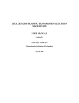
Electron Microscopy Centre
Title:
RUNNING Backscattered Detector (BSD) ON
LEO 1450VP SEM
Equipment: LEO 1450VP SEM Equipped with BSD
Revision:
1.0
Effective Date: 20/11/2005
Author:
X. Yang
Warning!
Before you attempt to operate this equipment for the first time, please make
sure you are aware of the precautions that you must take to ensure your own
safety.
Working Environment
Do not use electrical equipment in:
• Rain or excessive moisture environment
• The presence of flammable or explosive gases
The equipment is not designed to be water or splash proof, or to be used in
area where there are flammable or explosive gases or fumes.
1
Running BSD on the SEM: Basic Instructions
1. Logon LEO 1450VP user interface.
2. Check the BSD detector to make sure it is in the position to use. If it is not, contact
lab staff.
3. Load your specimen and standards (please wear provided powder free gloves only).
4. Turn on the filament and set accelerating voltage to desired operating conditions (if
not sure, set 20kV).
5. The default detector has been set to collect secondary electrons. You can now work
on obtaining a sharp image using stage position (focus), stigmators, image brightness
and contrast.
6. Set the working distance to desired value and adjust the spot size.
7. If you have found an interested spot, switch to BSD detector. The system will switch
to BSD imaging mode.
8. Adjust the brightness and contrast if necessary.
9. Save the desired image after scanning.
10. When done with collecting data, turn off the beam and wait for 30 min so the filament
is cooling down to room temp.
11. Remove specimen from chamber, close the door before pumping.
12. Log off the LEO user interface.
13. Remember to fill the log book.
2
Running miniCL on the SEM: Detailed Instructions
1. Logon LEO 1450VP user interface.
There are two computers on the SEM, but only one keyboard and mouse. The
monitor for the SEM computer is on the left - this is computer #1. The monitor for
the EDS system is on the right - this is computer #2. The red number on the gray box
in between the two monitors indicates which computer the monitor and keyboard are
working with. If the red number is 2, please switch the keyboard and mouse to
computer #1 by simply hitting Ctrl-Alt-1.
Click on the LEO icon on the SEM computer desktop and logon the software
interface using the username and password assigned to you.
2. Check whether the BSD is in position.
When in position for use, the BSD detector should be directly below the final lens as
shown in the picture. Please make sure that the specimen surface is always clear of
the BSD diode face to prevent detector damage. This could be achieved by limited
the value of Working Distance to be 10 mm or higher.
Final Lens
BSD
3. Load your specimen (please wear provided powder free gloves only).
The first thing you will want to do is to load your sample. Please make sure the
filament has been turned off at least 30 minutes so to increase the lifetime the
filament. Do this by right clicking on the Vac button in the bottom-right corner and
select Vent button at the popup widow. This will vent the sample chamber, and
allow you to open the sample chamber door. It will take approximately 5 minutes to
balance the pressure within the sample chamber with atmosphere. Never pull the
door during the 5 minutes venting period – another detector (EDS) has a fragile
3
and expensive window, a sudden pressure increase in the specimen chamber
would likely damage the EDS window.
Never reach into the sample chamber without gloves on! Always use the sample
exchange tool (tweezers and special designed screw-driver) to switch your sample
with the one that is already in the chamber. Put the sample you just removed into
covered box to prevent the dust. If you are not familiar with the sample exchange
procedure or how the sample stubs fit onto the stage, please ask for help.
WARNING: Any bare hand operation during the sample loading/unloading may
result in loss of privilege of using the system.
Once your sample is secure on the stage and positioned approximately where you
want it, close the door, latch it, and right click the Vac button on the computer then
select Pump to begin the pumping process. After about 5 minutes the Vac indicator
will shows a ready sign. At that point, the sample chamber is under vacuum,
and you are ready to begin viewing your sample. It is always a good idea to wait a
litter longer until the vacuum reading is approaching 10-6 torr.
While waiting for the vacuum to be ready, adjust the sample stage and locate your
first sample under the center of the column. In order not to hit on any detectors
within the chamber, it is always a good practice to move the sample at front end of
the stage under the column and rotate the stage to locate your interested one. It is
important to remember that the specimen must be flat or not tilted for BSD
detection.
4
4. Turn on the filament and set accelerating voltage to desired operating conditions
(if not sure, set 20kV).
Right-click on to Fil button (
) and select beam on to turn on the beam.
At this time, you should enter your information and the time-on into the SEM
logbook.
Set your operating conditions: The SEM accelerating voltage should be set to the
highest as long as the electron beam does not damage the specimen. No electrons
will be able to be detected by the BSD at a HV lower than about 4 kV. The BSD
performance is will increase progressively towards higher HVs. If you are not sure,
you could simply select 20 kV, at which you can have Atom No. resolution better
than 0.1Z between Atomic Number 29 and 30.
5. Work on obtaining a sharp image using stage position (focus), stigmators, image
brightness and contrast.
The default detector (SE1) has been set to collect secondary electrons. You can now
work on obtaining a sharp image using stage position (focus), stigmators, image
brightness and contrast.
In order to get an image, you will have to adjust the focus, contrast, and brightness.
It is always a good practice to start from a low magnification such 30x, 50x and
switch to higher magnification.
You could focus the image by selecting
focus/mg mode and turning the ‘FOCUS’
knob. In this case, the working distance (WD)
physically remains but the electron beam
focus on different level to match the WD. The
WD value becomes REAL only when the
beam is well focused on specimen.
You could also focus by changing the Zposition of your sample (this is the physical
distance between the sample holder and the
pole piece – the tapered column that hangs
above your sample). BE CAREFUL! You
might inadvertently ram your sample into the
pole piece. Therefore, you may want to
switch on the infrared camera (TV mode) so
that you can pay attention to how close your
sample is from the pole piece.
5
Stigmation: While you are still at high magnification, and once you have made your
final focus adjustments, you should adjust the 'Stigma-X' and 'Stigma-Y' to try to
improve the focus slightly. Moving between fine focus and Stigma adjustments at
high magnification may provide small, but sometimes significant improvements to
the quality of your image.
6. Set the working distance to desired height
(always focus using stage Z-drive) and spot
size.
A long working distance of 20 mm or more
will give the best atomic number contrast.
However, a working distance between 13 to
15 mm will give the best general purpose
signal. 9mm is optimum for acquiring
topographic information.
7. If you have found an interested spot, switch
to BSD detector. The system will switch to
BSD imaging mode.
Once you’ve found a spot you would like to
analyze, you are ready to switch your image
over to the BSD detector. Depending on your
sample type, you may also wish to get
elemental information from EDS detector
(please refer to different instruction manual).
In BSD mode, objects with a high atomic number will be brighter, where those with
low atomic numbers will be darker.
The QBSD detector provides 4 different
configuration viewing modes, which
could be selected by users. User could
access the control window by clicking on
“Tools” in the Menu Bar, and “Go To
Panel”, then “4QBSD Control”.
Two pre-defined configurations (COMPO
and TOPO) are available for user to
gather surface information. Various
setting combinations will give various
mixes of composition and topographical
representations, which are given in the
table listed below. In general, high topographical detail will result from selecting a
diametrically opposed pair of quadrants and summing them differentially, whereas
pure atomic number contrast is obtained with all quadrants set to Normal.
6
Q1
Normal
Normal
Q2
Normal
Off
Q3
Normal
Invert
Q4
Normal
Off
Normal
Normal
Invert
Invert
Normal
Normal
Normal
Invert
Function
Composition
Topographic, illuminated from one
particular angle
Topographic, illuminated at 45 degree to
angle obtained in set up above
Composition on 1,2, and 3 topographic
on 1 and 4
8. Adjust the brightness and contrast if necessary.
You will have to adjust your contrast and brightness to get a good image. You could
set the Brightness to 59% and adjust Contrast until an image is seen. Then adjust
Contrast together with Brightness to obtain the desired image.
9. Save the desired image after scanning.
You could save a desired BSD image under Menu Bar, click on “File” and “Save
Image” then choose the folder you would like to file to be saved.
10. When done with collecting image, turn off the beam and wait for 30 min so the
filament is cooling down to room temp.
11. Remove specimen from chamber, close the door before pumping.
12. Log off the LEO user interface.
In the Menu Bar, click on “File” and “Log Off”, then “OK” to log off the LEO
operation system.
You can also log off the system by closing the window.
Reminder: you have to log off the LEO system after finishing your research,
otherwise the computer log system would “mistakenly” consider you are still using
the machine and therefore extra charge may occur due to extended logon time to the
system
13. Remember to fill the log book.
Reference:
1. LEO 1400 Series Backscattered Electron Detector Operator’s/Installation Manual,
LEO Electron Microscopy Ltd, Cambridge, England, 1999
2. LEO 1400 Series Scanning Electron Microscopes Operator User Manual, LEO
Electron Microscopy Ltd, Cambridge, England, 1998
7