1
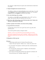
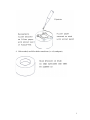
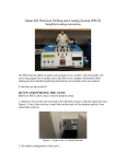
Electron Microscopy Centre
Title:
RUNNING COLD/HOT STAGE ON LEO 1450VP
SEM
Equipment: LEO 1450VP SEM Equipped with Gatan C1002
cold/hot stage and Oxford ITC 1502 temperature
controller
Revision:
1.0
Effective Date: January 10, 2006
Author:
X. Yang
Warning!
Before you attempt to operate this equipment for the first time, please READ
THIS INSTRUCTION and make sure you are aware of the precautions that
you must take to ensure your own safety.
Working Environment
Do not use electrical equipment in:
• Rain or excessive moisture environment
• The presence of flammable or explosive gases
The equipment is not designed to be water or splash proof, or to be used in
area where there are flammable or explosive gases or fumes.
Warning
Great care should be taken to ensure that when operating at high
temperature (200oC and above) EDS and back-scatter detectors should
be withdrawn or are not in close proximity to the heated specimen block.
Failure to adhere to this instruction may result in damage to the
detectors.
1
Running cold stage on the SEM: Detailed Instructions
1. Logon LEO 1450VP user interface.
There are two computers on the SEM, but only one keyboard and mouse. The
monitor for the SEM computer is on the left - this is computer #1. The monitor for
the EDS system is on the right - this is computer #2. The red number on the gray box
in between the two monitors indicates which computer the monitor and keyboard are
working with. If the red number is 2, please switch the keyboard and mouse to
computer #1 by simply hitting Ctrl-Alt-1.
Click on the LEO icon on the SEM computer desktop and logon the software
interface using the username and password assigned to you.
2. Check whether the cold stage is in position.
If you have previously informed the EMC
staff that you are going to use the cold/hot
stage for your research, the EMC staff
would install the cold stage for you before
your arrival. If not, please inform the
EMC staff immediately for a change of
stage.
When in position for use, the cold/hot
stage is ready for use. Please make sure
that the EDS and BSD detectors are
retracted if the hot stage is going to be
used.
Note: when the cold/hot stage is installed, the movement of the stage along Z
direction is much limited than that of regular stage.
3. Check the connections
Check the connection between high pressure nitrogen gas cylinder and the gas nozzle
on the Dewar top is ok. If not, inform EMC staff immediately.
Check the connection between the cold stage output and the temperature controller is
ok. If not, inform EMC staff immediately.
4. Switch on the temperature controller
2
The ITC temperature controller is located below the SEM monitor. Switch on the
ITC. At this stage the ITC will go through a short start-up sequence before displaying
a temperature.
Note: The ITC controller has been preset by EMC staff for general usage. Do not
attempt to change the ITC setting. If you do have a special need, contact the EMC
staff for a change of setting.
5. Cooling down the stage
A supply of pure dry nitrogen gas and liquid nitrogen are required to cool the cold
stage. Set the output of nitrogen gas to be between 0 and 20 p.s.i. and purge the
airway system with dry nitrogen gas for at least 5 minutes at about 10 p.s.i. before
filling with liquid nitrogen. This will flush out any water vapour in the air ways. The
actual gas flow could be fine adjusted by turning the nozzle on the top of the Dewar.
Set the nitrogen gas supply to 7 p.s.i.
(0.5 kg/cm2). Place the funnel into the
dewar and carefully pour in a small
amount of liquid nitrogen. Allow the
dewar to boil off and then continue to
fill slowly until the stage temperature
drops to about -180 oC. Top up the
dewar with liquid nitrogen and
regulate the gas flow as necessary to
maintain, increase or decrease cooling
requirement.
Note: please refer to a separate manual for detailed high pressure cylinder operation.
6. Freezing the Specimen
Pour liquid nitrogen into the insulated cup until it is full. Mount specimen onto stubs
and plunge the specimen stub with specimen mounted on it into the LN2 cup. Allow
specimen to freeze between 30 seconds and six minutes depending on specimen.
Warning: Personal Protective Devices such as goggles and gloves should be worn all
the time during the procedure. A cold burn could result is the splashed liquid
nitrogen is in contact with bare skin.
3
7. Load your specimen (please wear provided powder free gloves only).
Before you vent the chamber, please make sure the cold stage is at its ultimate
temperature. Please also make sure the filament has been turned off at least 30
minutes so to increase the lifetime the filament.
Vent the chamber by right clicking on the Vac button in the bottom-right corner and
select Vent button at the popup widow. It will take 3-5 minutes to balance the
pressure within the chamber with atmosphere. Never pull the door during the 5
minutes venting period – EDS detector has a fragile and expensive window, a
sudden pressure increase in the specimen chamber would likely break it.
Lift the frozen specimen from the LN2 cup and load it onto the cold stage by using the
sample exchange tool. This process should be immediate completed in order to
reduce the accumulation of condensation. If you are not familiar with the procedure
or how the sample stubs fit onto the stage, please ask for help.
WARNING: User should always wear gloves during the operation. Any bare hand
operation may result in loss of privilege of using the system.
Once your sample is secure on the cold stage, close the door, latch it, and right click
the Vac button on the computer then select Pump to begin the pumping process.
When the Vac indicator
shows a ready sign, the SEM system is ready for use.
It is always a good idea to wait a litter longer until the vacuum reading is
approaching 10-6 torr.
Caution: A cold burn could result is the cold plate or the liquid nitrogen is in contact
with bare skin when transferring the frozen specimen.
8. Turn on the filament and set accelerating voltage to desired operating conditions
(if not sure, set 20kV).
Right-click on to Fil button (
) and select beam on to turn on the beam.
At this time, you should enter your information and the time-on into the SEM
logbook. Set your operating conditions: the SEM accelerating voltage should be set
to the highest as long as the electron beam does not damage the specimen. If you are
not sure, you could simply select 20 kV.
9. Removal of surface ice
Ice is opaque in the SEM so any structure trapped within the ice of cryo prepared
specimen must have the ice above them removed before they can be viewed. User
4
can achieve the goal by selecting the sublimation temperature between -100 oC and
80 oC on the temperature controller.
When ready, select an area of interest on the SEM. Press the HEATER button. The
temperature will rise to the pre-selected sublimation temperature. You can use the
SEM to check on the effects of sublimation.
Note: Surface contrast and charging of the uncoated sample will be reduced by the
sublimation of surface atoms.
To halt the process of sublimation, the specimen must be cooled down rapidly. The
SEM cold stage heater is switched off and the gas flow rate may be increased
temporarily to increase the rate of cooling.
10. Set the working distance to desired height (always focus using stage Z-drive) and
spot size.
11. Work on obtaining a sharp image using stage position (focus), stigmators, image
brightness and contrast.
The default detector (SE1) has been set to
collect secondary electrons. You can now work
on obtaining a sharp image using stage position
(focus), stigmators, image brightness and
contrast.
In order to get an image, you will have to adjust
the focus, contrast, and brightness. Please read
a separate operation manual for detailed
adjustment operation.
Stigmation: While you are still at high
magnification, and once you have made your
final focus adjustments, you should adjust the
'Stigma-X' and 'Stigma-Y' to try to improve the
focus slightly. Moving between fine focus and
Stigma adjustments at high magnification may
provide small, but sometimes significant
improvements to the quality of your image.
12. If you have to view the specimen under low vacuum mode, switch to VPSE
detector. The system will switch to variable pressure imaging mode.
There are several settings required to be completed in order to view the specimen
under variable pressure mode, it is always a good idea to design your experiment
ahead and inform the EMC staff your needs. It will take EMC staff sometime to fit
5
the VP aperture, readjust the objective aperture and switch the detector channel from
CL to VPSE.
13. Adjust the brightness and contrast if necessary.
You will have to adjust your contrast and brightness to get a good image. You could
set the Brightness to 59% and adjust Contrast until an image is seen. Then adjust
Contrast together with Brightness to obtain the desired image.
14. Save the desired image after scanning.
You could save a desired BSD image under Menu Bar, click on “File” and “Save
Image” then choose the folder you would like to file to be saved.
15. When done with collecting image, turn off the beam and wait for 30 min so the
filament is cooling down to room temp.
16. Remove specimen from chamber, close the door before pumping.
17. Log off the LEO user interface.
In the Menu Bar, click on “File” and “Log Off”, then “OK” to log off the LEO
operation system.
You can also log off the system by closing the window.
Reminder: you have to log off the LEO system after finishing your research,
otherwise the computer log system would “mistakenly” consider you are still using
the machine and therefore extra charge may occur due to extended logon time to the
system
18. Remember to fill the log book.
Reference:
1. Liquid nitrogen and hot stages for Scanning Electron Microscope
C1002/C1003/C1005 Operator’s Manual, issue 3, Gatan (UK), Oxford, 1996
2. ITC 502 and ITC 503 temperature controllers Quick Start, Oxford Instruments
(UK) Ltd, 1996
3. ITC 502 temperature controller Operator’s Handbook, Oxford Instruments
Superconductivity, England, 2000
4. LEO 1400 Series Scanning Electron Microscopes Operator User Manual, LEO
Electron Microscopy Ltd, Cambridge, England, 1998
6
Appendix
Useful Specimen Mounting techniques provided by BIO-RAD
(The figures below are copied from BIO-RAD E7400 CRYOTRANS SYSTEM
Instruction Manual for educational purpose only)
1. Surface Mounting, useful for leaf surface, etc.
2. Edge Mounting, for edge observation and fracture
3. Mounting on nuclearpore filter on normal filter paper, Useful for liquid
suspensions.
4. Hole method, useful for thicker emulsions (i.e. oil, toothpaste)
1