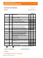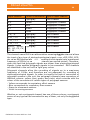
1
Bitmed eXea PSG User’s Manual EXEAPSG-534-400-MU2 Rev. 202 1 eXea PSG ELECTROENCEPHALOGRAPHY PORTABLE POLYSOMNOGRAPH User’s Manual EXEAPSG-534-400-MU2 Rev. 202 BITMED Clinical Manual eXea PSG Version: 534-400-MU2 Rev.202 All rights reserved. Please refer to the device’s Service Manual for additional information. This manual can be purchased through the After Sales Service. SIBEL S.A. Rosselló 500, 08026 Barcelona - Spain National Sales: Tel. 93 436 00 08 e-mail: [email protected] International Sales: Tel. +34 93 436 00 07 E-mail: [email protected] Technical service: Tel. +34 93 433 54 50 E-mail: [email protected] Fax: +34 93 436 16 11, Website: www.sibelmed.com SIBEL, S.A. belongs to SIBELGROUP COPYRIGHT No part of this publication may be reproduced, transmitted, transcribed, stored in a back-up system or translated into any language or computer language in any form or by any means, electronic, mechanical, optical, chemical or manual without the express written consent from SIBEL S.A. DISCLAIMER SIBEL S.A. is responsible for the security, reliability and performance of this equipment only if: • The place where the systrem is installed or used meets the requirements for electrical installations IEC and other applicable regulations. • All repairs, revisions or modifications, both in and out of the warranty period, are made by technical staff of Meditel Ingeniería Médica S.L. o SIBEL S.A. • The system is used by qualified staff in accordance with the recommendations stated in this User’s Manual. BRANDS Bitmed is a registered trademark of Sibel, S.A. PRODUCT IN COMPLIANCE WITH MEDICAL DEVICE DIRECTIVE 93/42/EEC (CLASS IIa). 0197 Thank you for choosing this product eXea PSG system is designed and manufactured with the best guarantees of quality. Applications eXea PSG and its related software will open a world of possibilities in the sleep study. If you have any possible improvement for this product, we welcome your suggestions may be directed to Customer Service Department. Revised Date: 2013-10 Antoni Picó Technical Director Approved Date: 2013-10 Carlos Recio Sales Director Bitmed eXea PSG 6 User’s Manual ÍNDICE 1. INTRODUCTION..............................................................8 1.1. THIS MANUAL..............................................................8 2. MEDICAL INFORMATION................................................9 2.1. INDICATIONS FOR USE...............................................9 2.2 LIMITATIONS IN USE. CONTRAINDICATIONS...............9 2.3. WARNINGS AND PRECAUTIONS.................................10 2.4. CERTIFICATIONS AND REGULATIONS.......................15 3. INITIAL INSPECTION...................................................17 3.1. SYSTEM REQUIREMENTS...........................................19 4. DESCRIPTION OF THE UNIT..........................................19 4.1. BRIEF DESCRIPTION OF THE eXea PSG MODELS.....19 4.2. DESCRIPTION OF THE eXea PSG UNIT. COMMON ELEMENTS........................................................................ 20 4.3. DESCRIPTION OF DIFFERENTIAL ELEMENTS OF THE eXea PSG Series 3............................................................ 24 4.4. DESCRIPTION OF DIFFERENTIAL ELEMENTS OF THE eXea PSG Series 4............................................................ 27 4.5. DESCRIPTION OF DIFFERENTIAL ELEMENTS OF THE eXea PSG Series 5............................................................ 30 5. CONNECTING THE eXea PSG SYSTEM...........................33 5.1. SELECTION OF THE INSTALLATION SITE...................33 5.2. CONNECTING............................................................. 34 6. MAKING A STUDY.........................................................36 6.1. BEFORE MAKING A STUDY.........................................36 6.2 PLACEMENT OF SENSORS AND ELECTRODES .............36 6.2.1. Electrodes for neurophysiologic signals (EEG, EOG, EMG, ECG and NEUTRAL electrode)...................................36 6.2.2. Polygraphy sensors................................................40 6.2.3. Specific sensors......................................................45 6.3 SYSTEM START-UP......................................................50 7. OPERATING MODES OF THE eXea PSG.........................50 EXEAPSG-534-400-MU2 Rev. 202 Bitmed eXea PSG User’s Manual 7 8. MAINTENANCE............................................................. 53 9. PROBLEM-SOLVING GUIDE...........................................55 10. TECHNICAL SPECIFICATIONS.....................................57 10.1 Specific technical specifications of the pulsioximeter.. 59 10.2 SIMBOLOGY.............................................................. 61 10.2 Manufacturer’s statement - EMC...............................62 EXEAPSG-534-400-MU2 Rev. 202 Bitmed eXea PSG User’s Manual 8 1. INTRODUCTION The eXea PSG is a polysomnograph/electroencephalograph that can be used to carry out sleep studies in ambulatory (residential) mode or in direct communication (real time) with a computer, in which the signals are observed at the same time as they are being captured. The connection to the computer is made through a USB port. The eXea PSG possesses a variable number of multi-purpose channels (from 12 to 20) depending on the model chosen. Moreover, it includes two DC channels which are suitable for non-differential signals (for example, position sensors) and four channels for specific use: pulse, oxygen saturation, ambient light detector and a user event-marking channel. Depending on the number of multipurpose channels, the eXea PSG can be one of the following models: eXea PSG Series 3 (12 multipurpose channels), eXea PSG Series 4 (16 multipurpose channels) or eXea PSG Series 5 (20 multipurpose channels). The characteristics that differentiate each model will be explained later on, with the term eXea PSG being used from now on to refer to these three models. The eXea PSG incorporates an advanced high-performance battery system giving it greater flexibility and mobility. It also has an internal memory to enable it to be used without being connected to a PC (ambulatory or residential mode). The multipurpose channels can work at various operating frequencies: 500Hz, 250Hz, 100Hz, 50Hz or 20Hz. The operating frequency for each channel is specified from the PC using the XGPLab software. Thus we can select a higher frequency for signals with a higher frequency range (for example, EEG), and a lower frequency for signals with a lower frequency range (for example bucconasal discharge). These frequencies must be specified by a qualified technician with experience in this type of signal. The DC channels and the specific channels (pulse, oxygen saturation, ambient light and user events) have a fixed sampling frequency of 10 Hz. 1.1. THIS MANUAL This manual refers to the eXea PSG. The BITMED product manuals are updated periodically to reflect all modifications. ! We ask you to read this manual carefully before use, paying special attention to the sections displaying an exclamation mark. EXEAPSG-534-400-MU2 Rev. 202 Bitmed eXea PSG User’s Manual 9 2. MEDICAL INFORMATION 2.1. INDICATIONS FOR USE The eXea PSG has been designed for maximum safety. All of the operating instructions must be read before proceeding to operate with the instrument. Failure to do so may result in injuries to the user or the patient and damage to the unit and/or accessories. The eXea PSG has been designed for use by a doctor or a technician trained in the acquisition of EEG and polysomnography (PSG) signals and transmission of these signals to a PC during neurophysiologic or sleep examinations. In these conditions it is possible for the user to configure the unit. However, it is not recommended that the configuration of the unit be modified without understanding the principles of signal digitalizing. The intended environments are hospitals, sleep centres and sleep clinics. Studies may also be carried out in the patient’s own home. In this case the patient is only authorized to start and stop the recording, and should be adequately informed by the doctor in this respect. The eXea PSG is not designed to be used outdoors, nor with other conditions or energy sources which have not been anticipated in this manual. The use of eXea PSG systems does not entail any supervision or diagnosis of the patient. 2.2 LIMITATIONS IN USE. CONTRAINDICATIONS. The interpretation of the tests and any ensuing treatment must be carried out by a doctor. The symptoms the patient presents before any polysomnographic tests are performed must be considered by the healthcare personnel. The acceptability of a test is the responsibility of the healthcare personnel. The polysomnograph must not be used when it is probable that the validity of the results can be doubtful due to external factors. EXEAPSG-534-400-MU2 Rev. 202 Bitmed eXea PSG User’s Manual 10 2.3. WARNINGS AND PRECAUTIONS ! • The eXea PSG is a polysomnograph for the study of sleep pathologies: recording, displaying, marking and counting events and episodes of polysomnographic signals during the patient’s sleep. It can also operate as an electroencephalograph. eXea PSG is designed to be used for medical staff only, trained and supervised by a physician. It’s recommended specific trainning on polysomnograph technique. User must be familiarized with the functioning of the device before using it with patients. All necessary information is available in this user manual. For additional information contact regarding polysomnograph tecnique or the device, contact SIBEL S.A. or your distributor. • The eXea PSG IS NOT CERTIFIED FOR USE IN CONTINUOUS MONITORING, where a failure in operation can cause injuries or the death of the patient. This product does not maintain nor does it help to maintain the life of the patient. The term CONTINUOUS MONITORING is specified in regulation EN606011. The eXea PSG is classified as Class IIa in accordance with Directive 93/42/EEC on medical devices. • The eXea PSG has an internal battery. • The system cannot be used for DIRECT CARDIAC APPLICATION. • The system is not an electrocardiograph, and thus cannot be used to produce a separable ELECTROCARDIOGRAM for diagnostic purposes, nor for electrocardiographic supervision, nor for ambulatory electrocardiography. • There is no means to indicate an INOPERATIVE ELECTROCARDIOGRAPH. • There are no parts applicable to the patient which produce stimulation. • DO NOT alter earth integrity of the power supply. Protection against electrical shocks is proportioned by connection to eartch . Earth protection is only effective when the power cable delivered with the device is connected to a electrical socket with earth. DO NOT remove the device or accessories cover. Servicing and repair of the apparatus must only be carried out by trained personnel. Contact with the voltage inside the system can cause serious injury. • The system does not have parts which can be replaced EXEAPSG-534-400-MU2 Rev. 202 Bitmed eXea PSG User’s Manual ! 11 by the user. Only the authorized Technical Service and replacement parts supplied by the manufacturer should be used. • The use of other cables, sensors or accesories different than the supplied ones, can affect patient safety and can degrade emissions and susceptibility of the device. • Contact of liquids with the internal parts of the unit and the connectors must always be avoided. • The device should NOT be used stacked or adjacent to other equipment. The equipment must be stored and used within the temperature, pressure and humidity ranges specified in this manual. • The system is only resistant to moderate splashes or drips (Protection level IPX1). • The cleaning instructions contained in this manual must be strictly observed, as must the instructions for use of the sensors included that are not manufactured by SIBEL S.A. • Keep the unit protected from impacts and vibrations. In transport, place all elements in the transport bag or case; their material provides sufficient protection against small accidental knocks. • Do not use the system in an MRI environment. • The system is not designed to work in an explosive environment or in the presence of anaesthetics or inflammable gases of any type. THIS MAY CAUSE AN EXPLOSION. • This product is intended for indoor use (e.g. in the patient’s home or in hospital) and is not suitable for use in the transport of patients. • The polysomnograph is not designed to be used outdoors, nor with other conditions or energy sources that are not provided for in this manual. • The eXea PSG is not protected against defibrillation. Therefore, never use a defibrillator on a patient to whom the eXea PSG has been connected. • Use the device in an acoustic enviroment wher the patient can sleep normally. • The device must be used in a stable position to avoid it can fall down and a damage is produced to the patiend and/or user. • While the eXea PSG is carrying out a recording, the use of mobile telephones, transmitters and similar equipment which generate radiofrequency emissions near the system is not permitted. Therefore, do not use the unit in presence of radio communication units (mobile telephones, walkie-talkies...) Follow the recommendations regarding separation distance indicated in the manufacturer’s statement concerning EMC in EXEAPSG-534-400-MU2 Rev. 202 Bitmed eXea PSG User’s Manual ! 12 this manual. • Remember that when there are several units connected to a patient, there exists a risk of accumulation of leakage currents. Reduce the number of units connected to the patient to a minimum. The use of the battery-powered eXea PSG helps considerably in reducing leakage currents. • To work with the eXea PSG powered by the electrical network, only use the power source supplied with the unit. The use of a different power source can put the safety of the patient at risk. Ensure that the electrical installation meets the safety requirements of the applicable legislation, and in particular that there is an earth connection. Remember that in certain places it is obligatory to have the electrical power supply coupled to isolation transformers for medical use. Where appropriate, consult the technical services of the hospital or centre. • Do not use network bases with multiple outlets, unless they comply with regulation EN-60601-1 as they can reduce electrical safety. • Do not remove the cover of the unit. The service and repair of the device should only be carried out by qualified personnel. Contact with the internal voltages of the system can cause serious injuries. • Do not use the unit if the power cable is damaged or cuts are observed. • Do not use the transducers if they are damaged. • Do not submerge the parts of the device in any unit. IT MAY CAUSE ELECTRIC DISCHARGE. • To ensure that the essential safety characteristics conform to regulation EN 60601-1, only units which comply with current electrical safety regulations may be connected to this instrument. If you use the eXea PSG connected to a computer, remember that it must comply with the applicable safety standard (EN 60601-1). If not, the system formed by eXea PSG plus computer will not comply with the pertinent safety regulations. If you use a computer connected to the eXea PSG system make sure that: o If you place the computer in the environment of the patient (“where the patient can reach the computer”), you must install an additional earth connection to the computer housing or else power it with a battery. • This is a medical electronic device and as such it needs special precautions regarding the electromagnetic compatibility (EMC) and it should be installed and setup according to the EMC information attached (See Appendix 1. ELECTROMAGNETIC COMPATIBILITY). EXEAPSG-534-400-MU2 Rev. 202 Bitmed eXea PSG User’s Manual ! 13 • The eXea PSG system is prepared to work at room temperature. Avoid exposing any component of the system to heat sources. Also avoid direct exposure to sunlight. Do not wrap the eXea PSG with any material that can prevent its natural ventilation. For this reason, do not put the unit on the patient’s bed. Changes in temperature and humidity can produce condensation. Before using the system, wait for the unit to adjust to the room temperature. As a reference, if the difference in temperature is above 10ºC, it would be advisable to wait 20 minutes in an intermediate room temperature. • Artefacts from the signal may be produced as a result of ESD. A trained operator should be able to recognize these artefacts easily. • The operator must be trained to recognize the differences between a biological signal and artefacts from the signal caused by movements by the patient, radio frequency interference or placement of the electrodes or sensors in a poor position. The presence of ESD or RF artefacts will not lead to mistaken conclusions. The unusable data is not considered a risk for the safety of the patient. • The conducting part of the electrodes and their connectors, including the earth electrode, must not touch other conducting parts, including the ground. • It must be ensured that neither the cables nor the sensors wrap around the neck of the patient, especially when the patient is a child. • The eXea PSG system does not increase the safety risk for patients with pacemakers as long as the pacemakers comply with standard EN50061 concerning the electrical safety of medical devices. Nevertheless, conducting a contact impedance test on the electrodes (measuring impedances) on patients fitted with a pacemaker is not recommended, so as to avoid any interference occurring in the normal operation of the pacemaker. Before using the system in patients with pacemakers, the operator must consult the documents accompanying the pacemakers with respect to their certifications and requirements for use and, where necessary, contact the manufacturer. • The patients must be insistently warned that they must not open the eXea PSG or try to adjust it. • Sensors or accessories that are in bad condition must not be used. • Use the eXea PSG only with accessories, sensors, and electrodes supplied by the manufacturer or an authorized dealer; or those who meet the specifications of this manual. The use of other sensors with the eXea PSG can damage the EXEAPSG-534-400-MU2 Rev. 202 Bitmed eXea PSG User’s Manual 0318 14 unit or the quality of the signal. • The sensors must be handled by their strongest parts, which are the connecting pins for connection to the units. Likewise, they must not get wet, or be exposed to very sudden changes in temperature, or cleaned with abrasive chemical products. Attention must be paid to how the sensors are used, ensuring that they are not submitted to forces for which they are not designed. The excessive bending of any of the parts of the sensors should particularly be avoided. This means that the material should not be bent more than is necessary in normal use. • With regard to the electrodes, correct contact of the skin with the electrodes must be achieved using conducting gel, as, if this is not the case, there is a danger that inaccurate measurements might be obtained. Avoid contact with the eyes or mucus membrane and gels, collodion, alcohol, acetone or any substance used in the placement or removal of electrodes. Be especially careful if you use collodion. Always follow the recommendations for use provided by your collodion manufacturer. • The polysomnograph is designed to be used exclusively by healthcare personnel, under the supervision and instructions of a doctor. PULSEOXIMETRY The pulse oximeter does NOT require calibration. The pulse oximeter has NO physiological WARNINGS. Pulse oximeter probes do NOT allow temporary immersion. The pulse oximeter waveform is NOT normalizzed. Patient movements or excessive lighting can affect the accuracy of the device. The pulse oximetry module has CE marking and can not reach temperatures> 42 º. The oxygen saturation and heart rate measures must be performed using the sensors manufactured by NONIN 8000J (Flexible) and 8000AA-3 which have been validated clinically. Otherwise, there may be significant measurement errors... The wavelengths used are 660 nm (red) and 910 nm (infrared) and the optical power is about 9.6mW. Consult the instructions for use of the probes, before use. Follow strictly manufacturer’s safety instructions, as well as those specified in this manual. See instructions for use of the sensor to evaluate the toxicity of SpO2 sensor. EXEAPSG-534-400-MU2 Rev. 202 Bitmed eXea PSG User’s Manual 15 The maximum recommended application’s time of an oximeter, in the same place, is 8 hours. The point of placement must be reviewed frequently to determine the position, the movement and the skin sensitivity of the patient. The reaction to the sensors by the patients may be different depending on their health and skin conditions. No adhesive material should be used if the patient has an allergic reaction to it. For log term monitoring, is recommended to use flexible or disposable sensors. The adult sensor is intended for use on people over 12 years old. The peadiatric probe is for children between 3 and 12 years. Although, the adults probes can be used on any pediatric patient whose fingertip reaches the finger stop of the sensor and on whom the sensor fits securely. The system can measure pulse and functional oxygen saturation. A functional meter can not be used to evaluate the accuracy of pulse oximeter probe or a pulse oximeter monitor. This oximetry sensor should be used by a physician or under the supervision of medical staff. The clip sensor can be cleaned by wiping it off with a soap or orthophthalaldehyde solution. SpO2 sensor equipment must be disconnected before cleaning. The SpO2 sensor must not be sterilized by autoclave, ETO or immerse in water or other solutions. 2.4. CERTIFICATIONS AND REGULATIONS The eXea PSG bears the EC conformity mark (CE 0318). The CE marking is a declaration certifying that the eXea PSG complies with the guidelines established by the EU for medical devices. The eXea PSG is manufactured by SIBEL and the system (including the software) complies with the following standards and regulations: European Directive 93/42 EEC (R.D: 1591/2009): • EC marking Class II a Safety: • EN 60601.1:2006+AC:2010 Electrical safety (3rd edition). EMC: • EN 60601.1.2:2007+AC:2010 EMC in medical devices (No life support) Software: EXEAPSG-534-400-MU2 Rev. 202 Bitmed eXea PSG User’s Manual 16 • EN 62304:2006+AC:2008 Medical device software Software life-cycle processes Usability: • EN 60601-1-6:2010 Medical Electrical Equipment – Part 1-6 General requirements for basic safety and essential performance - Collateral standard: Usability • EN 62366:2008 Medical Devices – Application of usability engineering to medical devices Pulse oximetry: • EN ISO 9919:2009 Special requirements for basic safety and essential operation characteristics of pulse oximeters for medical use Biocompatibility: • EN ISO 10993-1:2009+AC:2010 Biological evaluation of medical devices Vibration and temperature: • Tests according to EN 680068-2-27:1993 and EN 6800682-64:1994 Symbology: • EN ISO 15223-1:2012 Symbols to be used with medical device labels, labelling andi nformation to be suplplied. Packaging and labelling • Packaging and packaging waste directive 94/62/EC • Regulation EC 1272/2008 on classification, labelling and packaging of substances and mixtures (REACH) Information supplied by the manufacturer: • EN 1041:2008 Information supplied by the manufacturer of medical devices Electrical and electronic waste: • Electrical and electronic equipment and waste management. Transposition of the directive WEEE 2002/96/EC System quality: • Manufactured according to the SIBEL S.A. Quality Manual, which is compliant with EN ISO 13485:2012+AC:2012, EN ISO 9001:2008 and EN ISO 14971:2012 The user should follow: • Data protection: Directive 95/46/EC • 2002/96/EC Recommendations: • National consensus document on the apnea-hypopnea syndrome (SAHS). Spanish Sleep Group (GES), September 2005. • The AASM Manual for the Scoring of Sleep and Associated Events, 2007 3. INITIAL INSPECTION EXEAPSG-534-400-MU2 Rev. 202 Bitmed eXea PSG User’s Manual 17 Check to ensure that the unit is complete and has not been damaged in transport. If this is not the case, report it to your supplier immediately or straight to the manufacturer, whose address appears at the beginning of this manual. The unit should include at least the following components: • eXea PSG electroencephalograph / polysomnograph (amplifier box). • External power source to power the eXea PSG and recharge the battery. • USB connection cable linking the eXea PSG to a compatible computer system. • CDROM disc with XGPLab software. • Software protection key, also called HARDLOCK, for connection to the USB port or parallel port of the computer. • Transport bag or case. • User’s manual for the eXea PSG unit and the XGPLab Software. • The electrodes and sensors required by the user. 3.1. SYSTEM REQUIREMENTS Computer requirements The minimum requirements of the computer system must be consulted in the XGPLab Software manual or in section 10. TECHNICAL SPECIFICATIONS of this manual. Also pay special attention to section 2.2. WARNINGS AND PRECAUTIONS. Electrical installation The electrical installation which powers the eXea PSG and the computer must comply with all the safety regulations in force. Likewise, it is absolutely essential that the installation and the plug, both for the power supply for the eXea PSG and for the PC are earthed. For the connection of the eXea PSG to the power source, there must be a free power outlet less than 2m from where the unit is located. Read section 2.3. WARNINGS AND PRECAUTIONS carefully. EXEAPSG-534-400-MU2 Rev. 202 Bitmed eXea PSG 18 User’s Manual RELACIÓN DE CONTENIDO Página 1 de 1 EXEA PSG 534-408-010 REV. 3.00 MODELOS CÓDIGO CANT. DESCRIPCIÓN Serie 3 _________ 1 EXEA PSG 02132 1 FUENTE DE ALIMENTACIÓN / EXTERNAL POWER SUPPLY XGP (6V, MINI-SNAP) 01217 1 CABLE DE RED / POWER SUPPLY CORD 01661 1 CABLE DE CONEXIÓN / CONNECTION CABLE USB PC 03040 1 SOFTWARE XGPLAB (PROGRAMA 534-45A) 1 MANUAL DE USUARIO / USER’S MANUAL SOFTWARE XGPLAB (DOC. 534-450-MU_) 02479 1 MODULO BASE DE DATOS/ DATABSE MODULE 02470 1 MODULO ANALISIS AUTOM. FASES SUEÑO / AUTOMATIC SLEEP STAGES ANALYSIS MODULE 02469 1 MODULO ANALISIS AUTOM. EVENTOS SUEÑO / AUTOMATIC SLEEP EVENT ANALYSIS MODULE 02478 1 MODULO ANALISIS ESPECTRAL DE SEÑALES / SPECTRAL SIGNALS ANALYSIS MODULE 02480 1 MODULO CARTOGRAFIA CEREBRAL / BRAING MAPPING MODULE 02481 1 MODULO DETECCION DE PUNTAS / SPIKE DETECTION MODULE 02508 1 MODULO XGPVISION / XGPVISION MODULE 02324 1 MALETÍN DE TRANSPORTE PARA / CARRING CASE FOR EXEA PSG 02318 1 MALETÍN SENSORES / SENSORS CARRING CASE _________ 1 MANUAL DE USUARIO / USER’S MANUAL EXEA PSG (DOC. 534-400-MU_) _________ STANDARD 4 Serie 5 SN: 344- OPCIONAL / OPTIONAL PREPARADO/ PREPARED BY:........................... --- NO DISPONIBLE / NOT AVAILABLE REVISADO/REVISED BY:.............................. Fecha/Date: 4. DESCRIPTION OF THE UNIT EXEAPSG-534-400-MU2 Rev. 202 Serie Bitmed eXea PSG 19 User’s Manual All the controls, connectors and indicators of the eXea PSG are found on the upper and rear part of the amplifier module. 4.1. BRIEF DESCRIPTION OF THE eXea PSG MODELS eXea PSG Series 3 Model eXea PSG Series 3 has a total of 18 input channels which include: 12 differential AC bipolar channels, 2 DC channels and four permanent channels: pulse, oxygen saturation, ambient light and event marker. eXea PSG Series 4 Model eXea PSG Series 4 has a total of 22 input channels which include: 16 AC differential channels (8 of which are single-pole with a common reference and the other 8 bi-polar), 2 DC channels and four permanent channels: pulse, oxygen saturation, ambient light and event marker. eXea PSG Series 5 Model eXea PSG Series 5 has a total of 26 input channels which include: 20 AC differential channels (8 of which are single-pole with a common reference and the other 12 bipolar), 2 DC channels and four permanent channels: pulse, oxygen saturation, ambient light environment and event marker. 4.2. DESCRIPTION OF THE eXea PSG UNIT. COMMON ELEMENTS The various elements common to the different models of eXea PSG are described below. Upper panel EXEAPSG-534-400-MU2 Rev. 202 Bitmed eXea PSG 20 User’s Manual MEMORY INDICATORS Note: The striped zone corresponds to the specific front part of each of the eXea PSG models. Light indicator of the impedance meter, symbol : Colour-code indication the impedance for the electrodes connected in the red and black connectors of the impedance meter: • off: the electrodes are exposed or have an impedance greater than 80k . Very poor contact. • red: impedance between 25 k and 80 k . Bad contact. • yellow: impedance between 7 k and 25 k . Medium contact. • green: impedance less than 7 k . Good contact. Note: The indicated quality of contact should only be taken as a guideline: it should not be attributed the function of measurement. The use of the impedance meter is described in detail in section 6.1. PRIOR TO THE OPERATION OF THE eXea PSG. Red and black connectors of the impedance meter: The electrodes whose impedance is to be measured must be connected to these terminals. EXEAPSG-534-400-MU2 Rev. 202 Bitmed eXea PSG 21 User’s Manual Black connector marked with the symbol called ‘patient’s earth’). (‘NEUTRAL’ or ‘NEUTRO’, also The electrode marked ‘NEUTRAL’ (or patient’s earth), which is necessary for the correct acquisition of a signal from the channels, must be connected here. Normally this electrode is connected to the forehead of the patient. Dark grey connectors of the 2 DC channelsgrey: To these channels, known as +DC1, -DC1 and +DC2, DC2, are connected signals of direct current, such as, for example, the body position sensor, or the pressure level output for the CPAP. BATTERY/NETWORK CONNECTION/LOAD Indicator . . Three possible states are possible: • Indicator permanently on green: unit connected to the mains and the charging process is completed. The battery is charged after connection to the mains through a power supply. • Slow blink: activated when the unit is not connected to the mains. It shows the level of autonomy available by means of three colours: • Green: recording autonomy of more than 9 hours. • Yellow: recording autonomy of more than 1 hour. • Red: battery very low; autonomy of recording less than 10 minutes. • Rapid blinking in green: battery recharging by connection to the mains. MEMORY INDICATORS AVAILABLE: Depending on the number of channels, the frequency of work for each channel and the amount of memory installed for each channel, and the amount of memory installed in your unit, the system can store a different number of nights of approximately 9 hours. EXEAPSG-534-400-MU2 Rev. 202 Bitmed eXea PSG User’s Manual 22 Depending on the frequency of work chosen for each channel, this number of nights, (storage time) can vary significantly. The indicators show the available memory in the following manner: • Level of available memory (in groups of 9 hours ) up to 36 hours or more of recording time available up to 27 hours of recording time available up to 18 hours of recording time available up to 9 hours of recording time available • Number of hours of recording time available on each of the levels (for each of the light indicators) Green: between 6 and 9 hours available Yellow: between 3 and 6 hours available Red: between 0 and 3 hours available Off: 0 hours. The 4 yellow indicators: memory full. Thus, for example, a yellow indicator in together with two green indications in and , means that there is memory for a total of between 21 and 24 hours (9 + 9 + from 3 to 6). The indicators of available memory can be lit continuously or blinking (slowly or rapidly), depending on whether or not the unit is storing the data in its memory, or whether it is being recorded on the computer. Consult the section entitled OPERATING MODES for further details. Rear panel In this panel from left to right, the following elements: ON/OFF Switch: To turn on (position ON) or turn off the unit (position OFF). EXEAPSG-534-400-MU2 Rev. 202 Bitmed eXea PSG User’s Manual 23 Connector of the power source: For the power source supplied with the unit Connector for the pulsioximetry sensor (SpO2): To connect the sensor of the pulse and oxygen saturation channels. Connect for the events button (√) Connector of communications with the PC: One end of the communications cable should be connected at this point. The other end is connected to a USB port (Universal Serial Bus) of the PC in which the XGPLab Software must be installed. When the connection to the USB port of the PC is correct, the green light associated to the connector lights up. This light indicates that the operating system of the PC has correctly detected the eXea PSG unit; this is a necessary prior condition so that the XGPLab Software can communicate with the unit. If there is any problem in the connection of the USB cable or in the detection of the unit by the operating system, this light will not go on. When the unit is transmitting data to the PC, this light is orange. 4.3. DESCRIPTION OF DIFFERENTIAL ELEMENTS OF THE eXea PSG Series 3 Upper panel EXEAPSG-534-400-MU2 Rev. 202 Bitmed eXea PSG 24 User’s Manual This panel contains the following characteristic elements of the eXea PSG Series 3: 12 differential input channels (AC). All of them with two poles; 8 thanks to a single bipolar connector (labelled from 1 to 8) and 4 thanks to 2 single pole connectors (labelled from 9 to 12), which are as follows: Input channel Colour of connector Type of input channel 1, 2, 3 Light gray EXG Channel 4, 5 Yellow Polygraphic Channel 6 Blue Polygraphic Channel 7 Green Polygraphic Channel 8 Violet Polygraphic Channel +9/-9 Light gray EXG Channel +10/-10 Light gray EXG Channel +11/-11 Light gray EXG Channel +12/-12 Light gray EXG Channel The channel name EXG is an abbreviation meaning that the channel allows the input of any type of electrophysiological signal: EMG, EEG, EOG, ECG, etc. as an EXG channel allows the recording of such signals up to a maximum frequency of 500Hz (or samples per channel per second stored). Therefore, given the fact that polygraph signals are slower than electrophysiological signals, it also enables polygraph signals to be connected. EXG channels are identified in the unit with a light grey colour. Polygraph channels allow for the recording of signals up to a maximum frequency of 100Hz, and are therefore not adequate for recording electrophysiological signals. In order to simplify the task of connecting the polygraph sensors in the unit, the polygraph channels are equipped with connectors of different colours: yellow, blue, green, and violet, which coincide with the colour of the connectors of certain types of polygraph sensors: - Yellow corresponds to respiratory effort sensors - Blue corresponds to respiratory flow sensors - Green to movement sensors - Violet to snoring sensors Whether or not a polygraph channel has one of these colours, a polygraph sensor of any type can be connected to any of them, not only the suggested type. EXEAPSG-534-400-MU2 Rev. 202 Bitmed eXea PSG 25 User’s Manual Correspondence between the input connectors and the available channels in the XGPLab software Label Channel in the software 1 1 Differential EXG Channel (EEG, EOG, EMG, ECG or any polygraphic sensor). Recommended signal: EEG. 2 2 Differential EXG Channel (EEG, EOG, EMG, ECG or any polygraphic sensor). Recommended signal: EEG. 3 3 Differential EXG Channel (EEG, EOG, EMG, ECG or any polygraphic sensor). Recommended signal: EEG. 4 4 Polygraphic channel. Recommended sensor: Thoracic effort (the thoracicoabdominal sum is generated with software). 5 5 Polygraphic channel. Recommended sensor: Abdominal effort (the thoracicoabdominal sum is generated with software). 6 6 Polygraphic channel. Recommended sensor: Respiratory flow, thermocouple. 7 7 Polygraphic channel. movement (PLM). 8 8 Polygraphic channel. Recommended sensor: Snoring. 9 9 Differential EXG channel (EEG, EOG, EMG, ECG or any polygraphic sensor). Recommended signal: EOG. 10 10 Differential EXG channel (EEG, EOG, EMG, ECG or any polygraphic sensor). Recommended signal: EOG. 11 11 Differential EXG channel (EEG, EOG, EMG, ECG or any polygraphic sensor). Recommended signal: EMG. 12 12 Differential EXG channel (EEG, EOG, EMG, ECG or any polygraphic sensor). This channel would remain free in a typical configuration (recommended configuration for use of the unit). SpO2 13 Pulse. SpO2 14 Oxygen saturation. DC1 15 Generic DC channel. This channel remains free in the typical configuration of the unit (recommended configuration for use of the unit) and in it any DC sensor can be connected, such as a body position sensor or CPAP. DC2 16 9 The events marked by the user appear as notes in the upper part of the representation of the channels. EXEAPSG-534-400-MU2 Rev. 202 Description Recommended sensor: Limb Generic DC channel. Recommended signal: body position. User events marker. Bitmed eXea PSG User’s Manual 26 4.4. DESCRIPTION OF DIFFERENTIAL ELEMENTS OF THE eXea PSG Series 4 Upper panel In this panel we have the following characteristic elements of the eXea PSG Series 4 model: 8 Differential input channels (AC). All with two poles: 6 thanks to a single bipolar connector (labelled from 1 to 6) and 2 thanks to 2 single-pole connectors (labelled from 7 to 8). 8 Referential input channels, with a single single-pole connector, all referenced to the reference ‘REF’, in grey; all of them have a name based on standard EEG 10/20: EOGL, EOGR, A1, A2, C3, C4, O1, O2. EXEAPSG-534-400-MU2 Rev. 202 Bitmed eXea PSG 27 User’s Manual Input channel Colour of connector Type of input channel 1 Light gray EXG Channel 2,3 Yellow Polygraphic Channel 4 Blue Polygraphic Channel 5 Green Polygraphic Channel 6 Violet Polygraphic Channel +7 / -7 Light gray EXG Channel +8 / -8 Light gray EXG Channel E O G L , E O G R , Light gray A1, A2, C3, C4, 01, 02 EXG Channel The channel name EXG is an abbreviation meaning that the channel allows the input of any type of electrophysiological signal: EMG, EEG, EOG, ECG, etc. as an EXG channel allows the recording of such signals up to a maximum frequency of 500Hz (or samples per channel per second stored). Therefore, given the fact that polygraph signals are slower signals than electrophysiological signals, it also enables polygraph signals to be connected. EXG channels are identified in the unit with a light grey colour. Polygraph channels allow for the recording of signals up to a maximum frequency of 100Hz, and are therefore not adequate for recording electrophysiological signals. In order to simplify the task of connecting polygraph sensors in the unit, the polygraph channels have connectors of different colours: yellow, blue, green, and violet, which coincide with the colour of the connectors of certain types of polygraph sensors: - Yellow corresponds to respiratory effort sensors - Blue corresponds to respiratory flow sensors - Green to movement sensors - Violet to snoring sensors Whether or not a polygraph channel has one of these colours, a polygraph sensor of any type can be connected to any of them, not only the suggested type. EXEAPSG-534-400-MU2 Rev. 202 Bitmed eXea PSG User’s Manual EXEAPSG-534-400-MU2 Rev. 202 28 Bitmed eXea PSG User’s Manual 29 4.5. DESCRIPTION OF DIFFERENTIAL ELEMENTS OF THE eXea PSG Series 5 Upper panel In this panel we have the following characteristic elements of the eXea PSG Series 5 model: 12 Differential input channels (AC). All with two poles: 8 thanks to a single bipolar connector (labelled from 1 to 8) and 4 thanks to 2 single-pole connectors (labelled from 9 to 12). 8 Referential input channels, with a single single-pole connector, all with the reference ‘REF’, in grey; all of them have a name based on standard EEG 10/20: EOGL, EOGR, A1, A2, C3, C4, O1, O2. EXEAPSG-534-400-MU2 Rev. 202 Bitmed eXea PSG 30 User’s Manual Input channel Colour of connector Type of input channel 1 Light gray EXG Channel 2,3 Yellow Polygraphic Channel 4,5 Blue Polygraphic Channel 6,7 Green Polygraphic Channel 8 Violet Polygraphic Channel +9 / -9 Light gray EXG Channel +10 / -10 Light gray EXG Channel +11 / -11 Light gray EXG Channel Light gray EXG Channel E O G L , E O G R , Light gray A1, A2, C3, C4, 01, 02 EXG Channel +12 / -12 Power source The channel name EXG is an abbreviation meaningComunication that the channel allows cable the input of any type of electrophysiological signal: EMG, EEG, EOG, ECG, etc. as an EXG channel allowsPatient the recording of such signals up to a maximum frequency of 500Hz (or samples per channel per second stored). Therefore, given the fact that polygraph signals are slower than electrophysiological signals, it also enables polygraph signals to be connected. EXG channels are identified in the unit with a light grey colour. Polygraph channels allow the recording of signals up to a maximum frequency of 100Hz, and therefore are not adequate for recording of electrophysiological signals. In order to simplify the task of connection of polygraph sensors in the unit, the polygraph channels have connectors of different colours: yellow, blue, green, and violet, which coincide with the colour of the connectors of certain types of polygraph sensors: - Yellow corresponds to respiratory effort sensors - Blue corresponds to respiratory flow sensors - Green to movement sensors - Violet to snoring sensors Whether or not a polygraph channel has one of these colours, a polygraph sensor of any type can be connected to any of them, not only the suggested type. EXEAPSG-534-400-MU2 Rev. 202 Bitmed eXea PSG 31 User’s Manual Correspondence between input connectors and available channels in XGPLab software Label Channel in the software 1 1 EXG Channel (EEG, EOG, EMG, ECG or any polygraphic sensor). Recommended signal: ECG 2 2 Polygraphic signal. Recommended sensor: Thoracic effort (the thoracicoabdominal sum is generated with software). 3 3 Polygraphic signal. Recommended sensor: Abdominal effort (the thoracicoabdominal sum is generated with software). 4 4 Polygraphic signal. flow, thermocouple. 5 5 Polygraphic signal. Recommended sensor: Respiratory flow, transductor. (free in the standard or typical configuration) 6 6 Polygraphic signal. Recommended sensor: Limb movement (PLM). 7 7 Polygraphic signal. Recommended sensor: Limb movement (PLM). 8 8 Polygraphic signal. Recommended sensor: Snoring. 9 9 Differential EXG channel (EEG, EOG, EMG, ECG or any polygraphic sensor). Recommended signal: EMG 10 10 Differential EXG channel (EEG, EOG, EMG, ECG or any polygraphic sensor). This channel remains free in a typical configuration of the unit. 11 11 Differential EXG channel (EEG, EOG, EMG, ECG or any polygraphic sensor). This channel remains free in a typical configuration of the unit. 12 12 Differential neurophysiologic channel (EEG, EOG, EMG, ECG or any polygraphic sensor). This channel remains free in a typical configuration of the unit. C3 13 Referential EXG channel for EEG. C4 14 Referential EXG channel for EEG. O1 15 Referential EXG channel for EEG. O2 16 Referential EXG channel for EEG. A1 17 Referential EXG channel for EEG. A2 18 Referential EXG channel for EEG. EOGL 19 Referential EXG channel for EOG. EOGR 20 Referential EXG channel for EOG.. EXEAPSG-534-400-MU2 Rev. 202 Description Recommended sensor: Respiratory Bitmed eXea PSG User’s Manual 32 5. CONNECTING THE eXea PSG SYSTEM This section describes the connection operations necessary for the use of the eXea PSG. It explains the criteria for selecting the most appropriate installation site and also provides a list of recommendations for safe use in complianceof the regulations in force. 5.1. SELECTION OF THE INSTALLATION SITE The eXea PSG complies with the requisites concerning electromagnetic compatibility in the European Medical Device Directive. Hence, no waves are emitted that might interfere with other units or radio and telecommunication services, and ensures a level of immunity against interferences produced by other systems or devices. In any event, in order to protect the unit from an adverse electromagnetic environment, during operation we recommend the following courses of action: • Not using mobile telephones near the unit. • Placing the unit as far as possible from electricity lines or sources of static electricity. • Do not place the eXea PSG near other diagnostic or therapeutic units (for example, X-ray devices, ultrasound devices, beds with electric mechanisms, etc.), which can cause interference in the signals captured. • If it is not possible to place the unit far from other electrical devices, these should be switched off while making the recording. • It is recommended to take the recording in a room with a temperature of between 20 and 25 degrees Celsius. In this way, the sensation of cold is kept from increasing muscular trepidation in the patient. EXEAPSG-534-400-MU2 Rev. 202 Bitmed eXea PSG User’s Manual 33 • The unit should not be exposed to high temperatures and protected from dust, saline atmospheres, humidity, solvents or abrasive substances. 5.2. CONNECTING The following is a diagram of connection of the unit to the computer, with power source included. The electrodes and sensors placed on the patient are connected to the upper panel of the eXea PSG, with the exception of the pulsioximetric sensor, which is connected to the rear panel. These connections are explained in detail in section 6.2 PLACEMENT OF SENSORS AND ELECTRODES. Connection of the power source. The power source is connected to the rear panel of the eXea PSG. The connection of the service connection to the source and of the source to the eXea PSG must be made in such a manner that neither the patient nor the operator can trip accidentally on any of the cables. ! The connection of the service connection to the external power source, and of the source to the eXea PSG must be made in such a manner that neither the patient nor the operator can trip accidentally on any of the cables. To connect the power source to the eXea PSG, insert the end of the connector of the power source in the appropriate position upon the corresponding connector of the rear panel of the eXea PSG. Once slightly inserted, turn EXEAPSG-534-400-MU2 Rev. 202 Bitmed eXea PSG User’s Manual 34 the connector of the power source clockwise until it is correctly coupled to that of the unit. To disconnect, turn the connector of the source counter clockwise and pull lightly on the connector. Connection to PC The communication cable must be connected to the rear panel of the eXea PSG at one end and to a USB port of the PC at the other end. Different lengths of USB communication cable can be supplied on demand, although the normal length is 2 m. The connection of the eXea PSG to the USB port of the PC should be made in such a manner that neither the patient nor the operator can trip accidentally on any of the cables. ! The connection of the eXea PSG to the USB port of the PC must be made in such a manner that neither the patient nor the operator can trip accidentally on any of the cables. To connect the communication cable to the eXea PSG, insert the end of the cable with the standard USB connector in the chosen USB of the PC. Then, connect the end with the black connector in the appropriate position on the corresponding connector on the real panel of the eXea PSG. Once inserted slightly, turn the connector of the cable clockwise until it is correctly coupled to that of the unit. To disconnect, turn the cable connector counter clockwise and pull lightly on the connector. When the connection between the PC and the eXea PSG is correct (eXea PSG connected and recognized by the operating system of the PC), the USB indicator lights up permanently on green. Connection of the Hardlock to the PC The Hardlock (also called “piggyback”) makes available the software options that you have acquired (consult software manual XGPLab). The Hardlock connects in turn to another USB port. Once the Hardlock is connected correctly, its internal indicator lights up red, as can be seen in the following diagram. EXEAPSG-534-400-MU2 Rev. 202 Bitmed eXea PSG User’s Manual 35 For further details concerning the installation of the Hardlock, consult the User’s Manual for the XGPLab Software. Hardlock is connected to the USB port. In this case, make sure it is connected in the correct direction, with the arrow pointing toward the computer. If you already have the parallel port occupied by a printer, the parallel Hardlock can also be connected between the computer and the printer cable. 6. MAKING A STUDY 6.1. BEFORE MAKING A STUDY Read section 2 carefully. MEDICAL INFORMATION. ! The sensors and electrodes must be placed by a doctor or a technician trained in their installation in relation to the study to be carried out. 6.2 PLACEMENT OF SENSORS AND ELECTRODES 6.2.1. Electrodes for neurophysiologic signals (EEG, EOG, EMG, ECG and NEUTRAL electrode) It is recommended that the sensors supplied be used; if you wish to use other sensors, read their instructions carefully before using them and make sure that the sensors are compatible with the eXea PSG unit. If you have any doubts about the compatibility of the sensors, contact the technical department of the manufacturer. EXEAPSG-534-400-MU2 Rev. 202 Bitmed eXea PSG User’s Manual ! 36 Read the instructions for the electrodes carefully before using them and make sure that they are compatible with the eXea PSG unit. If you have any doubts about the compatibility of the electrodes, contact the technical department of the manufacturer. The connection of the spoon electrodes supplied with the unit is is now explained. The following steps should be taken to place these sensors: 1. Prepare the zone where the electrodes are to be placed. The skin must be cleaned adequately using abrasive paste. This will allow for a good contact impedance (typically less than 10K). 2. Affix the electrode to the skin of the patient. To do this, fill the electrode with conductive paste, stick it to the skin of the patient in the desired position and secure it with collodion. The drying process of the collodion can be accelerated using a hair dryer. Afterwards, when the study is finished, the electrode can be unstuck easily using acetone. Placement of the electrodes in a standard PSG study. 3. Check the impedance of the electrodes. The unit incorporates a hardware impedance meter with an indicator light which uses a three-colour code (see section 4. DESCRIPTION OF THE UNIT). It is recommended that the colour green be reached for a correct acquisition of the signal, although in environments with a low level of interference it may be sufficient to reach the colour yellow. To check the contact impedance of the electrodes, first connect the NEUTRAL electrode to the black connector of the impedance meter. Then connect the terminal of another electrode, preferably the reference one (REF), on the red connector of the impedance meter. Adjust both electrodes until the desired level of contact impedance is reached (green). The correct placement of EXEAPSG-534-400-MU2 Rev. 202 Bitmed eXea PSG User’s Manual 37 both electrodes, NEUTRAL AND REF, is very important, as the incorrect placement of either of these will affect the rest of the signals. Once a good impedance is obtained between both electrodes, return the REF electrode (in the PSG series 3 model, a REF electrode does not exist; therefore, any other can be used in its place) to its connector in the front of the unit and proceed in the same way with the rest of the electrodes, measuring their impedance one by one with respect to the NEUTRAL electrode. This process can also be carried out by activating the Impedance measurement section before carrying out a new recording with the XGPLab Software. Consult the XGPLab Software Manual for further information. 4. Connect the terminal of the NEUTRAL electrode to the NEUTRAL ( ) connector of the panel of the eXea PSG. For the correct operation of the impedance meter, no electrode must be connected to the NEUTRAL connector while such meter is being used. ! For correct operation of the impedance measurement hardware and software, no electrode must be connected to the NEUTRAL connector ( measurement. ) while carrying out this Moreover, take into account the following: Furthermore, the following points should also be taken into account: • For each differential channel, there are two poles: positive and negative; which electrode is connected to one or the other pole makes no difference. However, for EOG signals the same agreement must be used, so that the signals are received correctly in push-pull mode. Because of this, the two electrodes for the eyes are usually connected on the positive poles and the two electrodes for the ears or mastoids on the negative poles. • The NEUTRAL ( ) electrode acts as a “patient earth” (or ground). Its connection is necessary in order to obtain a good signal as it attenuates, to a great extent, the environmental noise which can affect the neurophysiologic signal which is to be monitored. This electrode is usually placed on the forehead of the patient. EXEAPSG-534-400-MU2 Rev. 202 Bitmed eXea PSG User’s Manual 38 • The cables of the electrodes can be braided to reduce interference. • In the case of differential channels, if you wish to use the same electrode as a reference for several channels, it will be necessary to use bridges to connect several poles with different differential channels between each other, creating a common reference. In this manner, the differential channels that have been bridged become referential channels. In the case of the eXea PSG Series 4 and eXea PSG Series 5 models, the negative poles of the differential channels required can be bridged to the REF connector, so that these channels will become referential channels with common reference in the REF connector. ! The NEUTRAL electrode must not be connected to any other measuring channel. A short-circuit from another electrode to the neutral electrode will severely degrade the signals that are being acquired Conditions for the use of electrodes In addition to the aforementioned explanations, the following precautions and considerations should be taken into account in using the electrodes: EXEAPSG-534-400-MU2 Rev. 202 Bitmed eXea PSG User’s Manual ! 39 CONDITIONS FOR US OF ELECTRODES • The conductive part of the electrodes plus connectors, including the NEUTRAL electrode, must never touch other conductive parts, including the earth connection. • To prevent the danger of burns: all of the electrodes must be removed from the patient before using a highfrequency (HF) surgical unit. • When there are several units connected to a patient, there is a risk of leakage currents accumulating, which can become dangerous for the patient. Reduce to the absolute minimum the number of units connected to the patient. The use of the battery-powered eXea PSG helps, to a great extent, to reduce leakage currents. • To protect the patient against the effects of electrical discharges, all of the electrodes must be removed from the patient before using a cardiac defibrillator. 6.2.2. Polygraphy sensors All polygraph sensors have connectors of a specific colour, depending on the type of sensor, so that they connect easily to their corresponding connector on the upper panel of the eXea PSG. In the following table, for each connector colour the associated type of sensor is specified: It is recommended to use the sensors supplied; if you wish to use other sensors, read the corresponding instructions carefully before using them and make sure that the sensors are compatible with the eXea PSG unit. If you have any doubts about the compatibility of the sensors, contact the EXEAPSG-534-400-MU2 Rev. 202 Bitmed eXea PSG User’s Manual 40 technical department of the manufacturer. ! Read the instructions for the sensors before using them and make sure that the sensors are compatible with the unit. If you have any doubts about the compatibility of the sensors, contact the technical department of the manufacturer. The following is a description of the connection of the standard sensors which are supplied with the unit. Respiratory effort sensors With the eXea PSG, piezoelectric effort sensors can be used. Piezoelectric respiratory effort sensors can be connected to any differential channel (polygraphic or neurophysiologic) which has the appropriate connector, although for simplicity of connection, the polygraph channels with the yellow connector are recommended. Piezoelectric respiratory effort sensors are used during studies of sleep disorders. On placing the respiratory effort sensors on a Velcro strip, the piezo crystal receives the pressure through the respiratory movements of the pectoral and abdominal walls. The pressure makes the piezo crystal emit a low-voltage signal which will be able to emit a signal of respiratory effort. These sensors help to distinguish between obstructive, central or mixed apnoea. To place these sensors, the following steps should be taken: 1. Place the belts around the abdomen and thorax of the patient and tighten the buckle in its place. 2. Adjust the belt to adapt to the patient, so that it will not be uncomfortable. To obtain better results, it is recommended that the patient should be lying down when the belt is tightened. 3. On placing the thoracic sensor, it is recommended NOT to place it in the central part of the chest. If the sensor is moved slightly, artefacts associated with obese patients or patients with large chests will be avoided. EXEAPSG-534-400-MU2 Rev. 202 Bitmed eXea PSG User’s Manual 41 4. Insert the connector of the respiratory effort sensor in the corresponding input of the eXea PSG (recommended channel with yellow connector). Other types of sensors can also be used: • Oesophageal pressure balloons with their pressure transducer Remember that you must follow the instructions of the sensor manufacturer. Respiratory flow sensors With the eXea PSG you can use thermocouple respiratory flow sensors to measure the air flow. The thermocouples can be connected to any differential channel (polygraphic or neurophysiologic) which has the appropriate connector, although, for simplicity of connection, the polygraph channels with the blue connector are recommended. The quality of the signal registered will depend on variables such as filters, placement, the patient’s respiration, room temperature, and CPAP pressure. To place a thermocouple respiratory flow sensor the following steps should be taken: 2. Place the thermocouple on the upper lip. Bend the nasal probes softly until the ends of the sensor are exposed to the maximum air flow possible. The probes must not touch the skin or penetrate the nasal orifice. EXEAPSG-534-400-MU2 Rev. 202 Bitmed eXea PSG User’s Manual 42 3. Wrap the sensor cable around the ears and place it under the chin. Surgical tape can be used to fasten the cables of the sensor to the cheeks of the patient. 4. If necessary, adjust the sensor cable on the chin. 5. Bend the oral probe softly in its position in front of the mouth, so that the end of the sensor is exposed to the greatest oral flow possible, approximately 1 cm. from the mouth. The probe should not touch the skin or the lips, nor penetrate the oral cavity. ! The ends of the sensor must not touch the skin, as such contact will significantly diminish the amplitude of the signal. Neither should they penetrate the nasal or oral cavity. EXEAPSG-534-400-MU2 Rev. 202 Bitmed eXea PSG User’s Manual 43 6. Insert the sensor connector in the corresponding input of the eXea PSG (recommended channel with blue connector). Other types of sensors can also be used: • Thermistors (however, these require battery power). • Pressure flow sensors with their associated tubes or masks (with or without CPAP input) Remember that the instructions of the sensor manufacturer must be followed. Limb movement sensors (PLM) With the eXea PSG, limb movement sensors can be used to detect the movements of the patient during sleep. The limb movement sensors can be connected to any differential channel (polygraphic or neurophysiologic) that has the appropriate connector, although for simplicity of connection the polygraph channels with the green connector are recommended. To place a limb movement sensor, follow these steps: 1. Place the sensor on the wrist or ankle of the patient, in an area which provides the maximum movement signal, as indicated in the diagram below. 2. Secure the sensor and its cable to the skin of the patient with hypoallergenic surgical tape or similar. 3. Insert the movement sensor connector in the corresponding input of the eXea PSG (recommended channel with green connector). Snoring sensors With the eXea PSG, piezoelectric snoring sensors can be used to detect the tracheal sounds of the patient during sleep. The snoring sensors can be connected to any differential channel (polygraphic or neurophysiologic) that has the appropriate connector, although, for simplicity of connection, the polygraph channels with the violet connector are recommended. EXEAPSG-534-400-MU2 Rev. 202 Bitmed eXea PSG User’s Manual 44 When the snoring sensor is placed on the throat of the patient, it generates a signal in response to a vibration produced during snoring. The piezoelectric sensors are based on the movement produced by the vibrations and not on the sounds, and thus, they eliminate artefacts associated with external noise. The snoring signal is converted into analogical voltage which can be measured. To place a piezoelectric snoring sensor, follow these steps: 1. Tell the patient to imitate a snore while you place your fingers, slightly off-centre, upon the throat. Try to select a point where the greatest vibration is produced that is associated with the snoring sound. Do not place the sensor directly upon the larynx. 2. Place the sensor with the flattest part in contact with the skin and secure it with surgical tape. 3. Insert the snoring sensor connector into the corresponding input of the eXea PSG (recommended channel with violet connector). Other types of sensors can also be used: • Dynamic microphones Remember that the sensor manufacturer’s instructions must be followed. 6.2.3. Specific sensors Internal pulsioximeter The system allows the measurement of pulse and the saturation of functional oxygen based on the technology of Nonin Medical Inc. The pulse and oxygen saturation sensor has a different connector which is connected to the upper part of the unit (see section 4.4. DESCRIPTION OF THE eXea PSG UNIT. COMMON ELEMENTS). Pay special attention to the sensor to be used. Do not use sensors other than those manufactured by Nonin Medical Inc. Please consult the manufacturer before using sensors which are not recommended. EXEAPSG-534-400-MU2 Rev. 202 Bitmed eXea PSG User’s Manual ! 45 The pulse measurement and SpO2 must be done only using the sensors manufactured by NONIN MEDICAL. Otherwise significant measurement errors may appear. The ideal point for the application of the oximeter sensor, in adults, is the index finger. However, it may also be placed on other fingers when the thickness is between 5mm and 21mm (0.2” and 0.8”), (dimension “H” in the following figure A). To place a NONIN flexible pulse oximeter sensor, follow these steps: 1. Fasten the blue tongue of the FlexiWrap adhesive (Figure B). Remove the protective paper. 2. Press the sensor firmly against the adhesive part of the FlexiWrap adhesive (Figure C). 3. Carefully place the finger of the patient on the adhesive part of the FlexiWrap (Figure D). Make sure that the dotted line is on the tip of the finger. (This position will help to align the light source and the light detector). EXEAPSG-534-400-MU2 Rev. 202 Bitmed eXea PSG User’s Manual 46 4. Wrap around the sides of the finger with the side flaps of the FlexiWrap as indicated by the arrows in Figure D. 5. Carefully, bend the flap of the FlexiWrap upon the finger (Figure E). Make sure that the light source and detector are aligned vertically (as shown with the dotted line of the axis Figure E). 6. Wrap the side of the finger with the short flap of the FlexiWrap and then wrap the other side of the finger with the long flap of the FlexiWrap (Figure F). 7. To obtain better results, fasten the cable of the sensor with surgical tape, preferably at the base of the finger. (Figure G). Make sure that the surgical tape does not cut off the blood circulation. 8. Insert the connector of the pulse oximeter sensor in the SpO2 input of the rear panel of the eXea PSG and check that the red sensor operation light comes on. To place a NONIN pulse oximeter finger clip sensor, follow these steps: EXEAPSG-534-400-MU2 Rev. 202 Bitmed eXea PSG User’s Manual 47 1. Insert a finger (preferably the index finger) in the sensor until the finger tip reaches the stop (Figure H). It is not recommended to place this sensor on the thumb. Keep the fingernail of the patient pointing toward the upper part of the sensor. Make sure that long fingernails do not interfere with the adequate positioning of the finger. 2. To obtain the best results, fasten the cable separately from the sensor, using surgical tape, preferably around the base of the finger (see Figure G above). Make sure that the tape fastening the cable does not restrict blood circulation. Furthermore, the following conditions of use should be observed provided by the manufacturer of the oximeter and the probes (NONIN): ! CONDITIONS FOR USE OF AN OXIMETER • The oximeter must not be used by itself to reach important medical conclusions. Medical caution must always be taken and other means must be used, whenever possible, for confirmation. • Incorrect use or inappropriate handling of the sensors can cause damage to the sensor or cable. This would lead to incorrect measurements and readings. • Remove nail polish or artificial nails before applying sensors, as they could cause incorrect readings. • Sterilization must not be carried out using an autoclave or ETO (ethylene oxide) as a sterilant, or by submerging the sensors in liquid. The sensors must be disconnected from the eXea PSG prior to cleaning or disinfection. • The operation of the oximeter sensors may be affected by the presence of strong room lighting. If necessary, the sensor area must be protected (for example, with surgical tape). • The maximum recommended time of application of an oximeter in the same place is 24 hours. The point of placement must be checked frequently to determine the position, circulation and coetaneous sensitivity of the patient. Reaction to the sensors by the patients may be different depending on their state of health and skin condition. Adhesive material EXEAPSG-534-400-MU2 Rev. 202 Bitmed eXea PSG User’s Manual 48 must not be used if the patient shows an allergic reaction to such material. • The oximeter may not be capable of detecting minimal values of saturation with the same degree of accuracy and precision as the maximum values of saturation. It is also possible that the degree of hyperoxemia existing cannot be quantified. • Interferences such as movement, abnormal haemoglobins, intravascular contrasts, states of low movement, abnormal haemoglobins, intravascular contrasts, states of low perfusion and skin pigmentation may affect the operation of the pulse oximeter. ! During the process of this sleep study always have the pulse oximeter connected to the unit. When quality of the signal is low or the finger is not present, SpO2 velues are showed as zero to avoid incorrect values. The device does not loose the data if the device is switched off. SpO2 values are updated every second, and are directly obtained withoud any processing from the SpO2 module. Ambient light sensor The eXea PSG unit has a built-in ambient light sensor. This sensor is located on the front panel, under the symbol . The purpose of this sensor is to determine whether the light in the patient’s room is turned on or off. With the XGPLab Software you may configure the threshold to define both states of light turned off or on (sensitivity). Also with the XGPLab Software you can configure the speed with which it changes state, depending on the light variations detected (response time). Events marker The event marker must be connected to the rear panel of the eXea PSG, to the connector marked with the symbol . It is a button with which the patient or user can enter their own marks in the recording, as the study is being carried out. The event marker is shown in the following illustration EXEAPSG-534-400-MU2 Rev. 202 Bitmed eXea PSG User’s Manual 49 The eXea PSG unit periodically checks the state of this switch, at the sampling frequency of said channel (10Hz). In the XGPLab Software these marks are displayed as NOTES. For further information, consult the XGP Lab SOFTWARE MANUAL. 6.3 SYSTEM START-UP Once all the sensors are connected, if you wish to make a recording in residential mode, you only have to flick the ON/OFF switch. In 15 seconds the eXea PSG will begin making the new recording, which it will store in the internal memory. ! Before making a recording in residential mode, make sure, that the unit has sufficient memory to perform the entire study. If necessary, delete the memory of the unit before commencing the study. If you wish to make a recording in real time, you will need to connect the communication cable to the PC (as stated in section 5. CONNECTING THE eXea PSG SYSTEM) and load the XGPLab Software in the PC. After following the preceding steps, you must only turn on the unit (ON/OFF switch). The remaining actions are carried out using the program in the computer (XGPLab Software). Remember that 15 seconds after turning on the eXea PSG, it will begin a recording in residential mode, which it will store in the internal memory. If the battery is drained, you will need to recharge it by connecting the power source supplied with the unit. In any situation (recording in residential mode, recording in real time, memory dump…), the eXea PSG can operate without any problem with the power source connected. 7. OPERATING MODES OF THE eXea PSG The eXea PSG system can carry out the following functions, some of which are carried out simply by flicking the switch of the unit, and others which EXEAPSG-534-400-MU2 Rev. 202 Bitmed eXea PSG 50 User’s Manual must be selected from the computer using the XGPLab Software: Recording in ambulatory or residential mode The unit automatically makes a recording in its internal memory. If you wish to make a recording in ambulatory or residential mode, you must only move the ON/OFF switch; after 15 seconds the eXea PSG will begin to make the new recording, which will be stored in the internal memory. The maximum duration of the recording depends on the memory installed in the eXea PSG, the number of input channels in the unit, and the work frequency of each of the channels. As a guideline, the duration of the memory for the three different models of the unit can be seen in the following table: OBSERVATIONS: Neurophysiologic and polygraphic channels configured at 100Hz. DC Channels and specific channels at 10Hz MODEL INTERNAL MEMORY MAXIMUM RECORDING TIME eXea PSG Series 3 256Mb 28h. 5 min. eXea PSG Series 4 256Mb 21h. 21min. eXea PSG Series 5 256Mb 17h. 14 min. It is advisable that before starting a recording in residential mode, the memory of the unit be deleted, because if, during the recording, the memory is filled up, the recording will automatically stop. The internal memory is a solid-state memory, and therefore will keep the memory even when the eXea PSG is turned off or without a battery. It is also advisable that if you are going to make a recording in residential mode with the unit operating on batteries (without the external power source connected), check as to whether the battery is completely charged before starting a new recording. When the battery is completely charged, the corresponding indicator lights up green permanently with the external power source connected. For your guidance, in the following table, the battery duration times are shown for each unit model: EXEAPSG-534-400-MU2 Rev. 202 Bitmed eXea PSG 51 User’s Manual OBSERVATIONS: Neurophysiologic and polygraphic channels configured at 100Hz. DC Channels and specific channels at 10Hz MODEL INTERNAL CAPACITY eXea PSG Series 3 6.8Ah 13h. 45 min. eXea PSG Series 4 6.8Ah 12h. 30 min. MEASURED TIME OF DURATION eXea PSG Series 5 6.8Ah 11h. 15 min. After moving the ON/OFF switch, the available memory indicator lights are lit steadily for approximately 15 seconds. After these 15 seconds, the indicator lights begin to flash, which indicates that a new recording has begun. The recording will continue until the battery is drained, the memory is used up, or until the user turns off the unit (ON/OFF switch). As the recording is being made, the available memory indicators show the recording time that is still usable (see section 4.4. DESCRIPTION OF THE eXea PSG. UNIT COMMON ELEMENTS for further information). It is possible to make as many recordings as the user wishes and of any duration, as long as the installed memory allows it. Recording in real time or direct communication This action must be selected from the computer, using the specific software which will be indicated below, with the unit being turned on and connected to the PC through the communication cable supplied with the unit. Its purpose is to see the signals received in real time on the computer screen, while these are stored in the PC memory. The recording is made simultaneously in the internal memory of the eXea PSG (unless it is full), which ensures that the recording is not lost if, for example, there is a failure in the electric power supply. If an order is given to stop direct communication, through the PC, the recording in memory will also stop. If during direct communication with the eXea PSG, its internal memory fills up, direct communication with the PC will stop. However, it can be restarted with no difficulty from the XGPLab Software even with the memory full. ! If during direct communication with the eXea PSG, its internal memory fills up, direct communication with the PC will stop. However, it can be restarted with no difficulty from the XGPLab Software. Once direct communication is finished, the unit must be turned off and on in order to make a recording with the unit working in residential mode. Deleting memory EXEAPSG-534-400-MU2 Rev. 202 Bitmed eXea PSG User’s Manual 52 When the user wishes to make a recording with a duration that is greater in time than the memory available allows, they must delete this memory. This action is selected from the XGPLab Software, with the unit turned on and connected to the PC. After deletion, the available memory indicator lights will remain green. In order to make a new recording, it will first be necessary to completely turn off the unit, by flicking the ON/OFF switch and disconnecting the unit from the power source if it is connected. ! After deleting the memory of the eXea PSG, in order to make a new recording, the unit must first be completely turned off, by flicking the ON/OFF switch and disconnecting the unit from the power source if it is connected Reading memory This action is also selected from the Software, the unit being turned on and connected to the PC through the communication cable supplied with the unit. It consists of dumping the content recorded in the memory of the unit into the computer. After dumping, the memory of the eXea PSG remains intact, and thus can again be read as many times as is necessary. For the duration of the memory dump, the battery and free memory indicator lights blink quickly. The USB connection indicator lights up orange. Consult the User Manual of the software for more information. 8. MAINTENANCE To clean the unit, only a damp cloth with water and mild (hand) soap is needed. Do not use other chemical products or detergents for domestic use. Read section 2 - MEDICAL INFORMATION - carefully. For the cleaning and disinfection of reusable sensors and electrodes, consult the information provided by the manufacturer. ! The unit cannot be sterilized. For the cleaning and disinfection of reusable sensors and electrode, consult the information provided by the manufacturer. EXEAPSG-534-400-MU2 Rev. 202 Bitmed eXea PSG User’s Manual 53 Environmental conditions The eXea PSG must be stored in a clean and dry place, with a temperature ranging from -5ºC to +60ºC. ! Some of the accesories can have storage conditions more restrictive. Consult the user’s manual of the accesories. The guarantee will be void if the unit is opened. Handle the eXea PSG with care. If you are going to store the unit for a long time at temperatures above 30ºC, make sure that the battery is only at 30% of the approximate charge, otherwise the battery capacity can diminish irreversibly (Lithium Ion Technology). The operating temperature range is between +5ºC a 40ºC, 85% relative humidity (non-condensing) and pressure of 525-800 mmHg (from 3000 to - 400 meters approx.). Avoid using the eXea PSG in places with elevated humidity, where there is a danger of condensation. Keep the connectors free of dust and dirt. Factory calibration The eXea PSG is calibrated during the manufacturing process with no further calibrations being necessary. Periodic controls To guarantee the operation of the unit throughout its lifetime, it is necessary to carry out periodic controls of functional and metrological verification; therefore, the manufacturer recommends annual verifications and in all cases not exceeding two years, in accordance with the standardized procedures specified by the manufacturer. eXea PSG Waste This symbol on the product indicates that the product cannot be thrown away with domestic waste. EXEAPSG-534-400-MU2 Rev. 202 Bitmed eXea PSG User’s Manual 54 On the contrary, if this type of waste must be eliminated, it is the responsibility of the user to take it to a collection point designated for recycling of electronic and electric devices. The recycling and separate collection of such waste at the time of elimination will help to preserve natural resources and to guarantee that recycling protects health and the environment. If you desire further information concerning the places where you can leave such waste for recycling, contact the local authorities in your city, the domestic waste management service or the distributor from whom you acquired the product. 9. PROBLEM-SOLVING GUIDE The computer cannot communicate with the unit 1) Check that the unit is turned on at the ON/OFF switch and that the battery indicator is lit, regardless of the colour and of whether it is steady or blinking. 2) Check the connections of the unit to the computer, making sure that the connectors are firm and tight, and that the cable has been connected to the appropriate port of the computer, so that the USB connection indicator on the unit is green. 3) Check that the USB driver has been installed correctly in the PC. For further information consult the user manual for the XGPLab Software . 4) Consult the technical service of the manufacturer or an authorized distributor. The signals on the screen appear with noise or distorted 1) In the case of the electrodes, make sure that the skin-electrode impedance is good. Use the impedance meter to find out the particular impedance level of each electrode so as to obtain the best results. When the indicator on the meter is green, the best impedance is obtained. Review section 6.2 PLACEMENT OF SENSORS AND ELECTRODES, with suggestions about the placement of the electrodes. 2) Check the NEUTRAL electrode connection. This electrode plays an important role in the quality of the signals. The same is the case with the electrode it is using as a reference (normally REF). 3) Braid the cables of the different electrodes to reduce the noise level. 4) Check in the Software that the bandwidth filters are appropriate for the type of signal that is being received. 5) Consult the technical service of the manufacturer or authorized distributor. EXEAPSG-534-400-MU2 Rev. 202 Bitmed eXea PSG User’s Manual 55 The pulsioximetry signals have null value 1) Make sure that the pulse oximeter sensor is connected to the patient correctly, as indicated in section 6.2.3. Specific sensors. 2) Make sure that the pulse oximeter sensor connector is properly connected to the connector of the eXea PSG rear panel. 3) Make sure that the red pulse oximeter sensor light comes on correctly when it is connected to the eXea PSG unit. 4) Consult the technical service of the manufacturer. EXEAPSG-534-400-MU2 Rev. 202 Bitmed eXea PSG User’s Manual 10. TECHNICAL SPECIFICATIONS EXEAPSG-534-400-MU2 Rev. 202 56 Bitmed eXea PSG 57 User’s Manual eXea PSG (exception: sensors and accesories: consult their user’s manual) +5 +85 % without condesation) Atmospheric pressure: 525 a 800 mmHg (3000 - 400 meters approx.) EXEAPSG-534-400-MU2 Rev. 202 -5ºC to 70ºC Bitmed eXea PSG User’s Manual 58 10.1 SPECIFIC TECHNICAL SPECIFICATIONS OF THE PULSIOXIMETER The pulse oximeter of the unit is based on the technology of NONIN MEDICAL, INC. ! The system allows pulse measurement and functional oxygen saturation. A functional meter cannot be used to evaluate the accuracy of a pulsioximetric probe or of a pulsioximetric monitor. EXEAPSG-534-400-MU2 Rev. 202 Bitmed eXea PSG User’s Manual 59 All specifications precision were determined by studies of hypoxia induced in adult healthy volunteers of both sexes and races Caucasian, Indian and African (technical information provided by NONIN MEDICAL, INC.) 1 EXEAPSG-534-400-MU2 Rev. 202 Bitmed eXea PSG User’s Manual 60 10.2 SIMBOLOGY SERIAL NUMBER MANUFACTURER (The date of manufacture, name and address of manufacturer) TEMPERATURE LIMITATION HUMIDITY LIMITATION PREASURE LIMITATION BF APPLICABLE PART DIRECTIONS FOR US LOT CAUTION DISPOSAL OF WASTE ELECTRICAL / ELECTRONIC AGREEMENT TO THE WEE DIRECTIVE EXEAPSG-534-400-MU2 Rev. 202 Bitmed eXea PSG 61 User’s Manual 10.2 Manufacturer’s statement - EMC Manufacturer’s guide and statement – electromagnetic emissions The eXea PSG is designed to be used in the electromagnetic environment specified below. The customer or user must ensure that it is used within this environment. Emissions Test Compliance level Radiated RF Emissions Group 1 CISPR 11 (EN 55011) Class B. Guide – Electromagnetic environment The eXea PSG uses RF energy only for internal use. Therefore its emissions are very low and unlikely to cause any interference to nearby electronic devices. Conducted RF Emissions Not applicable The eXea PSG runs on batteries. Not applicable The eXea PSG runs on batteries. CISPR 11 (EN 55011) Harmonic Emissions EN-IEC 61000-3-2 Flicker and tension fluctuations Not applicable The eXea PSG runs on batteries. EN-IEC 61000-3-3 EXEAPSG-534-400-MU2 Rev. 202 Bitmed eXea PSG User’s Manual EXEAPSG-534-400-MU2 Rev. 202 62 Bitmed eXea PSG User’s Manual EXEAPSG-534-400-MU2 Rev. 202 63 Bitmed eXea PSG User’s Manual EXEAPSG-534-400-MU2 Rev. 202 64