1
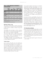
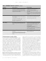
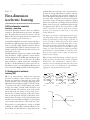
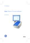
P A R T
I I .
F I R S T - D I M E N S I O N
Part II
First-dimension
isoelectric focusing
3.0 First-dimension isoelectric
focusing—overview
Amersham Pharmacia Biotech offers two different
systems for the first-dimension separation: the Multiphor II system with associated accessories, and the
IPGphor Isoelectric Focusing System. A comparison of
these two systems is given in section 1.2.
A useful first-dimension separation requires selecting a
first-dimension pH range appropriate for the sample as
well as a suitable sample application method. Choice
of immobilized pH gradient is discussed in section 3.2.
Sample application methods and their selection are
discussed in section 3.3.
The first-dimension separation procedure involves IPG
strip rehydration, sample application, and isoelectric
focusing. Preparation of the IPG strip rehydration solution is described in section 3.4. The protocols for IPG
strip rehydration, sample application, and IEF are
specific to the first-dimension system used and are
described in section 3.5 for the Multiphor II system, and
section 3.6 for the IPGphor Isoelectric Focusing System.
I S O E L E C T R I C
gradient where its net charge is zero. A protein with a
positive net charge will migrate toward the cathode,
becoming progressively less positively charged as it
moves through the pH gradient until it reaches its pI.
A protein with a negative net charge will migrate
toward the anode, becoming less negatively charged
until it also reaches zero net charge. If a protein should
diffuse away from its pI, it immediately gains charge
and migrates back. This is the focusing effect of IEF,
which concentrates proteins at their pIs and allows
proteins to be separated on the basis of very small
charge differences.
The degree of resolution is determined by the slope of
the pH gradient and the electric field strength. IEF is
therefore performed at high voltages (typically in
excess of 1,000 V). When the proteins have reached
their final positions in the pH gradient, there is very
little ionic movement in the system, resulting in a very
low final current (typically below 1 mA). IEF of a given
sample in a given electrophoresis system is generally
performed for a constant number of volt-hours. (Volthours is the product of the voltage and the hours
elapsed at that voltage.)
IEF performed under denaturing conditions gives the
highest resolution and the cleanest results. Complete
denaturation and solubilization achieved with a
mixture of urea and detergent ensure that each protein
is present in only one configuration and minimizes
aggregation and intermolecular interaction.
3.1 Background to isoelectric
focusing (IEF)
IEF is an electrophoretic method that separates
proteins according to their isoelectric points (pI).
Proteins are amphoteric molecules; they carry either
positive, negative, or zero net charge, depending on the
pH of their surroundings (see Figure 7). The net charge
of a protein is the sum of all the negative and positive
charges of its amino acid side chains and amino- and
carboxyl- termini. The isoelectric point is the specific
pH at which the net charge of the protein is zero.
Proteins are positively charged at pH values below
their pI and negatively charged at pH values above
their pI. If the net charge of a protein is plotted versus
the pH of its environment (see Figure 7), the resulting
curve intersects the abscissa at the isoelectric point.
The presence of a pH gradient is critical to the IEF
technique. In a pH gradient, under the influence of an
electric field, a protein will move to the position in the
1 4
|
U S I N G
I M M O B I L I Z E D
P H
G R A D I E N T S
F O C U S I N G
COO
COOH
COO
NH 3
NH 3
NH 2
COOH
COO
COO
NH 3
NH 3
pH < pl
NH 2
pH = pl
pH<pI
pH > pl
pH<pI
pH<pI
Net Charge
+3
+2
Isoelectric point (pl)
+1
0
3
-1
-2
-3
Figure 7
4
5
6
7
8
9
10
11 pH