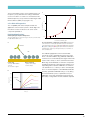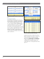
1
Protein Interaction Analysis
ProteOn™ XPR36 Experimental Design
and Application Guide
ProteOn XPR36 Experimental Design and Application Guide
Introduction Chapter 1 ProteOn XPR36 Technology 5
9
1.1 ProteOn XPR36 Technology Overview 1.2What Kind of Information is Obtained
with the ProteOn XPR36 System? 1.3How are Kinetic Parameters Obtained?
1.4 Advantages of the 6 x 6 Interaction Array
1.5 ProteOn Webinar Library
4.2.7Ligand Capture by Biotin Label or Histidine-Tag —
The NLC, HTG, and HTE Sensor Chip
4.2.8Summary
64
65
10
4.3Guide to Analyte Interaction
on the ProteOn XP36 System
66
11
12
13
17
4.3.1Introduction
4.3.2 Full Kinetic Profile
4.3.3 Determination of Analyte Concentrations
4.3.4 Analyte Preparation
4.3.5 Analyte Injection Parameters
4.3.6Analysis of Binding Results
66
66
66
66
66
67
Chapter 2 ProteOn Sensor Chips 19
2.1Overview
2.2 ProteOn Sensor Chip Surface Chemistry
2.3 Types of ProteOn Sensor Chips
20
20
21
4.4Guide to SPR Data Processing
on the ProteOn XPR36 System
68
4.4.1
4.4.2
4.4.3
4.4.4
4.4.5
68
68
69
70
72
2.3.1Amine Coupling ProteOn Sensor Chips:
GLC, GLM, and GLH
2.3.2ProteOn Sensor Chips for Site-Specific Attachment:
NLC, HTG, and HTE
2.3.3ProteOn Sensor Chips for Capturing Lipid
Assemblies: Modified GLC and LCP
21
23
24
2.4Guidelines for Choosing the Right
ProteOn Sensor Chip
26
Chapter 3 Applications
29
3.1Overview
3.2 Large and Small Molecule Screening
30
30
3.2.1 Antibody Kinetic Screening 3.2.2 Epitope Binding and Mapping
3.2.3Quantikinetics
3.2.4 Drug Compound Screening
30
33
34
35
3.3 Biomolecule Characterization
40
3.3.1
3.3.2
3.3.3
3.3.4
3.3.5
3.3.6
3.3.7
3.3.8
40
46
47
48
49
52
52
53
Structural Biology
Thermodynamics and Energetics
Histidine-Tagged Protein Analysis
Nucleic Acid Interaction Analysis
Lipid Membrane and Membrane Protein Analysis
Cell-Antibody Interaction Analysis
Regenerable Biotin-Capture Surface
SPR-MS Analysis
3.4 Biological Assays
54
3.4.1 Assay Design and Optimization
3.4.2 Biosimilar Assessment
54
56
3.5 Biomedical Applications
56
3.5.1 Vaccine Characterization
3.5.2 Clinical Diagnostics
56
57
Chapter 4 Experimental Design
59
4.1 Introduction to SPR Experimental Design 60
4.1.1 ProteOn XPR36 System
4.1.2 Checklist of Good Publication Standards
4.2Guide to Ligand Immobilization
on the ProteOn XPR36 System
4.2.1Conditioning
4.2.2Activation
4.2.3Immobilization
4.2.4Deactivation
4.2.5Stabilization
4.2.6Ligand Capture Using Capture Proteins —
Antibody Screening
60
60
62
62
62
63
64
64
64
Interaction Sensorgram Terms
Sensorgram Display
Sensorgram Processing
Sensorgram Referencing
Quality Standards for Processed Sensorgrams
4.5Guide to SPR Data Analysis
on the ProteOn XPR36 System
73
4.5.1
4.5.2
4.5.3
4.5.4
4.5.5
4.5.6
4.5.7
73
76
77
77
78
79
80
Kinetic Analysis
Equilibrium Analysis
Concentration Analysis
Report Point
Data Presentation
Sensorgram Appearance
Quality Standards for SPR Results
4.6How to Perform Excluded Volume
Correction on the ProteOn XPR36
Protein Interaction System
4.7Data Processing and Analysis Flowchart
4.8Options for Dataset Export
81
84
86
Chapter 5 Tips and Techniques
89
5.1Tips for Using ProteOn Sensor Chips 90
5.2Running Experiments with Sensor Chips 91
5.2.1Conditioning
5.2.2 Ligand Immobilization
5.2.3 Troubleshooting Ligand Immobilization
5.2.4Stabilization
5.2.5 Analyte Injection
5.2.6Regeneration
91
92
94
96
96
100
Chapter 6 Frequently Asked Questions 103
6.1Basics
6.2Sensorgram
6.3 Sensor Chips
6.4 Experimental Design
6.5 Experimental Tips
104
104
104
106
108
Chapter 7 Quick Guides 111
7.1Writing a ProteOn XPR36 Experiment
Protocol112
7.2Running an Experiment
with the ProteOn XPR36 System
114
7.2.1
7.2.2
7.2.3
7.2.4
Instrument Preparation
Running an Experiment
Instrument Maintenance
Import/Export Experiment Files
114
114
114
115
3
ProteOn XPR36 Experimental Design and Application Guide
4
Introduction
Introduction
Explore the World of
Parallel Surface Plasmon
Resonance Analysis
5
ProteOn XPR36 Experimental Design and Application Guide
Measuring Biomolecular
Interactions with SPR
Surface plasmon resonance, or SPR, is a biosensor
technology enabling label-free and real-time measurement
of biomolecular interactions. A typical SPR experiment
involves first immobilizing a ligand (a biomolecule such as
a protein or a nucleic acid) to the functionalized surface of
a sensor chip and then flowing an analyte (an interacting
biomolecule such as another protein or a small molecule)
over the chip surface to investigate the binding affinity
and binding kinetics between the analyte and the ligand.
The binding of the analyte to the ligand is tracked
in real time by following the change in SPR signal
over time, and this time-traced graph is called a
sensorgram. Fitting the sensorgram to a suitable
kinetic binding model allows for the calculation of
kinetic parameters such as the association rate
constant (ka in the unit of M-1s-1), the dissociation
rate constant (kd in the unit of s-1), and the
equilibrium constant (KD in the unit of M).
Biomolecular interaction in a new light. Two biomolecules, A and B, interact with each other to form a complex AB. Using an SPR
biosensor, along with the equilibrium constant K D, the association rate constant ka and the dissociation rate constant kd can be measured,
determining more details of the interaction compared to other methods.
6
Introduction
Advantages of the
ProteOn™ XPR36 System
The ProteOn XPR36 protein interaction array system is
an SPR biosensor platform that provides real-time labelfree analysis of the specificity, affinity, and kinetics of
biomolecular interactions. Using the XPR36 configuration,
the system features a novel 6 x 6 interaction array for the
simultaneous analysis of up to six ligands with up to six
analytes. The unique design increases the versatility of
experimental design and the productivity of experimental
workflow, enabling the completion of high quality SPR
experiments with high efficiency. The advantages position
the ProteOn XPR36 system as an optimal SPR platform
in label-free drug discovery and structural biology
applications with high quality results and excellent
cost-effectiveness.
The parallel-flow SPR biosensor platform:
■■
Analyzes up to 36 different protein interactions
in a single run on a single chip
■■
■■
■■
■■
easures a variety of experimental conditions
M
simultaneously using parallel-flow fluidics
Screens multiple panels of analytes
cquires the resonance angle shift as SPR response
A
units (RU) for accurate kinetics
mploys One-shot Kinetics™ technology, which
E
enables a complete kinetic analysis in a single run
This book describes how to apply the ProteOn XPR36 system for SPR analysis of biomolecular interactions. It includes technical
introductions, user guides, and tips and techniques.
7
ProteOn XPR36
Application
Guide
Experimental
for Process Design
Development
and Application
and Scale-Up
Guide
8
ProteOn XPR36 Technology
CHAPTER 1
ProteOn XPR36 Technology
™
Bio-Rad Laboratories has remained at the center of scientific discovery
for more than 60 years. The company is renowned worldwide among
hospitals, universities, and major research institutions, as well as
biotechnology and pharmaceutical companies, for its commitment
to quality and customer service. Among Bio-Rad's groundbreaking
contributions is the ProteOn XPR36 protein interaction array system,
an optical biosensor capable of simultaneously analyzing 36 individual
biomolecular interactions in a label-free manner. It has been widely
accepted and used as a fully automated, high-performance, and highthroughput surface plasmon resonance (SPR) biosensor platform.
9
ProteOn XPR36 Experimental Design and Application Guide
Surface plasmon resonance (SPR) is an optical
phenomenon that occurs when p-polarized light at
a certain wavelength and angle is reflected off a thin
metal film (the gold film coated on a sensor chip) under
the condition of total internal reflection (TIR). The light
excites surface plasmons in the metal at a certain
incident angle. The TIR field generates an evanescent
wave in the thin metal film that extends hundreds of
nanometers from the surface into the medium above, in
this case the molecules in contact with the chip surface.
The excited surface plasmons are very sensitive to
the refractive index change at the surface of the thin
metal film. Thus the incident angle of the light required
for SPR is impacted by the refractive index change of
the molecules in contact with the chip surface. In an
SPR binding experiment, this refractive index change is
brought about by binding of analyte in solution to ligand
immobilized on the chip surface; therefore, tracking the
change in the incident angle required for SPR allows
one to monitor biomolecular interactions in real time.
The change of the incident angle required for SPR is
defined as SPR response in the unit of response unit
(RU). 1 RU is 1/1,000,000 of 1 refractive index unit, and
is roughly equivalent to a surface density of protein
at approximately 1 pg/mm2. For a more in-depth
discussion of SPR, see a recent review that offers an
overview of SPR theory and different SPR configurations
(Daghestani et al. 2010). This reference describes the
theory and application of a number of optical biosensors,
including surface plasmon resonance biosensors, and
the different configurations for each.
Plotting the SPR response over time during the interaction
between an analyte and a ligand results in a sensorgram.
A sensorgram is a visual presentation of the interaction.
Figure 1.1 illustrates the terms for a sensorgram using,
for instance, an antibody-antigen interaction. The binding
response initially increases as analyte is flowed over the
sensor chip and associates with the immobilized ligand
and then decreases as the analyte solution is replaced
with buffer and the binding complex dissociates. If binding
equilibrium is reached during the association phase,
the sensorgram will reach a constant plateau before the
analyte solution is replaced with buffer and the binding
complex dissociates. Fitting the sensorgram data to a
binding model allows for the calculation of the association
10
and dissociation rate constants and determination of
the binding affinity. Traditionally, kinetic measurements
with SPR usually involve sequential injections of analyte
at increasing concentrations over the same ligand
surface, which requires complete removal of the analyte
or regeneration of the ligand surface between analyte
injections. In an ideal case, regeneration of the ligand
surface is observed in the sensorgram as a sharp
response change after dissociation to restore the baseline
to the original level. Regeneration is usually done with
a combination of dilute surfactants, salts, and acids or
bases; however, care must be taken during regeneration
to avoid denaturing the immobilized ligand or removing
ligand from the sensor chip.
The ProteOn XPR36 system characterizes the following
aspects of a biomolecular interaction:
■■
■■
■■
■■
Specificity of the interaction
Rate of the interaction (ka)
Stability of the complex (kd)
Strength of the interaction (K D [K D = kd / ka ])
The parameters are obtained from the data fitting of
the association, equilibrium (optional), and dissociation
phases of a sensorgram.
Baseline
Association
Equilibrium
Dissociation
Regeneration
Equilibrium
Response, RU
1.1 ProteOn XPR36 Technology
Overview
Association
Dissociation
Regeneration
Baseline
Time, sec
Fig. 1.1. SPR sensorgram. Surface ( ); ligand ( ); analyte ( ). RU,
response units.
ProteOn XPR36 Technology
The ProteOn XPR36 system offers a distinct advantage
over other SPR biosensor platforms because the unique
6 x 6 interaction array of the ProteOn sensor chips
enables the One-shot Kinetics™ approach measuring the
interaction of one ligand with a six-concentration series of
one analyte in a single injection. This approach eliminates
the need for traditional regeneration of the sensor chip
between analyte injections, which often deteriorates the
ligand surface. Using enhanced microfluidic delivery
and XPR™ technology, the ProteOn XPR36 system can
immobilize up to six separate ligands on a single sensor
chip in six separate flow cells and then rotate the sensor
chip 90 degrees to flow up to six separate analytes over
the ligand surfaces (Figure 1.2).
This unique feature of the ProteOn allows for the
detection of up to 36 separate interactions on a single
sensor chip and significantly increases the throughput of
SPR biosensing. In a recent study, the ProteOn XPR36
system was used to immobilize 36 different ligands
in a stepwise immobilization procedure designed for
the high-throughput epitope mapping and binding of
antibody-antigen interactions (Abdiche et al. 2011).
The 6 x 6 interaction array of the ProteOn sensor chips
also allows for inline referencing, whereby data from
unmodified spots in between the immobilized ligand
spots on the sensor chip are used to subtract out
artifacts such as noise and baseline drift. This inline
referencing is superior to referencing with a separate
flow cell and means the ProteOn XPR36 system can
collect high-quality SPR data at the low signal-to-noise
ratios often seen with small molecule analytes.
1.2 What Kind of Information is
Obtained with the ProteOn XPR36
System?
As an SPR biosensor platform, the ProteOn XPR36
system provides a wide variety of important information
on biomolecular interactions such as the specificity,
affinity, qualitative ranking, kinetics, and thermodynamics
of binding.
The ProteOn XPR36 system can be used in
pharmaceutical drug discovery, antibody characterization,
immunogenicity testing, the development and manufacture
of biologics, or for clinical research. It could also be
used in other fields where there is a need for labelfree characterization of biomolecular interactions. Key
applications include:
■■
■■
Quantification of binding affinity and kinetics
etermination of binding specificity and the number
D
of binding sites
■■
Characterization of the mechanism of action
■■
Confirmation of biomolecule binding to a target
■■
Screening of fragment libraries
■■
■■
Validation of IC50/EC50 values during hit-to-lead
optimization
Characterization of immune responses
Step 1
Step 2
Step 3
Immobilize up to 6 ligands
in the vertical direction.
Inject up to 6 analytes
in the horizontal direction.
Detail showing one
of 36 interaction spots.
Fig. 1.2. The ProteOn 6 x 6 interaction array on a sensor chip.
11
ProteOn XPR36 Experimental Design and Application Guide
Some classic applications of the ProteOn XPR36 system
are in antibody engineering. Epitopes on an antigen
can be characterized by epitope mapping, a process
by which the affinities of an antibody to site-directed
mutants of a single antigen help pinpoint the location of
an epitope. An investigation of the epitope specificity,
or epitope binding, of different antibodies can be done
on the ProteOn XPR36 system using the sandwich
assay. In this assay, a second antibody is injected over
a previously-formed antigen-antibody complex to see
whether or not the second antibody can still bind. Binding
of the second antibody to the antigen-antibody complex,
or the formation of a “sandwich” is an indication that the
second antibody recognizes a different epitope than the
first antibody.
SPR can also be used to determine the active
concentration of an analyte in a crude or impure
sample by probing the sample of interest under mass
transport control using a low flow rate and/or a highcapacity sensor chip bearing an analyte-specific
ligand. Under such mass transport limited conditions
the association rate of binding, or the initial binding
rate, is proportional to the concentration of analyte in
solution. The concentration of analyte in a crude sample
can be calculated by comparing the initial binding rate
to a standard curve of initial binding rates for known
concentrations.
1.3 How are Kinetic Parameters
Obtained?
By fitting sensorgram data from a ProteOn XPR36
system experiment to a suitable binding model, kinetic
parameters such as the association rate constant (ka),
dissociation rate constant (kd), and the equilibrium or
affinity constant (KD) can be extracted. Kinetic data are
crucial for characterizing an interaction, as they allow
for a thorough understanding of the nuances of binding.
Interactions with the same affinity can have markedly
different association and dissociation rates, as seen in
Figure 1.3. An antibody or small molecule that has a high
affinity (low KD value) for a protein target may be a poor
drug in vivo if it has a very high dissociation rate and thus
can be easily displaced by another molecule. This kind
of information is easily obtained from an SPR experiment
but would not be uncovered using a method such as
isothermal calorimetry (ITC) that measures binding affinity
based on binding at equilibrium. In addition, knowing
the kinetics of a small molecule interaction allows for
more accurate analysis of quantitative-structure activity
relationships, as different structures can be evaluated by
their separate effects on association and dissociation as
opposed to affinity alone.
Association
Dissociation
100
KD = 1.0 nM
[A] = 10 nM
Response, RU
80
60
ka
(M –1sec –1)
kd (sec –1)
1 x 10³
1 x 10⁴
1 x 10⁵
1 x 10 6
1 x 10⁷
1 x 10 –6
1 x 10 –5
1 x 10 –4
1 x 10 –3
1 x 10 –2
40
20
0
0
200
400
600 800 1,000 1,200 1,400
Time, sec
Fig. 1.3. Sensorgram plots showing the response (in RU)
versus time for five different interactions with the same affinity
(KD = 1.0 nM) but markedly different association (ka) and
dissociation (kd) rate constants. A, antibody.
Apply up to 6 unique target
molecules, such as mutant
or wild-type proteins.
Figure 1.4. The experimental workflow of the 6 x 6 interaction array.
12
Evaluate binding against
6 analytes, such as small
inhibitor molecules.
ProteOn XPR36 Technology
ProteOn Manager™ software gives you the option of
using seven different binding models to analyze your
sensorgram data:
■■
■■
■■
■■
■■
■■
■■
Langmuir — simple 1:1 bimolecular interaction
- Simultaneous fitting of ka and kd
- Fitting of kd only
Langmuir with drift — simple 1:1 biomolecular
interaction with a constant baseline drift taken into
account
Langmuir with mass transport limitations — simple
1:1 biomolecular interaction that takes into account
the rate of diffusion of analyte from the bulk to the chip
surface
1.4 Advantages of the
6 x 6 Interaction Array
When running an experiment using the ProteOn XPR36
system, the 6 x 6 interaction is formed. The experimental
workflow is shown in Figure 1.4.
The 6 x 6 interaction array brings these benefits
to SPR experiments:
■■
■■
■■
Experimental versatility
High productivity
High data quality
ivalent analyte — one analyte has two binding sites
B
to one ligand
eterogeneous analyte — two analytes compete for
H
binding to one ligand
eterogeneous ligand — one analyte binds to two
H
ligands
wo-state conformation — accounts for a change in
T
conformation of the binding complex that occurs after
the analyte binds. In addition, it is possible to calculate
the affinity value (KD) using equilibrium analysis, in
which the equilibrium responses at different analyte
concentrations are filled to a simple saturation binding
model
13
ProteOn XPR36 Experimental Design and Application Guide
Experimental Versatility — Multiple
Experimental Configurations and Fast
Qualitative and Quantitative Assays
Kinetic Characterization (1-to-1)
In kinetic characterization experiments, the optimization of experimental
protocols is usually the most labor-intensive and time-consuming step.
Probing at one time six ligand immobilization conditions together with six
analyte injection conditions, the ProteOn XPR36 system allows for full
optimization in a single run. This ensures the optimal experimental conditions
for the interaction between the ligand and the analyte. The method of using a
single run of 6 x 6 injections for a complete kinetic analysis is called the Oneshot Kinetics approach.
6 variations of a target
6 concentrations of the
same analyte
Kinetic Screening (6-to-1)
In kinetic screening experiments, each of the six ligand channels gives
a full kinetic analysis in a single run. This high throughput enables fast
processing of a large number of samples while accurate kinetics is
maintained. The ProteOn XPR36 system provides the best balance between
throughput and accuracy of kinetic screening.
6 different targets
6 concentrations of the
same analyte
Multiplex Screening (6-to-6) and Array Screening (36-to-1)
In multiplex or array screening experiments, the 6 x 6 interaction array
of the ProteOn XPR36 system is fully utilized for high throughput, which
enables multiplex or 36-ligand screening.
6 different targets
6 different analytes
36 different targets
1 analyte
14
ProteOn XPR36 Technology
High Productivity — Fast Protocol
Optimization and High Throughput
Sensor chip surface
Kinetic Characterization
■■
■■
roteOn XPR36 system: 1.1 hr, 6 full kinetics
P
(36 data points, surface regeneration not required)
onventional serial flow SPR system: 3.5 hr, 3 full
C
kinetics (18 data points, surface regeneration required)
Kinetic Screening
Captured Ligand Screening (for mAb supernatants)
■■
■■
roteOn XPR36 system: 11 hr, 96 full kinetics
P
(576 data points)
Conventional serial flow SPR system: 65 hr,
96 full kinetics (576 data points)
Analyte Screening
■■
■■
roteOn XPR36 system: 25 hr, 96 x 6 full kinetics
P
(3,456 data points)
Conventional serial flow SPR system: 5.7 days,
96 x 3 full kinetics (1,728 data points)
Multiplex Screening
■■
■■
roteOn XPR36 system: 0.7 hr, 6 x 6 binding matrix
P
(36 data points)
Conventional serial flow SPR system: 2.5 hr,
3 x 6 binding matrix (18 data points)
Array Screening
■■
■■
roteOn XPR36 system: 12 hr, 36 x 36 binding matrix
P
(1,296 data points)
Conventional serial flow SPR system: no equivalent
15
ProteOn XPR36 Experimental Design and Application Guide
High Data Quality — Four Factors of the
ProteOn XPR36 System for High-Quality SPR
Results
1
Sufficient Signal-to-Noise Ratio
Response, RU
48
32
16
0
0
50
Data Processing
Data Referencing
The key step in data processing is data referencing.
Data referencing corrects for the artifacts in SPR
experimental results.
SPR System
–4
–50
3
100
Time, sec
150
200
Fig. 1.5. ProteOn XPR36 system signal-to-noise ratio. ProteOn
XPR36 system noise is 1 RU and ~2 RU after double referencing. SPR
responses over three times signal-to-noise ratio (3 x SNR) are detectable.
RU, response units.
The ProteOn XPR36 system has two novel
advantageous referencing modes that no other SPR
system provides: an interspot reference to correct for
refractive index change (bulk effect) and nonspecific
binding, and a real-time injection reference to correct
for baseline drift resulting from the changes in the ligand
surface.
Note: For additional information about the referencing options in
the ProteOn XPR36 system, watch the video at www.bio-rad.com/
proteon/reference.
A
Baseline drift
6 ligand injections
5 analyte
injections
240
200
160
120
80
40
0
–200 0
500
1,000
1,500
2,000
Time, sec
2,500
3,000
3,500
Interspots
Fig. 1.6. Evaluation of kd value reproducibility using the ProteOn
One-shot Kinetics kit. 2 systems x 3 chips x 6 ligand channels x 6
analyte channels = 216 sensorgrams. CV = 6.1% (over 2 systems and 6
sensor chips). RU, response units.
2 Experiment Design
Baseline corrected
Buffer
injection
Response, RU
Instrument Stability
B
ProteOn XPR36 System Configuration Optimizes Multiple
Factors Simultaneously
Analyte
Ligand
Vertical channels
Optimize ligand immobilization
Capture reagent
Surface chemistry
Ligand conditions
Surface
Horizontal channels
optimize analyte injection
Analyte conditions
Referencing options
Flow rate
Sufficient SNR
Ligand is washed off in dissociation, causing exponential drift.
Fig. 1.7. Optimal experimental conditions are obtained in a single
run.
16
Fig. 1.8. Novel ProteOn XPR36 system references. The ProteOn
XPR36 system provides A, an interspot blank surface reference to save
interaction spots and provide immediate proximate referencing and B,
a real-time injection reference to correct the exponential baseline drift
when using ligand-capture surface chemistry.
ProteOn XPR36 Technology
4
Data Analysis
Software Advantages
ProteOn Manager software is a comprehensive, userfriendly tool for the analysis of biomolecular interactions.
Features include:
Ease of use
■■ Integration of data acquisition, data processing,
and data analysis
■■ Powerful graphic user interface
■■ Intuitive protocol writing interface
■■ Fast and accurate data processing
■■ Accurate fitting with 8 models
■■ Rapid data analysis
■■ Concise analysis reports
■■ Export functions for further data processing
in Excel or other software
■■
1.5 ProteOn Webinar Library
The ProteOn webinars feature presentations from
thought leaders in the field of label-free biomolecular
interaction analysis who use surface plasmon resonance
(SPR) technology.
Visit www.bio-rad.com/info/proteon to view Bio-Rad’s
extensive library of ProteOn webinars and to sign up for
future webinars.
References
Abdiche YN et al. (2011). Expanding the ProteOn XPR36 biosensor into
a 36-ligand array expedites protein interaction analysis. Anal Biochem
411, 139–151.
Daghestani HN et al. (2010). Theory and applications of surface
plasmon resonance, resonant mirror, resonant waveguide grating, and
dual polarization interferometry biosensors. Sensors 10, 9630–9646.
Fig. 1.9. ProteOn Manager software data analysis window. 17
ProteOn XPR36 Experimental Design and Application Guide
18
ProteOn Sensor Chips
CHAPTER 2
ProteOn Sensor Chips
™
Used with the ProteOn XPR36 system, ProteOn sensor chips are
built with an alginate polymer matrix (or a self-assembled monolayer
of organic molecules) bound to a thin gold film on a sensor prism.
The sensor chips can be functionalized with several different reactive
groups to achieve a variety of immobilization surface chemistries.
The surface chemistry of the ProteOn sensor chips allows the
ProteOn XPR36 system to detect tight binding interactions down
to picomolar concentrations of analytes, or analytes as small as
95 daltons (Da). The combination of ProteOn sensor chips with the
unique 6 x 6 interaction array allows the interaction analysis of up to
36 separate ligand/analyte pairs on a single chip, thereby increasing
the throughput of a single experiment.
19
ProteOn XPR36 Experimental Design and Application Guide
2.1 Overview
The ProteOn XPR36 system uses the 6 × 6 interaction
array of the ProteOn sensor chips. ProteOn sensor chips
contain more than just a gold layer; they are coated with
a modified alginate polymer that provides a solutionlike, biomimetic environment for ligand immobilization.
The general-use ProteOn GLC, GLM, and GLH chips
are functionalized with carboxyl groups that react with
surface-exposed amines on the ligand, tethering the
ligand to the chip surface in a random orientation.
The ProteOn NLC chip is coated with NeutrAvidin for
immobilization of biotinylated ligands, and the HTG and
HTE chips feature a tris-nitrilotriacetate (tris-NTA or
3 x NTA) surface for immobilization of histidine-tagged
proteins. The respective chips can be used to capture a
ligand at a site-specific location. The ProteOn LCP chip
is used with the LCP capturing reagent kit for liposome
capture. Although choosing a suitable sensor chip for
a particular interaction requires some research and
planning, this is time well spent, considering the highquality data that can be obtained with the right sensor
chip.
2.2 ProteOn Sensor Chip Surface
Chemistry
ProteOn sensor chips are built with an alginate polymer
matrix bound to a thin gold film on a sensor prism. The
alginate matrix can be functionalized with several different
reactive groups to facilitate different immobilization
surface chemistries. The hydrophilic nature of the alginate
layer creates a solution-like environment that prevents
denaturation of the immobilized ligand and nonspecific
adsorption of the analyte. The increased surface area
of the 3-D structure of the alginate layer provides more
attachment sites than would a completely flat surface
and results in more ligand molecules being immobilized
on the chip surface. In addition, the molecular weight
and structure of the alginate coating can be modified to
create sensor chips with different surface capacities. This
results in sensitive detection of interactions with minimal
surface effects on binding. The surface chemistry of the
ProteOn sensor chips allows the ProteOn XPR36 system
to detect tight binding interactions down to picomolar
concentrations of analytes, or analytes as small as 95 Da.
The ProteOn sensor chips, combined with the unique
6 x 6 interaction array, allow for the interaction of up to
36 separate ligand/analyte pairs on a single chip,
increasing the throughput of a single experiment.
The ProteOn sensor chips are based on innovative,
patent-protected surface chemistry. The general-use
ProteOn sensor chips (GLC, GLM, and GLH sensor
chips) come functionalized with carboxyl groups
to facilitate amine coupling of protein ligands via
surface-exposed amine groups. In addition to serving
as attachment sites, the carboxyl groups serve to
concentrate the ligand at the surface of the sensor
chip as the negatively charged carboxyl groups attract
proteins rendered positively charged via incubation in
an acidic buffer. Activation of carboxyl groups for ligand
immobilization is done using carbodiimide chemistry
with the reagents 1-ethyl-3-(3-dimethylaminopropyl)
carbodiimide (EDC) and N-hydroxysulfosuccinimide
(sulfo-NHS). Once activated, the resulting sulfo-NHS
esters are highly amine-reactive and react with free
amines exposed on the ligand to immobilize it to the
sensor chip. Any unreacted carboxyl groups that remain
activated during the immobilization step are deactivated
with ethanolamine to prevent immobilization of analyte
protein during the subsequent interaction step. The
chemical structure of the binding layers forms easilyactivated carboxyl groups, rendering especially high
binding capacity and ligand activity. It results in more
active ligand on the surface, thus higher analyte signals
and higher assay sensitivity.
Different amounts of ligand may be amine-coupled to
the GLC, GLM, and GLH sensor chips by controlling the
amount of ligand in solution during the immobilization,
by tuning the activation level, or by adjusting the length
of time of the immobilization step. The amount of ligand
bound is monitored by following the binding response
displayed on the sensorgram in real time. However,
specific applications may require a surface with a very
high or low ligand surface capacity. In such cases GLH
and GLC chips that have a very high or low density
alginate coating may be used.
For immobilizing targets through a site-specific tag,
ProteOn sensor chips such as the NLC, HTG, and
HTE chips can be used. The NLC chip is coated with
NeutrAvidin for immobilization of biotinylated ligands, and
the HTG and HTE chips are coated with a unique and
innovative tris-NTA surface for immobilization of histidinetagged proteins.
Sensor chips come packaged in a sealed pouch with an
inert gas, have a shelf life of two years if stored properly
at 4°C, and are guaranteed for six months from the date
of receipt. Sensor chips are continually monitored for
quality and have excellent spot-to-spot reproducibility
within the 6 x 6 interaction array.
Read more to learn about the different sensor chip
types (Figure 2.1) and which is best for your specific
application.
20
ProteOn Sensor Chips
2.3 Types of ProteOn Sensor Chips
There are seven types of ProteOn sensor chips that can
be used for a variety of different immobilization strategies
and the creation of different capacity surfaces. The GLC,
GLM, and GLH sensor chips are designed for general
amine coupling of ligands, whereas the NLC sensor chips
are designed for site-specific attachment of biotinylated
ligands and the HTG and HTE chips for histidine-tagged
proteins, respectively. Figure 2.1 shows an overview of the
different sensor chip types and the specific applications
in which they are used.
GLC Sensor Chip — Compact Binding Capacity
The GLC sensor chip is designed with an extremely
thin alginate layer for amine coupling of protein ligands
at a compact (~6 kRU) surface capacity. The compact
structure of the alginate layer helps mitigate mass
transport effects that are more often observed with
thicker layers of surface coating. This versatile sensor
chip is ideal for analyzing protein-protein interactions
(Figure 2.2).
A
Analyte
COOH
Prism
Incident light
Detector
B
Response, RU
160
120
80
40
0
Chip Selector
Compact capacity amine coupling for protein-protein
GLC
interactions
GLM
Medium capacity amine coupling for protein-protein
and protein–small molecule interactions
GLH
High capacity amine coupling for protein–small molecule
interactions
NLC
NeutrAvidin for biotinylated molecule capture
HTG
Compact capacity tris-NTA for histidine-tagged large molecule
capture
HTE
High capacity tris-NTA for histidine-tagged small molecule
capture
LCP
Used with the LCP capturing reagent kit for liposome capture
Fig. 2.1. An overview of the seven different types of ProteOn sensor
chips (GLC, GLM, GLH, NLC, HTG, HTE, and LCP) with the specific
application for each chip listed.
0
2
4
6
Time, min
8
10
12
Fig. 2.2. ProteOn GLC sensor chip. A, the thin alginate coating on the
GLC sensor chip responsible for creating a compact-capacity surface; B,
sensorgrams of the interaction between the cytokine IL-2 and the anti–IL-2
antibody using the GLC sensor chip. The IL-2 antibody was immobilized
to approximately 2,000 RU, and IL-2 was injected in a twofold dilution
series ranging from 80–2.5 nM. The nearly planar surface of the GLC
sensor chip allows for high-quality kinetic analysis. RU, response units.
GLM Sensor Chip — Medium Binding Capacity
The GLM sensor chip is coated with a thicker alginate
polymer that displays a higher amount of carboxylic acid
groups and is thus ideal for creating medium capacity
(~12 kRU) ligand surfaces via amine coupling. It can be
used for both protein-protein interactions and protein–
small molecule interactions (Figure 2.3).
2.3.1 Amine Coupling ProteOn Sensor Chips:
GLC, GLM, and GLH
Three ProteOn sensor chips are available for amine
coupling. The GLC, GLM, and GLH chips provide for
compact, medium, and high ligand surface capacities,
respectively. All three of these chips are functionalized
with easily-activated carboxylic acid groups that can be
reacted with the activation reagents EDC and sulfo-NHS
to react specifically with free surface amines of proteins.
Bio-Rad offers the amine coupling kit for use with the
GLC, GLM, and GLH sensor chips.
21
ProteOn XPR36 Experimental Design and Application Guide
A
A
Analyte
Analyte
COOH
COOH
Prism
Incident light
Prism
Detector
Incident light
B
B
160
120
40
0
012345678
Time, min
Fig. 2.3. ProteOn GLM sensor chip. A, the extended alginate coating
on the GLM sensor chip responsible for creating a medium-capacity
surface; B, sensorgrams of a TEM1 b-lactamase mutant interacting with
the b-lactamase inhibitor protein (BLIP) using the GLM sensor chip. TEM1
was immobilized to approximately 1,500 RU, and BLIP was injected in a
twofold dilution series ranging from 600–38 nM. RU, response units.
GLH Sensor Chip — High Binding Capacity
The GLH sensor chip is designed with a high density
alginate polymer that contains an increased number
of carboxylic acid groups to amine coupling ligands
at a very high (>20 kRU) surface capacity. This dense
alginate layer on the GLH chip is far superior at binding
high capacity ligand surfaces when compared to results
of ligand immobilizations done using the GLC and GLM
chips (Figure 2.4). This sensor chip is ideal for probing
protein–small molecule (<1000 Da) interactions, as
the high capacity surface gives an increased binding
response. A comparison between the GLH sensor chip
and a competitor’s high capacity sensor chip shows
the full advantage of the ProteOn chip’s easily-activated
carboxylic groups, rendering significantly higher binding
capacity and activity, and thus much higher analyte
response (Figure 2.5 and Table 2.1).
22
Response, RU
80
80
60
40
20
0
0
1
Time, min
2
Fig. 2.4. ProteOn GLH sensor chip. A, the dense alginate coating on
the GLH sensor chip responsible for creating a high-capacity surface;
B, sensorgrams of the interaction between the carbonic anhydrase II
(30 kD) and the inhibitor 4-carboxybenzenesulfonamide (CBS)
(201 Da) using the GLH sensor chip. Carbonic anhydrase II was
immobilized to approximately 24,000 RU, and CBS was injected in a
threefold dilution series ranging from 20–0.082 µM. RU, response units.
16,000
Immobilization level, RU
Response, RU
200
Detector
12,000
nConventional layer, NHS
nConventional layer, sulfo-NHS
nProteOn layer, NHS
nProteOn layer, sulfo-NHS
8,000
4,000
0
Standard-capacity
chip type
High-capacity
chip type
Fig. 2.5. Comparative coupling efficiency. Representative data for
immobilization of rabbit IgG. Ligand coupling efficiency of the ProteOn
chip’s easily-activated layers is higher than in conventional layers, and
activation of ProteOn chip layers is higher using sulfo-NHS instead of
NHS. RU, response units.
ProteOn Sensor Chips
Table 2.1. Representative immobilization efficiencies on ProteOn sensor chip surfaces designed for high protein binding capacity.
Non-Bio-Rad
GLM Chip,
GLM Chip,
GLH Chip,
Chip, NHS NHS
Sulfo-NHS
Sulfo-NHS
Activation, Activation,Activation,Activation,
Protein
pIRU
RU RU RU
Pepsin
3
Ovalbumin
70
750 2,050 2,470
4.52,800
3,400
6,700 6,800
Protein A
5.1
4,300
3,500 6,000 b2-microglobulin
5.3
2,600
3,250
3,650 12,400
Carbonic anhydrase II
5.9
6,600–2,300
6,000
9,000
21,200
6.9 –7.4
3,900
2,800 7,000 12,200
6 – 8
10,000
9,700 12,200 22,200
Myoglobin
Polyclonal IgG
2.3.2 ProteOn Sensor Chips for Site-Specific
Attachment: NLC, HTG, and HTE
NLC Sensor Chip — Immobilization of Biotinylated
Ligands
The NLC sensor chip is functionalized with NeutrAvidin
bound to the alginate polymer and can capture
biotinylated proteins, peptides, and nucleic acids. It can
capture ~2,000 RU of IgG or ~500 RU of DNA. The NLC
sensor chip is ideal for immobilizing ligands without
amine coupling but requires that the ligand be modified
with biotin prior to immobilization (Figure 2.6).
A
Analyte
NeutrAvidin
Prism
Incident light
Detector
B
Response, RU
200
160
120
80
40
0
0 1234 567 89101112
Time, min
18,800
HTE and HTG Sensor Chips — Immobilization
of Histidine-Tagged Proteins
The HTG and HTE sensor chips feature a novel tris-NTA
(3 x NTA) complex for improved capture of histidinetagged proteins. This tris-NTA complex has a significantly
higher binding stability than the traditional NTA, so minimal
ligand leaches off the surface and the sensorgram
baseline remains stable. NTA is the traditional method
used to capture histidine-tagged proteins but the binding
is less tight, causes ligand to leach off the surface, and
results in unstable baselines and distorted kinetic results,
all of which can lead to an inaccurate fit to a binding
model. The tris-NTA complex contains three NTA moieties
for improved binding stability and increased binding
selectivity to histidine-tagged proteins (Figure 2.7A). The
tris-NTA complex is attached to the alginate polymer
matrix on the sensor chip and is activated by injecting
nickel (II) ions. In order to achieve optimal performance
in various applications, the surface density of tris-NTA
complex is distinguished in the two different sensor chips,
HTG for compact density and HTE for high density (Figure
2.7B and C). The HTG and HTE sensor chips allow easy
surface regeneration, chip reuse, and capture of histidinetagged proteins directly from crude cell lysates. The HTG
sensor chip is an ideal choice for protein-protein and
protein-peptide interaction analysis, and the HTE sensor
chip for protein–small molecule interaction analysis (Figure
2.8). Bio-Rad offers the HTG and HTE reagent kit for use
with the HTG and HTE sensor chips.
Fig. 2.6. ProteOn NLC sensor chip. A, the NeutrAvidin-modified alginate
coating on the NLC sensor chip; B, sensorgrams of the interaction
between an antibody Fab fragment and biotinylated MHC I/Tyr antigen
using the NLC sensor chip. MHC I was captured to approximately
800 RU, and the Fab was injected in a twofold dilution series ranging
from 500–31 nM. RU, response units.
23
ProteOn XPR36 Experimental Design and Application Guide
A
A
300
HO
OH
O
OH
HO
O
O
NTA
Response, RU
N
O
HO
NTA
O
NTA
200
100
N
O
O
O
N
N
0
OH
O
O
–200–100 0
N
O
O
OH
N
OH
O
OH
OH
N
HO
O
N
O
NTA
O
100 200 300 400 500 600 700 800
Time, sec
OH
B
60
Analyte
tris-NTA
Response, RU
B
40
20
0
Prism
Incident light
Detector
C
Analyte
tris-NTA
Prism
Incident light
Detector
Fig. 2.7. ProteOn HTG and HTE sensor chips. A, structure of surfacebound NTA molecule (left) and tris-NTA molecule (right). Each individual
NTA group is circled; B, alginate coating modified with compact-density
tris-NTA on the HTG sensor chip; C, alginate coating modified with highdensity tris-NTA on the HTE sensor chip.
– 80– 40 0 40 80 120
Time, sec
Fig. 2.8. ProteOn HTG and HTE sensor chips. A, sensorgrams of
the interaction between the histidine-tagged protein A and human
IgG, showing the ability of the HTG sensor chip to resolve high-affinity
kinetics requiring long dissociation times. Protein A was captured to
approximately 60 RU, and human IgG was injected in a twofold dilution
series ranging from 100 nM to 6.3 nM; B, sensorgrams of the interaction
between histidine-tagged Erk2 (an MAP kinase) and the inhibitor
Purvalanol B (432.9 Da), showing that small molecules can be screened
using the HTE sensor chip. Erk2 was captured to approximately 12,800
RU, and Purvalanol B was injected in a threefold dilution series ranging
from 50–0.62 µM. RU, response units.
2.3.3 ProteOn Sensor Chips for Capturing
Lipid Assemblies: Modified GLC and LCP
Biological research focused on biomolecular interactions
involving lipid assemblies, such as liposomes and
lipoparticles, allows the study of native membrane
proteins as well as the role of the lipid bilayer of these
assemblies in the activity of the membrane proteins.
Analyzing lipid assemblies also provides insights
into lipid-protein or lipid–small molecule interactions,
answering critical questions in the fields of drug delivery,
virology, and signal transduction.
Modified GLC Sensor Chip — Lipophilic Surface
Chemistry
The GLC sensor chip can be modified for capturing
lipid assemblies. The surface lipophilicity of the chip is
adjusted through the amine coupling of an alkyl chain
for capturing lipid substances. This capture approach
provides the flexibility to control the lipophilicity of the
chip surface for customized lipid-based applications.
24
ProteOn Sensor Chips
The modified GLC sensor chip provides a traditional
lipophilic surface chemistry for capturing lipid assemblies,
which allows for typical lipid-based applications. Bio-Rad
offers the GLC lipid kit as an all-in-one kit composed of
the GLC sensor chip and the lipid modification kit. The
advantages of this application kit are summarized below:
■■
■■
■■
■■
■■
Lipophilic surface chemistry
Flexibility in adjusting surface properties
Good regeneration capability
Low cost
High throughput
A
Analyte
NeutrAvidin
Prism
Incident light
B
Kinetic
analysis
LCP chip
LCP chip +
Biotin-ssDNA
4
■■
■■
■■
■■
■■
Hydrophilic surface chemistry
Low nonspecific binding
Multiple layer capture capability
High regeneration capability
High throughput
Chol-dsDNA 1
liposome
Fig. 2.9. ProteOn LCP sensor chip. A, planar NeutrAvidin-modified
self-assembled monolayer on the LCP sensor chip; B, workflow for
liposome capture using the LCP chip and the LCP capturing reagent
kit. Chol-dsDNA 1 and single-stranded biotinylated DNA molecules
(biotin-ssDNA) contain complementary DNA sequences. The LCP
sensor chip surface is saturated with biotin-ssDNA, and then liposomes
incubated with chol-dsDNA 1 are captured to the surface through DNA
hybridization. For reagents and techniques used in this workflow, refer
to Bio-Rad bulletin 6161.
A
30,000
Liposome layer 4
Response, RU
Liposome layer 3
20,000
Liposome layer 2
Liposome layer 1
10,000
0
0
1,000 2,000 3,0004,000 5,000 6,000 7,000 8,000
Time, sec
B
120
Response, RU
The LCP sensor chip provides a novel hydrophilic
surface chemistry for capturing lipid assemblies, which
allows for novel lipid-based applications. Bio-Rad offers
the liposome capturing kit as an all-in-one kit composed
of the LCP sensor chip and the LCP capturing
reagent kit. The advantages of this application kit are
summarized below:
Hybridization of the
two DNA strands
Biotin-ssDNA
LCP Sensor Chip — Hydrophilic Surface Chemistry
The LCP sensor chip provides a surface functionalized
with NeutrAvidin in a planar configuration that is formed
on a self-assembled monolayer (Figure 2.9). It is
designed to be used with the ProteOn LCP capturing
reagent kit for lipid-protein, lipid–small molecule and
membrane protein-protein interaction analysis. The
reagent kit activates the chip surface by a biotinylated
DNA tag so that the chip is able to capture DNA-labeled
lipid assemblies through DNA hybridization. The reagent
kit attaches DNA tags to the lipid assemblies in order to
anchor them to the chip surface. It is possible to capture
two or more layers of lipid assemblies for additional
sensitivity. This method of capture allows for lipid-based
interaction analysis, including the analysis of membrane
proteins embedded in a lipid bilayer (Figure 2.10).
Detector
80
40
0
–200 0
200 400 600 8001,000
Time, sec
Fig. 2.10. ProteOn LCP sensor chip. A, sensorgram of the stable
capture of four 1-palmitoyl-2-oleoyl-sn-glycero-3-phosphocholine (POPC)
liposome layers. Tagging complementary single stranded DNA molecules
to the liposomes facilitates the multiple layer capture capability; B,
sensorgrams of the interaction between FITC-labeled 1,2-Distearoylsn-glycero-3-phosphocholine (DSPC) liposomes captured on the LCP
sensor chip and an anti-FITC antibody. FITC-labeled DSPC liposomes
were captured to approximately 330 RU, and the anti-FITC antibody
was injected in a twofold dilution series ranging from 10–0.63 nM. RU,
response units.
25
ProteOn XPR36 Experimental Design and Application Guide
2.4 Guidelines for Choosing
the Right ProteOn Sensor Chip
Which sensor chip you choose depends on a number
of experimental parameters. To help decide you may
consider the following questions:
■■
■■
■■
■■
■■
What is the application?
What kind of information do I want to get from my
ProteOn experiment?
What type of samples am I working with?
– Proteins?
– Peptides?
– Biotinylated or histidine-tagged proteins?
– Small molecules?
– Whole cells?
– DNA?
Do I know anything about the affinity (tightness)
of binding?
What are the molecular weights in my sample?
For routine examination of protein-protein interactions
such as antibody and antigen, the GLC chip is a good
first choice. The GLM or GLH chip can be used as
needed if troubleshooting your interaction with the GLC
chip suggests that you need a higher capacity surface.
Knowing the molecular weight of your analyte is also
crucial for choosing the right sensor chip, as low
molecular weight analytes such as small molecules need
a higher capacity ligand surface and will benefit from the
enhanced sensitivity afforded by the GLH chip.
An additional consideration to take into account when
choosing a sensor chip is whether or not your interaction
is mass transport limited — in other words, whether or
not your interaction has a fast association rate. High
capacity surfaces exacerbate a mass transport limited
interaction because high density of ligand on the sensor
chip depletes the analyte in the surrounding solution very
quickly, whereas compact density of ligand mitigates
the influence of mass transport by decreasing the rate at
which analyte is depleted from the surrounding solution.
Thus, the GLC chip with a compact capacity surface
may be more suitable than the high capacity GLH chip in
dealing with a mass transport limited interaction.
26
If the ligand is biotinylated or contains a histidine-tag, the
NLC, HTG, and HTE chips can be used to immobilize
ligand on the chip surface without the need to use amine
coupling. An additional benefit of using the NLC, HTG,
and HTE chips is that the ligand is immobilized on the
chip surface at a specific orientation, as opposed to
several random orientations with amine coupling.
ProteOn Sensor Chips
27
ProteOn XPR36 Experimental Design and Application Guide
28
Applications
CHAPTER 3
Applications
The ProteOn™ XPR36 system monitors many different biomolecular
interactions including those between antibodies and antigens, enzymes
and substrates, small molecules and drug discovery targets, and whole
cells and lipid membranes. The applications are vast and flexible,
and the experimental design can be tailored for individual needs.
The ProteOn XPR36 system has been widely accepted and used in
kinetic screening, protein characterization, protein quantitation, assay
optimization, and other novel research fields.
29
ProteOn XPR36 Experimental Design and Application Guide
3.1 Overview
Bio-Rad’s ProteOn XPR36 protein interaction array
system uses surface plasmon resonance (SPR)
technology to detect and monitor biomolecular
interactions in real time for label-free interaction
analysis. Binding events are detected by monitoring
the change in the SPR signal, which is proportional to
changes in mass at the sensor chip surface over time
as an analyte flows through a microfluidic channel and
interacts with a target immobilized to the sensor chip.
The ProteOn XPR36 system can be used to monitor
many different biomolecular interactions including
those between antibodies and antigens, enzymes
and their substrates/inhibitors, small molecules and
drug discovery targets, and whole cells and lipid
membranes. SPR technology is flexible; the applications
for SPR are vast, and experimental design can be
tailored to individual needs. An additional advantage
of the ProteOn XPR36 instrument is that unlike some
traditional SPR biosensors, the unique XPR™ technology
and 6 × 6 interaction array allow for the simultaneous
measurement of up to 36 biomolecular interactions
(Figure 3.1). XPR technology greatly speeds time to
results for traditional kinetic measurements by enabling
the patented One-shot Kinetics™ approach, whereby up
to six targets are immobilized to the sensor chip surface
followed by a single, orthogonal injection of six unique
analytes. In addition to traditional kinetic measurements,
SPR technology can be used to qualitatively assess
biomolecular interaction properties, monitor the quality
and/or concentration of biologics, and investigate
binding thermodynamics.
A
B
C
Fig. 3.1. Generation of the 6 x 6 ligand-analyte interaction array. A,
six ligands are immobilized in six parallel ligand channels; B, six analyte
samples are injected into six analyte channels orthogonal to the six ligand
channels; C, detail of a single ligand-analyte interaction spot (green)
showing the positions of the two interspot references (yellow).
The versatility of the microfluidics in the ProteOn XPR36
system makes it an ideal complement to the drug
discovery and development workflow for target screening
and characterization studies as well as assay design and
optimization. The ProteOn XPR36 system is capable of
monitoring up to 36 unique biomolecular interactions
simultaneously in a variety of configurations depending
on the experimental design. The ProteOn XPR36 system
can also be used in downstream processes such as
30
protein quantitation for quality control, as SPR can be
used to monitor the concentration of small molecules and
biologics in crude or impure samples.
3.2 Large and Small Molecule
Screening
The versatility of the ProteOn XPR36 platform expands
the capabilities of SPR analysis, making this technology
ideal for use in several stages of the drug development
pipeline. With the rise of antibody therapeutics, there is a
need for fast and accurate determination of the affinity of
candidate antibodies to their targets. SPR can be used for
both quantitative large and small molecule screening and
qualitative relative ranking of antibody therapeutics (binding
or no binding) during drug development. Small molecules
can also be screened for activity and desirable adsorption,
distribution, metabolism, and excretion (ADME) properties
using the ProteOn XPR36 system. This ability enables the
identification of undesirable compounds earlier in the drug
discovery process, before considerable time and effort are
invested in costly clinical trials.
3.2.1 Antibody Kinetic Screening
The ProteOn XPR36 system is ideal for screening
antibody-antigen interactions. The applications in
antibody engineering include kinetic screening, epitope
mapping, and epitope binding. Epitope mapping is a
process by which epitopes on an antigen are pinpointed,
and with SPR technology site-directed mutants of an
antigen against a given antibody are screened. The
ProteOn XPR36 system offers a distinct advantage over
other SPR platforms by facilitating One-shot Kinetics:
the rapid kinetic screening of antibody targets in a
single analyte (antigen) injection. The ProteOn XPR36
system can obtain kinetic constants of single antibodyantigen interaction by simultaneously injecting a full
concentration series of an analyte without having to
regenerate the ligand surface, in contrast to traditional
SPR instruments that allow only sequential injections. In
addition, the ProteOn sensor chip can be set up with 36
different target proteins for the acquisition of informationrich kinetic screening data in a high-throughput fashion.
This provides a novel method for screening analytes
simultaneously against 36 ligands, further increasing
the throughput of kinetic screening and enabling a rapid
comparison between many interactions. Advantages
include:
■■
■■
■■
■■
Efficient experimental optimization
Accurate kinetic analysis
Compatible with crude samples
High throughput screening
Applications
Published Applications
One-Shot Kinetics Approach for Antibody Screening
Bronner V et al. (2005). Rapid and efficient determination of kinetic rate
constants using the ProteOn XPR36 protein interaction array system.
Bio-Rad Bulletin 3172.
This technical note (Bronner et al. 2005) employs the
ProteOn XPR36 system to determine the kinetics of
the interaction between interleukin-2 (IL-2) and anti-IL-2
antibody. The experiment was performed using the
6L
iga
nd
s
n
6A
aly
One-shot Kinetics approach to monitor the interaction
between multiple concentrations of the analyte IL-2 and
the ligand anti-IL-2 antibody immobilized with multiple
conditions in a single analyte injection (Figure 3.2). The
6 x 6 interaction array is used to generate 36 sensorgrams
simultaneously. It not only increases throughput but also
provides novel referencing options.
Interaction at intersection
te s
2,053 RU ligand density
160
140
140
120
120
Response, RU
Response, RU
2,823 RU ligand density
160
100
80
60
40
20
100
80
60
40
20
0
0
0246810
12
Time, min
0246810
12
Time, min
1,468 RU ligand density
160
140
140
120
120
Response, RU
Response, RU
1,702 RU ligand density
160
100
80
60
40
100
80
60
40
20
20
0
0
0246810
12
Time, min
0246810
12
Time, min
1,368 RU ligand density
Reference channel
100
8
6
50
0
0246810
12
Time, min
Response, RU
Response, RU
4
2
0
–2
–4
–6
–8
0246810
12
Time, min
Fig. 3.2. One-shot Kinetics approach for the kinetic analysis of the IL-2 cytokine/IL-2 antibody interaction. Shown are the six sets of six
sensorgrams generated in a single analyte injection step. Each set of six sensorgrams displays the responses from the six IL-2 cytokine concentrations
[80 nM (—); 40 nM (—); 20 nM (—); 0 nM (—); 5 nM (—); 2.5 nM (—)] interacting with one immobilization level of IL-2 antibody. Sensorgrams are shown
for the five levels of IL-2 antibody immobilization (ligand density) and the reference channel. Black lines represent the global fit of the sensorgrams to a
1:1 kinetic interaction model. RU, response units.
31
ProteOn XPR36 Experimental Design and Application Guide
Yousef M (2007). Advances in rapid monoclonal antibody screening.
Am Biotech Lab 25, 26–28.
The article (Yousef 2007) describes an alternative method
for the rapid screening of monoclonal antibodies using
multiplexed SPR and the One-shot Kinetics approach of
the ProteOn XPR36 system. The ProteOn XPR36 system
was used to screen supernatants to identify high-affinity
mAb candidates against human IL-12 and hemoglobin E.
Over 250 supernatants were screened in 12.5 hr in one
experiment, using a single sensor chip. There was no
need to purify antibodies from the supernatants prior
to analysis.
and mapping were performed (Figure 3.4). In the
sandwich assay, an antibody is assessed as to whether
or not it can bind a preformed antigen-antibody complex
and form a sandwich on the surface of a sensor chip.
Formation of a sandwich means that the tested antibody
recognizes a separate epitope than the immobilized
antibody and the absence of a sandwich means that
the tested antibody recognizes the same epitope
as the immobilized antibody. All these experiments
were achieved on a single ProteOn sensor chip in
approximately one day.
260
Abdiche YN et al. (2011). Expanding the ProteOn XPR36 biosensor into
a 36-ligand array expedites protein interaction analysis. Anal Biochem
411, 139–151.
Lindquist K (2011). Enhancing throughput of the ProteOn biosensor in
antibody screening applications. Bio-Rad ProteOn Webinar 2011 Series.
BioRadiations 133. One Array, 36 Unique Protein Interactions. October
2011.
These articles (Abdiche et al. 2011 and BioRadiations
2011) and the webinar (Lindquist 2011) describe in detail
how to create a 36-ligand array for antibody screening
(Figure 3.3). This novel use of the ProteOn XPR36
system enables the immobilization of 36 individual
ligands to the array surface. In this assay, 36 antibody
targets to the same antigen were analyzed using the
classical sandwich method and both epitope binding
L1
L2
L3
L4
L5
L6
A1
A2
A3
A4
A5
A6
Fig. 3.3. A 36-Ligand array for high throughput screening.
32
240
Response, RU
36-Ligand Array for Antibody Kinetic Screening
and Epitope Binding
200
160
120
80
40
0
0 80 160240320400480560640720 800880960
Concentration, nM
Fig. 3.4. 36 equilibrium binding isotherms generated from doseresponse analysis of an analyte over the 36-ligand array. RU,
response units.
High Affinity Antigen-Antibody Interaction Analysis
Votsmeier C et al. (2012). Femtomolar Fab binding affinities to a protein
target by alternative CDR residue co-optimization strategies without
phage or cell surface display. mAbs 4, 341–348.
This article describes the affinity maturation of
adalimumab using an approach that employs
quantitative screening of soluble Fab fragments with
diversification to complementarity-determining region
(CDR) and alternative recombination to co-optimize
large sets of affinity-improving mutations. The approach
achieved ~500 fold affinity improvement and resulted
in the first reported femtomolar affinity antibody against
protein without display screening. The ProteOn XPR36
system was employed to characterize the binding
kinetics and affinity between the antigen and the
antibody variants. The result shows the capability of the
ProteOn XPR36 system to detect dissociation constants
less than 1 x 10⁻⁶ s-1 and equilibrium constants <1 pM,
which are the typical limits for label-free biosensors in
analyzing high affinity biomolecular interactions.
Applications
3.2.2 Epitope Binding and Mapping
■■
■■
■■
Flexible experimental configuration
Available for various types of assays
High-throughput screening
First Antibody
Second Antibody
Qualitative assessment of binding can be used to rank
the relative binding of antibody to targets, map or bind
epitopes, and define structure-activity relationships
for small molecules. The binding results obtained from
SPR analysis of biomolecular interactions can be used
to correlate to a particular property of the analyte or
the ligand, or to assess cross-reactivity to different
biomolecules. Screening antibody targets against
antigens with site-directed mutations is used in epitope
mapping that leads to improved antibody design.
Competitive binding assays such as the classic sandwich
assay are used to determine if different antibody
molecules recognize the same or different epitopes of an
antigen, a process called epitope binding.
2
1
7a
7b
2
1
0.2
5
6
1
0.3
1
3
4
7a
49
27
1
1
7b
44
19
1
1
Antibodies 1 and 2
Antibodies 7a and 7b
IL-9
Fig. 3.5. Table of epitope binding.
Published Applications
Epitope Binding and Mapping
Abdiche YN et al. (2009). Exploring blocking assays using Octet,
ProteOn, and Biacore biosensors. Anal Biochem 386, 172–180.
This article (Abdiche et al. 2009) describes the use of the
ProteOn XPR36 system to perform epitope binding using
competitive binding assays. Three different assays with
different molecular orientations called in-tandem blocking,
premix blocking, and classical sandwich assays were
performed. The results from the three assays showed
strong consistency. When comparing among different
label-free biosensors, the ProteOn XPR36 system also
showed strong consistency with other platforms utilized.
Bravman T et al. (2007). Screening, ranking, and epitope mapping
of anti-human IL-9 supernatants. Bio-Rad Bulletin 5540.
This technical note (Bravman et al. 2007) describes how
the ProteOn XPR36 system was applied to the selection,
ranking, and epitope mapping of 20 mAb supernatants.
The surface, immobilized with anti-mouse mAb, was
used to capture supernatant antibody as the ligand,
and the analyte IL-9 was injected to analyze the
mAb-Ag interactions (Figure 3.5). The throughput of the
system was capitalized by capturing five different ligands
in parallel and determining the binding kinetic constants
in a single injection of five analyte concentrations
(Figure 3.6). The four strong binding antibody samples
were purified and then epitope mapping was performed.
It should be noted that mAb supernatants containing
IgM were analyzed. Although the kinetics could not
be fitted to the simple Langmuir model, it showed the
possibility of qualitative analysis for IgM samples.
33
ProteOn XPR36 Experimental Design and Application Guide
Clone 5
90
90
70
Response, RU
Response, RU
Clone 1
115
65
40
15
–10
0
1
2
3
Time, min
4
50
30
10
–10
0
5
1
Clone 2
Response, RU
Response, RU
65
5
40
15
1
2
3
Time, min
4
80
50
20
–10
0
5
1
2
Time, min
Clone 3
3
4
Clone 7
115
110
90
Response, RU
Response, RU
4
Clone 9, lgM in supernatant
90
70
30
–10
0
3
Time, min
110
115
–10
0
2
1
2
3
Time, min
4
65
40
15
–10
0
5
1
2
3
Time, min
4
5
Fig. 3.6. Screening of antibody supernatants. RU, response units.
3.2.3 Quantikinetics
There are two major advantages of SPR over other
labeled techniques (such as ELISA) for concentration
analysis: (1) label-free SPR eliminates the effort involved
in labeling the analyte of interest and (2) using an SPR
sensor chip with binding specificity (either by using a
capture reagent or immobilizing ligands to the surface)
allows for the direct analysis of molecules from crude
samples without prior sample purification. The figure
illustrates the protein quantitation capability of the
ProteOn XPR36 system (Figure 3.7).
34
Concentration Analysis
40
Response, RU
30
20
10
0
0
4
8
12
16
20
Concentration, nM
24
28
Concentration Analysis
20
Standards
18
16
Response, RU
SPR technology is able to determine the concentration
of an analyte based on a set of standard samples of
known concentrations. Typically a ligand is immobilized
on the sensor chip at relatively high density and analyte
samples are injected. The initial binding rate of the
analyte is measured and correlated directly with analyte
concentration. The concentration of an unknown
sample is calculated by comparing the binding response
under these conditions to a standard curve of binding
responses for known concentrations. The parallel fluidics
and reproducibility of the ProteOn XPR36 system enable
reliable and high-throughput concentration analysis of
biological samples for both research applications and
manufacturing and quality control processes.
14
12
Control
10
8
Unknown
samples
6
4
6.4
7.2
8
8.8
9.6 10.4 11.2
Concentration, nM
12
12.8
Fig. 3.7. Protein quantitation capability of the ProteOn XPR36
system.
13.6
Applications
In a typical antibody production workflow, it is necessary
to monitor both concentration and kinetics of antibody
samples. Utilizing the capabilities of SPR technology,
the ProteOn XPR36 system maximizes the power of
the unique 6 x 6 experimental configuration to combine
concentration and kinetic analysis in a single experiment:
this is the so-called quantikinetics workflow. This new
workflow will significantly enhance the efficiency and
throughput in antibody production.
Published Applications
Analysis of Sample Quantitation and Kinetics in a Single
Experiment
Ross G (2012). Antibody quantitation and full kinetic analysis in a single
45 minute experiment using the ProteOn XPR36 system. Bio-Rad
ProteOn Webinar Series.
Ross G et al. (2013a). ProteOn XPR36 Quantikinetics: antibody
concentration and detailed kinetic analysis in a single experimental
cycle. Bio-Rad Bulletin 6411.
Ross G et al. (2013b). ProteOn XPR36 Quantikinetics: antibody
concentration and detailed kinetic analysis in a single experimental
cycle. Poster presented at Antibody Engineering & Therapeutics,
Huntington Beach, USA, Dec. 2013.
This article (Ross et al. 2013a), poster (Ross, 2013b),
and webinar (Ross 2012) describe how concentration
analysis is performed using an SPR biosensor and
how quantikinetics is realized with the ProteOn XPR36
system. Quantikinetics refers to the combination of
sample quantitation and kinetic analysis in a single
experiment to enhance efficiency and throughput in
antibody production (Figure 3.8). For proof of principle,
both purified antibody samples and supernatant samples
were used to show the workflow of quantikinetics and
verify its high performance (Figure 3.9).
A bulk
kt
Asurface + B
kt
d[AB]
dt
ka
kd
AB
≈ k t[Abulk]
Fig. 3.8. SPR technology for concentration analysis.
AB
nalysis
Immobilize AntiImmobilize
Mouse IgG Ab
anti-mouse IgG Ab
Quantify and Capture
Quantify and capture
Ab from Supernatant
Ab from supernatant
Inject Ag for
Inject Ag for
Kinetic Analysis
kinetic analysis
3.2.4 Drug Compound Screening
An important step in small molecule drug discovery
is the screening of large libraries of small molecules
for affinity and activity against a protein target. SPR
technology is extremely valuable in small molecule drug
discovery because it provides information on activity and
specificity that allows for the quantitative ranking of lead
compounds. The ProteOn XPR36 system has very good
sensitivity that allows for the detection of molecules as
small as 100 Da. High capacity sensor chips such as
the GLH and HTE chips are ready for analyzing small
molecules since their high surface capacity for ligand
immobilization increases the binding response for a
given analyte. In addition, the high throughput and the
versatility in experimental design facilitated by the novel
6 x 6 interaction array allows for the production of high
quality SPR results using the ProteOn XPR36 ystem.
■■
■■
■■
High sensitivity (>95 Da)
High-throughput screening
Available for fragment screening
Published Applications
Screening of Carbonic Anhydrase Inhibitors
Bravman T et al. (2006). Exploring “One-shot” Kinetics and small
molecule analysis using the ProteOn XPR36 array biosensor. Anal
Biochem 358, 281–288.
Bravman T et al. (2008). The ProteOn XPR36 array system - high
throughput kinetic binding analysis of biomolecular interactions. Cell
Mol Bioeng 1, 216–228.
Turner B et al. (2008). Applications of the ProteOn GLH sensor chip:
Interactions between proteins and small molecules. Bio-Rad Bulletin
5679.
These articles (Bravman et al. 2006, Bravman et al. 2008,
and Turner et al. 2008) describe how the ProteOn XPR36
system was used to investigate the interaction between
carbonic anhydrase isozyme (CAII) and a series of known
small molecule sulfonamide inhibitors. Kinetic screening
results were obtained quickly using the One-shot Kinetics
approach (Figure 3.10). The measured affinity of nine
inhibitors were consistent with those determined using
isothermal titration calorimetry (Table 3.1). Temperature
dependence of the kinetics was also measured and Van’t
Hoff plot was obtained to determine the thermodynamics
of the CAII/carboxybenzenesulfonamide (CBS) interaction.
In addition, the binding of a 95 Da methylsulfonamide
inhibitor to the ligand CAII was measured, showing the
high sensitivity of the ProteOn XPR36 system for small
molecule interaction analysis.
Fig. 3.9. Workflow of quantikinetics.
Workflow of “Quantikinetics”
35
ProteOn XPR36 Experimental Design and Application Guide
120
100
80
60
40
20
0
–20
–100–50
50
0
–50
–50
0 50 100 150 200
Time, sec
0
50
100 150 200 250 300
Time, sec
Methylsulfonamide
180
140
100
60
20
65
110
Time, sec
155
78
56
34
12
200
Acetazolamide
Sulfanilamide
120
30
20
10
0
–10
–50
–10
–6072 204336468600
Time, sec
0
50
100
Time, sec
150
108
Response, RU
94
68
42
16
–10
–70 28 126224322420
Time, sec
88
68
48
28
8
–12
–34 16 66116166216
Time, sec
100
80
60
40
20
0
–20
–50 0 50 100 150 200250
Time, sec
Sulpiride
Response, RU
164
114
64
14
–36
–48 2 52 102152
Time, sec
Fig. 3.10. Screening of sulfonamide inhibitors to carbonic anhydrase isozyme. RU, response units.
Table 3.1. Results of the interactions of CAII (MW 29,000) with ten different inhibitors.
Highest
Concentration Analyte
MW
Used, μM
ka , M –1sec –1 kd,KD,Rmax,
sec –1M RU
Sulpiride 2.52 x 10³
Sulfanilamide
Furosemide
CBS
341
172
331
201
0.26
1.0 x 10 –4188
50
4
2.40 x 10 0.12
4.8 x 10 –6112
50
4
0.04
7.1 x 10 –7180
2.83 x 10
4
0.03
1.2 x 10 –6105
5
0.09
6.5 x 10 –7105
250
50
5.15 x 10
Dansylamide
250
10
1.33 x 10
1,3-benzenedisulfonamide
236
10
1.11 x 10⁵
0.09
8.1 x 10 –799
Benzenesulfonamide 157
50
1.17 x 10⁵
0.12
1.0 x 10 –6114
7
-fluoro-2,1,
0.01
2.8 x 10 –8
82
3-benzoxadiazole-
217
2
4.64 x 10⁵
4-sulfonamide
36
200
1,3 -benzenedisulfonamide
Response, RU
20
Response, RU
100
Response, RU
Response, RU
150
100
7-fluoro-2,1,
3-benzoxadiazole-4-sulfonamide
Dansylamide
Response, RU
200
Response, RU
120
100
80
60
40
20
0
–20
–100 0 100 200 300400
Time, sec
–20
–25
Furosemide
Benzenesulfonamide
Response, RU
Response, RU
CBS
Acetazolamide
222
2
9.28 x 10⁵
0.02
2.6 x 10 –899
Methylsulfonamide
95
2,500
—
—
3.2 x 10 –422
Applications
Screening of Kinase Inhibitors
Miura T et al. (2010). High-throughput profiling of kinase inhibitors
selectivity using the ProteOn XPR36 protein interaction array system.
Bio-Rad Bulletin 5960.
This technical note (Miura et al. 2010) describes the use
of the One-shot Kinetics approach of the ProteOn XPR36
system to generate KD values during the screening of
small molecule kinase inhibitors against five known
important kinase targets. The biotinylated kinases and
one ATPase were immobilized and a concentration
series of the inhibitors were injected (Figure 3.11). The
assay was evaluated using Z'-factor analysis and it was
shown that an Rmax of 17 RU was sufficient to achieve
consistent results.
Inhibitor A
Response, RU
50
38
26
14
2
–10
–100–20 60 140220300
Time, sec
50
38
26
14
2
–10
–100–20 60 140220300
Time, sec
50
38
26
14
2
–10
–100–20 60 140220300
Time, sec
Kinase E
Response, RU
Kinase D
50
38
26
14
2
–10
–100–20 60 140220300
Time, sec
Response, RU
Kinase C
Response, RU
Kinase B
Kinase A
Response, RU
Response, RU
ATPase
50
38
26
14
2
–10
–100–20 60 140220300
Time, sec
50
38
26
14
2
–10
–100–20 60 140220300
Time, sec
KD, nMka, M -1s-1kd, s-1
Kinase B
Kinase C
8
34
1.75 x 10⁵
1.60 x 10⁵
1.4 x 10 –3
5.5 x 10 –3
Inhibitor B
Kinase A
25
18
11
4
–3
–10
–100–20 60 140220300
Time, sec
25
18
11
4
–3
–10
–100–20 60 140220300
Time, sec
Response, RU
Response, RU
25
18
11
4
–3
–10
–100–20 60 140220300
Time, sec
25
18
11
4
–3
–10
–100–20 60 140220300
Time, sec
Kinase E
Kinase D
Kinase C
Response, RU
Kinase B
Response, RU
Response, RU
Response, RU
ATPase
25
18
11
4
–3
–10
–100–20 60 140220300
Time, sec
25
18
11
4
–3
–10
–100–20 60 140220300
Time, sec
KD, nMka, M -1s-1kd, s-1
Kinase C
8.4
3.9 x 10⁵
3.2 x 10 –3
Fig. 3.11. Screening of two inhibitors’ binding kinetics to the six different ligands. Inhibitors A and B concentrations are 1,000 (—), 333 (—),
111 (—), 37 (—), and 12 μM (—). RU, response units.
37
ProteOn XPR36 Experimental Design and Application Guide
Tabul M et al. (2010). Rapid, high-throughput screening of protein kinase
inhibitors using the ProteOn XPR36 protein interaction array system.
Bio-Rad Bulletin 5965.
This technical note (Tabul et al. 2010) describes the
rapid optimization of immobilization conditions for
protein kinase targets (p38 and Erk2) to ensure that
the immobilized molecules remain in an active state.
0 RU
2. P38 WT + SB203580 – 21600 RU
11700 RU
P38WT
WT– –11700
11700RU
RU
1.1.P38
RURU
6060
A1
12_SB
60
p38 + SB203580
1 2 70
_70SRU
B 1
A1
Compounds from two small molecule kinase inhibitor
libraries were screened against each kinase target to
identify binding hits (Figures 3.12 and 3.13). Detailed
kinetic analysis of the positive binding compounds
was conducted to determine the binding constants.
Screening of 110 compounds took only 5 hr, and all the
work was completed on a single sensor chip.
A 2 1 2 _52S B2.1 P38 WT + SB203580 – 21600 RU
SB203580 A 1
SB203580
34
_BS B1 1
A 4 A1221_2S52
_BS B1 1
A 5 A1321_2S34
A4
A 6 1 2 _ S16
B 1
sec
156
sec
200
RU
-20
A 1 1 570
_PD 1
Response, RU
A 2 1 5 _ P70
DRU
1
RU
A 1 1 80
570
_PD 1
A 3 1 552
_PD 1
A 2 15_PD 1
52
A 4 A135 1_60
5P52
_DP D1 1
316
400
sec
400
sec
sec
250
16
A 6 1-2
2
A6 12_
sec
sec
sec
34
88
142
ERK2 –p3821700
RU
+ PD169316
196
-20
-20
sec
–20 24 68 112 156200
-20
24
68
112
156
200
-20
24
68 Time, sec
112
156
200
sec
sec
Time, sec
250
sec
R
1_5P7_D
A 1A 11 570
6060
A 2 19_PD
PD169316 AA 33
PD169316
A 2A 12 51_5P_D
1_5P5_D
A 3A 13 552
A 4A 14 51_5P_D
2
AP
4TDP1 19 _ P D
1 29_A
_28
343
2828
AA 44 2
1 29_A
_AP5TDP1 19 _ P D
20
-2 –2
-2
0
-20–20
-20
-20
34
88
142
196
-20
–20
34 88
88
142196
-20
34
142
196
-20
4
28Time, sec 52
76
APTDP 1
AA 55 2
1 29_A
_12
6 19_PD
1212
A
6
2
2
_
A 6 1 9 _APTDP 1
–4
-4-4
sec
sec sec
-20-
p38
RU
A60
1RU1 9 _ P D
AA 11 2
1 29__APTDP 1
44
44
AA 22 2
1 44
_AP3TDP1 19 _ P D
29_A
34
A 5A 15 51_5P_D
A 6A 16 51_5P1_D
16
R eq [RU]
80
-2 -
sec
sec
–20
-20
-20
secsec
60148
–20
64
148
232316
400
-20
148
232
316
400
-20
6464
232
316
400
Time, sec
sec
250
250 sec
100
sec
-20
-20
-2
Fig 3.12. Ligand-protected immobilization (left), with the addition of inhibitors SB203580 and PD169316
shows preserved p38
40
kinase activity compared to direct immobilization (right). RU, response units.
RU
80
80
ERK2 + ATP – 19300
RU
Erk2 + ATP
ERK2 – 21700 RU
RU
A 1 80
2
2_A TP 1
80
A 2 22_A TP 1
60
60
A3 2
2_A TP 1
60
60
Response, RU
232
316
A 5A 15 21_2
sec –20
A 5 A145 1_5P34
_DP D1 1
34
5P_DP D1 1
A 6 A155 1_40
1616
A 6 15_PD 1
16
sec
A 4 12_SB 1
A 6 12_SB 1
-20
-20
34
A 4A 14 21_2
A 5 1 2 _ S-4
B -4
1
1
-2
34 88
88142
142196
34
196
A 6 1 2–20
_-20
SB 1
sec
28
A 2 12_SB 1
–4
A 5 1–20
2_SB 1
sec
A 4 28
12_SB 1
A 4 22_A TP 1
40
40
A 5 22_A TP 1
Response, RU
112
200
52
A 3A 13 21_2
1 2 _ S28
B 1
Response, RU
156
A 2A 12 21_2
A 5 12_SB 1
A 3 1 2 _ S12
B 1
12
A 12
6 12_SB 1
16
16
–2
1-2
2_SB
Response, RU
Response, RU
A
A 1A 11 2170
_2
1
A 2 12_SB 1
4444
A 44
3 12_SB 1
52
3 A1121_2S70
_BSRU
B1 1
34
2
p38
40
40
20
Erk2
A 1 22_A
0
0.0e+0
A 2 22_A
2.0e-5
4.0e-5
6.0e-5
A 5 22_A
A 6 22_A
A 6 20
2
2_A TP 1
20
20
20
A 3 22_A
8.
A 4 22_A
0
0
0
0
–20
-20
sec
–20
4
28 52
5276
76100
-20
4
28
100
Time, sec
sec
sec
–20
-20
sec
–20 4 28 52 76100
-20
4
28
52
76
100
Time, sec
Fig. 3.13. Ligand-protected immobilization (left), with the addition of ATP does not impact Erk2 kinase activity compared to direct
immobilization (right). RU, response units.
RU
80
ERK2 + ATP – 19300
RU
A 1 22_A
A 2 22_A
60
40
20
38
0
A 3 22_A
A 4 22_A
A 5 22_A
A 6 22_A
Applications
Luo R et al. (2011). Novel ProteOn sensor chips with high stability and
high selectivity for label-free polyhistidine-tagged protein interaction
analysis. Poster presented at Antibody Engineering and Therapeutics,
San Diego, Dec. 2011.
The histidine tag is one of the most widely used tags in
protein purification. Recombinant proteins containing the
histidine tag are easily captured by a tris-nitrilotriacetate
(tris-NTA) surface. Bio-Rad offers the HTG and HTE chips
for capturing histidine-tagged proteins for interaction
analysis. The chips employ a novel tris-NTA surface
chemistry, which provides higher stability and selectivity
Staurosporin*
20
0
60
Amino Purvalanol A
Response, RU
Response, RU
Purvalanol B
40
20
0
20
40
0 102030405060708090100
Concentration, µM
0
MW, Da
KD, µM
ATP (positive control)
507.2
16.8
41.3
Staurosporin
466.5
12.7
156.7
JAK3 Inhibitor VI
383.4
10.2
103.6
Purvalanol B
432.9
1.5
56.2
Amino Purvalanol A
403.9
9.5
93.7
20
1-Naphthyl PP1
317.4
18.7
55.1
10
*W
ith some inhibitors, the highest concentration signal was omitted from the
analysis since it was higher than the theoretical Rmax. This could result from
aggregate formation at high analyte concentrations.
1-Naphthyl PP1*
Response, RU
Response, RU
–80–40 0 40 80 120160
Time, sec
40
0
JAK3 Inhibitor VI*
80
60
40
20
0
60
80
–80–40 0 40 80 120160
Time, sec
–80–40 0 40 80 120160
Time, sec
ATP
Staurosporin
JAK3 inhibitor VI
Purvalanol B
Amino purvalanol A
1-Naphthyl PP1
80
–80–40 0 40 80 120160
Time, sec
–80–40 0 40 80 120160
Time, sec
Equilibrium analysis
100
200
160
120
80
40
0
Response, RU
Response, RU
Response, RU
ATP
40
in capturing histidine-tagged proteins, compared to the
traditional NTA surface chemistry, and allows for surface
regeneration. This surface intercalates at three points with
a histidine-tagged protein rather than a single point for
the NTA surface. The HTG chip is ideal for the analysis of
protein-protein and protein-peptide interactions, and HTE
chip for protein–small molecule interactions. In this work
(Luo et al. 2011), small molecules binding to histidinetagged Erk2 were screened based on the equilibrium
analysis (Figure 3.14).
40
–80–40 0 40 80 120160
Time, sec
Inhibitor Rmax, RU
Fig. 3.14. Experiment screening small molecule inhibitors against a polyhistidine-tagged enzyme. The high-quality results show the potential of
using the HTE chip in drug screening. The ligand was captured on the HTE chip surface and the response reached ~12,700 RU. The capture uniformity
and the baseline stability were excellent. Fixation of the ligand to the surface is not necessary. Analyte concentrations, ATP: 100, 50, 25, 12.5, and 6.2
μM; Staurosporin: 50, 16.7, 5.6, 1.9, and 0.6 μM; JAK3 Inhibitor VI: 16.7, 5.6, 1.9, 0.6, and 0.2 μM; Purvalanol B: 50, 16.7, 5.6, 1.9, and 0.6 μM; Amino
purvalanol A: 50, 16.7, 5.6, 1.9, and 0.6 μM; 1-Naphthyl PP1: 50, 16.7, 5.6, 1.9, and 0.6 μM. RU, response units.
39
ProteOn XPR36 Experimental Design and Application Guide
Fragment Screening
Dolezal O (2013). Lead discovery: Screening and characterization using
multiplexed SPR. Bio-Rad ProteOn Webinar Series.
Peat TS et al. (2012). Small molecule inhibitors of the LEDGF site of
Human Immunodeficiency Virus Type 1 Integrase identified by fragment
screening and structure based design. PLoS ONE 7: e40147.
Popplewell J (2013). SPR-based fragment screening and small molecule
affinity analysis using ProteOn XPR36 system. BioRadiations Sep. 2013.
Fragment-based lead discovery, or fragment screening,
is a method used for finding lead compounds in drug
discovery. It is based on identifying small chemical
fragments, which may bind weakly to the biological target,
and deriving or combining them to build a lead compound
with higher binding affinity. An SPR biosensor is one of
the major tools used in fragment screening. These articles
and the webinar report different fragment screening
experiments performed with the ProteOn XPR36 system
(Figure 3.15).
Response, RU
■■
■■
■■
■■
ighly efficient mutagenesis workflow for structural
H
biology
Compatible with crude samples
Flexible experimental configuration
Efficient experimental optimization
Published Applications
Characterization of the Interaction of TEM1 and BLIP
Reichmann D et al. (2005). The modular architecture of protein-protein
binding interfaces. Proc Natl Acad Sci USA 102, 57–62.
60
40
20
0
–20
–20 0 20 40 60 80100120140160180 200
Time, small
sec
Screening of 260 - 360 Dalton
molecules for fragmentbased lead discovery
Fig. 3.15. Screening of 260–360 Dalton small molecules for
fragment-based lead discovery. RU, response units.
3.3 Biomolecule Characterization
Unlike endpoint assays such as ELISA that measure
a binding response after reaching equilibrium,
SPR allows for the detection of binding events in
real time and the accurate measurement of both
association (ka) and dissociation (kd) rate constants.
This additional information can be helpful in designing
potent agonists/antagonists with a fast ka and slow
kd or for understanding quantitative structure-activity
relationships of small molecules. The unique
One-shot Kinetics approach of the ProteOn XPR36
system allows for robust kinetic characterization from
a single analyte injection. In addition, biomolecular
interaction thermodynamics can be characterized by
using the ProteOn XPR36 system to quantify kinetics at
different temperatures.
40
The ProteOn XPR36 system has wide applications in
structural biology and biophysics to pinpoint structures
in small molecules and regions on proteins that
are responsible for binding. Because SPR obtains
information on both the association and dissociation
phases of the biomolecular interaction, it allows for
discovery of quantitative structure-activity relationships
based on the effects of specific structures. SPR benefits
include:
Bronner V et al. (2005). Mechanisms of protein-protein binding:
double-mutant cycle analysis using the ProteOn XPR36 system.
Bio-Rad Bulletin 5358.
80
3.3.1 Structural Biology
This technical note (Bronner et al. 2005) and article
(Reichmann et al. 2005) employ the ProteOn XPR36
system to uncover important residues for the interaction
of two proteins, TEM1 beta-lactamase (TEM1) and its
inhibitor beta-lactamase inhibitor protein (BLIP). In this
study, double-mutant cycle (DMC) analysis was used
along with the innovative One-shot Kinetics approach
in the ProteOn XPR36 system. The DMC analysis is an
excellent tool to investigate the structure, mechanism,
and dynamics of protein-protein interactions. Multiple
mutants of TEM1 and BLIP were analyzed against each
other to determine the contribution of residues toward
the stability of the TEM1/BLIP interaction.
Bronner V et al. (2005). Analysis of multiple protein-protein interactions
using the ProteOn XPR36 protein interaction array system. Bio-Rad
Bulletin 5368.
This technical note (Bronner et al. 2005) employs the
ProteOn XPR36 system to uncover important residues
for the interaction of two proteins, TEM1 beta-lactamase
(TEM1) and its inhibitor beta-lactamase inhibitor protein
(BLIP) (Figure 3.16). In this study, the interaction analysis
of TEM1 mutants and BLIP was used to construct a
picture of the TEM1/BLIP binding interface. The innovative
One-shot Kinetics approach in the ProteOn XPR36
system was employed to achieve kinetic analysis with high
accuracy and efficiency (Table 3.2). The kinetic analysis
could be used to derive the binding energetics and obtain
the architecture of the binding interface.
Applications
TEM1 K234A
250
250
200
200
150
150
Response, RU
Response, RU
TEM1 R243A/S235A
100
100
50
50
0
0
0246810
12
14
Time, min
0246810
12
14
Time, min
200
200
150
150
100
100
50
50
0
0
0246810
12
14
Time, min
0246810
12
14
Time, min
TEM1 S130A/S235A
250
200
Response, RU
TEM1 E104A
250
Response, RU
Response, RU
TEM1 R243A/S130A
250
150
100
Fig. 3.16. Kinetic analysis for the interactions between BLIP and
five TEM1 mutant proteins. Each set of six sensorgrams displays
the responses from the six BLIP analyte concentrations 600 nM (—);
300 nM (—); 150 nM (—); 75 nM (—); 37.5 nM (—); 18.75 nM (—)
interacting with one (2 µM) TEM1 mutant protein ligand. Black lines
represent the global fit of the sensorgrams to a 1:1 kinetic interaction
model. See Table 3.2 for the kinetic constants derived from these data.
RU, response units.
Table 3.2. Kinetic and equilibrium constants for the interactions
between BLIP and TEM1 mutants.
TEM1 Mutant
50
0
0246810
12
14
Time, min
ka (M –1sec –1)kd (sec –1)KD (nM)
R243A/S235A
1.51 x 10⁴
5.09 x 10 –433.8
R243A/S130A
S130A/S235A
K234A
E104A
1.27 x 10⁴
3.10 x 10⁴
2.01 x 10⁴
1.70 x 10⁵
1.24 x 10 –397.6
9.33 x 10 –4 30.1
8.50 x 10 –4 42.3
7.40 x 10 –343.5
41
ProteOn XPR36 Experimental Design and Application Guide
Taylor M et al. (2010). Development of a proteolytically stable retroinverso peptide inhibitor of beta-amyloid oligomerization as a potential
novel treatment for Alzheimer’s disease. Biochemistry 49, 3261–3272.
These articles (Di Fede et al. 2012, Stravalaci et al.
2010, and Taylor et al. 2010) and the webinar (Gobbi
2012) outline an SPR-based approach to investigate the
elongation of Ab fibrils and an immunoassay to analyze
the formation of Ab oligomers.
The interaction between Ab monomer, Ab oligomers,
and Ab fibrils could be characterized with kinetic
analysis using the ProteOn XPR36 system. When Ab
fibrils were immobilized on the surface of sensor chips,
Ab monomers and the mixture with inhibitors could be
applied to investigate the elongation of Ab fibrils and
evaluate the inhibitors.
Using the ProteOn XPR36 system, aliquots of a solution
of Ab(1-42) monomer were taken at different time points,
and injected to the system to analyze the interaction with
4G8 antibody. As time passed, the aliquot contained
varying concentrations of synthetic Ab(1-42) monomer,
and spontaneously formed Ab(1-42) oligomers and
Ab(1-42) fibrils. The interaction sensorgrams were
deconvoluted to obtain the concentration ratio between
42
A
t=0
6.3 x 10 –3 s –1
1.1 x 10 –4 s –1
B
600
400
200
0
0
400
200
0
0
300 600
Time, sec
C
t = 5 hr
D
600
300 600
Time, sec
t = 8 hr
600
400
200
0
0
E
t = 2 hr
600
Response, RU
Stravalaci M et al. (2010). Use of surface plasmon resonance to
study the elongation kinetics and the binding properties of the highly
amyloidogenic Ab(1-42) peptide, synthesized by depsi-peptide
technique. Biosens Bioelectron 26, 2772–2775.
Fast koff
Slow koff
Response, RU
Gobbi M (2012). Novel application of SPR to study amyloidogenic
peptides and proteins. Bio-Rad ProteOn Webinar Series.
Ab(1-42) aliquots
400
200
0
300 600
Time, sec
t = 24 hr
0
F
600
300 600
Time, sec
t = 72 hr
600
Response, RU
Di Fede G et al. (2012). Good gene, bad gene: New APP variant may be
both. Prog Neurobiol 99, 281–292.
4G8 antibody
immobilized
Response, RU
This article (Di Fede et al. 2009) reports the
characterization of an amyloid precursor protein
mutation (A673V) that causes disease only in the
homozygous state whereas heterozygous carriers are
unaffected. The ProteOn XPR36 system was used to
determine the binding of wild-type and mutated
Ab(1-40) or Ab(1-6) to Ab(1-40)wt fibrils. The results
showed no difference between Ab(1-40)wt and
Ab(1-40)mut binding to Ab(1-40)wt fibrils. However, the
amino-terminal fragment Ab(1-6)mut showed greater
ability to bind to Ab(1-40)wt fibrils than did Ab(1-6)wt,
indicating that the A-to-V substitution in the amino acid
sequence facilitates the interaction between mutant and
wild-type Ab peptides. The observation is consistent
with the previously reported molecular mechanism of Ab
aggregation.
Response, RU
Di Fede G et al. (2009). A recessive mutation in the APP gene with
dominant-negative effect on amyloidogenesis. Science 323, 1473–1477.
Ab(1-42) monomer and Ab(1-42) oligomers that was
verified by combining SPR with chromatography
(Figure 3.17). It was shown that the immunoassay could
be used to analyze the formation of Ab oligomers, thus
allowing the evaluation of inhibitors and effects of Ab
mutations, as well as the potential detection of native Ab
oligomers in biological samples.
Response, RU
Characterization of the b-Amyloid Peptide Assemblies
in Alzeheimer’s Disease Research
400
200
0
0
300 600
Time, sec
400
200
0
0
300 600
Time, sec
Fig. 3.17. Binding of Ab(1-42) aliquots taken from the same solution
at different time points (0–72 hr) resolving the fast koff Ab(1-42)
monomers and slow koff Ab(1-42) oligomers. Experimental data:
binding species 1 + binding species 2 ( ); binding species 1 (fast koff )
( ); binding species 2 (slow koff ) ( ). RU, response units.
Applications
investigated. In order to characterize the binding site on
PrPC, the PrPC fragments PrP23-230, N1 terminus, and
C1 terminus were captured on the chip surface using
an anti-Myc antibody. Ab monomer and Ab oligomers
were injected in the parallel flow channels for interaction
analysis (Figure 3.18). Further characterization showed
the interaction between the N1 terminus and transient
Ab assemblies. It was discovered that the N1 terminus
of PrPC was necessary and sufficient to facilitate
the binding to Ab oligomers and could block some
detrimental effects, thus it represents a new generation
of therapeutic agents for Alzeheimer’s disease.
Balducci C et al. (2010). Synthetic amyloid-beta oligomers impair longterm memory independently of cellular prion protein. Proc Natl Acad Sci
USA 107, 2295–2300.
Biasini E (2013). Using SPR to characterize the interaction between
the cellular prion protein and Ab oligomers. Bio-Rad ProteOn Webinar
Series.
Fluharty BR et al. (2013). An N-terminal fragment of the prion protein
binds to amyloid-b oligomers and inhibits their neurotoxicity in vivo.
J Biol Chem 288, 7857–7866.
These articles (Balducci et al. 2010 and Fluharty et al.
2013) and the webinar (Biasini 2013) describe research
on a pathway related to Alzeheimer’s disease using
the ProteOn XPR36 system. The interaction between
Ab assemblies and cellular prion protein (PrPC) was
Ab Oligomers, μM
Ab
Monomer
0.5
1
24
PBST
PrP23-230
N1
C1
Ctr Buffer
PBST
PBST
N1
C1
100
100
80
80
80
60
40
20
0
inject
wash
0 200400600800
Time, sec
60
40
Response, RU
100
Response, RU
Response, RU
PrP 23-230
60
40
20
20
0
0
0 200400600800
Time, sec
0 200400600800
Time, sec
Fig. 3.18. Interaction analysis between PrPc fragments PrP23-230, N1 terminus, C1 terminus, and Ab assemblies. 4 μM Ab oligomers ( );
2 μM Ab oligomers ( ); 1 μM Ab oligomers (•); 0.5 μM Ab oligomers (•). RU, response units.
43
ProteOn XPR36 Experimental Design and Application Guide
Characterization of the Interaction Between Proteins
and Phosphoinositides
Lekomtsev S et al. (2012). Centralspindlin links the mitotic spindle to the
plasma membrane during cytokinesis. Nature 492, 276–279.
Sommer L (2012). Identification and characterization of novel
phosphoinositide binding proteins using SPR. Bio-Rad ProteOn
Webinar Series.
The article (Lekomtsev et al. 2012) describes the
research on the molecular mechanism of cytokinesis.
A set of tools were used to study the connection
between mitotic spindle and plasma membrane during
cytokinesis. It was discovered that the C1 domain of the
centralspindlin subunit MgcRacGAP associated with
the plasma membrane by interacting with polyanionic
phosphoinositide lipids. The ProteOn XPR36 system
was used in the analysis of the binding of MgcRacGAP
C1 domain to phosphatidylethanolamine vesicles
containing 5% PtdIns(4)P or PtdIns(4,5)P2. The model
of MgcRacGAP C1 domain-membrane interaction was
established (Figure 3.20).
Sommer L (2012). Using DNA hybridisation to capture liposomes for
SPR based detection of phosphoinositide-protein interactions. Poster
presented at PEGS, Boston, USA, Apr. 2012.
Phosphoinositides are important lipid signaling
molecules found in all cellular membranes and also
in a non-membranous (endo-nuclear) form within
the nucleus. Phosphoinositide signaling is mediated
through interaction with proteins that contain specific
phosphoinositide binding domains. The ProteOn XPR36
system was used to analyze some phosphoinositideprotein interactions of interest (Figure 3.19).
GST-2xFYVE-HRS 250 nM
Response, RU
800
600
400
+
PC(0.2 mg/ml)
PI3P
2x PI5P
PI94,5)P2
2x PI(4,5)P2
PI5P
Mixed phospholipid
liposome
Phosphoinositide
200
0
–200
–100 0 100200300400500
Time, sec
Interaction between FYVE-motif of HRS and endosomal PI3P
GST-PH-PLCd1 125 nM
Response, RU
2,000
1,600
1,200
PC(0.2 mg/ml)
PI3P
2x PI(4,5)P2
PI(4,5)P2
2x PC(0.2 mg/ml)
PI(3,4,5)P3
800
400
0
–200
–100 0 100200300400500
Time, sec
Interaction between PH domain of PLCd1 and PI(4,5)P2
Fig. 3.19. Interaction of phosphoinositides with various proteins
containing phosphoinositide binding domains and proteins that lack
a phosphoinositide binding domain. RU, response units.
44
MgcRacGAP C1
domain
Fig. 3.20. Analysis of the binding of MgcRacGAP C1
domain to phosphatidylethanolamine vesicles containing
5% phosphatidylinositol-4-phosphate (Ptdlns(4)P) or
phosphatidylinositol-4,5-bisphosphate (Ptdlns(4,5)P2).
Characterization of the Interaction of IL-1 R1 and IL-1b
in the Presence of RAcP
Issafras H (2011). Antibody characterization using the ProteOn XPR36
system, a multiplexed SPR biosensor. Bio-Rad ProteOn Webinar Series.
This webinar reports the work that investigated
the binding of interleukin-1b (IL-1b) and the receptor
(IL-1 R1) in the presence of the receptor accessory protein
(RAcP) (Issafras 2011). Utilizing the parallel channel
experimental configuration in the ProteOn XPR36 system,
interaction surfaces with different mixing ratios of the two
proteins and reference surfaces were established and
simultaneously analyzed with IL-1b was implemented.
The synergy of the two proteins was observed in the
sensorgrams. The synergy resulted in the fitting to the
1:1 Langmuir model while the single receptor showed
more complicated binding mechanisms (Figure 3.21).
Applications
1:0
1:2
sRI biotin ± AcP biotin
1:3
1:5
1:10
AcP
Sensor chip
Response
Models
ratio
R:co-R
1:0
NeutrAvidin coated surface
Ligand-bound receptor exists
in open and closed states in
the absence of co-receptor
Time
One-shot Kinetics
Fit with heterogeneous ligand binding model
IL-1b
R:co-R
1:10
Closed state of receptor
stabilized by co-receptor
Experimental Design
ExperimentalDesign
Design
Experimental
Design
Experimental
Experimental Design
Binding Mechanism
Response
6 concentrations
Sensorgrams with
Fitting
Time
Mechanism
Binding Mechanism
Mechanism
Binding
Binding Mechanism
Fit with simple 1:1 Langmuirwith
Sensorgrams
Fitting
Sensorgrams binding
withmodel
Fitting
Sensorgrams
Sensorgrams with with
Fitting Fitting
Fig. 3.21. The experimental design, binding mechanism, and sensorgrams with fitting in the analysis between IL-1b, IL-1 R1, and RAcP.
Characterization of the Interaction of Ubiquitin
and Ubiquitin Binding Domains
Marchese A (2011). Identification of a novel ubiquitin binding domain
by SPR. Bio-Rad ProteOn Webinar Series.
Ubiquitin modification of proteins is involved in
proteasomal degradation and also serves many nonproteasomal functions in a wide variety of biological
functions. The functional outcome of ubiquitin modification
is dependent upon noncovalent interactions with ubiquitin
binding domains. This webinar outlines SPR experiments,
in which a biotinylated peptide, encompassing a novel
ubiquitin binding domain, was immobilized on an NLC
chip, and single and polymerized forms of ubiquitins
were used as analytes (Marchese 2011). Dose-response
sensorgrams of the interaction of the peptide and the
analytes were observed (Figure 3.22).
High Affinity Proteasome-Activator Interaction Analysis
Stadtmueller BM et al. (2010). Structural models for interactions
between the 20S proteasome and its PAN/19S activators. J Biol Chem
285, 13–17.
This article (Stadtmueller et al. 2010) describes the
structural models of the binding between the PAN/19S
activators and the 20S proteasomes from T. acidophilum
and S. cerevisiae. The models were determined by
analyzing the interactions between PA26 variants and
each proteasome in a workflow based on label-free
analysis. The ProteOn XPR36 system was employed to
characterize the binding kinetics and affinity between the
two types of biomolecules. The result shows the ability
of the ProteOn XPR36 system to detect dissociation
constants at the level of 1 x 10⁻⁶ s-1 and equilibrium
constants at the level of 1 pM, which are the typical
limits for label-free biosensors in analyzing high-affinity
biomolecular interactions.
Response, RU
200
100
0
–100 0 100200300400
Time, sec
Fig. 3.22. Sensorgrams showing dose-response analysis of the
interaction between a peptide and ubiquitins. RU, response units.
nsorgrams showing dose-response analysis of
interaction between a peptide and ubiquitins
45
ProteOn XPR36 Experimental Design and Application Guide
3.3.2 Thermodynamics and Energetics
Published Applications
Knowing the thermodynamics of biomolecular
interactions, in addition to the kinetics, leads to a better
understanding of the mechanisms behind binding and
helps improve rational drug design. SPR technology is
capable of measuring the energetics of a biomolecular
interaction by both equilibrium-based and kinetic-based
analyses. The typical experiment of thermodynamics
involves measuring equilibrium constants under different
temperatures and calculating the dissociation constant
(KD) at each temperature. The experimental results
are plotted with the Van’t Hoff equation to obtain the
enthalpy, entropy, and free energy changes of the
interaction. When transition state thermodynamics, or
energetics, are desired, kinetic constants are measured
at different temperatures and plotted with the Eyring
equation. It is a useful method for understanding detailed
characteristics of biomolecular interactions. Because of
the parallel injection feature, the ProteOn XPR36 system
allows for high-quality and high-throughput experimental
results at different temperatures and greatly enhances
the performance of the SPR technology in obtaining
thermodynamics of biomolecular interactions. The
ProteOn XPR36 system's benefits include:
Characterization of the VWF A1 Domain Epitope
for an Antibody
■■
■■
Kulman J (2010). A calcium-dependent immunocapture strategy
for enhanced-throughput SPR. Bio-Rad ProteOn Webinar Series.
In order to resolve the binding epitope of VWF
(von Willebrand factor) A1 domain against a monoclonal
Ab of interest, the interaction was analyzed using the
ProteOn XPR36 system (Kulman 2010). A series of A1
mutants were designed and expressed in cell lines and
were screened against the antibody using the
One-shot Kinetics approach. The measurement
was taken over a temperature range to achieve
thermodynamic analysis (Figure 3.23). From this
experiment, both kinetic constants and transition state
thermodynamic parameters were obtained. According to
the values of free energy, enthalpy, and entropy changes
in the interactions, the contribution of different residues
at the potential epitope region was resolved. Compared
to conventional biosensors, the high throughput of the
ProteOn XPR36 system shortens the experiment time
from a month to days.
ighly efficient thermodynamic analysis workflow
H
for structural biology
Stable performance and high reproducibility
Energy
unbound
32.5°C
DH≠d
30.0°C
27.5°C
DH
25.0°C
bound
22.5°C
20.0°C
Binding coordinate
17.5°C
15.0°C
Response, RU
ka and kd
WT
KD
Equilibrium
kd
Q628A
N633A
Association
Dissociation
Baseline
K660A
Regeneration
L664A
Time, sec
Fig. 3.23. Thermodynamic analysis for epitope mapping in the Ag-Ab interaction.
46
Run 5
Run 4
Run 3
35.0°C
L664A
K660A
Run 2
N633A
Q628A
DH≠a
Run 1
transition state
Applications
■■
■■
■■
tability of histidine-tagged protein capture for
S
accurate analysis
Selectivity for efficient online purification process
Regeneration capability for low cost
Published Applications
A Wide Range of Applications for the HTG and HTE Sensor
Chips
Bronner V et al. (2012). The ProteOn HTE sensor chip: novel surface for
stable capture of histidine-tagged proteins for protein-small molecule
interaction analysis. Bio-Rad Bulletin 6254.
Luo R et al. (2012). Label-free drug screening against histidine-tagged
proteins using novel ProteOn sensor chips. Poster presented at Drug
Discovery Chemistry, San Diego, USA, Apr. 2012.
Rabkin E et al. (2012). The ProteOn HTG sensor chip: novel surface for
stable capture of histidine-tagged proteins for protein-protein interaction
analysis. Bio-Rad Bulletin 6132.
The histidine tag is one of the most widely used tags in
protein purification. Recombinant proteins containing
the histidine tag are easily captured by an NTA surface.
Bio-Rad offers the HTG and HTE chips for capturing
histidine-tagged proteins for interaction analysis. The
chips employ a novel tris-NTA surface chemistry, which
provides higher stability and selectivity in capturing
histidine-tagged proteins, compared to the traditional
NTA surface chemistry, and allows for surface
regeneration. This surface intercalates at three points
with a histidine-tagged protein rather than a single point
for the NTA surface (Figure 3.24). The HTG chip is ideal
for the analysis of protein-protein and protein-peptide
interactions, and HTE chip for protein–small molecule
interactions (Figure 3.25). The technical notes and the
poster describe both protein-protein and protein–small
molecule interactions performed using the HTG and HTE
chips.
A
B
NTA
(Traditional surface)
Tris-NTA
(Novel surface)
Protein
Histidine
Ni-NTA
Percent of protein that remained bound 5 min after end of
injection
Tris-NTA
NTA
Protein A
100%
97%
Protein A/G
96%
88%
Ubiquitin
92%
45%
Fig. 3.24. Description of tris-NTA technology.
A
300
Response, RU
The ProteOn XPR36 system, combined with the HTG
and HTE chips, offers the capability to achieve highquality interaction analysis of histidine-tagged proteins.
The HTG and HTE chips feature a novel Tris-NTA
technology that provides the binding stability and binding
selectivity to histidine-tagged proteins, and surface
regenerability of the sensor chips. In the field of labelfree biomolecular interaction analysis, this is a significant
advance from the traditional approaches, which exhibit
considerable baseline drift due to low binding stability.
Benefits of HTG and HTE chips include:
Comparison of NTA and tris-NTA binding to histidine-tagged
proteins
200
100
0
–200–100 0
100 200 300 400 500 600 700 800
Time, sec
B
60
Response, RU
3.3.3 Histidine-Tagged Protein Analysis
40
20
0
– 80– 40 0 40 80 120
Time, sec
Fig. 3.25. ProteOn HTG and HTE sensor chips. A, sensorgrams of
the interaction between the histidine-tagged protein A and IgG, showing
the ability of the HTG chip to resolve high-affinity kinetics requiring long
dissociation times. Protein A was captured to approximately 60 RU,
and human IgG was injected in a twofold dilution series ranging from
100–6.3 nM; B, sensorgrams of the interaction between histidinetagged Erk2 (a MAP kinase) and the inhibitor Purvalanol B (432.9 Da),
showing that small molecules can be screened using the HTE chip.
Erk2 was captured to approximately 12,800 RU, and Purvalanol B was
injected in a threefold dilution series ranging from 50 µM to 0.62 µM.
RU, response units.
47
ProteOn XPR36 Experimental Design and Application Guide
Stephen A et al. (2009). Determining the binding kinetics of HIV-1
nucleocapsid protein to six densities of oligonucleotide using the
ProteOn XPR36 protein interaction array system. Bio-Rad Bulletin 5846.
Characterization of the Interaction of ODC and Az
Cohavi O et al. (2009). Docking of antizyme to ornithine decarboxylase
and antizyme inhibitor using experimental mutant and double-mutant
cycle data. J Mol Biol 390, 503–515.
ProteOn XPR36 system is used to analyze the binding
kinetics of the HIV-1 nucleocapsid protein (NC) with a
short deoxynucleotide d(TG)5 (Stephen et al. 2009).
Six different surface densities of d(TG)5 were achieved
in the six ligand channels of a sensor chip, and five
concentrations of NC were tested in the analyte
channels. The low ligand density channels showed
excellent sensorgrams (Figure 3.27). The results are
consistent with previous work.
3.3.4 Nucleic Acid Interaction Analysis
A
Response, RU
The interactions between proteins and nucleic acids
have increasingly drawn attention in the biological
research community, as the understanding of these
interactions often bridges genomics and proteomics.
The ProteOn XPR36 system is capable of analyzing
protein-nucleic acid interactions with high performance.
20
10
0
60
40
20
0
0200 400600800 1,000
Time, sec
Channel 3
Channel 4
D
30
25
150
20
15
100
10
5
50
0
0
–5
0200 400600800 1,000
0200 400600800 1,000
Time, sec
Time, sec
Response, RU
Response, RU
This technical note (Cohen et al. 2006) describes the
specificity and stability of biotinylated ligands captured
to the ProteOn NLC sensor chip. Three common
biomolecular interaction models representing antigenantibody, DNA-protein, and protein-protein interactions
were analyzed using the One-shot Kinetics approach.
These three interaction types were analyzed in high
throughput with the 6 x 6 interaction array (Figure 3.26).
E
F
Channel 5
300
200
100
Response, RU
Response, RU
400
0
0200 400600800 1,000
Time, sec
Trp repressor and trp operator
60
Response, RU
80
200
Cohen S et al. (2006). Applications of the ProteOn NLC sensor chip:
antibody-antigen, DNA-protein, protein-protein interaction analysis.
Bio-Rad Bulletin 5449.
Channel 6
600
400
200
0
0200 400600800 1,000
Time, sec
Fig. 3.27. Interaction kinetics of NC binding to different densities
of d(TG)5 are compared to the Langmuir 1:1 model. The black trace
represents the global fit of the sensorgrams to the 1:1 interaction model.
The interactions between six different d(TG)5 ligand densities with five
concentrations [300 nM (—); 100 nM (—); 33 nM (—); 11 nM (—); 3.7 nM
(—)] of NC were tested. RU, response units.
40
20
Interaction Analysis of Pt Squares to G-Quadruplexes
0
1
2
Time, min
Fig. 3.26. Interaction analysis of an oligonucleotide containing the
trp operator sequence and the trp repressor protein. Trp repressor
concentrations; 8 nM (—), 6 nM (—), 4 nM (—), 2 nM (—), 1 nM (—), and
0.5 nM (—). RU, response units.
48
30
C
A Wide Range of Applications for the NLC Sensor Chip
Channel 2
100
0200 400600800 1,000
Time, sec
Published Applications
0
B
Channel 1
40
Response, RU
This article (Cohavi et al. 2009) uses the ProteOn XPR36
system to analyze the interaction between ornithine
decarboxylase (ODC) and a regulatory protein antizyme
(Az). The binding sites of ODC on Az were mapped
using high-throughput mutagenesis and computational
docking. Double-mutant cycle (DMC) analysis between
residues on Az and ODC was used to obtain further
insights on the structure and function of the complexes.
3
Zheng X et al. (2012). Platinum squares with high selectivity and affinity
for human telomeric G-quadruplexes. Chem Commun 48, 7607–7609.
Two new Pt squares with quinoxaline-bridges selectively
stabilize human telomeric G-quadruplexes with high
binding constants (10-7–10-9 M) and an unprecedented
binding stoichiometric ratio of Pt square/G-quadruplex
(6:1) (Zheng et al. 2012). The binding affinity is measured
using the parallel SPR analysis in the ProteOn XPR36
system.
Applications
3.3.5 Lipid Membrane and Membrane
Protein Analysis
The ProteOn XPR36 system allows for interaction
analysis of lipid bilayer membranes or membrane
proteins with other biomolecules. Membrane proteins
encapsulated in liposomes or lipoparticles may
be immobilized to sensor chips to investigate the
interactions. In addition, because of its flexibility for
experiment design, the ProteOn XPR36 system is used
as a platform for solution-based membrane protein
research with soluble recombinant proteins. The benefits
include:
■■
■■
■■
ydrophilic surface chemistry allowing for high
H
performance for capturing lipid assemblies
Edri M et al. (2013). Novel liposome-capture surface chemistries
to analyze drug-lipid interaction using the ProteOn XPR36 system.
Bio-Rad Bulletin 6449.
Luo R et al. (2013). Novel Liposome-Capture Surface Chemistries to
Analyze Drug-Lipid Interaction Using the ProteOnTM XPR36 System.
Poster presented at AAPS, San Antonio, USA, Nov. 2013.
The ProteOn liposome capturing kit used within the
ProteOn XPR36 system provides a novel hydrophilic
surface chemistry that allows for advanced applications
such as drug-lipid interaction analysis (Edri et al. 2013).
This technical note and the poster show that it is
possible to establish an evaluation tool using this kit for
small molecule drug delivery, including measuring the
properties of drugs and evaluating drug carrier systems
(Figure 3.29).
ydrophilic surface chemistry allowing for easy
H
regeneration
Real-time referencing for reliable experimental results
Published Applications
Applications of Liposome Capturing Kit and GLC Lipid Kit
Luo R et al. (2012). Novel lipid/membrane protein application kits for
label-free biomolecular interaction analysis. Poster presented at PEGS,
Boston, USA, May 2012.
The ProteOn liposome capturing kit and the ProteOn
GLC lipid kit are the novel lipid/membrane protein
application kits designed for capturing lipid assemblies
such as liposomes for lipid-protein, lipid–small molecule,
and membrane protein-protein interaction analysis (Luo
et al. 2012). The kits have been used in a variety of
applications that characterize lipid-based biomolecular
interactions, including (1) antibody interacting with small
molecules on liposomes, (2) small molecule drugs
partitioning between aqueous solution and lipid bilayer,
and (3) membrane disruptive peptides interacting with
liposomes (Figure 3.28). The poster shows the value of
using these products for lipid membrane and membrane
protein research.
Ligand
Analyte
Ligand
+
+
FITC-labeled
DSPC liposome
Analyte
Anti-FITC Ab
Plain POPC liposome
Melittin
Fluorescein group
Ligand
Analyte
+
Plain POPC
liposome
At a low concentration
melittin binds to the surface
of the lipid membrane.
Small molecule drug
At a higher concentration
melittin forms pores that
span the lipid membrane.
At an even higher
concentration melittin
ruptures the liposome to
produce micellar structures.
Fig. 3.28. Use the lipid/membrane protein application kits to analyze liposome-protein, liposome-peptide and liposome–small molecule
interactions.
49
ProteOn XPR36 Experimental Design and Application Guide
A
POPC
DSPC/CHOL (NH4)2SO4
800
600
Response, RU
Response, RU
800
400
400
200
0
0
–100 0 100 200 300400
Time, sec
n Ketoprofen (1)
n Dibucaine (8)
n Pindolol (2)
n Metoprolol (9)
–100 0 100200 300400
Time, sec
n Tolmetin (3)
n Sulpiride (4)
n Naproxen (5)
n Tetracaine (6)
n Hydroclorothiazide (10) n Propanolol (11) n Verapamile (12) n Imipramine (13)
n Alprenolol (7)
n Desipramine (14)
Normalized max. signal, RU/MW
B
2.5
2
1.5
1
0.5
0
–0.5
12345678910
1112
13
14
Drug number
Fig. 3.29. The results of the screening of 14 small molecule drugs against five liposomes using the 6 x 6 configuration in the ProteOn
XPR36 system. A, sensorgrams obtained from the ligand channels of POPC and DSPC/CHOL (NH4)2SO4. Weak or no responses were observed
in the other ligand channels, indicating no interactions between the small molecule drugs and the other three liposomes; B, normalized maximum
signals of the interactions of the drugs and the liposomes. Responses were normalized by dividing by the compound’s MW. Strong responses were
observed when using POPC liposomes and ammonium sulfate gradient DSPC/CHOL (NH4)2SO4 liposomes. POPC (n); DSPC/CHOL (67/33) (n);
DSPC/CHOL (55/45) (n); DSPC/CHOL (NH4)2SO4 (n); DSPC/CHOL/PEG (50/45/5) (n). RU, response units; MW, molecular weight.
Membrane Protein Analysis Using Lipoparticles
Willis S (2009). ProteOn XPR36 and lipoparticle technology – A powerful
combination for screening antibody therapeutics against membrane
proteins. Bio-Rad ProteOn Webinar Series.
This webinar (Willis 2009) provides an overview of the
Integral Molecular lipoparticle technology. Lipoparticles
are virus-like particles that contain high levels of the
protein of interest. The protein is correctly folded,
homogenous, stable, and highly active. This offers a
unique solution for those researchers looking to work
with membrane proteins. Combining this technology
with the ProteOn XPR36 system offers an easy-to-use
robust approach to screen antibody therapeutics against
membrane proteins in a high-throughput manner.
50
Bronner V et al. (2011). Highly efficient lipoparticle capture and SPR
binding kinetics of a membrane protein using the ProteOn XPR36
protein interaction array system. Bio-Rad Bulletin 6161.
The technical note (Bronner et al. 2011) provides an
introduction to the novel liposome immobilization
technology called the MemLAYER technology.
Immobilization protocols for liposomes and lipoparticles
using this technology in the ProteOn XPR36 system
were shown. The method of membrane protein-antibody
interaction analysis is investigated by comparing other
lipoparticle immobilization approaches. The screening
of drug candidates against malaria proteins using the
novel SPR-based approach is discussed. MemLAYER
technology is employed in the ProteOn liposome
capturing kit (Figure 3.30).
Applications
Amine Coupling
D
MemLAYER Capture (Single Layer)
100
250
80
200
60
150
Response, RU
Response, RU
A
40
20
50
0
0
–50
–20
–200
100
–200
0 200 400600800
Time, sec
B
CD81 Antibody Capture
E
200
Response, RU
Response, RU
60
40
20
0
150
100
50
0
–50
–20
C
MemLAYER Capture (Double Layer)
250
80
–200
0 200 400600800
Time, sec
0 200 400600800
Time, sec
–200
0 200 400600800
Time, sec
Undecylamine Capture
300
250
Response, RU
200
150
100
50
0
–50
–200
0 200 400600800
Time, sec
Fig. 3.30. Kinetic analysis for comparing the different lipoparticle immobilization approaches. CXCR4 lipoparticles were immobilized on
GLM using the four described methods, followed by One-shot Kinetics injection of CXCR4 antibody. A through E show the sensorgrams for the five
analyte concentrations with the overlaid 1:1 model fit. 30 nM ( ■); 15 nM ( ■ ); 7.5 nM ( ■ ); 3.75 nM ( ■ ); 1.875 nM ( ■ ). RU, response units.
51
ProteOn XPR36 Experimental Design and Application Guide
Identification of Membrane Protein Interactions
Through Domain Screening
Jiang L et al. (2010). Identification of leucocyte surface protein
interactions by high-throughput screening with multivalent reagents.
Immunology 129, 55–61.
This article (Jiang et al. 2010) describes a method to
screen the interactions between membrane protein
extracellular domains. It uses the 36-ligand array
approach of the ProteOn XPR36 system to attain
high-throughput results. Approximately 36 unique
domains were immobilized to a sensor chip surface
and qualitative binding results were determined against
the same 36 domains injected as analytes across the
surface. A few interactions were discovered between
different membrane protein extracellular domains and
specific research on the membrane proteins containing
these extracellular domains.
3.3.6 Cell-Antibody Interaction Analysis
A new application of the ProteOn XPR36 system is
the investigation of whole cell-protein interactions.
Mammalian or bacterial cells are captured on sensor
chips immobilized with cell-specific antibodies, allowing
for the interaction of these cells with different proteins.
Published Applications
Analysis of Cell-Antibody Interactions
Adachi S et al. (2011). Interaction analysis of cell-protein using surface
plasmon resonance (SPR) method. Medical Science Digest (Japanese),
37, 33–38.
This article (Adachi et al. 2011) employs the
ProteOn XPR36 system to identify the binding of
bacterial cells to a sensor chip surface. Staphylococcus
aureus bacteria, expressing a high level of protein A on
their surfaces, were prepared in a single cell suspension.
The cells have a diameter of approximately 1 µm. An IgG
surface was used to successfully capture bacterial cells
from the suspension through the high-affinity binding
between Protein A and the IgG Fc fragment. In addition,
two experiments were performed to show the possibility
of analyzing non-adherent mammalian cells using the
same method (Figure 3.31).
140
120
Response, RU
100
80
60
40
20
0
–20
–200 0 200 400600 800
Time, sec
nal IgG
Ligand: rabbit IgG
Analyte:
Staphylococcus
aureus
Analyte: Staphylococcus
aureus
p
Dilution
A1: x10 A2: x30 A3: x90
Flow rate: 100Ratio:
ul/min
Fig. 3.31.
KineticA5:
analysis
of the IgG Fc fragment — Staphylococcus
A4:
x270
x810
Dilution ratio: A1: x10; A2: x30; A3: x90; A4: x270; A5: x810
aureus interaction.
Flow Rate: 100 uL/min
mL
52
Contact Time : 180 s
3.3.7 Regenerable Biotin-Capture Surface
Ligand capture method is a strategy often employed in
an SPR experiment to increase the data quality and the
reusability of sensor chips. To apply the ligand capture
method, it is required that an affinity tag is linked to
the ligand in order to facilitate the binding between the
ligand and the capture reagent. Widely used affinity tags
include biotin, histidine-tag (polyhistidines), etc. Biotin
is particularly preferred in some cases due to the ease
of biotinylation to biomolecules and its high affinity to
the avidin family proteins, such as avidin, streptavidin,
and NeutrAvidin. However, the high affinity between
biotin and streptavidin also results in difficulty of surface
regeneration. A new method of biotin-based ligand
capture with surface regeneration capability is possible.
This new method takes two affinity tags, biotin and
histidine tag, to build a multi-layer ligand immobilization
configuration. It incorporates the advantages of both
affinity tags: the regeneration capability of the histidine
tag surface chemistry and the high-affinity biotin surface
chemistry.
Published Applications
Proof of Principle for Regenerable Biotin-Capture Surface
Zhu M et al. (2013). A novel biotinylated ligand-capture method with
surface regeneration capability for label-free biomolecular interaction
analysis. BioRadiations Feb. 2013.
This article (Zhu et al. 2013) describes the workflow of
using both HTG and HTE chips to achieve regenerable
biotin-capture surface. As the proof of principle, two
steps were implemented in an SPR experiment: (1)
histidine-tagged streptavidin was captured to the chip
surface through the interaction between the histidine
tag and the activated tris-NTA on the chip surface, and
(2) a biotinylated ligand was captured to the streptavidin
to prepare the ligand surface for interaction analysis. In
this approach, the histidine-tagged streptavidin is the
primary capture reagent and the activated tris-NTA is
the secondary (Figure 3.32). When it comes to surface
regeneration, a chelate such as EDTA is typically used
to break the binding between histidine tag and tris-NTA,
and the original surface with only tris-NTA is restored.
Excellent results were shown for this approach.
Applications
st
1 capture
reagent
1st Capture
Reagent
nd
nd
2 capture
reagent
2nd Capture
Reagent
Capture
Capture
ChipSurface
surface
Chip
2 capture
reagent
Reagent
2nd Capture
ChipSurface
surface
Chip
Regenerate
Ligand
Ligand
Regenerate
Analyte
Analyte
Analyte
Analyte
Ligand
Ligand
Ligand
Ligand
1st capture
reagent
1st Capture
Reagent
1st capture
reagent
Reagent
1st Capture
2nd capture
reagent
2nd Capture
Reagent
2nd capture
reagent
Reagent
2nd Capture
ChipSurface
surface
Chip
Chip
surface
Chip
Surface
Analyte — rabbit IgG Ab
Analyte – Rabbit IgG Ab
Ligand
goat
anti-rabbit
IgG AbIgG Ab
Ligand—–biotinylated
Biotinylated
Goat
Anti-Rabbit
1stst capture
— histidine-tagged
1
Capturereagent
Reagent
– His-Tagged streptavidin
Streptavidin
nd capture
2
reagent
— Tris-NTA
2nd
Capture
Reagent
– Tris-NTA
Fig. 3.32. Workflow of the regenerable biotin-capture surface based on HTG and HTE chips.
3.3.8 SPR-MS Analysis
Published Applications
SPR-matrix-assisted laser desorption/ionization (MALDI)MS coupling has been a constant demand in biology.
It combines the advantages of SPR and MS for analyte
characterization and identification in a single experiment.
Traditionally the analytes are collected by elution from
the sensor chip surface after kinetic analysis. However,
it is possible to directly detect surface-captured analytes
on an SPR sensor chip with no elution steps, since an
SPR sensor chip is coated with a conductive gold layer
that makes it highly adaptable to MALDI-MS. A new
method is possible for SPR-MALDI-MS coupling using
the ProteOn XPR36 system that allows direct detection
from the chip surface.
Proof of Concept for SPR-MS Analysis
Luo R et al. (2012). Analyzing binding kinetics with surface plasmon
resonance complemented with direct mass spectrometry on the same
sensor chip. BioRadiations Aug. 2012.
Roth S et al. (2011). Secondary analysis of SPR based arrays by direct
use in MALDI time of flight mass spectrometer. Poster presented at
HUPO World Congress, Geneva, Switzerland, Sep. 2011.
This article and poster describe the proof of concepts
of SPR-MS analysis using the ProteOn XPR36 system.
In the experiment, b-amyloid peptide fragments were
first analyzed and captured by SPR and subsequently
analyzed by MALDI-MS to identify the mass of each
individual peptide fragment. For kinetic characterization,
One-shot Kinetic analysis of the interaction of 6E10 Ab
and b-amyloid 1–40 was performed. The surface was
regenerated with phosphoric acid, and reloaded with
b-amyloid 1–40 (80 nM) for MS analysis. The chip was
rinsed in water and mounted to a customized adaptor,
and then MALDI-MS analysis was carried out
in a mass spectrometer (Figure 3.33).
53
ProteOn XPR36 Experimental Design and Application Guide
Desalt Array
Insert Array in Adapter
Insert Adapter in Holder
Read in Mass Spec
1,500
50
40
Intensity
Response, RU
Add Matrix
30
20
1,000
500
10
0
–10
–200 –100
0
100
0
200 300 400 500 600 700 800 900 1,000
Time, sec
500 1,000 1,5002,000 2,5003,0003,500 4,000 4,500 5,000
m/z
Fig. 3.33. SPR-MS analysis using direct MALDI-MS analysis from the chip surface. RU, response units.
3.4 Biological Assays
Due to the vast array of applications for SPR, label-free
interaction protocols for the ProteOn XPR36 must be
tailored to specific experimental needs to obtain highquality SPR data. With the deposition of six or more
ligands on one sensor chip, the ProteOn XPR36 system’s
unique 6 × 6 interaction array allows for flexibility in the
design of biomolecular interaction assays, streamlining
assay design. Interrogating these different ligands with
six separate analytes facilitates experimental optimization
by enabling the real-time detection of up to 36 different
biomolecular interaction events simultaneously.
Typical assay optimization involves screening different
experimental conditions for ligand immobilization and
analyte injection, including ligand density, immobilization
pH, analyte concentration, ionic strength, choice of
additives, etc. The combination of different parameters
results in a large matrix of experimental conditions for
optimization. Utilizing the novel 6 x 6 interaction array,
the ProteOn XPR36 system has the power to achieve
an entire optimization experiment in a short period of
time and provides the highest efficiency and accuracy
compared to other SPR platforms available. Optimization
advantages include:
■■
3.4.1 Assay Design and Optimization
Assay design is essential for obtaining high quality
SPR results. Selecting and optimizing the most suitable
experimental conditions is of utmost importance
for assay accuracy and reproducibility. The novel
6 x 6 interaction array of the ProteOn XPR36 system
provides the versatility for many different types of
experiments on a single platform at high throughput.
This advantage allows for simultaneous investigation
of multiple experiment conditions of an interaction, and
also rapid reproducibility testing. Because of the unique
advantages in assay design, the ProteOn XPR36 system
provides the highest efficiency and accuracy compared
to other SPR platforms available. Advantages include:
■■
■■
■■
■■
54
Rapid screening for reagents
Flexible assay configuration
Multiple surfaces for different assays
Unattended running for assay validation
■■
6 x 6 experiment conditions in an injection
Efficient data analysis by the software
Published Applications
Design and Optimization of Antibody Analysis Assays
Bronner V et al. (2009). Rapid screening and selection of optimal
antibody capture agents using the ProteOn XPR36 protein interaction
array system. Bio-Rad Bulletin 5820.
This technical note (Bronner et al. 2009) describes how
the One-shot Kinetics approach was used to rapidly
screen the binding of four antibody-binding proteins
and seven types of antibody targets. The selection
of antibody-binding proteins that provide the optimal
binding characteristics for the capture of each antibody
type was achieved rapidly in the ProteOn XPR36 system.
The One-shot Kinetics approach allows for the analysis
of multiple experimental conditions in a single experiment
(Figure 3.34).
Applications
Human IgG1l
IL-6 cytokine/IL-6 antibody
0
80 360640 920
Time, sec
40
16
–8
Protein G
Response, RU
Response, RU
Protein L
80 360640 920
Time, sec
Response, RU
80 360640 920
Time, sec
150
2,000
100
50
0
4,000
0246810
12
Time, min
2,000
IL-18 cytokine/IL-18 antibody
0
80 360640 920
Time, sec
Fig. 3.34. Screening of human IgG1l to the immunoglobulinbinding proteins. RU, response units.
Bronner V et al. (2006). Rapid and detailed analysis of multiple antigenantibody pairs using the ProteOn XPR36 protein interaction array
system. Bio-Rad Bulletin 5360.
This technical note (Bronner et al. 2006) describes
the rapid and detailed characterization of four Ag-Ab
interactions by the ProteOn XPR36 system to fully exploit
the 6 x 6 interaction array. Four different antibody targets
and a negative control were immobilized in five ligand
channels, leaving one ligand channel blank to serve as
reference (Figure 3.35). A concentration series of each
antigen was injected in six analyte channels. Ligand
surface regeneration was performed before each new
analyte injection.
200
150
Response, RU
1,100
200
4,000
100
50
0
0246810
12
Time, min
Negative control (TEM1)
50
Response, RU
2,700
Protein A/G
Response, RU
Response, RU
Protein A
0
–50
0246810
12
Time, min
IL-2 cytokine/IL-2 antibody
200
Reference channel
50
100
Response, RU
Response, RU
150
50
0
0246810
12
Time, min
IL-4 cytokine/IL-4 antibody
200
Response, RU
150
0
–50
0246810
12
Time, min
Fig. 3.35. Kinetic analysis for four Ag-Ab pairs on a single sensor
chip. RU, response units.
100
50
0
0246810
12
Time, min
55
ProteOn XPR36 Experimental Design and Application Guide
3.4.2 Biosimilar Assessment
Antibody drugs have constituted the major part of
biotherapeutics. Biosimilars are generic versions of
existing approved antibody drugs. Determination of
the Ab-Fc receptor binding affinity of biosimilars is
essential for understanding the mechanism by which
the drug functions and the similarity between biosimilars
and the corresponding original drugs. The N-linked
oligosaccharide side chain of the Fc region varies in
different cell lines and even in different batches, which
compels biopharmaceutical manufacturers to analyze
Ab-Fc receptor binding affinity during the discovery and
manufacturing phases (Figure 3.36). The ProteOn XPR36
system has been used as an essential tool for biosimilar
assessment in Ab-Fc receptor binding affinity analysis.
A
3.5 Biomedical Applications
Recently, the uses of the ProteOn XPR36 system have
extended to a number of biomedical applications, which
were not typically performed by the SPR community,
including vaccine evaluation and clinical diagnostics.
These new applications have benefited from the usability
and experimental convenience of the ProteOn XPR36
system. They have also broadened the scope of SPR
technology.
3.5.1 Vaccine Characterization
The compatibility with clinical samples enables the
ProteOn XPR36 system to provide both concentration
analysis and kinetic profiling of active components in
clinical samples. Therefore, it is an efficient tool in the
evaluation of vaccines for immunogenicity analysis and
potency determination.
Light chain k or λ
N
Published Applications
N
N
Vaccine Immunogenicity Analysis
N
V
L
VL
Khurana S et al. (2010). Properly folded bacterially expressed H1N1
hemagglutinin globular head and ectodomain vaccines protect ferrets
against H1N1 pandemic influenza virus. PLoS ONE 5, e11548.
V
H
VH
Antigen
binding
Fab
–S–S–
–S–S–
C H2
CHO
H
–
C
C H2
C
–S
C
–S
S–
CHO
C H3
C H3
Fc region
C
C
B
a2–6
b1–4
(±)
(±)
b1–2
b1–4
(±)
a2–6
(±)
b1–4
b1–4
b1– 4
These articles describe how SPR technology provided
by the ProteOn system was used to facilitate vaccine
production against pandemic diseases. For example, in
the first article, the H1N1 A/California/07/2009 virus was
the target. A recombinant protein approach was used
to achieve rapid large-scale production of vaccines.
The ProteOn system was used as a quality control tool.
H1N1-HA (1-330) and H1N1-HA (1-480) were expressed
in E. coli under controlled redox refolding conditions.
H1N1-HA0 was a mammalian cell-derived recombinant full
length H1N1-HA virus. The ProteOn system was used to
verify the expression of conformational ‘native’ antigenic
epitopes. The articles illustrate a promising workflow for
the rapid large-scale production of vaccines.
Vaccine Potency Determination
a1–6
(±)
Asn
b1–2
(±)
Fig. 3.36. Human antibodies and their variable oligosaccharide
side chains. A, structure of a human antibody, depicting the aminoterminal (N), carboxy-terminal (C), heavy and light chain variable
domains (V H and V L, respectively), heavy and light chain constant
domains (CH1/2/3 and CL, respectively), disulfide bonds (S-S), and
oligosaccharide side chains (CHO); B, example of an oligosaccharide
side chain, with the following components: ( ) N-acetylglucosamine;
( ) bisecting N-acetylglucosamine; ( ) mannose; ( ) galactose;
( ) sialic acid; ( ) fucose.
56
Khurana S et al. (2010). Vaccines with MF59 adjuvant expand the
antibody repertoire to target protective sites of pandemic avian H5N1
influenza virus. Sci Transl Med 2, 15ra5.
1
C
L
CL
1
CH
Biological
activity
–S–
(Fab')2
Khurana S et al. (2014) Novel antibody-independent receptor-binding
SPR-based assay for rapid measurement of influenza vaccine potency.
Vaccine 32, 2188–2197.
An antibody-independent, simple, high-throughput
receptor-binding SPR-based potency assay is proposed
using the ProteOn XPR36 system. The assay measures
the binding between influenza vaccine strains in sample
flow and synthetic glycans immobilized on the surface
of a chip. The active forms of hemagglutinin in vaccine
samples are quantified by the initial binding slopes and
thus vaccine potency is determined. The advantages of
this SPR-based potency assay are: (1) it does not require
any reference antiserum sample and (2) it can be used
for rapid hemagglutinin quantitation and vaccine release.
Applications
The ProteOn XPR36 system shows high performance in
the potency assay and allows the testing of multivalent
vaccines. Excellent concordance is shown between the
SPR-based potency assay and the standard single-radial
immunodiffusion (SRID) assay (Figure 3.37).
C
1.5
The compatibility with clinical samples extends the
applications of the ProteOn system to clinical diagnostics.
It has been used as an efficient tool in active serum
component quantitation.
Normalized Rate
1.0
3.5.2 Clinical Diagnostics
0.5
Published Applications
Antibody Drug Companion Diagnostics
Thoren K (2014). SPR as a new technology in clinical research. Bio-Rad
ProteOn Webinar 2014 Series.
A
0.0
0.1 1 10100
Concentration of Infliximab, µg/ml
Fig. 3.37. Quantitation of infliximab in serum samples. A, illustration
of the SPR surface chemistry; B, quantitation based on the initial rate
of the ligand-analyte interaction, ( n ) 20 µg/ml; ( n ) 10 µg/ml; ( n ) 5 µg/ml;
( n ) 1 µg/ml; ( n ) 0.6 µg/ml; ( n ) 0.3 µg/ml; C, the initial rate obtained from
standard analyte samples is plotted against analyte concentrations to
form a standard curve.
Infliximab
TNF
Surface
Ligand: TNF
– Density 2000–2500 RU
– pH 5.0
– Concentation 5 µg/ml
Analyte: Infliximab
– Spiked serum samples
– Patient samples
B
Response, RU
800
600
Initial rate
This webinar highlighted how the ProteOn XPR36
system was used to quantitatively measure the serum
levels of the TNFa inhibitor drug, infliximab. Infliximab is
a chimeric monoclonal antibody that targets TNFa and is
used to treat a variety of chronic autoimmune disorders.
Monitoring serum infliximab concentrations is important
in guiding management, especially when a patient is not
responding well to treatment. Infliximab concentrations
can determine if loss of efficacy is due to an inadequate
dose or to the development of anti-infliximab antibodies.
Currently, quantitative infliximab testing is costly. Using
the ProteOn XPR36 system, a quantitative assay for
infliximab in serum was developed, and in general, the
practicality of using SPR in the clinical lab was explored.
400
200
0
–40 0 40 80 120 160200
Time, sec
57
ProteOn XPR36 Experimental Design and Application Guide
58
Experimental Design
CHAPTER 4
Experimental Design
A surface plasmon resonance (SPR) experiment, designed to investigate
biomolecular interactions, incorporates an entire workflow including
data acquisition, data processing, and data analysis. High-quality SPR
results can be obtained when all the steps in this workflow are designed
and executed appropriately.
59
ProteOn XPR36 Experimental Design and Application Guide
4.1 Introduction to
SPR Experimental Design
Data processing includes sensorgram processing
and sensorgram referencing, where referencing is the
main task. Data analysis includes parameter setting
followed by sensorgram fitting (for kinetic analysis) or
value plotting (for equilibrium analysis and concentration
analysis) to yield SPR results.
4.1.1 ProteOn™ XPR36 System
The ProteOn XPR36 system is an SPR biosensor
platform consisting of the ProteOn XPR36 instrument
and ProteOn Manager™ software. The system features
a novel 6 × 6 experimental configuration for multiplexed
interactions among multiple targets and analytes. This
unique, patented design facilitates the simultaneous
investigation of 36 different interactions using the
One-shot Kinetics™ approach. The ProteOn XPR36
system facilitates the generation of high-quality SPR
results by providing versatility in experimental design,
reproducible instrument performance, comprehensive
referencing options, and a powerful software user
interface. In addition, this platform allows for
high-throughput sample processing. Thus, the
ProteOn XPR36 system is positioned as an optimal
SPR biosensor platform for high data quality and
cost-effective experiments.
60
Ligand Immobilization
RU
Deactivation
Activation
Ligand
RL
Time
Raw Interaction Sensorgram
Analyte Injection
RU
Four steps are typically performed to complete an SPR
experiment: ligand immobilization, analyte injection,
data processing, and data analysis, as shown in
Figure 4.1. The ligand immobilization step refers to the
immobilization of a ligand (the first interaction partner)
onto a sensing surface, and the analyte injection
step refers to the injection of an analyte (the second
interaction partner) in a flow to interact with the ligand.
These two steps are the data-generating process in
an SPR experiment. Thus, together they are termed
data acquisition. The data collected are presented as
a time trace to form a sensorgram. Data acquisition,
processing, and analysis form the data flow leading to
SPR results.
Ligand Immobilization Sensorgram
Time
Processed Interaction Sensorgram
Data Processing
RU
An SPR experiment to investigate biomolecular
interactions incorporates an entire workflow including
data acquisition, data processing, and data analysis.
High-quality SPR results can be obtained when all
the steps in this workflow are designed and executed
appropriately. The workflow should be optimized to
achieve this goal. This chapter introduces the SPR
workflow and provides optimization approaches to
facilitate high-quality SPR results.
Time
Data Analysis
SPR Result
k a kd K D Concentration
Fig. 4.1. SPR workflow for biomolecular interaction analysis. RU,
response units.
In order to provide a user-friendly guide for the use of the
ProteOn XPR36 system, the steps of data processing
and data analysis using ProteOn Manager software are
outlined in section 4.7, and the options for exporting SPR
results for presentation are described in section 4.8.
4.1.2 Checklist of Good Publication Standards
Because implementing biomolecular interaction
analysis with SPR biosensors incorporates a multiplestep workflow, all the factors of data acquisition,
processing, and analysis should be considered upfront in
experimental design. An excellent resource regarding the
factors to consider is the checklist of good publication
standards developed by the key opinion leaders in the
SPR community. These standards help present SPR
results in a clear and organized manner for publications,
facilitating effective information sharing among
researchers using SPR biosensors. This checklist, The
Bare Minimum Requirements for an Article Describing
Optical Biosensor Experiments (TBMRFAADOBE),
is presented here (Rich and Myszka 2011). We highly
recommend consulting this checklist prior to submitting
SPR results for publication.
Experimental Design
TBMRFAADOBE
■■
■■
■■
■■
■■
■■
■■
■■
■■
■■
■■
■■
■■
Instrument used in analysis
Identity, source, MW of ligand and analyte
Surface type
Immobilization condition
Ligand density
Experimental buffers
Experimental temperatures
Analyte concentrations
Regeneration condition
Figure of binding responses with fit
Overlay of replicate analyses
Model used to fit the data
Binding constants with standard errors
Reference
Rich, RL and Myszka DG (2011). Survey of the 2009 commercial optical
biosensor literature. J Mol Recognit 24, 892–914.
61
ProteOn XPR36 Experimental Design and Application Guide
4.2 Guide to Ligand Immobilization
on the ProteOn XPR36 System
SPR has revolutionized the study of biomolecular
interaction by providing a platform that does not
require the ligand or analyte to be labeled. SPR
measures the interaction between a ligand immobilized
to the surface of a sensor chip and an analyte in
solution. This measurement takes place in real time,
providing kinetic, equilibrium, and concentration
data. Performing interaction analysis on an active
and stable ligand surface is key to generating robust
data. The ProteOn XPR36 system is a multiplexed
SPR instrument that utilizes novel fluidics to monitor
the interaction of up to six ligands and six analytes.
This allows for the simultaneous study of up to 36
interactions on the surface of the sensor chip, greatly
increasing experimental throughput and reducing assay
development time.
In the simplest SPR experiment using the ProteOn XPR36
system, a ligand is covalently immobilized to the surface
of the sensor chip and interacts with an analyte present
in the running buffer that flows over the surface of the
sensor chip. This is known as direct immobilization.
In another commonly used method, a biomolecule is used
to capture the ligand prior to analyte interaction. In this
case the ligand is not covalently immobilized on the chip
surface but is captured through biomolecular interactions.
The advantages of ligand capture are as follows:
■■
reates a homogenous ligand surface (well-defined
C
orientation)
■■
Purifies ligand on the chip surface
■■
Allows regeneration of the ligand surface
Both methods have different advantages, depending on
the type of interaction analysis that is being carried out.
The major steps of each method are discussed in this
guide.
62
Part 1: Direct Immobilization
of Ligand
Methods of direct immobilization include amine coupling,
thiol coupling, and aldehyde coupling. While all these
methods are applicable on the general-use GLC, GLM,
and GLH chips, the amine coupling method is the most
typical in SPR experiments. The ProteOn amine coupling
kit contains all the reagents needed for amine coupling
of proteins or peptides to the sensor chip.
Major Steps for Covalent Immobilization
1. Conditioning of sensor chip.
2.Immobilization of ligand (activation, immobilization,
and deactivation).
3. Stabilization of ligand surface.
4.2.1 Conditioning
Conditioning is recommended for new sensor chips
and can generally improve data quality by cleaning the
new chip surface, encouraging rapid stabilization of the
baseline prior to the start of the experiment. Sensor
chip conditioning is optional. Conditioning is performed
following the sensor chip initialization process using
ProteOn regeneration solutions (refer to Chapter 5).
4.2.2 Activation
In this step reactive groups are formed on the sensor
chip surface. The ligand of interest, such as a protein, is
then attracted to the surface and binds through amine
coupling. Any primary amine within a protein sequence
can bind (lysine residues and the N-terminus).
To create these reactive groups an activation solution is
applied to the surface. This activation solution consists
of an equivolume mixture of two reagents, 1-ethyl3-(3-dimethylaminopropyl) carbodiimide (EDC) and
N-hydroxysulfosuccinimide (sulfo-NHS). These two
reagents are part of the ProteOn amine coupling kit and
are prepared by addition of 7.5 ml water to each reagent
bottle to make 400 mM EDC and 100 mM sulfo-NHS,
which are stored at –20ºC until needed. It is important to
make this mixture fresh every time, as it has a half life of
30–60 min and should therefore be used immediately.
After thawing and mixing, you may dilute the equivolume
activation solution prior to use, depending on which
application you are working with.
Experimental Design
Chemistry
Ligand Conditions
After addition of EDC and sulfo-NHS to the chip, the
carboxyl groups react and become sulfo-NHS esters.
During the ligand injection step, the ligand preferentially
binds to the esters and is amine-coupled to the chip
surface.
The concentration of the ligand will also affect the
total amount immobilized. Typically, concentrations of
5–100 µg/ml should be sufficient to attain a good level
of immobilization.
4.2.3 Immobilization
Many factors affect ligand immobilization, including chip
type, level of surface activation, ligand concentration,
size, and injection parameters such as contact time,
injection flow rate, and electrostatic attraction of the
ligand to the surface. Electrostatic attraction is one
of the most important factors because if the ligand
is not attracted to the surface, there will be very little
immobilization.
Optimizing Immobilization Conditions
After amine coupling, the sensor chip surface will have
an overall negative charge; the ligand therefore must
have an overall positive charge. This is achieved by
determining the optimal immobilization buffer. Since
the ProteOn XPR36 system has six ligand channels,
you can easily test multiple immobilization conditions
(immobilization buffers of different pH) to determine
which gives the highest level of immobilization. For
example, BSA has a pI of approximately 5.5. To have
a positive charge the protein must be dissolved in a
buffer of pH less than 5.5. Therefore, one might wish
to try a series of buffers with a pH of 5.5, 5.0, 4.5, and
4.0 and monitor which results in the highest level of
immobilization. Care must be taken to ensure that the
immobilization conditions used result in an immobilized
ligand that retains its activity. When using buffers of
extreme pH, the ligand may be denatured or unfolded
and therefore lose its activity (see Table 4.1). Detergents
may also be added into the immobilization buffer but salt
should be kept to a minimum, just enough to keep the
ligand soluble.
The ligand stock buffer should have a high
concentration of the ligand so that when it is diluted
with the immobilization buffer any salts or other additives
present in the stock buffer will also be diluted.
A good recommendation for ligand stock buffer is
0.5–1 mg/ml. Avoid (or minimize) any other aminecontaining compounds (or any strong nucleophilic
groups) such as azide or Tris buffer, as these amines will
compete with the ligand amines.
Guidelines for Immobilization Levels
What level of ligand immobilization to use depends on
the type of interaction under study. However, “less is
more” is a good guide and this is generally followed
for kinetic binding measurements. With a high-density
surface, mass transport issues
and crowding effects may result in altered kinetics (see
Table 4.1).
An easy way to help determine which ligand level to use
is to calculate the theoretical Rmax of the interaction to be
studied.
Using Rmax to Determine Ligand Immobilization Levels
The theoretical Rmax is the maximum analyte response,
assuming all of the ligand is active, ligand is 100% pure,
and all binding sites are available. When using amine
coupling, assume that not all ligand binding sites will
be available after immobilization, since this is a random
coupling of the ligand to the sensor chip and therefore
the ligand is not present in a homogenous orientation at
the sensor chip surface (see Figure 4.2).
Ligand Injection Parameters
Determining the ideal ligand injection parameters
is important. Flow rate and contact time can have
significant effects on immobilization. Default injection
parameters are 30 µl/min for 5 min. The low flow rate will
help to increase immobilization, as will an increase in the
injection/contact time.
Rmax, maximum theoretical response of the analyte for a given ligand level.
RL , amount of ligand immobilized.
MW, molecular weight.
n, stoichiometry of the reaction.
Fig. 4.2. Determining theoretical Rmax. The standard analyte response
that gives the best kinetic analysis is between 100–200 RU. RU, response
units.
63
ProteOn XPR36 Experimental Design and Application Guide
Choosing a Sensor Chip for Amine Coupling
Which sensor chip is used to study the interaction will
depend on the level of immobilization of ligand that is
required and on the specific application. A complete
guide to the different ProteOn sensor chips is given in
Chapter 2. In short, for protein-protein interactions, GLC
and GLM chips are sufficient. For protein–small molecule
interactions, GLM and GLH chips are the best choice.
4.2.4 Deactivation
The injection of 1 M ethanolamine follows the ligand
immobilization step and deactivates any unreacted
sulfo-NHS ester groups. Default injection parameters
are 30 µl/min for 5 min.
4.2.5 Stabilization
This step is generally performed to ensure that any
noncovalently attached proteins that may still be
electrostatically held at the sensor surface are removed
prior to the analyte injection and interaction analysis.
Stabilization buffer is injected across the surface. The
type of buffer ranges from running buffer to harsher
solutions like 50 mM NaCl and 50 mM NaOH. Care
should be taken not to use a stabilization buffer that is
so harsh that the immobilized ligand is denatured. This
will reduce its activity, affect the interaction analysis,
and reduce analyte response. Use short injections
of 30–60 sec with high flow rates of 100 µl/min to
reduce this possibility. After the stabilization injection is
complete, look for a stable baseline. If the baseline drifts,
a second stabilization injection may be needed or a
harsher buffer may be used.
Part 2: Capture of Ligand
For some applications, such as antibody screening
or capturing proteins using tags, immobilization of the
ligand of interest directly to the chip surface may not be
desired. In such a case, selective capture of the ligand
from a crude sample for subsequent analysis with an
analyte may be preferred.
Major Steps for Ligand Capture
2. Immobilization of capture reagent or biomolecule
(activation, immobilization, and deactivation) using a
general-use sensor chips such as GLC, GLM, and
GLH chips. If working with biotinylated molecules or
histidine-tagged proteins, NLC, HTG, or HTE chips
may be used and this step may be skipped.
3. Injection of solution containing ligand to be captured,
such as crude supernatant or tissue culture lysate.
4. R
emoval of nonspecifically captured biomolecules
and stabilization of ligand capture prior to interaction
analysis.
4.2.6 Ligand Capture Using Capture Proteins
— Antibody Screening
In some cases, capture of the ligand of interest from a
crude sample, such as a hybridoma supernatant or phage
display supernatant, prior to analysis with an analyte may
be the method of choice. This type of noncovalent capture
is ideally suited to the ProteOn XPR36 system, as the
6 x 6 array allows for the rapid screening of hundreds
of antibodies.
Ligand Capture Conditions for mAb Screening
To capture a mAb from a crude hybridoma supernatant,
create a sensor chip that contains a relevant capture
protein to capture the mAb, such as an anti-IgG antibody
or protein A/G. These anti-IgG and protein A/G surfaces
can be created using the direct amine coupling method
described previously.
Using Rmax to Determine Ligand Capture Conditions for
mAb Screening
Consideration must be taken to ensure that enough of
the mAb is captured to be able to interact with its
analyte. The level of mAb captured is dependent on the
amount of mAb available in the supernatant and on the
efficacy and immobilization level of the capture protein.
In this case, the Rmax equation must be used twice.
First, determine how much mAb must be captured to be
able to see an analyte response of ~200 RU. Second,
calculate how much of the protein capture anti-IgG or
protein A/G would need to be immobilized to attain the
required mAb level.
For a customized ligand capture chip surface:
1. C
onditioning of sensor chip — conditioning is
performed following the sensor chip initialization
process using ProteOn regeneration solutions
(refer to Chapter 5.2)
64
4.2.7 Ligand Capture by Biotin Label or
Histidine Tag — The NLC, HTG, and HTE
Sensor Chip
The NLC chip allows for the selective capture of
ligands that contain a biotin tag, such as proteins,
DNA, or liposomes. The NLC chip comes prepared
with NeutrAvidin immobilized to its surface. This chip
is suitable for subsequent protein-protein and protein–
nucleic acid interaction analysis.
Experimental Design
The HTG and HTE chips feature a novel tris-NTA (3
x NTA) surface for stable capture of histidine-tagged
proteins. The HTG chip has a compact capacity for
protein-protein interaction analysis and the HTE chip has
a high capacity for protein–small molecule interaction
analysis. (Refer to Chapter 5, section 5.2 for the ligand
capture conditions for NLC, HTG, and HTE chips.)
Table 4.1. Troubleshooting.
Problem
Possible Causes
Solution
Working with acidic proteins
Acidic proteins are difficult to immobilize by
amine coupling, as they require buffer conditions
that may be denaturing and may neutralize the
activated negative sulfo groups on the chip
surface and prevent attraction
Try a capture method or biotinylation of the acidic
protein
Enhance immobilization
The amount of protein immobilized
is too low
Increase contact time
Lower flow rate
Increase protein concentration
Optimize pH
Ligand immobilized but no interaction
Protein may no longer be active because the
immobilization conditions are too harsh (too strong
pH or salts)
Use positive control to gauge the activity of the
immobilized protein
The active site on the protein may be buried
because of the random immobilization orientation
Try a capture method to ensure correct orientation
Enzymes may be active only if immobilized in the
presence of another molecule or cofactor or to
protect the active binding site
Immobilize in presence of protecting molecule
or cofactor
This occurs when the rate of diffusion of the
analyte from the flow is slower than the rate of
association of the analyte to the ligand
Reduce ligand density or increase analyte flow rate
Mass transport
4.2.8 Summary
The protocols and methods provided in this chapter are
meant as a foundation to create your own experimental
protocols and methods specific to your individual
research projects.
Exact optimal experimental conditions will vary
according to the specific application. We strongly
recommend the conditions be optimized and
determined, as this will lead to consistent and highquality SPR results.
65
ProteOn XPR36 Experimental Design and Application Guide
4.3 Guide to Analyte Injection
on the ProteOn XPR36 System
4.3.1 Introduction
A simple binding interaction analysis by SPR starts with
the immobilization of ligand to the sensor chip surface,
as described in section 4.2. This is followed by the
addition of the analyte of interest to the buffer flowing
over the ligand surface. The interaction of the ligand and
analyte is measured by the SPR instrument as a change
in refractive index over time. From this, the association
(ka or kon ), dissociation (kd or koff ), and equilibrium (KD )
constants can be derived. These data are valuable
to those studying biomolecular interactions in many
applications from binding site interface analysis and
concentration determination to thermodynamic analysis.
cause bubbles and destroy proteins. Samples may be
centrifuged for about 15 seconds to ensure that all of the
solution is at the bottom of the tubes prior to loading into
the instrument.
4.3.5 Analyte Injection Parameters
Typically, analyte injections are performed at a high
flow rate of about 50–100 µl/min; this helps to reduce
any mass transport effects that may be present if a
lower flow rate is used. Injections should be performed
once the baseline is stable. A stabilization step may be
needed in some cases. In order to calculate correct
binding constants, the amount of time alloted to the
association phase should be enough to observe a
curvature of the binding response, and the dissociation
phase should be long enough to observe decay in the
response (Figure 4.3).
4.3.2 Full Kinetic Profile
To generate a full kinetic profile for the interaction of an
analyte with a ligand and obtain the binding constants
above, one must measure the interactions at multiple
analyte concentrations. Typically, multiple analyte
concentrations are required for good model fitting. The
benefit to using the ProteOn XPR36 array system for
kinetic analysis is that its 6 x 6 array lends itself perfectly
to the simultaneous injection and analysis of up to six
analyte concentrations at once. One of these analyte
concentrations can be sacrificed for use as a real-time
double reference.
4.3.3 Determination of Analyte
Concentrations
As a rough guide, the range of concentrations needed
for an analyte injection should span 10x greater than and
10x less than the expected KD. If you are starting with
an unknown system and you have no prior knowledge
of the KD, search the literature to discover if a similar
interaction system has been previously studied to obtain
guidance on where to start. If this is not possible, then
consider what type of biomolecules you are working
with. For example, if it is an antibody-antigen interaction,
you would expect something within the nanomolar to
subnanomolar KD range for a tight interaction. If this is a
completely novel system, then choose a very large range
of concentrations for an initial scouting experiment.
This allows you to home in on the concentration range,
gradually decreasing the concentration range to span
10x above and below the KD.
4.3.4 Analyte Preparation
Knowing your analyte concentration is key, as it directly
affects ka and kd. Analyte samples should be created
using serial dilution into running buffer to minimize
bulk effects. Take care to avoid vortexing, as this will
66
Concentration
range and
injection
length are long
enough to see
curvature.
Concentrations
are too low or
injection length
is too short, so
only the linear
phase of the
response is
measured.
Concentrations
are too high;
most of them
are saturating
and excess
sample is
used.
Fig. 4.3. Analyte concentrations and injection length. Extracting
reliable binding kinetic constants requires: (1) the use of several analyte
concentrations that bracket the KD value and (2) injection length long
enough to see a curvature of the binding response.
Experimental Design
Initial analyte injection times can be guided by the
strength of the interaction you are examining. If you are
working with a small molecule that has fast binding and
dissociation from the ligand surface, then you can keep
the times short: between 1–2 min association and 1–2
min dissociation. See the table below for guidance on
analyte injection times.
Table 4.2. Analyte injection time.
Type of interaction
Association, Dissociation,
min
min
Fast binding/fast dissociation
1–2
Slow binding/slow dissociation
5
10–60
Fast binding/slow dissociation
1–2
10–60
1–10
4.3.6 Analysis of Binding Results
Once you have optimized the analyte injection
parameters and concentrations to generate good
quality reproducible data, you are ready to perform
data processing and analysis. This analysis allows you
to determine the type of interaction you are dealing
with and to obtain binding kinetics, concentration, or
thermodynamic parameters. Please refer to sections
4.4 and 4.5.
67
ProteOn XPR36 Experimental Design and Application Guide
4.4 Guide to SPR Data Processing
on the ProteOn XPR36 System
Baseline
Association
Dissociation
An SPR sensorgram is a graph of time-traced SPR
responses during a biomolecular interaction analysis.
The x-axis is time in seconds and the y-axis is SPR
response in response units (RU). It should be noted
that both ligand immobilization and analyte injection
steps generate sensorgrams, but the sensorgram in
the analyte injection step is more often used because
it contains the ligand-analyte interaction information
for affinity or kinetic analysis. Therefore, where not
otherwise specified, “sensorgram” hereinafter refers to
that generated in the analyte injection step.
As illustrated in Figure 4.4, an SPR sensorgram can be
divided into three different phases:
Baseline is the phase before the injection of the
analyte. Running buffer flows over the sensor chip
surface bearing the immobilized ligand and the baseline
response is recorded.
Association is the phase during the injection of the
analyte. The analyte flows over the sensor chip surface
and binding occurs between the ligand and the analyte.
A concave increasing response curve is produced.
Depending on the binding kinetics of the interaction
partners and the experimental conditions, the increasing
response curve may or may not reach a plateau, which
indicates that interaction equilibrium has been reached.
Dissociation is the phase after the injection of the
analyte is finished. Running buffer flows over the
sensor chip surface and washes off the analyte bound
to the ligand. A convex decreasing response curve is
produced. Depending on the binding kinetics of the
interaction partners and the experimental conditions,
the decreasing response curve may or may not return to
baseline, which indicates complete dissociation of the
analyte from the ligand surface.
68
RU
4.4.1 Interaction Sensorgram Terms
Time
Fig. 4.4. A typical SPR sensorgram displaying the three sensorgram
phases. RU, response units.
Kinetic analysis requires the presence of all three
phases, whereas equilibrium and concentration
analyses do not. However, note that (1) the association
phase must reach the interaction equilibrium plateau
for an equilibrium analysis, and (2) the beginning of
the association phase must be linear to reveal masstransport limitations to kinetics in a concentration
analysis.
4.4.2 Sensorgram Display
In the 6 × 6 experimental configuration of the
ProteOn XPR36 system, 36 sensorgrams are
produced simultaneously in each SPR experiment.
In the sensorgram display, by default the sensorgrams
are grouped by ligand in six sensorgram windows, or
graphs. In the One-shot KineticsTM approach, each graph
contains a set of six dose-response sensorgrams for a
dilution series of the analyte interacting with six identical
ligand surfaces. During data processing, the grouping
of sensorgrams can be adjusted in the Data Grouping
screen, and individual sensorgrams can be removed
from or added back to the sensorgram display in the
Interaction screen.
The sensorgram graphs can be selected or deselected
by clicking their blue title bars. In the menu bar, click
View and Select All Graphs to select all the sensorgram
graphs. While Auto data processing activities apply to
all the sensorgram graphs, Selected data processing
activities apply only to the selected sensorgram graphs.
Experimental Design
4.4.3 Sensorgram Processing
A lign the Sensorgram Set
o perform reliable kinetic analysis, a set of doseT
response sensorgrams for the same interaction
are typically analyzed together to minimize system
deviations. This action requires the alignment of the
graphs in a sensorgram set in both the vertical and
horizontal dimensions. In scientific experiments, it is
common to perform diagram or sensorgram alignment
when processing multiple data groups in a dataset.
The alignment action for SPR sensorgrams is similar to
those performed for other bioanalytical technologies
such as spectroscopy. ProteOn ManagerTM software
offers automatic injection alignment along the x-axis
and baseline alignment along the y-axis available in the
Process menu, as shown in Figure 4.5.
Injection alignment adjusts all the sensorgrams to share
the same starting point along the x-axis, thus removing
time differences among the different sensorgrams.
Unlike traditional serial-flow SPR systems, the
ProteOn XPR36 system has a parallel flow design that
allows for synchronized analyte injection across different
flow channels. Therefore, injection alignment is typically
achieved with very good accuracy.
Baseline alignment adjusts all the sensorgrams to
the same zero-baseline level along the y-axis, thus
removing slight baseline-level differences among the
sensorgrams resulting from previous steps. Although
the automatic baseline alignment function processes
entire sensorgrams by default, it is possible to align
the sensorgrams based on the values in a selected
region. To define the selected region, click and drag in
the sensorgram graph. Click Process, select Baseline
Alignment, and choose Selected in the submenu to
perform baseline alignment for the selected region.
In addition to the alignment options described above,
manual correction may be applied to fine-tune the
sensorgrams. To perform the manual sensorgram
alignment, click Process and select Manual Alignment.
Individual sensorgrams can then be moved when
selected with the mouse.
Remove the Artifacts
ensorgram artifacts, usually spikes caused by tiny air
S
bubbles in the analyte solution, are sometimes present
and should be removed. Note that artifact here refers to
a response deviation over a very small time period. If a
significant portion of the sensorgram deviates from the
expected response, the trial should be rerun.
ProteOn Manager software offers automatic artifact
removal that flattens these artifacts to restore
sensorgram integrity, as illustrated in Figure 4.6.
Although the automatic artifact removal processes
entire sensorgrams by default, it is possible to process
a selected region of the sensorgrams. To define the
selected region, click and drag in the sensorgram graph.
Click Process, select Artifact Removal, and choose
Selected in the submenu to perform artifact removal
within the selected region.
or ease of use, ProteOn Manager software allows the
F
user to conduct all the sensorgram processing steps
with a single command. Selecting Auto Process in
the Process menu sequentially performs injection
alignment, baseline alignment, and artifact removal.
Fig. 4.5. The automatic injection alignment and baseline alignment
functions offered in ProteOn Manager software.
Fig. 4.6. The automatic artifact removal function offered in
ProteOn Manager software.
69
ProteOn XPR36 Experimental Design and Application Guide
4.4.4 Sensorgram Referencing
Sensorgram referencing is the most important step in
data processing. The subtraction of references removes
artifacts of refractive index change (bulk effect) from the
analyte sample, nonspecific binding (NSB) of the analyte
and impurities on the sensor chip surface, and changes
of the ligand surface. There are two types of referencing in
SPR analysis: blank surface referencing and blank buffer
referencing. A blank surface reference is used to correct
for bulk effect and NSB, and a blank buffer reference
is used to correct for baseline drift resulting from the
changes of the ligand surface.
Ligand Surface + Analyte Injection
Buffer Flow
Analyte
Ligand
Interaction surface
Blank Surface + Analyte Injection
The novel 6 × 6 experimental configuration in the
ProteOn XPR36 system offers a comprehensive set of
referencing options. The referencing options can be
selected from the Process menu, as shown in Figure 4.7.
The available referencing options are listed below.
Buffer Flow
Analyte
Reference surface
Fig. 4.8. Schematic diagram of a blank surface reference.
The ProteOn XPR36 system offers two blank surface
referencing options, as illustrated in Figure 4.9.
Channel referencing is the reference method
traditionally used in commercial SPR biosensors. It
involves reserving a portion of the potential interaction
surfaces for use as blank surfaces.
Fig. 4.7. ProteOn Manager software presents all the referencing
options available in the ProteOn XPR36 system for selection.
Blank surface referencing (also known as channel
referencing) is performed on a blank surface (either an
empty or an irrelevant protein-coated surface) with an
analyte solution flowing over it. The reference responses
are collected on blank surfaces during the analyte
injection (that is, blank surface reference = blank surface
+ analyte solution), as shown in Figure 4.8. Blank surface
referencing is used to correct for bulk effect and NSB.
70
Interspot referencing is unique to the ProteOn XPR36
system. Instead of consuming potential interaction
surfaces, this reference method employs the interval
surfaces adjacent to interaction surfaces. Compared
with the traditional channel reference, the interspot
reference has the advantages of immediate proximity
to interaction spots and the conservation of interaction
spots. The immediate proximity enhances the
referencing quality.
Experimental Design
Ligand Surface + Analyte Injection
Ligand channels
Raw Data
(Analyte Injection)
Analyte channels
RU
Buffer Flow
Analyte
Time
Ligand
RU
Reference Subtracted
(Analyte Injection)
Interaction surface
Ligand Surface + Blank Injection
Time
Option 1 — Channel reference
Buffer Flow
Ligand channels
Analyte channels
RU
Raw Data
(Analyte Injection)
Ligand
Time
Reference Subtracted
(Analyte Injection)
Reference surface
RU
Fig. 4.10. Schematic diagram of a blank buffer reference.
Time
Option 2 — Interspot reference
Fig. 4.9. The two blank surface referencing options in the
ProteOn XPR36 system. RU, response units.
Blank buffer referencing (also known as double
referencing) is performed on a ligand surface with a
blank buffer (either a running buffer or a negative control
sample) flowing over it. The reference responses are
collected on ligand surfaces during a blank buffer injection
(that is, blank buffer reference = ligand surface + blank
buffer), as shown in Figure 4.10. Blank buffer referencing
is used to correct for baseline drift resulting from the
changes of the ligand surface.
The ProteOn XPR36 system offers two blank buffer
referencing options, as illustrated in Figure 4.11.
Injection referencing is the reference method
traditionally used in commercial SPR biosensors. It
requires a blank buffer injection performed prior to the
analyte injection.
Real-time double referencing is unique to the
ProteOn XPR36 system. This method employs a blank
buffer injection in parallel with the analyte injection.
Compared with the traditional injection reference,
the real-time double reference has the advantages of
accurate monitoring of possible changes on ligand
surfaces and saving time by eliminating the additional
blank buffer injection. The accurate monitoring of
ligand surfaces greatly enhances the referencing
quality, especially in the cases of capture surfaces,
where reversible capture of the ligand is employed and
exponential baseline decay is often observed.
71
ProteOn XPR36 Experimental Design and Application Guide
Ligand channels
Analyte channels
RU
Blank Surface
Reference Subtracted
Time
Prior Blank
Buffer Injection
RU
Double Referenced
Sensorgram
Time
Option 1 — Injection reference
4.4.5 Quality Standards for Processed
Sensorgrams
The following standards are used to judge the quality of
processed sensorgrams:
Ligand channels
RU
Blank Surface
Reference Subtracted
Analyte channels
Excluded volume correction (EVC) is not an
independent referencing option but rather a calibration
with a blank surface reference. This calibration is applied
when a cosolvent with a high refractive index, such as
DMSO, is used in an analyte solution to increase the
analyte solubility. A high refractive index cosolvent may
produce a larger bulk effect on a reference surface than
on an interaction surface due to the volume exclusion of
the cosolvent by the ligand on the interaction surface. This
inconsistency can be resolved by the EVC calibration.
Please refer to section 4.6 for a detailed explanation of
this calibration and experimental guidance.
Time
1. Processed sensorgrams — the sensorgrams are
aligned in both dimensions, and artifacts, such as
air bubbles, are removed. This processing requires
both good-quality raw sensorgrams and appropriate
software functions.
2. Good choice of referencing — both blank surface
referencing (channel referencing) and blank buffer
referencing (double referencing) are appropriately
performed. The experimental design must ensure
the incorporation of the correct referencing options.
The referenced sensorgrams should not show bulk
Time
effects or baseline drift. Although it is not required,
Option 2 — Real-time reference
the best practice is to have no response jump
Fig. 4.11. The two blank buffer referencing options in the ProteOn XPR36 present between the end of the association and the
system. RU, response units.
beginning of the dissociation phases.
RU
Double Referenced
Sensorgram
Referencing options should be selected in the experiment
design phase, as the reference surfaces are created in
the ligand immobilization and analyte injection steps. A
combination of blank surface and blank buffer referencing
is usually applied to yield high-quality SPR results. This
combination is implemented by sequentially subtracting
one blank surface reference and one blank buffer
reference. The ProteOn XPR36 system offers the flexibility
of selecting any combination of the four references to
optimize data processing.
3. Sufficient interaction time — the interaction time or
the time of analyte injection in the association phase
is long enough to show curvature, and the running
buffer injection time in the dissociation phase is long
enough to show adequate response decrease to
resolve the dissociation rate constant. The choice of
appropriate injection conditions, including interaction
time, analyte concentration, and injection flow
rate, is based on the user’s understanding of the
interaction. For example, the user can determine the
binding affinity and ligand-analyte complex stability
by obtaining this information from either preliminary
experimental trials or literature values. This
consideration is essential for accurate sensorgram
fitting.
Note: The steps of data processing and data analysis using
ProteOn Manager software are outlined in section 4.7.
72
A+B
AB
kd
ka
A+B
Rt =
4.5 Guide to SPR Data Analysis
on the ProteOn XPR36 System
[
Rmax [A]
KD + [A]
KD + [A]
4.5.1 Kinetic Analysis
Binding Models — Langmuir
The most commonly used binding model for SPR
biosensors is the Langmuir model. It describes a 1:1
interaction in which one ligand molecule interacts with
one analyte molecule. In theory, the formation of the
ligand-analyte complex follows second-order kinetics.
However, because the majority of SPR biosensors are
fluidics-based and capable of maintaining a constant
analyte concentration in a continuous liquid flow,
complex formation actually follows pseudo-first-order
kinetics. In addition, this model assumes that the binding
reactions are equivalent and independent at all binding
sites. It is also assumed that the reaction rate is not
limited by mass transport. Many interactions adhere
to this model, in which the interaction is described by
the simple equation shown below, where B represents
the ligand, and A is the analyte. The rate of complex
formation is represented by the association constant
(ka, in the unit of M-1s-1) and the rate of complex decay is
represented by the dissociation constant (kd, in the unit
of s-1), as given by Equation 1:
Dissociation:
[A] + k d)t
[
]
d
]
a
kd
AB
Equation 2
Rt =
AB
kd
Experimental Design
a
d
a
AB
1 – e–(kA +[A]B + k )tk
Rt = Rk0e–k t
A+B
Rmax [A]
KD + [A]
Rt = R0e–k t
[
Equation
3
a [A] + k d )t
1 – e–(k
]
d
Equation 2
Rt =
Rmax [A]
KD + [A]
Req =
RU
To determine the kinetic constants of a biomolecular
interaction through SPR analysis, the sensorgram
must be fitted to a kinetic model using a mathematical
algorithm. In ProteOn Manager software, the user may
choose among seven different binding models with
which to perform the interaction analysis. However, it
is recommended that SPR interactions are fitted to the
simplest model possible.
a
Rmax [A]
Rt =
Association:
1 – e–(k
kd
[
R
1 – e–(k
a
[A] + k d)t
[ ]
max A
K + [A]
AssociationD
]
Rt = R0e–k t
d
Rmax [A]
Rt = R0e–k t
Req =
d
Equation 3
Req =
KD + [A]
[A]
Rmax
Dissociation
KD + [A]
Baseline
Req =
Rmax [A]
] Binding Rate = k [A]
K + [A] Rate = kt [AInitial
Initial Binding
D
t
Time
Fig. 4.12. An idealized sensorgram showing the baseline,
association, and dissociation phases.
t
Initial Binding Rate = k [A]
Initial Binding Rate = kt [A]
Analysis of the sensorgram curve in the association
phase, in which binding is measured while the analyte
solution flows over the ligand surface, allows the
determination of the rate of complex formation. There
is an associated increase in response units over time
as the complex forms on the chip surface. Figure 4.13
outlines the derivation of Equation 2.
[AB] a Rt
[B]max a Rmax
[A] = constant
[B] = [B]max – [AB]
Association Phase
ka
A+B
kd
d[AB]
= ka[A][B] – kd[AB]
dt
AB
dRt
= ka[A](Rmax – Rt) – kdRt
dt
Equation 1
In a kinetic analysis, the equilibrium constant (KD, in the
unit of M) is calculated from the two kinetic constants
Rmax [A]
through
= ak[A]
the
d )t
d /k+a.kRelating
Rt the
= defining relation
1 –KeD–(k
interaction state
to
the
SPR
sensorgram
is
accomplished
KD + [A]
by applying specific equations relevant to the different
sensorgram phases, as illustrated in Figure 4.12.
[
]
Rt =
Rmax [A]
KD + [A]
Determines the
equilibrium level
[
1 – e–(k
a
[A] + k d)t
]
Determines the time
to reach equilibrum
Fig. 4.13. The derivation of Equation 2.
Rt = R0e–k t
d
Req =
Rmax [A]
KD + [A]
73
ProteOn XPR36 Experimental Design and Application Guide
As can be inferred from this derivation, the change in the
amount of complex formed over time is linearly related to
ka, kd, and the analyte concentration, [A]. The complex
formation can be further described in terms of response
units, where the change in response units over time is
again linearly related to ka, kd, and [A]. Thus, Equation 2
describes the level of response at equilibrium and also
the time taken to reach a certain response level during
the association phase.
Dissociation Phase
In the dissociation phase, the analyte concentration in
the flow is suddenly reduced to zero by the injection
of running buffer. The rate of complex dissociation
follows simple exponential decay, or first-order kinetics.
Equation 3 is derived in a manner similar to Equation 2.
It describes the time taken to reach a certain response
level during the dissociation phase, as outlined in
Figure 4.14.
d[AB]/dt = ka[A][B] – kd[AB]
[A] = 0
d[AB]/dt = – kd[AB]
dRt /dt = – kdRt
Rt = R0e–k t
d
R 0 is the signal level at the beginning of dissociation.
R0 is the signal level at the end of association.
Fig. 4.14. The derivation of Equation 3.
ProteOn Manager software offers two options for the
Langmuir model: simultaneous ka /kd or off-rate analysis.
The first option is the default choice, which fits both the
association and dissociation phases for the full set of
constants (ka, kd, and KD), whereas the second option
analyzes only the dissociation phase for kd.
Langmuir with Drift or Mass Transport
There are two other kinetic interaction models based
on the Langmuir equations: Langmuir with drift and
Langmuir with mass transport. Langmuir with drift is
commonly used in experiments that use a capture
surface, for example, the reversible antibody or histidinetag capture surface. In such cases the captured ligand
may escape from the capture reagent on the chip
surface, leading to baseline drift before the analyte
injection and during the association and dissociation
phases. Note that this model calculates only a linear
drift that is constant with time. Blank buffer referencing
should be used to gain accuracy when correcting for a
large baseline drift showing an exponential curvature as
described in section 4.4.
74
The second model is Langmuir with mass transport.
Mass transport is the process whereby an analyte
diffuses from the bulk solution to the chip surface. To
determine whether a particular interaction is limited by
mass transport and thus whether this model should be
used, inject an analyte sample at different flow rates. If
the association curves are different, then this interaction
is mass-transport limited. In contrast, if the association
curves are independent of the flow rate (all binding
curves overlap), then diffusion is not the rate-limiting
factor, and the simple Langmuir model can be applied.
Other Binding Models
There are four complex binding models for analyzing
non-Langmuir interactions: the heterogeneous analyte,
heterogeneous ligand, two-state, and bivalent analyte
models. When choosing a model to determine the binding
kinetics of interactions, the Langmuir or Langmuir with
mass transport model should be selected by default
since the majority of biological interactions occur in a
1:1 stoichiometry. It is necessary to provide a biological
justification for the use of other models, and conclusions
based on analyses with these complex models should be
confirmed with additional experiments.
Langmuir Model with Drift
The Langmuir model with drift is used when a
biomolecular interaction follows simple 1:1 binding but
exhibits a persistent baseline drift that interferes with
data interpretation. This is applied in SPR experiments
using capture agents, as the captured ligand may leach
from the surface over time. The Langmuir model with
drift uses the same kinetic equations as the simple
Langmiur model but calculates the drift as a linear
drift with time, D*t, where D is the slope of the drift. It
should be noted that this model should be applied to
the experiments with slow baseline drift because fast
baseline drift caused by the rapid decay of the captured
ligand usually shows an exponential curvature and does
not fit with this model. The optimal solution is correcting
the baseline by the subtraction of a blank buffer
reference (reference of blank analyte buffer over ligand
surface).
Experimental Design
Langmuir Model with Mass Transport Limitations
Heterogeneous Analyte Model
The Langmuir model with mass transport limitations
assumes a 1:1 binding model, as is the case with the
simple Langmuir model, but it takes into account the
rate at which analyte is brought from the bulk solution
to the sensor chip surface, which is governed by mass
transport. Some biomolecular interactions may be mass
transport limited if the rate of association is faster than
the rate at which analyte diffuses to the sensor chip
surface. The following equation describes Langmuir
binding with mass transport limitations:
When an analyte is heterogeneous, analyte may bind
to the ligand in two different locations. This can occur
naturally if a sample is not completely pure or if there
are two different types of analyte in solution. Thus,
a sensorgram of a heterogeneous analyte binding to
immobilized ligand represents the sum of two separate
binding interactions. If one analyte has a naturally higher
affinity than the other analyte, the two may compete for
binding of the ligand and the sensorgram data will reflect
the binding kinetics of the higher affinity ligand. The
following equations are used to describe and model the
binding of a heterogeneous analyte:
Abulk
kt
kt
Asurface + B
ka
kd
AB
Equation 4
where k t is the mass transport rate constant for the
diffusion of analyte A from the bulk solution to the
surface. A good test of whether an interaction is mass
transport limited is to run the experiment at different
flow rates and calculate the association rate constant.
Diffusion to the surface of the sensor chip will be faster
at higher flow rates; thus, if the association rate of a
given interaction increases with higher flow rates and
decreases with low flow rates, most likely the interaction
is mass transport limited. Usually one can get around
a mass transport limited interaction by running the
ProteOn XPR36 system at high flow rates or by using
low ligand density; however, there are certain situations
when even these adjustments cannot eliminate the mass
transport effect, and modeling the interaction using a
Langmuir model with mass transport limitations is more
attractive.
Bivalent Analyte Model
The bivalent analyte model is used when an analyte
has two separate binding sites. The following equation
describes binding of a bivalent analyte:
A+B
ka1
kd1
AB + B
ka2
kd2
ABB
Equation 5
where A is the analyte and B is the ligand. The
association and dissociation of the first binding event is
described by ka1 and kd1, respectively, while ka2 and kd2,
respectively, describe the association and dissociation
of the second binding event. The first event will yield
a traditional 1:1 kinetic fit where the second binding
event will cause the ligand-analyte complex to stabilize,
thus changing the kinetics of the reaction. Therefore, a
sensorgram of a bivalent analyte binding to ligand is the
result of two separate kinetic processes occurring in
tandem.
A1 + B
ka1
kd1
A1B; A2 + B
ka2
kd2
A2B
Equation 6
Heterogeneous Ligand Model
A heterogeneous ligand model assumes that there
are two sites on the ligand that bind analyte. This can
occur if ligand binds to the sensor chip in different
orientations, resulting in different binding faces being
presented to the analyte. Polyclonal antibodies
recognize different epitopes on the same antigen and
thus would be considered a heterogeneous ligand. The
following equation describes binding of analyte to a
heterogeneous ligand:
A + B1
ka1
kd1
AB1; A + B2
ka2
kd2
AB2
Equation 7
where B1 and B2 are the two separate binding sites
on the ligand and A is the analyte. Note that there are
two separate sets of association and dissociation rate
constants (ka1/kd1 and ka2/kd2) to describe each binding
event. The binding response of a sensorgram from a
heterogeneous ligand then, is the sum of the binding
response of two separate binding events.
Two-State Conformation Model
The two-state conformation model accounts for the
existence of two conformations of the bound complex.
This can happen if binding of the analyte to ligand
triggers a change in conformation of the bound complex.
Equation 8 describes the two-state confirmation binding
model:
A+B
ka1
kd1
AB
ka2
kd2
(AB)*
Equation 8
75
ProteOn XPR36 Experimental Design and Application Guide
In Equation 8, AB is the first conformation of the bound
complex and (AB)* is the second conformation of the
bound complex. Once the complex AB forms it can
either dissociate to unbound ligand (B) and free analyte
(A) or change to the new conformation (AB)*. However,
the complex (AB)* must return to the first complex AB
before dissociating into unbound ligand and free analyte.
The two-state conformation model is very useful for
describing an allosteric binding effect where binding
of analyte to ligand (a substrate or inhibitor binding to
an enzyme, for example) results in a conformational
change.
Parameter Setting
In ProteOn Manager software, there are two parameter
setting options for kinetic analysis: (1) the choice of
Fitted or Constant parameters, and (2) the choice of
Global, Grouped, or Local sensorgram fitting scopes,
as indicated in Figure 4.15.
The fitted parameters are variables in the sensorgram
fitting, whereas constant parameters are fixed at their
initial values. The three sensorgram fitting scopes —
global, grouped, and local — are defined and compared
in Table 4.3. When setting the parameter fitting types,
the initial values of all parameters can be changed
to start the sensorgram fitting from a closer point to
the result. This option may be applied in sensorgram
fitting with complex models to reduce computational
demands.
Table 4.3 shows the results of an experiment with the
ProteOn protein–small molecule kit. In this experiment,
six identical ligand channels were prepared so that the
global fitting of all 36 sensorgrams and the grouped
fitting of a set of six sensorgrams in each ligand channel
are comparable.
Table 4.3. Results of an experiment using the ProteOn proteinsmall molecule kit.
Parameterk a (1/Ms) kd (1/s)
K D (M)
R max (RU) Chi2 (RU)
Scope
GlobalGlobal Global Global All
1.51 x 10⁴
3.63 x 10 -2 2.41 x 10 -680.52
6.0
Scope
GroupedGrouped Grouped Grouped
All
L1
L2
L3
L4
L5
L6
1.58 x 10⁴
1.60 x 10⁴
1.54 x 10⁴
1.54 x 10⁴
1.42 x 10⁴
1.36 x 10⁴
5.3
5.0
5.1
5.8
5.8
8.1
3.76 x 10 -2
3.75 x 10 -2
3.67 x 10 -2
3.54 x 10 -2
3.56 x 10 -2
3.48 x 10 -2
2.38 x 10 -684.9
2.35 x 10 -678.7
2.38 x 10 -679.3
2.30 x 10 -678.6
2.51 x 10 -680.4
2.56 x 10 -681.3
Global: Parameters are identical for all sensorgrams.
Grouped: Parameters are identical for a certain ligand channel.
4.5.2 Equilibrium Analysis
The equilibrium constant, KD, can be calculated directly
from a sensorgram using Equation 9:
Req=
Fig. 4.15. The parameter setting options for kinetic analysis.
76
Rmax [A]
KD + [A]
Equation 9
Equation 9 describes the response at the steady-state
or equilibrium phase of the interaction, as shown
in Figure 4.16. In this phase, the rate of association
equals the rate of dissociation. To determine the KD,
the response at equilibrium, Req, is measured over a
given range of analyte concentrations and the values
are plotted as shown in Figure 4.17. Req is proportional
to the analyte concentration at the low concentration
range, but as the analyte concentration is increased, it
approaches the theoretical maximum response Rmax,
Experimental Design
4.5.3 Concentration Analysis
the limiting value. When performing an equilibrium
analysis, use data in which the responses of all analyte
concentrations have reached equilibrium and confine the
fitted region to the areas where the responses are flat.
Although SPR biosensors can be used to determine
analyte concentrations at binding equilibrium in a
manner similar to an enzyme-linked immunosorbent
assay (ELISA), concentration analysis in SPR
biosensors is usually implemented in a different
approach for higher efficiency and convenience. Here,
the initial binding rate of a biomolecular interaction is
measured under mass transport limited conditions, in
which the binding rate is directly proportional to the
bulk analyte concentration, as shown in Equation 10.
The concentration of an unknown sample is calculated
by comparing the binding response under these
conditions to a standard curve of binding responses for
known concentrations, as shown in Figure 4.18.
Note that in ProteOn Manager software, the equilibrium
analysis also presents the choices of fitted or constant
and global or grouped for parameter setting. The
definitions are the same as those described in the
previous section.
Equation 4
R eq =
R max [A]
K D + [A]
RU
Equilibrium
Initial Binding Rate = k t [A]
Baseline
Equation 10
Response, RU
1,200
Time
Fig. 4.16. An idealized sensorgram displaying the equilibrium phase.
1,000
800
600
400
200
0
RU
–40–20 0
ka
A+B
Time
Rt =
Rmax [A]
KD + [A]
AB
kd
[
1 – e–(k
a
Fig. 4.18. The mechanism of concentration analysis. k t is the mass
transport coefficient in the unit of RU M-1s-1.
]
In ProteOn Manager software, the standard fourparameter logistic equation is employed to determine
the unknown concentration. Note that the concentration
analysis again presents the scopes of fitted or constant
and global or grouped for parameter setting. The
definitions are the same as those described in the
previous section.
[A] + k d)t
For [A]>>K D ————
> R eq = R max
Rt = R0e–k t
Req
d
Req =
20 40 60 80 100120
Time, sec
Rmax [A]
KD + [A]
4.5.4 Report Point
A report point is created to directly read the average
value of sensorgrams within a specified time range.
R max [A]
Initial Binding Rate = k [A]
For [A]<<K D ————
> R eq =
Sensorgram fitting is not involved in this procedure. A
KD
report point is often used to measure the immobilization
Fig. 4.17. Determination of the equilibrium constant.
level of ligands or qualitatively compare the responses
with different analyte injections. A report point is created
in two steps: (1) right click and drag to select a time
range, and (2) right click the selected time range to
create a report point. The report point values are shown
in a new column of the data table, as shown in
Figure 4.19. The report point created in another dataset
may be imported to the current dataset by right clicking
the data table and choosing Add Report Point.
[A]
t
77
ProteOn XPR36 Experimental Design and Application Guide
Fig. 4.19. Report point values are shown in a new column of the data
table.
4.5.5 Data Presentation
ProteOn Manager software presents the results of an
interaction analysis as a data table. The data table shows
all the fitted parameters by default. The parameter list
can be changed by dragging the parameter columns
in or out of a parameter database Column Chooser to
customize the parameter list. This option is available in
the pop-up menu by right clicking the data table.
The data table is automatically grouped in the same way
as the sensorgram sets (typically by ligand channels) and
may be ungrouped to display all the values, as shown
in Figure 4.20. In addition, the data table panel can be
resized by clicking the arrow buttons at the top-left
corner. The values in the data table can be selected and
copied to spreadsheets.
Fig. 4.20. The grouping and ungrouping of the data table.
In drug discovery research, isoaffinity and screening
graphs are frequently used to view the screening results
for positive-hit pickup. ProteOn Manager software
can be used to create either graph. In the Analysis
menu, choose Isoaffinity Graph or Screening Graph
to display the graph. Both graphs allow the selection
of target datasets in the left panel. Examples of an
isoaffinity graph and a screening graph are presented in
Figure 4.21.
78
Experimental Design
Fig. 4.21. Examples of an isoaffinity graph (top) and a screening
graph (bottom).
4.5.6 Sensorgram Appearance
To customize the sensorgram visualization to meet
user preferences, ProteOn Manager software offers
two appearance functions: sensorgram smoothing and
sensorgram appearance setting.
Fig. 4.22. The sensorgram smoothing and sensorgram appearance
setting functions in ProteOn Manager software.
Note: Please refer to section 4.8 for the different options of exporting
SPR results to external software platforms from ProteOn Manager
software.
Sensorgram smoothing is available in the View menu
and may be removed by deselecting it. This function
smoothes the baseline noise to better display the
curvature of sensorgrams. Note that the sensorgram
smoothing is only a display-related feature. It does not
affect sensorgram fitting or SPR results because the data
analysis is always based on the raw data.
The sensorgram appearance setting is available in the
Tools menu and allows the user to choose sensorgram
color and line thickness. The appearance change
will be applied to the sensorgram series (a particular
sensorgram in all sensorgram sets) rather than a single
sensorgram. To change the color of a single sensorgram,
open the Interaction screen in the Data tab, and right
click an interaction spot to select a color. The sensorgram
appearance functions in ProteOn Manager software are
shown in Figure 4.22.
79
ProteOn XPR36 Experimental Design and Application Guide
4.5.7 Quality Standards for SPR Results
Visual inspection — the lines of the resulting fit
should pass through the experimental sensorgrams.
Both the fitted and original data should be displayed
for publication
Parameter results — fitted parameters should be
within an expected and reasonable range. The Rmax
value should be within the range of a few hundred
RU, ideally less than 200 RU, to minimize the impact
of mass transport effects. ProteOn Manager software
provides the choices of global, grouped, or local
sensorgram fitting. When available, it is recommended
to compare the results of global and grouped analyses,
to demonstrate the reliability of the sensorgram fitting.
If it is possible to perform both kinetic and equilibrium
analyses on the same dataset, the calculated KD value
obtained from the equilibrium analysis should be similar
to the KD value calculated from the individual ka and kd
values obtained from the corresponding kinetic analysis.
These two comparisons are usually applied to determine
the confidence level of the fitted parameters. The fitted
parameters must be recorded when publishing SPR
results
Chi2 — Chi2 is the average of the squared residuals
(the average of the squared differences between the
measured data points and the corresponding fitted
values). The lowest value that can be expected is the
baseline noise. The Chi2 value should also be published,
as it indicates the fitting confidence. Empirically, these
values should be less than 10% of Rmax regardless
of units
Residuals — a plot of the residuals should form a
random scattering of the same order of magnitude as
the noise level. It is helpful to display the residual data
along with the fitted data when publishing your work
Standard errors — standard errors determine how
sensitive the sensorgram fitting is to changes in the
parameters and should be included in publications
Signal-to-noise ratio — the responses in both the
association and dissociation phases must show an
adequate signal-to-noise ratio (SNR), typically over 3.
Given that the baseline noise is ~1 RU in the
ProteOn XPR36 system, the sensorgram rise in the
association phase and fall in the dissociation phase must
be greater than 3 RU to guarantee the quality of the SPR
results
80
Response, RU
The following standards are used to judge the quality
of SPR results. An example of high-quality SPR results
is shown in Figure 4.23, as a reference for applying the
standards.
40
20
0
–80–40 0 40 80 120 160200240 280
Time, sec
kakakdkdK D
(1/Ms) Error (1/Ms) (1/s)
Error (1/s)
(M)
R max Chi2
(RU) (RU2)
1.79 x 10⁴ 2.09 x 10² 4.00 x 10 -2 2.84 x 10 -4 2.23 x 10 -637.5 2.66
Fig. 4.23. An example of high-quality SPR results. Sensorgrams of
three repeated analyte injections are overlaid and fitted together using
the Langmuir model (top); table of fitting results (bottom). If the same
experiment is repeated multiple times, the standard deviations of the
fitting results should also be shown.
Note: The steps of data processing and data analysis using
ProteOn Manager software are outlined in section 4.7.
Experimental Design
4.6 How to Perform Excluded Volume
Correction on the ProteOn XPR36
Protein Interaction System
In experiments where analytes are dissolved in a
cosolvent with a high refractive index, such as DMSO,
the reference surface produces a larger bulk solvent
response than the ligand surface. Normally this bulk
effect can be cancelled out after reference subtraction.
However, the bulk effect is not equal on the interaction
and reference spots. The reference surface produces
a larger bulk shift/effect because of the larger
concentration of cosolvent near the chip surface, caused
by the exclusion by the ligand of cosolvent from the chip
surface of the interaction spot. This is known as the
excluded volume (EV) effect (Figure 4.24).
Active spot
Normal
Subtracted data
Binding
RI
DMSO
Reference spot
RI
Binding
RI
RI
Binding
Wrong signal
Binding
Fig. 4.24. Explanation of the EV effect when using cosolvents with
high refractive index, such as DMSO. Normally the bulk effect will be
cancelled out after reference subtraction. However, the bulk effect is not
equal on both the active and reference spots due to DMSO exclusion by
the ligand from the surface on the active channel.
Small differences in the concentration of DMSO in the
analyte and running buffers also lead to large changes
in response. This bulk effect is ~100 RU for every 0.1%
difference in DMSO concentration.
Performing an EV correction step can cancel out these
differences and lead to more reliable binding results.
To Run an Experiment With a Highly
Refractive Cosolvent (DMSO)
1.Flush the instrument with ligand immobilization buffer
in buffer position B. The ligand immobilization buffer
usually does not contain cosolvent unless it is known
not to interfere with immobilization.
2.Immobilize the ligand onto the sensor chip.
3.Determine the cosolvent concentration to be used in
the experiment (for example, DMSO 5%) that will keep
the analyte soluble. DMSO concentrations up to 10%
are acceptable.
or example, prepare 4–6% DMSO EVC standards,
F
5% DMSO running buffer, and 5% DMSO stock
analyte from 10x PBS buffer as shown in Table 4.4.
Dilute the stock analyte solution using the fresh 5%
DMSO interaction running buffer (Figure 4.25).
Table 4.4. Preparation of DMSO solutions. Analyte and DMSO running
buffer concentrations are given as an example and may change according
to individual experimental needs.
DMSO, ml
4% DMSO, ml
6%
DMSO, ml Analyte, ml
5%
20 µM
10x PBS
DMSO
Water
Analyte
(2 mM, 100% DMSO)
Final Volume
1.0
0.4
8.6
—
1.0
0.6
8.4
—
200
0.2
1000.08
1,7001.7
—0.02
10
10
2,000
2.0
Stock
solution
Concentration:
Analyte, µM
20 10 5 2.5 1.250.625
DMSO, % 555555
Fig. 4.25. Analyte preparation. Dilute the stock analyte solution with
the highest concentration of analyte using the freshly prepared DMSO
running buffer. The analyte concentration will be reduced but the
DMSO concentration will stay the same (for example, DMSO 5%/PBS).
5. F
lush the instrument twice with the interaction
analysis buffer containing the cosolvent at the
preferred concentration in buffer position A.
6.Prepare six different concentrations of DMSO in
fresh running buffer (Figure 4.26).
Estimated
concentration
DMSO, % 4 4.4 4.8 5.25.6 6
DMSO 4%
6%
10 ml
0 ml
8 ml
2 ml
6 ml
4 ml
4 ml
6 ml
2 ml
8 ml
0 ml
10 ml
Fig. 4.26 Preparation of DMSO dilutions for EV calibration. Prepare
two dilutions of DMSO in fresh running buffer, one above and one below
the concentration used for the DMSO running buffer. In this example
the running buffer contains 5% DMSO. Mix the two dilutions at the
ratios described in the diagram to create a concentration series that has
concentrations that cross over the DMSO concentration in the running
buffer.
4.Prepare an analyte stock solution, EV calibration
standards, and running buffer. These solutions should
be prepared similarly to make the EV correction (EVC)
the most accurate.
81
ProteOn XPR36 Experimental Design and Application Guide
Note: Blank injections that are used for double referencing must be
made from the running buffer with the cosolvent.
5. Double click the thumbnail plots with low R2 values
and then click on the bad data point to remove it.
The excluded data point is represented by an empty
circle and will not be included in the analysis. At least
three solid data points must be selected from each
calibration plot (Figure 4.29).
8. Place the six DMSO dilutions into the instrument at
the positions shown in the sample layout.
A
7.In the Protocol tab, after creating your protocol, click
and drag the EV correction step group to the end of
your protocol. The EV correction step group contains
six injections by default.
0
Processing and Applying EV Correction Data
1.Use the controls in the Data screen to select and
group the analyte data for processing.
2.Select Channel Reference and choose EVC
Calibration (Figure 4.27).
–5
RU ligand–RU reference
When processing SPR data collected using a buffer that
includes high refractive index cosolvents, the data’s bulk
reference (primary reference) must first be corrected for
excluded volume effects. The data should be processed
as follows:
–10
–15
–20
–25
–400 –200 0
RU reference
200400
B
0
–5
3.A wizard opens at the bottom of the Data screen.
Select a row, column, or interspot reference. If you are
using a Column or Row reference, use the associated
dropdown menu to identify which channel the
reference data are in.
4.In the step list, select a minimum of three EVC injections,
if they are not already selected (Figure 4.28). The
wizard displays EVC calibration data as thumbnail plots
that show a best-fit line. These plots are accompanied
by a table that lists the R2 values for the best-fit lines.
Choose reference
Select EVC injections
R2 value
Fig. 4.28. Choosing reference and EVC injections, and viewing the
R2 value of the fit.
82
RU ligand–RU reference
Fig. 4.27. Opening the EVC calibration wizard.
–10
–15
–20
–25
–400 –200 0
RU reference
200400
Fig. 4.29. Viewing the quality of the data for calibration. A, original
data showing all five data points included in the calibration plot; B,
modified calibration plot after removal of a data point.
Experimental Design
6.In the second wizard step, select all the analyte steps
for which you want to apply the EV correction. Click
Finish to apply the reference and display the corrected
data (Figure 4.30).
Fig. 4.30. Choosing the steps for applying EV calibration.
7.Apply a double reference, if desired.
8.Autoprocess the data.
9.Save the processed dataset using the Create dataset
option.
83
ProteOn XPR36 Experimental Design and Application Guide
4.7 Data Processing and Analysis Flowchart
Raw data
In the menu bar, click File and select Open to open the database
browser. Select an experiment and press Open to display the
sensorgrams. In the Navigator sidebar, select the Data tab to
display the sensorgrams. The software may automatically complete
this step.
Open file
Step III — create dataset
Step II — reference raw data
Data Processing
Step 1 — view raw data
Note: If the analysis is performed immediately following the
experiment, start from the next step.
Panel type
Protocol step
Open the Protocol Step screen, and select the desired step to
display. Click Apply and then Close.
Data grouping
pen the Data Grouping screen and select the appropriate
O
grouping option. Press Apply and then Close.
Auto process
In the menu bar, click Process and select Auto Process to
automatically process the sensorgrams. This processing includes
artifact removal, injection alignment, and baseline alignment.
1st reference
In the menu bar, click Process, select Channel Reference, and
choose the appropriate blank surface reference (namely, Channel
Reference) in the submenu.
2nd reference
In the menu bar, click Process, select Double Reference, and
choose the appropriate blank buffer reference (namely, Double
Reference) in the submenu.
Interaction
selection
Open the Interaction screen and deselect the unused or outlier
interaction spots to be excluded from sensorgram fitting. Click
Apply and then Close.
Create dataset
Good
sensorgrams?
YES
84
Open the Panel Type screen and select the step type chosen for
analysis. Click Apply and then Close.
Open the Create Dataset screen, type a dataset name in
the dialog box, and click Create to create a dataset with the
processed sensorgrams.
NO
Troubleshooting with the data processing steps.
Experimental Design
Processed data
Choose dataset
In the Navigator sidebar, select the Analysis
Dataset tab, and choose a dataset to fit.
What type
of analysis?
Step 1 — choose analysis type
Select type
Choose model
Step II — run analysis
Data Analysis
KINETICS
Set parameter
Review result
Good SPR
result?
EQUILIBRIUM
Select Analysis and choose
Kinetic in the submenus.
Select type
Select Analysis and
choose Equilibrium in
the submenus.
CONCENTRATION
Select type
Select Analysis and
choose Concentration
in the submenus.
hoose the appropriate kinetic fitting
C
model in the Choose Model box. If
the Langmuir model is used, select
Simultaneous ka /kd in the Model
Options box for full sensorgram
fitting or Off-Rate Analysis for
dissociation fitting. Click Next.
Inspect the displayed regions selected for analysis and the settings of
analysis parameters. Click Next to analyze the sensorgrams.
he calculated SPR results are displayed in the report table. Click Finish
T
to save the fitted sensorgram set under the dataset.
NO
Troubleshoot with the data processing steps.
YES
SPR result
85
ProteOn XPR36 Experimental Design and Application Guide
4.8 Options for Dataset Export
Three different ways of exporting a dataset are explained
in the following.
Option 1 – Print a ProteOn Manager Report
1.In the Navigator sidebar, enter the Analysis Dataset
tab and choose a dataset. When a graph with
overlaid fitted curves is preferred, select an analyzed
sensorgram set under the dataset. If the “Unsaved
processed data will be lost” dialog box appears,
select Yes. Choose Print in the submenu.
Option 2 – Copy Sensorgrams to a
Presentation
1.In the Navigator sidebar, enter the Analysis Dataset
tab and choose a dataset. When a graph with
overlaid fitting curves is preferred, select an analyzed
sensorgram set under the dataset.
2.Click to select the graph to be copied. Hold down the
Ctrl key to select multiple graphs, or press Ctrl+A to
select all graphs.
2.In the Report Options dialog box, select the items
to be included in the report and then click OK.
The experiment report is generated in the Report
Preview screen.
3.Right click any selected graph and choose Copy
Graph. A status bar is shown. When the copy process
is completed, paste the graph into the presentation.
Option 3 – Export Data to a Spreadsheet
1.In the Navigator sidebar, enter the Analysis Dataset
tab and choose a dataset. If the curve-fitting data are
needed, select an analyzed sensorgram set under the
dataset.
2.Click to select the graph to be copied. Hold down the
Ctrl key to select multiple graphs, or press Ctrl+A to
select all graphs.
3.Right click any selected graph and choose Copy
Data. A status bar is shown. When the copy process
is completed, paste the graph into a spreadsheet.
The first column lists time values, and the other
columns list the sensorgram response values and the
corresponding curve-fitting data points.
3.In the menu bar, select File > Export Document
and choose the report format in the submenu. Click
OK in the dialog box, select the target folder in
which to store the report, and click Save to save the
experiment report.
86
Experimental Design
87
ProteOn XPR36 Experimental Design and Application Guide
88
Tips and Techniques
CHAPTER 5
Tips and Techniques
The tips and techniques for using ProteOn™ sensor chips, including
experimental conditions and troubleshooting guides, are essential for
successful surface plasmon resonance (SPR) experiments. The tips and
techniques are organized by the surface chemistries of ProteOn sensor
chips.
89
ProteOn XPR36 Experimental Design and Application Guide
5.1 Tips for Using ProteOn
Sensor Chips
All ProteOn sensor chips are designed for use with the
ProteOn XPR36 protein interaction array system.
Each sensor chip is a gold-coated glass prism containing
surface chemistry used for the immobilization of
biomolecules (ligands) to the chip surface. This enables
interactions with other biomolecules (analytes) to create
a measurable SPR response used in kinetic analysis and
other applications.
The sensor chip cartridge label contains the following
information:
Expiration date
Barcode
Chip type
Catalog number
Refer to Chapter 2 for details on the surface chemistry of
each chip.
For more information about the ProteOn XPR36
instrument and instructions for running experiments,
refer to Chapters 1 and 4.
The ProteOn family of chips features outstanding
performance in kinetic analysis, high binding capacities,
high sensitivity for the detection of low molecular weight
analytes, uniform spot-to-spot response, minimal
baseline drift, barcodes, and long-term storage stability.
Each ProteOn sensor chip is suitable for particular
applications, including the following:
■■
■■
■■
■■
■■
■■
■■
roteOn GLC sensor chip — for protein-protein
P
interaction analysis
roteOn GLM sensor chip — for protein–small
P
molecule and protein-protein interaction analysis
roteOn GLH sensor chip — for protein–small
P
molecule interaction analysis
roteOn NLC sensor chip — for DNA-protein
P
and protein-protein interaction analysis
Storing Sensor Chips
Store chips at 4°C. To avoid condensation on the chip
surface, which can lead to inaccurate results, keep
sensor chips in the sealed nitrogen-filled pouch during
storage. Sensor chips should also remain in the pouch
until reaching room temperature before use. Temperature
equilibration takes from 30 to 60 min.
Opening a Sensor Chip
1. After temperature equilibration, cut the top seal
of the aluminum pouch.
2. Hold the black end of the cartridge up inside the
pouch to ensure the sensor chip slide remains inside
the cartridge.
3. Press the sensor chip slide firmly into place within
the cartridge.
4. Remove the sensor chip cartridge from the
aluminum pouch.
roteOn HTG sensor chip — for protein-protein
P
and protein-peptide interaction analysis
roteOn HTE sensor chip — for protein–small
P
molecule interaction analysis
roteOn LCP sensor chip — for capturing lipid
P
assemblies for lipid-protein, lipid–small molecule, and
membrane protein-protein interaction analysis
Sensor chip slide
Cartridge
Initializing a Sensor Chip
New sensor chips must be initialized in the instrument
the first time they are used in an experiment. Follow
these steps to initialize the sensor chip, using either air or
glycerol.
90
Tips and Techniques
5.2 Running Experiments
with Sensor Chips
1. Insert the temperature-equilibrated sensor chip
into the instrument chip loader. The chip ID, chip
chemistry, and chip expiration date populate the
Chip Details area of the Sensor Chip box in
ProteOn Manager™ software.
2. Choose one of the initialization options, using either
air or glycerol.
An interaction analysis experiment comprises five major
steps:
1. Conditioning
2. Ligand immobilization
3. Stabilization
3. The additional Use Last initialization option is
available for reuse of the sensor chip. If the used
sensor chip is taken out and reinserted in the
instrument, glycerol initialization must be used.
4. Analyte injection
5. Regeneration
5.2.1 Conditioning
Setting Up a Protocol
1. Choose New or Open from the menu bar to open the
database browser.
2. Choose a Template, Protocol, or Experiment.
Edit the name as needed for your new experiment.
Conditioning prepares the chip surface for use. Although
optional, it is highly recommended because it can increase
baseline stability. The conditioning protocols for all sensor
chips are listed in Table 5.1.
3. In the Protocol screen, edit the configuration,
samples, and protocol steps as needed.
4. In the Instrument Control screen, set the chip
temperature and sample temperature.
Table 5.1. Conditioning parameters.
GLC, GLM, and GLH Chips
Injection
Reagent
Orientation
1
2
3
4
5
6
0.5% SDS
50 mM NaOH
100 mM HCl
0.5% SDS
50 mM NaOH
100 mM HCl
Horizontal
Horizontal
Horizontal
Vertical
Vertical
Vertical
Injection
Reagent
Orientation
1
2
3
4
50 mM NaOH
1 M NaCl
50 mM NaOH
1 M NaCl
Horizontal
Horizontal
Vertical
Vertical
Injection
Reagent
Orientation
1
2
3
4
5
6
7
8
0.5% SDS
50 mM NaOH
100 mM HCl
300 mM EDTA
0.5% SDS
50 mM NaOH
100 mM HCl
300 mM EDTA
Horizontal
Horizontal
Horizontal
Horizontal
Vertical
Vertical
Vertical
Vertical
Volume, µl
Flow Rate, µl/min
30 30
30
30
30
30
30
30
30
30
30
30
Volume, µl
Flow Rate, µl/min
30
30
30
30
30
30
30
30
Volume, µl
Flow Rate, µl/min
30
30
30
30
30
30
30
30
30
30
30
30
30
30
30
30
NLC Chip
HTG and HTE Chips
Note: 1. In conditioning, it is recommended to use the same buffer used for running experiments. When working with buffers containing metal ions
that form hydroxide precipitates, however, eliminate the NaOH injections. 2. Trehalose is used as a protective layer for the dry NeutrAvidin on the
NLC chip surface. It will be completely removed by continuous buffer flow over the chip surface. However, conditioning is highly recommended
to ensure complete removal of the protective layer. 3. For the LCP chip used with the ProteOn liposome capturing kit, perform conditioning after
biotin-ssDNA capture and before liposome capture. For the GLC chip used with the ProteOn GLC lipid kit, perform conditioning after surface
modification and before liposome capture. Refer to Section 5.2.2, Ligand Immobilization, for more details.
91
ProteOn XPR36 Experimental Design and Application Guide
5.2.2 Ligand Immobilization
Ligand immobilization refers to the attachment of a
ligand to the chip surface either by irreversible covalent
bonding or by reversible capture using a capture
reagent. Refer to Chapter 4, section 4.2 for details on
achieving optimal ligand immobilization.
Desired Ligand Immobilization Level
The immobilization level (RL), or amount of ligand
immobilized on the chip surface, should be determined.
The desired immobilization level is calculated using the
following equation:
M
Rmax = n A RL
ML
Rmax is the desired maximum response when the ligand
interacts with an analyte, MA is the molecular weight of
the analyte, ML is the molecular weight of the ligand, and
n is the stoichiometric coefficient of the interaction (the
analyte/ligand ratio). For kinetic analysis, aiming for an
analyte response with Rmax ≤200 RU is recommended.
The approximate capacity of the amine coupling (GLX:
GLC, GLM, and GLH) chips, the biotin capture (NLC)
chip, and the histidine-tag capture (HTX: HTG and HTE)
chips is as follows:
GLC ~8 kRU
GLM ~12 kRU
NLC ~2 kRU
HTG ~5 kRU
GLH ~20 kRU
HTE ~12 kRU
Optimizing Immobilization Conditions
In the ProteOn XPR36 system, the experimental
conditions can be conveniently optimized by injecting
reagents across multiple channels with each channel
having different conditions, for example, varied
concentrations of ligand and analyte.
GLX Sensor Chips
Activation
When using the amine coupling or GLX chips, mix
1-ethyl-3-(3-dimethylaminopropyl) carbodiimide
hydrochloride (EDC) and N-hydroxysulfosuccinimide
(sulfo-NHS) to produce the activation solution. The
activation reagents are typically prepared as a mixture
of 1x EDC/sulfo-NHS and should be mixed immediately
before the injection. It is recommended to use a contact
time of 60 sec for moderate activation and 300 sec for
high activation levels. For the GLH chip, avoid using a high
activation level because it can lead to overactivation and
subsequent multivalent linking of the ligand.
92
It is possible to eliminate the manual mixing of the
activation reagents by using the Co-inject function of
the ProteOn XPR36 system. Refer to the article “Ligand
Immobilization in Protein Interaction Studies – An
Unattended Amine Coupling Protocol with Automatic
Co-Injection Activation” in the May 2012 online issue
of BioRadiations.
Note: 1x EDC/sulfo-NHS contains a 1:1 mixture of EDC and sulfo-NHS,
components that are included with the ProteOn amine coupling kit.
Follow the instructions shipped with the kit to prepare the activation
reagents. The final concentrations of the activation reagents are 20 mM
EDC and 5 mM sulfo-NHS.
Ligand Immobilization
It is recommended to prepare the ligand in a
concentration range of 0.5 µg/ml to 25 µg/ml. Typically,
using a slow flow rate of 30 µl/min is suggested to
reduce ligand use during ligand injection; the contact
time may vary from 1 min to over 10 min, depending on
the immobilization level needed. The ligand coupling
buffer is ~1 pH unit lower than the ligand pI to facilitate
charge attraction between the positive ligand and the
negative chip surface. Low ionic strength is also required
to enhance charge attraction. These conditions provide
a starting point for further optimizing experimental
conditions.
To achieve the desired immobilization level, choose from
the two following approaches:
1. Set two consecutive ligand injection steps with a
pause step in between. The first ligand injection step
is short and measures the ligand immobilization rate.
During the pause step, you may fine-tune the second
ligand injection according to the ligand immobilization
rate determined in the first step to achieve the desired
immobilization level.
2. Set a single long ligand injection step and monitor
the ligand immobilization process. When the desired
immobilization level is reached, press the Abor t
button to end the ligand injection step.
Deactivation
Deactivation uses 1 M ethanolamine HCl at pH 8.5 to
block any remaining activated carboxyl group on the
chip surface. It is performed in the vertical direction,
the same direction as the activation and ligand
immobilization injections.
Tips and Techniques
NLC Sensor Chip
LCP Sensor Chip and Liposome Capturing Kit
Ligand Capture
Refer to the product insert (part number 10024332) of
the liposome capturing kit for details on how to use this
kit.
It is recommended to prepare the ligand in a
concentration range of 0.5 µg/ml to 25 µg/ml. Typically,
using a slow flow rate of 30 µl/min is suggested to
reduce ligand use during ligand injection; the contact
time may vary from 1 min to over 10 min depending
on the immobilization level needed. These conditions
provide a starting point for further optimization of
experimental conditions.
Note: The NLC chip does not need activation, and deactivation
with biotin is optional.
HTX Sensor Chips
Activation and Ligand Capture
Refer to the product insert (part number 10021524)
included with the HTG and HTE reagent kit for details
on how to use this kit.
When using the histidine-tag capture chips or HTX
chips, it is recommended to prepare the ligand in a
concentration range of 0.5 µg/ml to 25 µg/ml and using
a slow flow rate of 30 µl/min for the ligand injection to
reduce ligand use. The contact time may vary from
1 min to over 10 min depending on the immobilization
level needed. These conditions provide a starting point
for the further optimization of experimental conditions.
Perform ligand injection immediately after the activation
step to avoid nickel (II) ion leakage and consequently a
reduced immobilization level.
Table 5.2. HTX chip activation and ligand capture parameters.
Injection
Reagent
Orientation Volume, µl
1
2
Vertical
10 mM NiSO4
0.5–25 µg/ml ligand Vertical
60 Flexible
Flow Rate,
µl/min
30
30
The ProteOn HTG and HTE chips are designed
to capture histidine-tagged proteins directly from
crude media and purified proteins. When capturing
ligand from crude samples, dilute the ligand sample
before the capture to reduce nonspecific binding. It
is recommended to perform a significant dilution (for
example, by 100-fold), depending on the amount of
active ligand in the sample.
Note: The HTG and HTE chips require activation but not deactivation.
Ligand injection should be performed immediately after the activation
step to avoid nickel (II) ion leakage and consequently a reduced binding
level.
Activation, Conditioning, and Liposome Capture
Inject the biotin-ssDNA solution for surface activation.
Then precondition the chip surface with the lipid
modification conditioning solution (20 mM CHAPS)
before liposome capture. Next, inject the chol-dsDNA 1
tagged liposome solution. If an additional liposome layer
is needed, inject the chol-dsDNA 2 solution (0.4 µM)
and allow the signal to stabilize for 5 min: then inject the
chol-dsDNA 1 tagged liposome solution. Repeat this
step to form multiple liposome layers.
Table 5.3. LCP chip activation, conditioning, and liposome capture
parameters.
Injection Reagent
1.3 µM
1
biotin-ssDNA
2
20 mM CHAPS
1 mg/ml
3
chol-dsDNA 1
tagged liposomes
0.4 µM
4
chol-dsDNA 2
1 mg/ml
5
chol-dsDNA 1
tagged liposomes
Volume,
µl
Flow Rate,
µl/min
Vertical
50
30
Vertical
150
30
Vertical
150
30
Vertical
50
30
Vertical
150
30
Orientation
Note: Injections 4 and 5 are optional.
GLC Lipid Kit
Refer to the product insert (part number 10023826) of
the GLC lipid kit for details on how to use this kit.
Surface Modification
Use the activation and deactivation reagents from the
ProteOn amine coupling kit. Inject 1x EDC/sulfo-NHS,
lipid modification solution, and 1 M ethanolamine HCl
sequentially.
Table 5.4. GLC surface modification parameters.
Injection Reagent
1x EDC/
1
sulfo-NHS
2
4.6 mM alkylamine
1 M ethanolamine
3
HCl
Orientation
Volume,
µl
Flow Rate,
µl/min
Vertical
150
30
Vertical
150
30
Vertical
150
30
Note: 1x EDC/sulfo-NHS contains a 1:1 mixture of EDC and sulfo-NHS.
These components are included with the ProteOn amine coupling kit.
It is recommended to follow the instructions shipped with the kit to
prepare the activation reagents for this protocol. The final concentrations
of the activation reagents are 20 mM EDC and 5 mM sulfo-NHS.
93
ProteOn XPR36 Experimental Design and Application Guide
The extent of surface modification can be used to control
the liposome capture level and configuration. Lower
surface modification will reduce the capture capacity
and in many cases improve the resistance to nonspecific
binding. Lower modification will also increase the chances
of capturing intact liposomes, while higher modification
will increase the tendency of liposomes to deform and
even open into lipid bilayers spread over the surface.
Partial surface modification refers to varying the
immobilization level of undecylamine by controlling
the immobilization conditions. This can be achieved
by controlling the activation level, undecylamine
concentration, or injection volume. Otherwise, mixing
undecylamine with ethanolamine HCI (the deactivation
solution from the amine coupling kit) is an easy and
efficient alternative. For fine-tuned control of the
surface modification level, it is recommended to
vary the undecylamine concentration by diluting the
lipid modification solution (undecylamine) in the lipid
modification conditioning solution (CHAPS).
5.2.3 Troubleshooting Ligand Immobilization
Amine Coupling with GLX Chips
The degree of success in ligand immobilization can
be visualized by observing the sensorgram during the
procedure (see Figure 5.1).
Typical Ligand Buffer Conditions
The typical ligand buffer conditions are listed below:
1. The pH should be one unit below the pI value.
2. The ligand buffer ionic strength should be low, and
the ligand concentration should be above 0.1 μg/ml.
3. If a disulfide bond reduction reagent is needed in the
ligand buffer, TCEP is preferred over DTT because
TCEP is compatible with the amine coupling protocol.
A
20,000
Response, RU
Partial Surface Modification
Activation
10,000
5,000
Immobilization
0
Liposome Capture
Before liposome capture, condition the chip surface
with the lipid modification conditioning solution
(20 mM CHAPS), and then inject the liposome solution.
Deactivation
15,000
–5,000
–200
0
500
Time, sec
1000
1,500
B
Table 5.5. Liposome capture parameters.
Flow Rate,
µl/min
30
30
25,000
When injecting ligand, concentrations between 0.5 and
25 μg/ml are typically used with a flow rate of 30 μl/min
and a contact time of 1–14 min. These conditions will
produce signals of up to 5,000 RU on the HTG chip and
12,000 RU on the HTE chip.
8,200
Injection Reagent
1
20 mM CHAPS
2
1 mg/ml liposomes
Orientation
Vertical
Vertical
Volume,
µl
150
150
Deactivation
19,400
13,800
Activation
94
Immobilization
2,600
–3,000
–200
180 560 940 1,3201,700
Time, sec
Please note that when capturing ligands from crude
media, various other proteins may also be adsorbed to
the chip surface, mainly due to nonspecific interaction
with nickel (II) ions. Fortunately, these proteins will
not interfere with the kinetics because ligand-analyte
interactions are specific.
Fig. 5.1. Sensorgram examples of failed (A) and successful (B) ligand
immobilization by amine coupling.
However, the presence of nonspecifically bound lysate
proteins will make accurate determination of ligand
binding levels difficult given that the observed signal is
the sum of the ligand signal and the signal from other
bound proteins.
2. Immobilizing another protein that was previously used
under the same conditions with success.
Activation Quality
Verify the activation quality by:
1. Using fresh activation reagents.
Ligand Activity
If a sufficiently high Rmax is predicted based on the RL
value, but the binding response is much lower than the
expected response, this indicates low ligand activity
on the surface. Because the amine coupling method
randomly links any free amine group on the ligand
molecule to the chip surface, the ligand activity is usually
not very high.
Tips and Techniques
If low ligand activity is caused by incorrect ligand
orientation in amine coupling, there are multiple
alternative ligand immobilization methods typically used
as solutions.
Table 5.6. Aldehyde coupling on GLX chip.
Injection
Reagent
Reaction to Chip Surface
1
EDC/sulfo-NHS
Activation of carboxyl groups
2
Carbohydrazide
Creation of amine groups
3
Ethanolamine
2
Cysteine
Creation of thiol groups
3
Ethanolamine
Deactivation of carboxyl
groups
4
DTNB (5,5’-dithiobis(2-nitrobenzoic acid))
Formation of disulfides
5
Ligand
Immobilization of ligand by
substitution of disulfides
Deactivation of carboxyl
groups
1. Capture surface — use a chip surface functionalized
Immobilization of ligand by
with some capture agent such as antibody, biotin4
Ligand
Schiff base reaction
binding proteins (avidin family proteins), histidineNaB(CN)H3 (sodium
Stabilization of ligand by
5
tag-binding reagents, etc. It should be noted that
cyanoborohydride)
reduction of Schiff base
the ligand must be biotinylated or histidine-tagged to
use biotin or histidine-tag binding surface chemistry
4. Thiol coupling — if the ligand contains thiol groups
(Figure 5.2A).
not located at the analyte-binding site, thiol coupling
2. Protected immobilization — premix the ligand with
can be used to increase the ligand activity. It is
a known reagent binding to the analyte-binding site,
typically used when the ligand contains cysteine
for example premix kinase (ligand) with a known
residues far from the analyte-binding site. Thiol
inhibitor in the case of screening new inhibitive
coupling can be achieved on GLX chips by sequential
compounds. The ligand-reagent complex formed in
injections of the reagents in Table 5.7.
the solution guarantees the right orientation of the
Table 5.7. Thiol coupling on GLX chip.
ligand when it is immobilized on the chip surface. The
Injection
Reagent
Reaction to Chip Surface
ligand surface is then regenerated and ready for the
1
EDC/sulfo-NHS
Activation of carboxyl groups
ligand-analyte interaction analysis (Figure 5.2B).
3. Aldehyde coupling — if the ligand contains
aldehyde groups not located at the analyte-binding
site, aldehyde coupling can be used to increase
the ligand activity. It is typically used to increase
the activity of human antibody, in which case the
polysaccharide side chain of an antibody is oxidized
by NaIO4 (sodium peroxide) to create aldehyde
groups. Aldehyde coupling can be achieved on GLX
chips by sequential injections of the reagents found in
Table 5.6.
A. Capture surface method
Capture reagent
Surface
Surface
Analyte
Ligand
Ligand
Capture reagent
Capture reagent
Surface
Surface
B. Protected immobilization method
Protected ligand
Surface
Surface
Analyte
Ligand
Ligand
Surface
Surface
Fig. 5.2. The capture surface method (A) and protected immobilization method (B) for higher ligand activity.
95
ProteOn XPR36 Experimental Design and Application Guide
Tips for capturing ligands with NLC chips:
analysis. Three useful tips for obtaining reliable
interaction analysis results are listed below:
1. It is important to assess the result of biotinylation after
ligand preparation. If the biotinylation step was not
successful, ligand capture will not be observed on
NLC chips.
1. Prepare the analyte samples as a concentration
series, typically a two- or threefold dilution series
centered around the expected KD.
Ligand Capture with the NLC Chip
2. If excess biotin is not removed properly from the
ligand sample, it will occupy the available binding sites
on the chip surface and result in low binding levels,
typically of a few tens of RU.
3. If the ligand is biotinylated and captured on the chip
surface but does not show a binding response with
the analyte, over-biotinylation may have occurred.
To avoid this, prepare the ligand in a stoichiometry
of 1:1 (one biotin molecule per ligand molecule). This
also prevents cross-linking of the ligand. Alternatively,
carry out the biotinylation reaction in a low pH buffer
(50 mM acetate at pH 5.5) to favor the selective
biotinylation of alpha-amino groups, which leaves the
lysine residues unblocked.
5.2.4 Stabilization
Stabilization is the step between ligand immobilization
and analyte injection. Injecting running buffer or
regeneration solutions removes any noncovalently
bound ligand molecules from the chip surface. Thus,
stabilization creates the stable baseline required to
perform the interaction analysis.
Stabilization involves performing one or more injections of
either the running buffer or regeneration solutions that do
not affect the immobilized ligand. Allowing the baseline to
stabilize for 30 min is recommended to obtain high-quality
kinetic analysis. For the NLC chip, it is recommended to
inject 1 M NaCl in the stabilization step.
2. If needed, set up a double reference. Replace one
of the six analyte channels with running buffer for
use as a real-time double reference (row reference).
Alternatively, set up an injection of running buffer into
all six analyte channels prior to the injection of analyte
samples (injection reference). Double referencing
is needed for baseline drift correction and is used
mostly when the ligand is reversibly captured by a
capture reagent such as an antibody, NeutrAvidin
(NLC and LCP chips), or a tris-NTA complex (HTG
and HTE chips).
3. Set the association time of the interaction to be
long enough to observe curvature in the association
phase. Similarly, allow the dissociation time of the
interaction to be long enough to observe a signal
drop in the dissociation phase.
The concentration range of analyte should span
10x above and 10x below the expected KD. A buffer
blank can also be injected as a real-time double (row)
reference.
Note: In protein–small molecule interaction analysis, the small molecule
analyte is sometimes prepared with a high refractive index cosolvent
such as DMSO. In such a case, excluded volume correction is typically
used to account for the excluded volume effect while maintaining the
accuracy of referencing. Refer to Chapter 4, section 4.6 for details on
how to apply this correction.
Troubleshooting Analyte Injection
Nonspecific Binding
Nonspecific binding (NSB) is defined as the direct binding
of an analyte or sample components other than the target
ligand to the sensor chip surface. NSB is characterized
5.2.5 Analyte Injection
by significant binding responses that occur on reference
In the ProteOn XPR36 system, ligands and analytes
spots and do not return to baseline at the end of the
are typically injected at perpendicular directions in the
injection (Figure 5.3). These events can potentially skew
6 x 6 configuration to perform the interaction analysis.
This patented One-shot Kinetics™ approach allows up to experimental results. In theory, if the NSB responses on
36 individual interactions to be performed simultaneously the interaction surface and on the reference are similar,
subtracting the reference from the interaction response
in a single analyte injection, providing high efficiency in
will correct the data and lead to accurate fitting to the
experiment optimization and high throughput in data
binding model. In practice, however, it is very difficult to
production. Ligands and analytes are typically injected
in the vertical and horizontal directions, respectively. For determine whether NSB is similar on the interaction and
kinetic analysis, analyte injections are usually performed reference surfaces. There are cases where the ligand
molecules on the interaction surface block NSB on the
at a high flow rate (for example, 100 µl/min), but a lower
chip surface. This leads to a higher NSB response on the
flow rate may be used to reduce sample consumption.
reference surface and results in incorrectly referenced
The injection conditions, including association and
data or even negative responses.
dissociation time, flow rate, and analyte concentrations,
should be optimized to obtain high-quality interaction
analysis. Refer to Chapter 4, section 4.3 for further
details on achieving optimal ligand-analyte interaction
96
Tips and Techniques
At present, NSB is one of the most difficult factors to
optimize in label-free biomolecular interaction analysis.
There are two main strategies used in SPR biosensors to
overcome NSB:
1. Using a reference surface that is as similar to the
ligand surface as possible. Optimally, a reference
protein unrelated to the ligand should be bound at the
same density as the ligand.
Electrostatic NSB
NSB is most commonly caused by the electrostatic
attraction of a positively charged analyte or other sample
components to the negatively charged surface layer of
the sensor chip (Figure 5.4). This type of NSB, termed
electrostatic NSB, is common when the analyte is a
protein with a pI higher than the pH of the running buffer.
2. Minimizing NSB by optimizing the buffer conditions
and surface chemistry.
B. NSB
A. No NSB
(i)
Analyte on ligand channel
(i)
600
160
Response, RU
Response, RU
200
120
80
40
0
400
200
0
–40 0 40 80 120 160200240
Time, sec
(ii)
–100 0 100200 300 400
Time, sec
Analyte on reference channel
(ii)
200
Analyte on reference channel
600
160
Response, RU
Response, RU
Analyte on ligand channel
120
80
40
0
400
200
0
–40 0 40 80 120 160200240
Time, sec
–100 0 100 200 300400
Time, sec
Fig. 5.3. Comparison of responses on reference surfaces showing only bulk effect (a refractive index difference between the sample buffer
and running buffer) (A) or exhibiting NSB (B). A (ii), for the analyte injected in the reference channel showing no NSB, the analyte response is flat
during the injection and returns to zero at the end of the injection. B (ii), for the analyte injected in the reference channel showing NSB, the analyte
response exhibits curvature during the injection and does not return to zero at the end of the injection. RU, response units.
(+)
(+)
Analyte
Electrostatic NSB
Electrostatic NSB
Ligand
Ligand-analyte
interaction
(–)
(–)
(+)
Ligand-analyte
interaction
Analyte
(–)
Ligand
Surface layer
Surface layer
Gold substrate
Surface layer
Gold substrate
Fig. 5.4. Electrostatic NSB on the chip surface.
97
ProteOn XPR36 Experimental Design and Application Guide
Non-Electrostatic NSB
Sources of NSB also include chemical interactions of
the analyte or other components with the binding layer,
such as hydrophobic interactions, hydrogen bonding,
or binding to nanoscopic areas of exposed gold on the
surface (Figure 5.5). This type of NSB, termed nonelectrostatic NSB, is usually observed when “sticky”
or crude analyte samples are applied. When using the
HTG or HTE chips, non-electrostatic NSB can also be
caused by proteins with a sequence containing a few
adjacent histidine residues, which have a low affinity for
the nickel (II)-activated tris-NTA surface. Molecules that
can potentially exhibit electrostatic and non-electrostatic
NSB are listed in Table 5.6.
Analyte
Non-electrostatic NSB
Ligand-analyte
interaction
Non-electrostatic
NSB
Ligand-analyte
interaction
Analyte
Ligand
Ligand
Surface layer
Surface layer
Gold substrate
Gold substrate
Fig. 5.5. Non-electrostatic NSB on the chip surface.
Table 5.6. Molecules with a propensity for NSB.
98
Molecules in Samples
Type of NSB
Positively charged proteins with
a pI > pH of the running buffer
Electrostatic NSB
Techniques for Reducing or Eliminating NSB on All Sensor Chips
Suggested changes in experiment design to reduce
NSB:
Relatively small molecules containing
thiol groups or related forms of sulfur, such
Non-electrostatic NSB
as cysteine-containing peptides,
known to show high affinity to gold
1. Optimize the appropriate running buffer and sample
buffer for the application.
Molecules that naturally bind
polysaccharides, such as lectins, may
exhibit NSB to the alginate-containing
binding layer
Non-electrostatic NSB
3. Try different immobilization surface chemistries;
for example, use a biotinylated ligand.
Biological solutions such as serum, crude
lysates, or supernatants
Electrostatic NSB and
Non-electrostatic NSB
2. Purify the analyte or sample when possible.
4. Swap the ligand and the analyte if only the analyte
shows NSB.
5. Retest your binding assay with a fresh chip, preferably
from a different lot, to eliminate chip-specific defects.
Tips and Techniques
Table 5.7. Techniques for reducing or eliminating NSB on all sensor chips.
Non-electrostatic
NSB
Electrostatic NSB
Increase the buffer salt
concentration
Increase the salt concentration to up to 500 mM in the analyte
buffer and running buffer, using NaCl for example, to shield the
electrostatic charges. It is important to verify that the high salt
concentration does not affect the ligand or analyte activity.
—
•
Increase the buffer pH
Increase the pH of the analyte buffer and running buffer to
reduce the positive charges contributing to electrostatic NSB. It
is important to verify that the high pH does not affect the ligand
or analyte activity.
—
•
Add 0.05% Tween 20 and/
or 0.1% BSA
Add 0.05% Tween 20 and/or 0.1% BSA to the running buffer to
reduce both electrostatic and non-electrostatic NSB; 0.1% BSA
may also be used to saturate the chip surface to block potential
NSB sites.
•
•
Create an appropriate reference
surface
Create an appropriate reference surface by capturing a reference
protein unrelated to the ligand, such as BSA, to the same level as
the ligand. The reference protein does not reduce NSB directly,
but it shields the charges on the chip surface as much as the
ligand. NSB will be corrected for by subtracting the reference.
—
•
Dilute the analyte
When using a complex analyte sample like serum or crude
lysate, dilute the sample with the running buffer — a five- to
tenfold dilution is usually recommended. Higher dilution rates
should be used if the sample is very concentrated.
•
•
Use prepurification
Use simple prepurification methods to remove the majority of the
contaminants in the analyte sample.
•
•
Remove albumin from the ligand
sample
If a ligand sample contains albumin, inject a pulse of 1 M salt
solution to remove the albumin bound to the chip surface before
the analyte injection.
•
•
Methods
Details
Note: Refer to Bio-Rad bulletin 6302 for specific troubleshooting tips for HTX chips.
Bulk Effect
The bulk effect refers to a spurious SPR response that
is caused by changes in the refractive index of the
solution near the sensing surface rather than the binding
of biomolecules to the surface. Such refractive index
changes typically occur during the sequential injection
of two solutions with different compositions such as
different salt, detergent, or biomolecule concentrations.
A small bulk effect can be completely removed by
applying proper referencing, but a large bulk effect
may cause inaccuracy in data processing and analysis.
To minimize the bulk effect, the refractive index of the
running buffer should match that of the sample buffer.
Correct for Bulk Effect
1.Apply proper referencing.
2.Match the refractive index of the sample buffer and
running buffer.
3.In experiments where analytes are dissolved in a
cosolvent with a high refractive index, such as DMSO,
the reference surface produces a larger bulk solvent
response than the ligand surface because of the larger
concentration of cosolvent near the reference surface.
This effect is due to the exclusion of cosolvent by the
ligand near the ligand surface. The resulting difference
in bulk effect causes inaccurate reference subtraction.
To correct for the difference in bulk effect between
interaction and reference surfaces, excluded volume
correction is applied; refer to Chapter 4, section 4.6 for
more details.
Bubble Formation
Separation air bubbles are intentionally created between
the sample and running buffer to prevent mixing during
sample aspiration. No bubbles should be injected into
the ligand or analyte channels. Spikes in the sensorgram
usually indicate the injection of bubbles. Small spikes
can be completely removed in sensorgram processing,
but large spikes may cause inaccuracy in experimental
results.
Problems with bubbles during the injection step:
1.Bubbles at the beginning of an injection are usually
coupled with an injection delay in one or more
channels.
2.Responses may vary in intensity among different
channels, injections, and times.
99
ProteOn XPR36 Experimental Design and Application Guide
Techniques for Reducing Bubble Formation
1.Use prepierced vial caps and microplate sealing films
to prevent vacuum formation inside the container
during aspiration.
2.Degas the sample and reagent solutions.
3.Verify that the volumes of sample and reagent
solutions are sufficient before injection.
EDTA may not completely remove nonspecifically
adsorbed proteins because they are adsorbed to the
surface not only via the nickel (II) ions but, for example,
also by electrostatic interactions. In such cases, other
ProteOn regeneration solutions, such as 50 mM NaOH
and 100 mM HCl, may be needed along with EDTA to
regenerate the surface.
LCP Chip and Liposome Capturing Kit
5.2.6 Regeneration
Regeneration removes the analyte or ligand-analyte
complex on the chip surface to prepare the surface for
the next experiment without damaging the ligand and/
or chip surface. Regeneration conditions should be
optimized for each interaction. The reproducibility of
repeated analyte injections is typically used to check the
performance of regeneration. With a good regeneration
protocol, the sensorgrams of repeated analyte injections
should overlap when viewed in the same window.
GLX and NLC Sensor Chips
For GLX and NLC chips, the ligand is bound to the chip
surface by covalent or very high affinity noncovalent
linking. Regeneration is used to remove the analyte
while keeping the ligand active on the chip surface.
The regeneration conditions should be optimized to a
balance that is strong enough to completely remove
the analyte but not so harsh as to damage the ligand.
Some recommended conditions for different interaction
systems are listed below.
Table 5.8. GLX and NLC chip regeneration reagents.
Ligand
Analyte
Recommended Reagent
Protein/antibody
Protein/peptide
10 mM glycine pH 1.5–3.0,
1% phosphoric acid
Peptide/
nucleic acid
Protein/peptide
0.01–0.5% SDS/
5–10 mM NaOH
Nucleic acid
Nucleic acid
5–10 mM NaOH/
deionized water
HTX Sensor Chips
The captured ligand can be stripped off and replaced
with fresh ligand by a highly efficient regeneration
step using 300 mM EDTA, pH 8.5. Once the ligand is
removed, the chip can be reactivated to capture new
ligands.
Table 5.9. HTX chip regeneration conditions.
100
Injection
Reagent
1
300 mM
EDTA, pH 8.5
Orientation
Volume, ul
Flow Rate,
ul/min
Vertical
400
30
Regeneration is accomplished by DNA dehybridization
using the following conditions. Injection 2 is optional
because it is used to remove the remaining lipid
assemblies if the regeneration is incomplete with
injection 1. For the first injection, use a freshly prepared
8 M solution of urea in deionized water.
Table 5.10. LCP chip regeneration conditions.
Injection
Reagent
Orientation
Volume, ul
Flow Rate,
ul/min
1
8 M urea*
Vertical
150
30
2
20 mM
CHAPS
Vertical
150
30
* If urea is not available, inject deionized water.
GLC Lipid Kit
When using the GLC lipid kit, injecting lipid modification
solution (20 mM CHAPS) will regenerate the chip
surface.
Table 5.11. GLC chip regeneration conditions.
Injection
Reagent
1
20 mM
CHAPS
Orientation
Volume, ul
Flow Rate,
ul/min
Vertical
150
30
Tips and Techniques
101
ProteOn XPR36 Experimental Design and Application Guide
102
Frequently Asked Questions
CHAPTER 6
Frequently Asked Questions
Answers are provided to frequently asked questions on how to use the
ProteOn™ XPR36 system.
103
ProteOn XPR36 Experimental Design and Application Guide
6.1 Basics
What is SPR? What does SPR measure?
SPR is surface plasmon resonance. It senses the
refractive index change (mass change) within a thin
layer on the surface of a metal that is in contact with
a dielectric medium.
What applications can the ProteOn XPR36
system be used for?
■■
■■
■■
Real-time kinetic analysis of biomolecular interactions
Equilibrium analysis for affinity constant determination
Protein concentration quantitation
What do the SPR terms sensor chip surface,
ligand, analyte, and capture reagent mean?
For the association kinetic constant (ka), the analyte
concentration is necessary for the calculation. For the
dissociation kinetic constant (kd), no parameter
is needed.
6.3 Sensor Chips
What types of sensor chips does Bio-Rad
offer?
ProteOn GLC sensor chip — for general amine
coupling, polymer matrix layer with compact binding
capacity of approximately one protein layer.
Sensor chip surface (or surface) — a metal surface
coated with a polymer where SPR is measured. The
surface is located on an SPR sensor chip.
ProteOn GLM sensor chip — for general amine
coupling, polymer matrix layer with intermediate binding
capacity.
Ligand — an interaction reagent immobilized on the
surface, also often referred to as the target.
ProteOn GLH sensor chip — for general amine
coupling, polymer matrix layer with high binding
capacity.
Analyte — an interaction reagent flowed over the ligand
immobilized to the surface.
Capture reagent — a reagent immobilized to the
surface that is used to capture the ligand by biological
interactions. It is used to reversibly immobilize the ligand
to the surface.
6.2 Sensorgram
What are the descriptors used to define
specific regions of an SPR sensorgram?
Which regions are used to calculate the
kinetic and equilibrium constants ka, kd,
and KD?
The association phase shows second-order kinetics
and the dissociation phase shows first-order kinetics.
The regions used to calculate the constants are shown
in the graph (Figure 6.1).
Response, RU
ka and kd
Kd
Equilibrium
Association
kd
Dissociation
Baseline
Regeneration
Time, sec
Fig. 6.1. Descriptors of an SPR sensorgram and regions to calculate
the kinetic and equilibrium constants. RU, response units.
104
In an SPR sensorgram, which parameters
are necessary for the calculation of kinetic
constants?
ProteOn NLC sensor chip — for capturing biotinylated
molecules, polymer matrix layer containing NeutrAvidin
with compact binding capacity.
ProteOn HTG sensor chip — for capturing histidinetagged proteins, polymer matrix layer containing tris-NTA
complexes with compact binding capacity.
ProteOn HTE sensor chip — for capturing histidinetagged proteins, polymer matrix layer containing tris-NTA
complexes with high binding capacity.
ProteOn LCP sensor chip — for capturing lipid
assemblies such as liposomes, for use with LCP
capturing reagent kit.
On a single SPR sensor chip, can I use
one spot/channel at a time for ligand
immobilization and reserve the blank spots/
channels for a future experiment?
Yes, it is possible to immobilize ligands in individual
spots/channels with any of the amine coupling, histidinetag capture, and lipid assembly capture sensor chips.
This may not apply to streptavidin or NeutrAvidin sensor
chips if an additive is used to stabilize these proteins on
the surface.
Frequently Asked Questions
Can I reuse a spot/channel after a ligand
is immobilized to the surface of the sensor
chip?
It is possible to reuse a spot/channel after a ligand has
been immobilized to the surface of the sensor chip, if the
chip surface is regenerated and preserved properly to
keep the ligand activity.
How many times is it possible to regenerate
a sensor chip surface?
HO
N
OH
O
OH
O
N
O
HO
O
N
N
NTA
N
OH
O
O
N
O
O
OH
O
OH
The nickel(II)-activated nitrilotriacetate (NTA) surface
chemistry is often used for capturing histidine-tagged
proteins in SPR experiments. Tris-NTA (3 x NTA) surface
chemistry is derived from NTA with improved capture
stability and selectivity. It results in minimal ligand drift
and improves sensorgram baseline stability. Compared
to NTA sensor chips, the tris-NTA sensor chips allow
easy surface regeneration, chip reuse, and capture of
histidine-tagged proteins directly from crude samples.
The Bio-Rad ProteOn HTG and HTE sensor chips are
tris-NTA sensor chips.
N
O
What are the differences between NTA and
tris-NTA surface chemistry in capturing
histidine-tagged proteins?
OH
OH
OH
NTA Structure
Glycoproteins can be immobilized using amine coupling
or captured on an NLC chip if the glycoprotein is
biotinylated.
O
O
N
O
Yes. The GLH chip has high ligand binding capacity in
the amine coupling chips, and the HTE chip has high
capacity for capturing histidine-tagged targets. They are
both designed for this type of analysis.
Are glycoproteins compatible with the
ProteOn sensor chips?
HO
NTA
O
NTA
O
NTA
HO
Is the ProteOn XPR36 system able to screen
small molecules? Which sensor chip is
suggested for analyzing small molecules?
The HTG and HTE sensor chips feature a novel trisNTA surface for improved capture of histidine-tagged
proteins. The functional group, tris-NTA, is unique to
the ProteOn HTG and HTE sensor chips and has a
significantly higher binding stability compared to that of
the traditional NTA surface (Figure 6.2). Bio-Rad offers
two ProteOn sensor chips for various histidine-tagged
protein applications: HTG for compact-density (large
molecule) applications and HTE for high-density (small
molecule) applications. Both the HTG and HTE sensor
chips allow easy surface regeneration, chip reuse, and
capture of histidine-tagged proteins directly from crude
samples.
O
The extent of regeneration depends on the immobilized
protein ligand. In the case of small molecule screening,
running buffer is often the regeneration solution.
Therefore, the protein ligand remains quite stable
and active for multiple rounds of analyte injections.
However, when an acid, base, or detergent is required
for regeneration, the ligand may lose its activity and a
positive control is recommended to monitor the ligand
activity.
What are the applications of ProteOn HTG
and HTE sensor chips?
Tris-NTA Structure
Fig. 6.2. Structures of NTA and tris-NTA bound to SPR sensor chips.
The challenge in working with membrane
proteins is finding methods to capture the
membrane proteins while keeping them
active. Is there any information about the
study of membrane proteins with the ProteOn
XPR36 system?
If the soluble form of membrane proteins is available for
use, you may immobilize the membrane proteins with
the same methods as for other protein targets, such as
amine coupling and antibody capture. However, many
membrane proteins require a lipophilic environment to
maintain the ability to react with biomolecules. A common
method of maintaining lipophilic environments is to embed
proteins in lipid assemblies such as liposomes.
Biomolecular interactions involving lipid assemblies such
as liposomes is an interesting direction in biological
research today. An essential purpose of using lipid
assemblies is attaining native membrane proteins
embedded in the lipid bilayer of these assemblies, by
which the activity of the membrane proteins is maintained.
In order to facilitate the interaction analysis with lipid
assemblies, two ProteOn kits have been developed: GLC
lipid kit and the liposome capturing kit. These kits facilitate
105
ProteOn XPR36 Experimental Design and Application Guide
Liposome
EDC/NHS
Alkylamine
Ethanolamine
CHAPS
GLC chip
Liposomes
Modified GLC chip
Kinetic
Analysis
Modified GLC chip
Fig. 6.3. Workflow for liposome capture using the ProteOn GLC lipid kit, based on the traditional lipophilic surface chemistry.
The lipophilicity of the GLC chip surface is adjusted through surface modification in order to capture lipid assemblies such as liposomes.
membrane-involved interactions analysis such as lipidprotein interactions,
lipid–small molecule interactions, and membrane proteinprotein interactions, which are usually considered difficult
targets in label-free interaction analysis.
ProteOn GLC lipid kit — this kit is based on the
traditional approach of capturing liposomes using
a modified lipophilic GLC sensor chip surface
(Figure 6.3). It also provides the flexibility to adjust
the surface chemistry for a particular application.
ProteOn liposome capturing kit — This kit includes
a new LCP sensor chip that is designed for use with
the ProteOn LCP capturing reagent kit (Figure 6.4). This
kit provides a novel hydrophilic surface chemistry that
allows for advanced applications, such as minimizing
lipophilicity-based nonspecific binding and capturing
liposomes that are difficult to analyze with the traditional
approach. It is possible to capture multiple layers of lipid
assemblies for additional sensitivity.
Kinetic
analysis
LCP chip
LCP chip +
Biotin-ssDNA
Hybridization of the
two DNA strands
Biotin-ssDNA
Chol-dsDNA 1
liposome
6.4 Experimental Design
How do I design an SPR experiment and what
factors should be taken into consideration?
There are two major steps in an SPR experiment: ligand
immobilization and analyte injection. All the factors
affecting these two major steps, including the pre-steps
and post-steps to enhance the performance of these
two major steps, should be taken into consideration. The
subsequent data processing and analysis should also
be taken into consideration during experimental design.
Please refer to Chapter 4 for details.
For an SPR experiment, how do I estimate
the theoretical maximum analyte-ligand
interaction response Rmax?
First, measure the ligand immobilization response R L
from the ligand step. Second, use the equation below to
calculate theoretical maximum analyte-ligand interaction
response Rmax.
Rmax = n
MA
ML
RL
n — stoichiometric number of the analyte-ligand
interaction
4
MA — analyte molecular weight
ML — ligand molecular weight
Fig. 6.4. Workflow for liposome capture using the ProteOn liposome
capturing kit, based on a novel hydrophilic surface chemistry. The
LCP chip surface is saturated with single-stranded biotinylated DNA
molecules, and liposomes tagged with cholesterol-labeled doublestranded DNA molecules are captured to the surface through DNA
hybridization. For the details of reagents and techniques in this graph,
refer to Bio-Rad bulletin 6161.
For example, if an antibody (ligand) of 150 kD is
immobilized to 1,000 RU, an antigen (analyte) is 30 kD,
and the interaction ratio is 1:1. The Rmax is calculated as
follows.
Rmax = 1 x 30/150 x 1,000 = 200 RU
There are two channel referencing and
two double referencing options. What do
they mean and which should I use for my
experiment?
Channel referencing is the minimum referencing required
for SPR analysis. Double referencing is the secondary
referencing that is used with the primary referencing in
certain applications such as ligand-capture surfaces
(Figure 6.5).
106
Frequently Asked Questions
SPR Reference
Secondary (2°) Reference
Primary (1°) Reference
Channel Reference
Double Reference
Option 2
Option 2
Option 1
Option 1
Interspot Reference
Injection Reference
Novel
Traditional
Channel Reference
Real-Time Double Reference
Traditional
Novel
Fig. 6.5. SPR referencing options provided by the ProteOn XPR36 system.
A
Interspot referencing:
■■ Is closest to interaction spots
■■ D oes not require a ligand channel (all 36 spots are available
for interaction analysis)
Ligands
Interspots
Analytes
Ligands
Analytes
The ProteOn XPR36 system offers two channel
referencing options: interspot referencing, the novel
referencing mechanism utilizing the blank surfaces
between interaction spots, and channel referencing,
the traditional SPR referencing mechanism that uses a
dedicated blank channel. The innovative fluidics design
of the ProteOn XPR36 system also offers two double
referencing options: injection referencing, the traditional
referencing mechanism that uses a blank running
buffer injection prior to analyte injections, and real-time
double referencing, a blank real-time running buffer
injection performed in parallel with analyte injections.
The ProteOn XPR36 system is the only SPR biosensor
to feature real-time double referencing that runs
simultaneously with the ligand-analyte interactions.
B
Real-time double referencing:
■■ Provides higher data quality, especially important when using
a capture surface
Interspot and real-time double referencing are unique
to the ProteOn XPR36 system and their advantages are
listed in Figure 6.6.
Analytes
Ligands
Blank analyte channel for
real-time double reference
Fig. 6.6. The unique interspot referencing (A) and real-time double
referencing (B) in the ProteOn XPR36 system. Saving interaction spots
and providing high data quality.
107
ProteOn XPR36 Experimental Design and Application Guide
What are the advantages of using a capture
surface for ligand immobilization compared
to direct amine coupling of the ligand?
The captured ligand has a better recovery yield because
the capture mechanism results in the correct orientation
of the ligand molecules. It is also easy to remove the
ligand and generate a new ligand surface. Capture
surfaces can be used for capturing ligands from crude
samples, where amine coupling surfaces should not.
Sometimes capture surfaces exhibit drift, or leaching, of
the captured ligands from the chip surface. This can be
completely resolved using the unique real-time double
referencing in the ProteOn XPR36 system.
How do I correct the baseline drift when
using a capture surface?
A capture surface uses a reagent to reversibly capture a
ligand to the surface instead of covalently immobilizing
the ligand. It has two main advantages: ease of surface
regeneration and compatibility with non-purified ligand
samples. Sometimes capture surfaces exhibit drift, or
leaching, of the captured ligands from the chip surface.
This can be completely resolved using the unique realtime double referencing in the ProteOn XPR36 system.
The solution to baseline drift is referencing the
sensorgram to a blank running buffer injection
(Figure 6.7). The ProteOn XPR36 system features
unique real-time double referencing to correct this
effect, providing the best referencing accuracy. The
ProteOn XPR36 system is the only SPR biosensor
to feature real-time double referencing that runs
simultaneously with ligand-analyte interactions.
For robust kinetics, how many concentration
points should be analyzed?
Kinetic analysis requires at least three dose-responsive
sensorgrams. A single analyte injection on the
ProteOn XPR36 system collects a set of six sensorgrams
(five sensorgrams if the real-time double referencing is
applied), that is one per analyte concentration. In the
data analysis section of the software, the user may
select the three to five best sensorgrams for analysis
using the following criteria:
■■
■■
Good sensorgram reproducibility
An analyte concentration in the range of 0.1KD –10KD
6.5 Experimental Tips
Should I degas the buffers and samples?
It is not required to degas the running buffer because
the ProteOn XPR36 system has an inline degasser.
However, samples should be degassed if they contain
air bubbles.
How do I prevent evaporation of samples
when working with microplates? Should I
cover the plate with a simple seal?
Please use the sealing film that is provided with the
ProteOn XPR36 system for microplates. Additional film
can be ordered from Bio-Rad Laboratories.
I have experiment protocols from sensor
chips coated with a carboxylated dextran
layer. Can I apply them on ProteOn sensor
chips that are coated with a carboxylated
alginate layer?
Yes. In rare cases some protocols may need to be
slightly adjusted due to the different surface chemistry.
Analyte
Interaction
Ligand
Capture reagent
Surface
Reference
Ligand is washed off in dissociation, causing
baseline drift.
Corrected Interaction
Fig. 6.7. The baseline drift, when using a capture surface, can be
completely resolved using the real-time double referencing option
in the ProteOn XPR36 system.
108
The carboxylated dextran surface is a highly charged
polymer, which may be difficult to activate and require
high concentrations of activation reagents. This highsurface charge can cause nonspecific binding of some
analytes.
The carboxylated alginate surface of ProteOn sensor
chips is easily activated and has a low charge density.
Compared to the carboxylated dextran surface, the
carboxylated alginate surface usually requires fewer
activation reagents.
Frequently Asked Questions
Why does the ProteOn XPR36 system
intentionally create separation air bubbles
during sample uptake?
Separation air bubbles are intentionally created between
the sample and the running buffer to prevent mixing. You
may choose how many bubbles are generated based on
the injection quality required.
Which sensor chips are compatible with
running buffer containing free amine salts,
such as Tris-HCl?
All sensor chips are compatible with running buffer with
free amine salts. However, avoid using these salts when
immobilizing a ligand by amine coupling.
When and why do I apply excluded volume
correction?
Refer to Chapter 4, section 4.6. In experiments where
analytes are dissolved in a cosolvent with a high
refractive index, such as DMSO, the reference surface
produces a larger bulk solvent response than the ligand
surface because of the larger concentration of cosolvent
near the reference surface. It is due to the exclusion of
cosolvent by the ligand near the ligand surface. This
difference in bulk effect causes inaccurate reference
subtraction. Excluded volume correction uses a dilution
series of DMSO solutions to correct for the difference of
bulk effect between interaction and reference surfaces.
What is the mass transport effect and how
can it be minimized?
The mass transport effect refers to the conditions where
the transport rate or accessibility of the analyte to the
ligand on the surface is restrained. Thus, the kinetic
constant measurement is affected by the transport rate.
This effect is typically due to fast on-rate and/or very
high surface density of the ligand. Common solutions
include reducing the ligand surface density and/or
increasing the analyte injection flow rate. It should be
noted that the mass transport effect is not a problem if
its influence is insignificant to the data fitting. Normally
in SPR experiments, biochemical factors such as ligand
immobilization chemistry or analyte sample preparation
have more influence on the accuracy of SPR analysis.
To quickly check for the mass transport effect: (1)
Inject the analyte at different flow rates. If the same ka
is measured at all flow rates, there is no influence from
the mass transport effect. But if the ka decreases with
decreasing flow rates, the system is mass transport
limited. (2) Analyze data first with the Langmuir model
and then with the Langmuir with mass transport model.
If the same ka values are obtained, there is no influence
from the mass transport effect. If the ka is lower with
Langmuir analysis, then the system is mass transport
limited.
109
ProteOn XPR36 Experimental Design and Application Guide
110
Quick Guides
CHAPTER 7
Quick Guides
The quick guides outline the workflows for writing
an experimental protocol and running an experiment
on the ProteOn™ XPR36 system.
111
ProteOn XPR36 Experimental Design and Application Guide
7.1 Writing a ProteOn XPR36
Experiment Protocol
NLC Chip
Surface plasmon resonance (SPR) is a biosensor
technology that measures biomolecular interactions in
a real-time and label-free manner. The ProteOn XPR36
protein interaction array system is an SPR platform that
utilizes the novel technology “XPR36” to enable parallel
flow channels and crisscross microfluidics. It enables the
creation of a 6 x 6 interaction array on a sensor chip.
This guide is written for the regular experiment layout
in the ProteOn XPR36 system, which includes injecting
ligands in vertical channels and analytes in horizontal
channels.
A ProteOn Manager™ software protocol for an
experiment consists of seven basic phases as shown
below.
■■
Setting (optional)
■■
Conditioning
■■
Immobilization
■■
Stabilization
Step
1
2
3
4
Orientation
Horizontal
Horizontal
Vertical
Vertical
Volume
30 µl
30 µl
30 µl
30 µl
Flow Rate
30 µl/min
30 µl/min
30 µl/min
30 µl/min
HTG and HTE Chips
Step
Reagent
Orientation
Volume
Flow Rate
1
2
3
4
5
6
7
8
0.5% SDS
50 mM NaOH
100 mM HCl
300 mM EDTA
0.5% SDS
50 mM NaOH
100 mM HCl
300 mM EDTA
Horizontal
Horizontal
Horizontal
Horizontal
Vertical
Vertical
Vertical
Vertical
30 µl
30 µl
30 µl
100 µl
30 µl
30 µl
30 µl
100 µl
30 µl/min
30 µl/min
30 µl/min
30 µl/min
30 µl/min
30 µl/min
30 µl/min
30 µl/min
LCP Chip (Used in ProteOn Liposome Capturing Kit)
Step
Reagent
Orientation
Volume
Flow Rate
1
2
1.3 µM biotin-ssDNA
20 mM CHAPS
Vertical
Vertical
50 µl
150 µl
30 µl/min
30 µl/min
Step Type
Regenerate
Orientation
Refer to the tables
■■
Interaction
3. Immobilization: This phase immobilizes a ligand
to the chip surface through either direct covalent
attachment or binding to a capture reagent. Consider
the following factors before immobilizing ligands:
■■
Regeneration (optional)
■■
■■
VC calibration (for applications with DMSOE
containing running buffer)
1. Setting: This phase ensures the system is ready to
perform the experiment. It is used to set the chip
temperature, flush the system with running buffer,
and allow time for the system to come to thermal
equilibrium after instrument startup.
Step Type
Step Details
GLC ~ 8 kRU
NLC ~ 2 kRU GLC, GLM, and GLH Chips
Orientation
Horizontal
Horizontal
Horizontal
Vertical
Vertical
Vertical
Volume
30 µl
30 µl
30 µl
30 µl
30 µl
30 µl
Flow Rate
30 µl/min
30 µl/min
30 µl/min
30 µl/min
30 µl/min
30 µl/min
ML
RL
The approximate capacity of the amine coupling chips
(GLX: GLC, GLM, and GLH), the biotin capture chip
(NLC chip), and the histidine-tag capture chips
(HTX: HTG and HTE) are listed as follows:
Note: For the LCP chip used in the ProteOn liposome capturing kit,
conditioning is performed after the biotin-ssDNA capture step and
before the liposome capture step.
Reagent
0.5% SDS
50 mM NaOH
100 mM HCl
0.5% SDS
50 mM NaOH
100 mM HCl
MA
R max is the theoretical maximum response when the
ligand interacts with an analyte, MA is the molecular
weight of the analyte, ML is the molecular weight of
the ligand, and n is the stoichiometric number of the
interaction (analyte/ligand). For high-quality kinetic
analysis, it is recommended to aim for analyte
response with R max ≤ 200 RU.
Set Temperature
Set Buffer
2. Conditioning: This phase prepares the chip surface
for use. It is optional, but highly recommended
because it increases baseline stability. The protocols
of conditioning are listed as follows.
Step
1
2
3
4
5
6
esired ligand immobilization level — determine
D
the immobilization level (RL ), or amount of ligand
immobilized on the chip surface, using the following
equation:
Rmax= n
Step Details
112
Reagent
50 mM NaOH
1 M NaCl
50 mM NaOH
1 M NaCl
■■
GLM ~ 12 kRU
HTG ~ 5 kRU
GLH ~ 20 kRU
HTE ~ 12 kRU
ptimize the immobilization conditions — in a
O
ProteOn XPR36 system, immobilization conditions are
optimized by injecting ligand across multiple channels
with each channel containing the ligand at a different
condition, such as concentrations or pH. Kinetic
analysis is performed for all ligand channels at once.
Quick Guides
■■
or amine coupling chips (GLC, GLM, and GLH chips)
F
— typically, activation reagents for amine coupling
chips are used at concentrations of 20 mM
1-ethyl-3-(3-dimethylaminopropyl)
carbodiimide hydrochloride (EDC) and 5 mM
N-hydroxysulfosuccinimide (sulfo-NHS), but could be
diluted in distilled water when a low immobilization
level is needed. Activation reagents should be mixed
immediately prior to injection. Contact time is 60
sec for moderate activation and 300 sec for high
activation levels. Usually the ligand is prepared in the
concentration range of 0.5–25 µg/ml. Ligand coupling
buffer is typically 1 pH unit lower than the ligand pI to
facilitate charge attraction between the positive ligand
and the negative chip surface. Low ionic strength is
also required to enhance the charge attraction.
Note: It is possible to use the co-inject function in the ProteOn XPR36
system to remove the manual mixing of the activation reagents. Refer
to the article “Ligand Immobilization in Protein Interaction Studies —
An Unattended Amine Coupling Protocol with Automatic Coinjection
Activation” at bioradiations.com (May 16. 2012).
Step Details
Step TypeEDC/sulfo-NHS (for amine coupling chip) or
NiSO4 (for HTG and HTE chips) − Activate
Ligand − Ligand
Ethanolamine (for amine coupling chip) or
biotin (for NLC and LCP chips) − Deactivate
OrientationVertical
Note: NLC and LCP chips do not need activation, and deactivation
with biotin is optional. HTG and HTE chips need activation but not
deactivation.
4. Stabilization: This phase removes unattached ligand
from the chip surface and stabilizes the baseline
after multichannel module (MCM) rotation. Allowing
the baseline to stablize for 30 min is recommended
to obtain high-quality kinetic analysis. The phase
includes performing one or more injections, which
may consist of either or both of the following. For NLC
chip, injection of 1 M NaCl following the running buffer
is recommended.
✓Running buffer
✓Any regeneration solution compatible with the ligand
Step Details
Step Type
Running buffer − Blank
Regeneration solution − Regenerate
Step Details
Step Type
EVC Calibration
OrientationHorizontal
6. Interaction: This phase analyzes the interaction
between the ligand and the analyte. The following factors
should be considered.
1. Prepare the analyte samples in the running buffer
to form a dilution series, typically a two- or threefold
dilution series centered at the expected K D value.
2. When needed, set up a double reference. Replace
one of the six analyte channels with running buffer for
use as a real-time double reference (row reference).
Alternatively, set up an injection of running buffer to
all six analyte channels prior to the injection of analyte
samples (injection reference). Double referencing is
needed to correct for baseline drift, and is used mostly
when the ligand is captured reversibly by a capture
reagent, such as antibody, NeutrAvidin (NLC and LCP
chips), or tris-NTA complex (HTG and HTE chips).
3. Ensure that the contact time of the interaction is
long enough to observe curvature in the association
phase.
4. Ensure that the dissociation time of the interaction is
long enough to observe adequate signal drop in the
dissociation phase.
5. Optimize flow rate to reduce the mass transport effect
while minimizing sample consumption.
Step Details
Step Type
Injection reference − Blank
Analyte − Analyte
Orientation
Horizontal
Note: The injection parameters of analyte samples may be customized,
and those of the injection reference should be the same for correct
referencing.
7. Regeneration: This phase regenerates the chip
surface with ligand or capture reagent.
Step Details
Step Type
Regenerate
OrientationHorizontal
Note: Conditions should be optimized for each interaction.
Regeneration should remove all the bound analyte but not damage the
ligand.
Orientation
Horizontal
5. E VC Calibration: This phase accounts for excluded
volume effect. Follow Chapter 4, section 4.6 for setting
up the steps. Calibration is needed when DMSO is
used as cosolvent in sample and running buffer in
small molecule application.
113
ProteOn XPR36 Experimental Design and Application Guide
7.2 Running an Experiment
with the ProteOn XPR36 System
B. The software will automatically go to the Protocol
tab. Select Configuration. Edit the protocol name,
sample container, and other information as needed.
7.2.1 Instrument Preparation
C. Select Samples and input the information of all the
samples and reagents in the corresponding sample
wells.
1. Start the instrument. (Skip this step if the instrument is
in continuous use.)
A. Press the power button on the left side of the
instrument to the 1 position to turn on the instrument.
B. Turn on the user computer.
C. Wait until all five instrument LEDs turn yellow.
D. Launch ProteOn Manager™ software.
E. Wait until all five instrument LEDs and
Communication state in the software turn green.
F. If the instrument is started after long-term shutdown,
follow the pop-up instruction in the software to flush
the fluidic system.
2. Initialize the sensor chip.
Note: If the sensor chip to be used is already in the instrument, start
from step D if it is not initialized. Press Resume if the Instrument State
is in Standby.
A. In the Navigator panel, go to the Instrument tab
and select Instrument Control.
B. If there is a sensor chip in the instrument, press
Eject to eject it.
C. Insert the sensor chip to be used. Wait until the
Initialization Status box shows Chip Not Initialized.
D. Select the chip initialization method and press
Initialize Chip. For glycerol initialization, follow the
pop-up instruction to place the normalization solution
in the instrument.
E. Wait until the Initialization Status box shows Chip
Initialized (Type), indicating that the chip initialization is
completed.
7.2.2 Running an Experiment
1. Write a protocol.
Note: Refer to section 7.1.
A. In the menu bar, click File and select New to open
the database browser. Select New Protocol and
press the New Protocol button to start with a blank
protocol, or select an existing protocol and press
New Protocol to copy the selected protocol.
Note: In the database browser, Protocol means the set of instrument
parameters, sample information, and running steps for an experiment.
Template means a saved protocol intended for reuse, and Experiment
means an implemented protocol. You are able to create a new protocol
from all these file types.
114
D. Select Steps. In the Protocol Editor panel, drag
steps one by one from the left side. In the Step
Details panel, click the arrow in the sample source
table and select the correct sample. If needed, adjust
the step setting such as Flow Rate, Contact Time,
and Volume.
E. Select Protocol Check and review the protocol
steps.
F. If a printed copy of the protocol or the sample layout
is needed for sample preparation, select Protocol
Report or Sample Report.
2. Run a protocol.
A. Prepare and load the samples in the sample
container. The sample container must be either a
rack or microplates and consistent with the sample
container information in the Configuration screen.
Place the sample container in the instrument correctly
positioned with sample vial/well A1 at bottom-left
corner.
B. In the Navigator panel, go to the Run tab and select
the protocol in the Selected Protocol/Experiment box.
C. Click Run to start the experiment.
Note: If there is any non-timeout pause step in the protocol, you will
have to click Run in that step to continue the experiment.
7.2.3 Instrument Maintenance
1. Run post-experiment maintenance. (Skip this step if the
sensor chip will be reused immediately.)
A. In the Navigator panel, go to the Instrument tab and
select Instrument Control. Click Eject to eject the
sensor chip.
B. Insert an MNT chip. Wait until the Initialization Status
box shows Maintenance Chip. The software will
automatically choose the Maintenance screen.
C. Click Post-Experiment. Follow the pop-up
instruction to load the reagents and click Next to
start the maintenance protocol.
Note: The ProteOn Manager software also indicates in the Maintenance
Status table whether other maintenance protocols are required at this
time.
Quick Guides
2 . S
hut down the instrument. (Skip this step if the
instrument is in continuous use.)
NeutrAvidin is a trademark of Thermo Fisher Scientific, Inc.
A. In the Navigator panel, go to the Instrument tab and
select Instrument Control.
Integral Molecular is a trademark of Integral Molecular, Inc.
B. Click Shutdown and select either Immediate
Shutdown or Long Term Shutdown. An MNT chip
in the instrument is required for long-term shutdown.
Follow the pop-up instruction to load the reagents,
and click Next to start the shutdown process.
Tween is a trademark of ICI Americas, Inc.
C. Wait until the shutdown process is completed. Press
the power button on the left side of the instrument to
the 0 position to turn off the instrument.
The ProteOn XPR36 protein interaction array system is covered by
Bio-Rad patents, including United States patent numbers 8,111,400,
8,105,845, 7,999,942, and 7,443,507.
Note: If the instrument is in continuous use, it is recommended to keep
the system in distilled water during the idle time; for example, overnight
or over the weekend.
Excel is a trademark of Microsoft Corporation.
MemLAYER is a trademark of Layerlab AB.
For technical support, call your local Bio-Rad office, or in the U.S.,
call 1-800-424-6723. This product is for research use only.
©2014 Bio-Rad Laboratories, Inc. Reproduction in any form,
either print or electronic, is prohibited without written permission
of Bio-Rad Laboratories, Inc.
This product or portions thereof is manufactured and sold under license
from GE Healthcare under United States patent numbers 5,492,840,
5,554,541, 5,965,456, 7,736,587, and 8,021,626, and any international
patents and patent applications claiming priority.
7.2.4 Import/Export Experiment Files
1. Export experiment files.
A. In the menu bar, click File, select Export, and
choose Experiment/Protocol File in the submenu
to open the database browser. Select the experiment
file to export. Hold the Ctrl key to select multiple
experiment files.
B. Click Export and select the target folder to store
the exported experiment files.
C. Click Save to start the export process. When
completed, click Close. The exported file has the
extension name “pomexp” and it can be imported
only to ProteOn Manager software.
Note: It is possible to export the data in an experiment file in a
spreadsheet format. Please refer to the user manual for details.
2. Import experiment files.
A. In the menu bar, click File, select Import, and
choose Experiment/Protocol File in the submenu
to open the file browser. Select the experiment
file to import. Hold the Ctrl key to select multiple
experiment files.
B. Click Open to start the import process. When the
import process is completed, click Close to close
the database browser. The imported experiment file
is added to the database.
Note: If the Import and Export options are grayed out, check that the
ProteOn Manager USB key is in place.
115
Bio-Rad
Laboratories, Inc.
Web site www.bio-rad.com USA 800 424 6723 Australia 61 2 9914 2800 Austria 01 877 89 01 Belgium 09 385 55 11 Brazil 55 11 3065 7550
Canada 905 364 3435 China 86 21 6169 8500 Czech Republic 420 241 430 532 Denmark 44 52 10 00 Finland 09 804 22 00
France 01 47 95 69 65 Germany 089 31 884 0 Greece 30 210 9532 220 Hong Kong 852 2789 3300 Hungary 36 1 459 6100 India 91 124 4029300
Israel 03 963 6050 Italy 39 02 216091 Japan 81 3 6361 7000 Korea 82 2 3473 4460 Mexico 52 555 488 7670 The Netherlands 0318 540666
New Zealand 64 9 415 2280 Norway 23 38 41 30 Poland 48 22 331 99 99 Portugal 351 21 472 7700 Russia 7 495 721 14 04
Singapore 65 6415 3188 South Africa 27 861 246 723 Spain 34 91 590 5200 Sweden 08 555 12700 Switzerland 026 674 55 05
Taiwan 886 2 2578 7189 Thailand 1800 88 22 88 United Kingdom 020 8328 2000
Life Science
Group
Bulletin 6414 Rev B
US/EG
14-1372
0814
Sig 1213