1
2. Protocols
1. Description
of lymphocytes1, including CD4+ RTEs (recent thymic emigrants)2. CD31
is central to the transendothelial migration of leukocytes.3 Cell-to-cell
interactions via CD31 occur homophilically, with CD38 or with αvβ3
integrin as ligands. CD31 is also involved in angiogenesis.4
Endothelial cells form the layer of thin, flat cells that line the inside of
blood vessels, forming a barrier between blood in the vessel lumen and
the rest of the vessel wall. In addition to the regulation of transendothelial
migration of leukocytes, endothelial cells play a key role in the inhibition
of inflammation, thrombosis, vascular smooth muscle proliferation
and the promotion of vasodilation by its release of nitric oxide (NO)5.
The functional integrity of the endothelium is an important area of
cardiovascular research, especially its dysfunction in the formation of
atherosclerotic lesions. The isolation of endothelial cells from foreskin
biopsies and umbilical cord vein tissue using CD31 MicroBeads permits
the in vitro study of highly pure endothelial cell cultures.
1.2 Product applications
●
Purification of CD31+ cells, isolated from Rhesus monkey (Macaca
mulatta) tissue.
2. Protocols
The following section describes the extraction of CD31+ endothelial
cells from two different tissue sources for the subsequent purification
with CD31 MicroBeads. Dermal microvascular endothelial cells can
be extracted from human foreskin biopsies (section 2.1), while venous
endothelial cells can be obtained from the umbilical cord (section 2.2).
2.1 Preparation of human dermal microvascular
endothelial cells (HDMECs)
2.1.1 Principle of HDMEC purification
●
Purification of CD31+ HDMECs, isolated from human foreskin
tissue.
●
Purification of CD31+ HUVECs, isolated from human umbilical
cord tissue.
●
Enrichment of HDMECs or HUVECs in fibroblast-contaminated
cell cultures.
This protocol describes the purification of CD31+ HDMECs from foreskin
tissue. Firstly, neutral protease treatment of the biopsy using Dispase II
enables the separation and removal of the epidermal layer from the
dermis. Once separated, the dermis itself is digested by Collagenase
Type 1a to leave a cell suspension containing HDMECs, which are then
cultivated for a minimum of 24 hours. Cultivation facilitates an increased
purity and yield of CD31+ HDMECs after CD31 MicroBead selection by
removal of non-adherent CD31+ cells.
●
Purification of other CD31+ cell types, such as CD4+ RTEs after
prior untouched enrichment of T cells using the Naive CD4+ T Cell
Isolation Kit (# 130-091-894).
After treatment of the cultured cells with trypsin, HDMECs, in a single
cell suspension, are purified using the CD31 MicroBead Kit. Briefly,
CD31+ HDMECs are immunolabeled with CD31 MicroBeads, before
140-001-341.04
4
140-001-341.04
5
2. Protocols
2. Protocols
being loaded onto a column placed in the magnetic field of a MACS®
Separator. The magnetically labeled CD31+ cells are retained within the
column. The unlabeled cells run through; this cell fraction is thus depleted
of CD31+ cells. After removing the column from the magnetic field, the
magnetically retained CD31+ cells can be eluted as the positively selected
cell fraction and directly taken into culture or analyzed for purity by flow
cytometry.
2.1.2
Experimental overview
Foreskin biopsy (not older than 1 day)
Ø
Optional: Braunol® treatment for samples with inflammation
Ø
Dispase II digestion overnight, 4 °C
Ø
Separation of dermis from epidermis
Ø
Collagenase Type 1a digestion, 1–2 h 37 °C
Ø
Filtration of cell suspension (70 μm nylon mesh)
Ø
Cultivation in EndoGMMV, 24 h
Ø
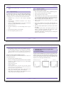
Figure 1: Epidermis, dermis, and hypodermis form a three-tiered structure. To access HDMECs
within the dermal layer (middle), the epidermis can be removed by peeling the top layer from
the dermis and hypodermis.
Trypsination
Ø
HDMEC purification with the CD31 MicroBead Kit
6
140-001-341.04
140-001-341.04
7