1
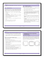
2. Protocols
2. Protocols
●
(Optional) Pre-Separation Filters (# 130-041-407) to remove cell
clumps.
2.3.3
Magnetic separation
Magnetic separation with LS Columns
2.3.2
Magnetic labeling
▲ Volumes given are for up to 1×10⁷ cells. When working with fewer
than 1×10⁷ cells, use the same volumes as indicated. When working
with higher cell numbers, scale up all reagent volumes and total volumes
accordingly (e.g. for 2×10⁷ total cells use twice the volume described).
1.
Place an LS Column in the magnetic field of a MidiMACS Separator.
2.
Prepare column by rinsing with 3 mL of medium.
3.
Apply cell suspension onto the column.
4.
Collect unlabeled cells which pass through and wash column with
3×3 mL of medium. Perform washing steps by adding medium three
times. Only add new medium when the column reservoir is empty.
Collect total effluent; this is the unlabeled cell fraction.
1.
Centrifuge cells at 300×g for 3 min. Aspirate supernatant
completely.
2.
Resuspend cells to a maximum concentration of 1×10⁷ cells per
60 μL of medium.
5.
Remove column from the separator and place it on a suitable
collection tube.
3.
Add 20 μL of FcR Blocking Reagent per 1×10⁷ cells. Vortex briefly,
then add 20 μL of CD31 MicroBeads to the mixture.
6.
Pipette 5 mL of medium onto the column. Immediately flush out
the magnetically labeled cells by firmly pushing the plunger into the
column.
4.
Incubate 15 min at 4 °C.
7.
5.
Add 1 mL of medium per 1×10⁷ and centrifuge cells at 300×g for
3 min.
Cells can be directly analyzed by flow cytometry for purity or taken
into culture.
Resuspend the cell pellet in 1 mL of medium.
7.
Proceed to magnetic separation (see 2.3.2).
For HUVECs, yield of CD31+ cells will depend upon cord length and
efficiency of enzyme digestion.
140-001-341.04
20
140-001-341.04
21
3. Examples of separations using the CD31 MicroBead Kit
2. Protocols
Magnetic separation with the autoMACS® Separator
▲ Refer to the autoMACS® user manual for instructions on how to use
the autoMACS Separator.
1.
Prepare and prime autoMACS Separator.
2.
Place tube containing the magnetically labeled cells in the autoMACS
Separator. For a standard separation, choose one of the following
separation programs:
Positive selection: "Possel"
Depletion: "Depletes"
▲ Note: Program choice depends on the isolation strategy, the strength of magnetic
labeling and the frequency of magnetically labeled cells. For details see autoMACS user
manual, section autoMACS Cell Separation Programs.
3.
3. Examples of separations using the CD31
MicroBead Kit
3.1 Separation of HDMECs
Separation of CD31+ HDMECs from a preparation of dermal-layer cells
of human foreskin using CD31 MicroBeads and a MidiMACS Separator
with an LS Column. The cells are fluorescently stained with CD31-APC
(# 130-092-652). Cell debris and dead cells were excluded from the
analysis based on scatter signals and PI fluorescence.
HDMECs before separation
CD31-APC
CD31+ cells
CD31- cells
Relative cell number
▲ Note: Should fibroblast contamination outgrow endothelial cells upon prolonged
cultivation, endothelial cells should be re-purified using CD31 MicroBeads, or
alternatively fibroblasts can be removed using Anti-Fibroblast MicroBeads (# 130-050601).
Relative cell number
Cells should be seeded at a density of approximately 150,000 per
T-75 flask. Culture conditions: 37 °C, 5% CO2 and >95% humidity.
Relative cell number
6.
For HDMECs, yield of CD31+ cells depends upon the size and
thickness of foreskin used. Generally, cells isolated from a single,
4 cm2 biopsy should be cultured in one T-75 flask.
CD31-APC
CD31-APC
When using the program "Possel", collect positive fraction from
outlet port pos1. This is the purified positive cell fraction.
When using the program "Depletes", collect unlabeled fraction from
outlet port neg1. This is the negative cell fraction.
22
140-001-341.04
140-001-341.04
23