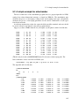
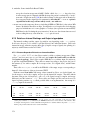
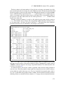
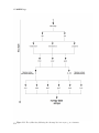
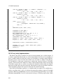
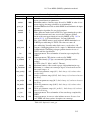
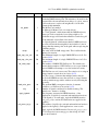
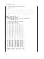
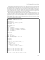
1
AmberTools13 Reference Manual AmberTools13 Reference Manual AmberTools consists of several independently developed packages that work well with Amber itself. The main components of AmberTools are listed below. NAB (Nucleic Acid Builder) sqm Thomas J. Macke, W.A. Svrcek-Seiler, Russell A. Brown, István Kolossváry, Yannick J. Bomble, Ramu Anandakrishnan, David A. Case Ross C. Walker, Michael F. Crowley, Scott Brozell, Tim Giese, Andreas W. Götz, Tai-Sung Lee, David A. Case LEaP Wei Zhang, Tingjun Hou, Christian Schafmeister, Wilson S. Ross, David A. Case antechamber Junmei Wang amberlite Romain M. Wolf ptraj Thomas E. Cheatham, III, et al. (see http://ambermd.org/contributors.html) cpptraj Daniel R. Roe, et al. (see http://ambermd.org/contributors.html) pbsa Jun Wang, Qin Cai, Wesley M. Botello-Smith, Xiang Ye, Meng-Juei Hsieh, Chuck Tan, Ray Luo CHAMBER Michael F. Crowley, Mark Williamson, Ross C. Walker 3D-RISM Tyler Luchko, David A. Case, Sergey Gusarov, Andriy Kovalenko mdgx David S. Cerutti MMPBSA.py Jason Swails, T. Dwight McGee Jr., Bill Miller III MTK++, MCPB Martin Peters, Kenneth Ayers, Andrew Wollacott, Duane E. Williams, Benjamin P. Roberts, Kenneth M. Merz, Jr. paramfit Ross C. Walker, Robin Betz 1 Notes • Most of the programs included here can be redistributed and/or modified under the terms of the GNU General Public License; a few components have other open-source licenses. See the amber12/AmberTools/LICENSE file for details. The programs are distributed in the hope that they will be useful, but WITHOUT ANY WARRANTY; without even the implied warranty of MERCHANTABILITY or FITNESS FOR A PARTICULAR PURPOSE. • Some of the force field routines were adapted from similar routines in the MOIL program package: R. Elber, A. Roitberg, C. Simmerling, R. Goldstein, H. Li, G. Verkhivker, C. Keasar, J. Zhang and A. Ulitsky, "MOIL: A program for simulations of macromolecules" Comp. Phys. Commun. 91, 159-189 (1995). • The "trifix" routine for random pairwise metrization is based on an algorithm designed by Jay Ponder and was adapted from code in the Tinker package; see M.E. Hodsdon, J.W. Ponder, and D.P. Cistola, J. Mol. Biol. 264, 585-602 (1996) and http://dasher.wustl.edu/tinker/. • The "molsurf" routines for computing molecular surface areas were adapted from routines written by Paul Beroza. The "sasad" routine for computing derivatives of solvent accessible surface areas was kindly provided by S. Sridharan, A. Nicholls and K.A. Sharp. See J. Computat. Chem. 8, 1038-1044 (1995). • Some of the “pb_exmol” routines for mapping molecular surface to finite-difference grids were adapted from routines written by Michael Gilson and Malcolm Davis in UHBD. See Comp. Phys. Comm. 91, 57-95 (1995). • The cifparse routines to deal with mmCIF formatted files were written by John Westbrook, and are distributed with permission. See cifparse/README for details. Recommended Citations: • When citing AmberTools 13 in the literature, the following citation, along with the citation of the corresponding program, should be used: D.A. Case, T.A. Darden, T.E. Cheatham, III, C.L. Simmerling, J. Wang, R.E. Duke, R. Luo, R.C. Walker, W. Zhang, K.M. Merz, B. Roberts, S. Hayik, A. Roitberg, G. Seabra, J. Swails, A.W. Götz, I. Kolossváry, K.F. Wong, F. Paesani, J. Vanicek, R.M. Wolf, J. Liu, X. Wu, S.R. Brozell, T. Steinbrecher, H. Gohlke, Q. Cai, X. Ye, J. Wang, M.-J. Hsieh, G. Cui, D.R. Roe, D.H. Mathews, M.G. Seetin, R. Salomon-Ferrer, C. Sagui, V. Babin, T. Luchko, S. Gusarov, A. Kovalenko, and P.A. Kollman (2012), AMBER 13, University of California, San Francisco. Cover Illustration The cover shows a slice through a crystal simulation of a designed peptide; see Aravinda, S.; Shamala, N.; Das, C.; Sriranjini, A.; Karle, I. L.; Balaram, P. J. Am. Chem. Soc., 2003, 125, 5308–15. 36 unit cells are stacked in a 4 x 3 x 3 arrangement in the triclinic super-system; each unit cell comprises two decapeptide helices arranged roughly parallel to one another, with water molecules forming channels perpendicular to the plane of the illustration. Figure by Dave Cerutti. 2 Contents Contents 1 Getting started 1.1 1.2 1.3 1.4 1.5 1.6 1.7 2 4 9 Information flow in Amber . . . . . . . . . . . . . . . Installation . . . . . . . . . . . . . . . . . . . . . . . Combining AmberTools13 with Amber11 or Amber10 Testing the installation . . . . . . . . . . . . . . . . . Applying Updates . . . . . . . . . . . . . . . . . . . . Contacting the developers . . . . . . . . . . . . . . . . List of programs . . . . . . . . . . . . . . . . . . . . . . . . . . . . . . . . . . . . . . . . . . . . . . . . . . . . . . . . . . . . . . . . . . . . . . . . . . . . . . . . . . . . . . . . . . . . . . . . . . . . . . . . . . . . . . . . . . . . . . . . . . . . . . . . . . . . . . . . . . . . . . . . . . . . . . . . . . . . . . . . . . . . . . . . . . . . . . . . . . . . . . . . . . . . . . . . . . . . . . . . . . . . . . . . . . . . . . . . . . . . . . . . . . . . . . . . . . . . . . . . . . . . . . . . . . . . . . . . . . . . . . . . . . . . Specifying a force field 2.1 2.2 2.3 2.4 2.5 2.6 2.7 2.8 2.9 2.10 2.11 2.12 3 3 9 12 15 16 16 19 19 25 Specifying which force field you want in LEaP . . . . The ff12SB force field . . . . . . . . . . . . . . . . . The AMOEBA potentials . . . . . . . . . . . . . . . . The Duan et al. (2003) force field . . . . . . . . . . . The Yang et al. (2003) united-atom force field . . . . . Force fields related to semi-empirical QM . . . . . . . The GLYCAM force fields for carbohydrates and lipids Lipid Force Fields . . . . . . . . . . . . . . . . . . . . Ions . . . . . . . . . . . . . . . . . . . . . . . . . . . Solvent models . . . . . . . . . . . . . . . . . . . . . CHAMBER . . . . . . . . . . . . . . . . . . . . . . . Obsolete force field files . . . . . . . . . . . . . . . . 26 27 30 30 30 31 31 39 43 44 45 52 Reading and modifying Amber parameter files 57 3.1 3.2 3.3 57 67 86 Understanding Amber parameter files . . . . . . . . . . . . . . . . . . . . . . ParmEd . . . . . . . . . . . . . . . . . . . . . . . . . . . . . . . . . . . . . . paramfit . . . . . . . . . . . . . . . . . . . . . . . . . . . . . . . . . . . . . . LEaP 4.1 4.2 4.3 4.4 4.5 4.6 97 Introduction . . . . . . . . . . . . . . . . . . . . . . Concepts . . . . . . . . . . . . . . . . . . . . . . . Running LEaP . . . . . . . . . . . . . . . . . . . . . Basic instructions for using LEaP to build molecules Commands . . . . . . . . . . . . . . . . . . . . . . Building oligosaccharides and lipids . . . . . . . . . . . . . . . . . . . . . . . . . . . . . . . . . . . . . . . . . . . . . . . . . . . . . . . . . . . . . . . . . . . . . . . . . . . . . . . . . . . . . . . . . . . . . 97 97 103 109 111 130 3 CONTENTS 5 Antechamber and MCPB 5.1 5.2 5.3 5.4 5.5 5.6 6 Introduction . . . . . . . . . . . . . . . . . . Coordinates and Parameter-Topology Files . . pytleap: Creating Coordinates and ParameterTopology Files . . . . . . . . . . . . . . . . 6.4 Energy Checking Tool: ffgbsa . . . . . . . . 6.5 Energy Minimizer: minab . . . . . . . . . . . 6.6 Molecular Dynamics "Lite": mdnab . . . . . 6.7 MM(GB)(PB)/SA Analysis Tool: pymdpbsa . 6.8 Appendix A: Preparing PDB Files . . . . . . 6.9 Appendix B: Atom and Residue Selections . . 6.10 Appendix C: Examples and Test Cases . . . . . . . . . . . . . . . . . . . . . . . . . . . . . . . . . . . . . . . . . . . . . . . . . . . . . . . . . . . . . . . . . . . . . . . . . . . . . . . . . . . . . . . . . . . . . . 140 145 148 153 155 156 159 . . . . . . . . . . . . . . . . . . 159 . . . . . . . . . . . . . . . . . . 162 . . . . . . . . . . . . . . . . . . . . . . . . . . . . . . . . . . . . . . . . . . . . . . . . . . . . . . . . . . . . . . . . . . . . . . . . . . . . . . . . . . . . . . . . . . . . . . . . . . . . . . . . . . . . . . . . . . . . . . . . . . . . . . . . . . . . . . . . . . . . . . . . Available Hamiltonians . . . . . . . . . . . . . . . . . . . . . . . . . . Charge-dependent exchange-dispersion corrections of vdW interactions Dispersion and hydrogen bond correction . . . . . . . . . . . . . . . . Usage . . . . . . . . . . . . . . . . . . . . . . . . . . . . . . . . . . . . . . . . . . . . . . . . . . . . . . . . . . . . . . . . . . . . . . . . . . . . . . . . . . . . . . . . . . . . . . . . . . . . . . . . . . . . . . . Running cpptraj . . . . . . . . . . . . . . . . . General Concepts . . . . . . . . . . . . . . . . Data Sets and Data Files . . . . . . . . . . . . Coordinates as a Data Set (COORDS Data Sets) General Commands . . . . . . . . . . . . . . . Parameter File Commands . . . . . . . . . . . Trajectory File Commands . . . . . . . . . . . Data File Commands . . . . . . . . . . . . . . Actions that Modify Topology/Coordinates . . Action Commands . . . . . . . . . . . . . . . Matrix and Vector Actions . . . . . . . . . . . Data Set Analysis Commands . . . . . . . . . Coordinate Analysis Commands . . . . . . . . Matrix and Vector Analysis . . . . . . . . . . . Matrix/Vector Analysis Examples . . . . . . . 163 167 167 169 170 178 181 184 195 cpptraj 8.1 8.2 8.3 8.4 8.5 8.6 8.7 8.8 8.9 8.10 8.11 8.12 8.13 8.14 8.15 4 . . . . . . sqm: Semi-empirical quantum chemistry 7.1 7.2 7.3 7.4 8 . . . . . . amberlite: Some AmberTools-Based Utilities 6.1 6.2 6.3 7 139 Principal programs . . . . . . . . . . . . . . . A simple example for antechamber . . . . . . . Programs called by antechamber . . . . . . . . Miscellaneous programs . . . . . . . . . . . . New Development of Antechamber And GAFF Metal Center Parameter Builder (MCPB) . . . 195 197 198 199 207 . . . . . . . . . . . . . . . . . . . . . . . . . . . . . . . . . . . . . . . . . . . . . . . . . . . . . . . . . . . . . . . . . . . . . . . . . . . . . . . . . . . . . . . . . . . . . . . . . . . . . . . . . . . . . . . . . . . . . . . . . . . . . . . . . . . . . . . . . . . . . . . . . . . . . . . . . . . . . . . . . . . . . . . . . . . . . . . . . . . . . . . . . . . . . . . . . . . 207 208 212 214 215 220 223 229 230 238 265 267 272 277 280 CONTENTS 9 ptraj 9.1 9.2 9.3 9.4 9.5 9.6 283 ptraj coordinate input/output commands . . . . . . . . . . . . . . ptraj commands that override the molecular information specified ptraj action commands . . . . . . . . . . . . . . . . . . . . . . . Correlation and fluctuation facility . . . . . . . . . . . . . . . . . Hydrogen bonding facility . . . . . . . . . . . . . . . . . . . . . rdparm . . . . . . . . . . . . . . . . . . . . . . . . . . . . . . . . . . . . . . . . . . . . . . . . . . . . . . . . . . . . . . . . . . . . . . . . . . 10 PBSA 10.1 10.2 10.3 10.4 10.5 299 Introduction . . . . . . . . . . . . . . . . . . . . . . Usage and keywords . . . . . . . . . . . . . . . . . Example inputs and demonstrations of functionalities Visualization functions in pbsa . . . . . . . . . . . . pbsa in sander and NAB . . . . . . . . . . . . . . . . . . . . . . . . . . . . . . . . . . . . . . . . . . . . . . . . . . . . . . . . . . . . . . . . . . . . . . . . . . . . . . . . . . . . . 11 Reference Interaction Site Model 11.1 11.2 11.3 11.4 11.5 11.6 Introduction . . . . . . . Practical Considerations Work Flow . . . . . . . rism1d . . . . . . . . . . 3D-RISM in NAB . . . . rism3d.snglpnt . . . . . . . . . . . . . . . . . . . . . . . . . . . . . . . . . . . . . . . . . . . . . . . . . . . . . . . . . . . . . . . . . . . . . . . . . . . . . . . . . . . . . . . . . . . . . . . . . . . . . . . . . . . . . . . . . . . . . . . . . . . . . . . . . . . . . . . . . . . . . . . . . . . . . . . . . . . . . . . . . . . . . . . . . . . . Introduction . . . . . . . . . . . . . . . . . . Preparing for an MM/PB(GB)SA calculation Running MMPBSA.py . . . . . . . . . . . . Python API . . . . . . . . . . . . . . . . . . . . . . . . . . . . . . . . . . . . . . . . . . . . . . . . . . . . . . . . . . . . . . . . . . . . . . . . . . . . . . . . . . . . . . . . . . . . . . . . . . . . . . . . . . . . . . . . . . . . . . . . . . . . . . . . . . . . . . . . . . . . . . . . . . . . . . . . . . . . . . . . . . . . . . . . . . . . . . . . . . . . . . . . . . . . . . . . . . . . . . . . . . . . . . . . . . . . . . . . . . . . . . . . . . . . . . . . . . . . . . . . . . . . . . . . . . . . . . . . . . . . . . . . . . . . . . . . . . . . . . . . . . . . . . . . . . . . . . . . . . Input and Output . . . . . . . . . . . . . . . . . Installation . . . . . . . . . . . . . . . . . . . . Special Algorithmic Features of mdgx . . . . . . Customizable Virtual Site Support in mdgx . . . Restrained Electrostatic Potential Fitting in mdgx Bonded Term Fitting in mdgx . . . . . . . . . . . Thermodynamic Integration . . . . . . . . . . . Future Directions and Goals of the mdgx Project ambpdb . . . . . . . . . . . . . . . . . . . reduce . . . . . . . . . . . . . . . . . . . . elsize . . . . . . . . . . . . . . . . . . . . Utilities for Molecular Crystal Simulations . MdoutAnalyzer.py . . . . . . . . . . . . . 349 350 353 369 377 14 Miscellaneous utilities 14.1 14.2 14.3 14.4 14.5 329 335 337 337 341 344 349 13 mdgx: A Developmental Molecular Simulation Engine 13.1 13.2 13.3 13.4 13.5 13.6 13.7 13.8 299 303 314 317 326 329 . . . . . . 12 MMPBSA.py 12.1 12.2 12.3 12.4 283 284 285 289 293 295 377 379 379 380 383 386 388 388 391 . . . . . . . . . . . . . . . 391 393 396 397 400 5 CONTENTS 15 NAB: Introduction 15.1 Background . . . . . . . . . . . . . 15.2 Methods for structure creation . . . 15.3 Compiling nab Programs . . . . . . 15.4 Parallel Execution . . . . . . . . . . 15.5 First Examples . . . . . . . . . . . 15.6 Molecules, Residues and Atoms . . 15.7 Creating Molecules . . . . . . . . . 15.8 Residues and Residue Libraries . . . 15.9 Atom Names and Atom Expressions 15.10Looping over atoms in molecules . . 15.11Points, Transformations and Frames 15.12Creating Watson Crick duplexes . . 15.13Structure Quality and Energetics . . 401 . . . . . . . . . . . . . . . . . . . . . . . . . . . . . . . . . . . . . . . . . . . . . . . . . . . . . . . . . . . . . . . . . . . . . . . . . . . . . . . . . . . . . . . . . . . . . . . . . . . . . . . . . . . . . . . . . . . . . . . . . . . . . . . . . . . . . . . . . . . . . . . . . . . . . . . . . . . . . . . . . . . . . . . . . . . . . . . . . . . . . . . . . . . . . . . . . . . . . . . . . . . . . . . . . . . . . . . . . . . . . . . . . . . . . . . . . . . . . . . . . . . . . . . . . . . . . . . . . . . . . . . . . . . . . . . . . . . . . . . . . . . . . . . . . . . . . . . . . . . 16.1 Introduction . . . . . . . . . . . . . . . . . . . . . 16.2 Language Elements . . . . . . . . . . . . . . . . . 16.3 Higher-level constructs . . . . . . . . . . . . . . . 16.4 Statements . . . . . . . . . . . . . . . . . . . . . . 16.5 Structures . . . . . . . . . . . . . . . . . . . . . . 16.6 Functions . . . . . . . . . . . . . . . . . . . . . . 16.7 Points and Vectors . . . . . . . . . . . . . . . . . . 16.8 String Functions . . . . . . . . . . . . . . . . . . . 16.9 Math Functions . . . . . . . . . . . . . . . . . . . 16.10System Functions . . . . . . . . . . . . . . . . . . 16.11I/O Functions . . . . . . . . . . . . . . . . . . . . 16.12Molecule Creation Functions . . . . . . . . . . . . 16.13Creating Biopoloymers . . . . . . . . . . . . . . . 16.14Fiber Diffraction Duplexes in NAB . . . . . . . . . 16.15Reduced Representation DNA Modeling Functions 16.16Molecule I/O Functions . . . . . . . . . . . . . . . 16.17Other Molecular Functions . . . . . . . . . . . . . 16.18Debugging Functions . . . . . . . . . . . . . . . . 16.19Time and date routines . . . . . . . . . . . . . . . 16.20Computational resource consumption functions . . . . . . . . . . . . . . . . . . . . . . . . . . . . . . . . . . . . . . . . . . . . . . . . . . . . . . . . . . . . . . . . . . . . . . . . . . . . . . . . . . . . . . . . . . . . . . . . . . . . . . . . . . . . . . . . . . . . . . . . . . . . . . . . . . . . . . . . . . . . . . . . . . . . . . . . . . . . . . . . . . . . . . . . . . . . . . . . . . . . . . . . . . . . . . . . . . . . . . . . . . . . . . . . . . . . . . . . . . . . . . . . . . . . . . . . . . . . . . . . . . . . . . . . . . . . . . . . . . . . . . . . . . . . . . . . . . . . . . . . . . . . . . . . . . . . . . . . . . . . . . . . . . . . . . . . . . . . . . . . . . . . . . . . . . . . . . . . . . . . . . . . . . . . . . . . . . . . . . . . . . . . . . . . . . . . . . . . . 16 NAB: Language Reference 441 17 NAB: Rigid-Body Transformations 17.1 17.2 17.3 17.4 17.5 6 Transformation Matrix Functions . . . . . . . . Frame Functions . . . . . . . . . . . . . . . . Functions for working with Atomic Coordinates Symmetry Functions . . . . . . . . . . . . . . Symmetry server programs . . . . . . . . . . . 402 404 407 407 408 411 412 413 415 417 418 420 430 441 441 443 451 454 456 457 458 459 459 459 463 464 465 466 466 468 469 470 470 473 . . . . . . . . . . 473 473 474 474 477 CONTENTS 18 NAB: Distance Geometry 18.1 18.2 18.3 18.4 481 Metric Matrix Distance Geometry . . . . . . . . . . . . Creating and manipulating bounds, embedding structures Distance geometry templates . . . . . . . . . . . . . . . Bounds databases . . . . . . . . . . . . . . . . . . . . . . . . . . . . . . . . . . . . . . . . . . . . . . . . . . . . . . . . . . . . . . . . . . . . . Basic molecular mechanics routines . . . . . . . . . . . . NetCDF read/write routines . . . . . . . . . . . . . . . . . Typical calling sequences . . . . . . . . . . . . . . . . . . Second derivatives and normal modes . . . . . . . . . . . Low-MODe (LMOD) optimization methods . . . . . . . . Using the Hierarchical Charge Partitioning (HCP) method . . . . . . . . . . . . . . . . . . . . . . . . . . . . . . . . . . . . . . . . . . . . . . . . . . . . . . . . . . . . . . . . . . . . . . . . . . . . . . . . . . . . . . . . . . . . . . . . . . . . . . . . . . . . . . . . . . . . . . . . . 19 NAB: Molecular mechanics and dynamics 19.1 19.2 19.3 19.4 19.5 19.6 493 20 NAB: Sample programs 20.1 20.2 20.3 20.4 20.5 Duplex Creation Functions . . nab and Distance Geometry . . Building Larger Structures . . Wrapping DNA Around a Path Other examples . . . . . . . . 481 482 487 490 493 505 508 509 511 526 529 . . . . . . . . . . . . . . . . . . . . . . . . . . . . . . . . . . . . . . . . . . . . . . . . . . . . . . . . . . . . . . . . . . . . . . . . . . . 529 530 540 548 556 Bibliography 557 Bibliography 557 Index 577 7 1 Getting started AmberTools is a set of programs for biomolecular simulation and analysis. They are designed to work well with each other, and with the “regular” Amber suite of programs. You can perform many simulation tasks with AmberTools, and you can do more extensive simulations with the combination of AmberTools and Amber itself. Most components of AmberTools are released under the GNU General Public License (GPL). A few components are in the public domain or have other open-source licenses. See the README file for more information. We hope to add new functionality to AmberTools as additional programs become available. If you have suggestions for what might be added, please contact us. Everyone should read (or at least skim) Section 1.1. Even if you are an experienced Amber user, there may be things you have missed, or new features, that will help. If you are installing this package see Section 1.2. There are also tips and examples on the Amber Web pages at http://ambermd.org. Although Amber may appear dauntingly complex at first, it has become easier to use over the past few years, and overall is reasonably straightforward once you understand the basic architecture and option choices. In particular, we have worked hard on the tutorials to make them accessible to new users. Thousands of people have learned to use Amber; don’t be easily discouraged. If you want to learn more about basic biochemical simulation techniques, there are a variety of good books to consult, ranging from introductory descriptions,[1, 2] to standard works on liquid state simulation methods,[3–5] to multi-author compilations that cover many important aspects of biomolecular modelling.[6–10] Looking for "paradigm" papers that report simulations similar to ones you may want to undertake is also generally a good idea. 1.1 Information flow in Amber Understanding where to begin in AmberTools is primarily a problem of managing the flow of information in this package — see Fig. 1.1. You first need to understand what information is needed by the simulation programs (sander, pmemd, mdgx or nab). You need to know where it comes from, and how it gets into the form that the energy programs require. This section is meant to orient the new user and is not a substitute for the individual program documentation. Information that all the simulation programs need: 1. Cartesian coordinates for each atom in the system. These usually come from X-ray crystallography, NMR spectroscopy, or model-building. They should be in Protein Data Bank (PDB) or Tripos “mol2” format. The program LEaP provides a platform for carrying out many of these modeling tasks, but users may wish to consider other programs as well. Topology: Connectivity, atom names, atom types, residue names, and charges. This information comes from the database, which is found in the $AMBERHOME/dat/leap/prep directory, and is 9 1 Getting started antechamber, tleap, MCPB pdb LES info prmtop prmcrd NMR or XRAY info MMPBSA.py, amberlite sander, nab, mdgx, pmemd ptraj, cpptraj Figure 1.1: Basic information flow in Amber described in Chapter 2. It contains topology for the standard amino acids as well as N- and Cterminal charged amino acids, DNA, RNA, and common sugars. The database contains default internal coordinates for these monomer units, but coordinate information is usually obtained from PDB files. Topology information for other molecules (not found in the standard database) is kept in user-generated “residue files”, which are generally created using antechamber. 1. Force field: Parameters for all of the bonds, angles, dihedrals, and atom types in the system. The standard parameters for several force fields are found in the $AMBERHOME/dat/leap/parm directory; see Chapter 2 for more information. These files may be used “as is” for proteins and nucleic acids, or users may prepare their own files that contain modifications to the standard force fields. 2. Commands: The user specifies the procedural options and state parameters desired. These are specified in “driver” programs written in the NAB language. 1.1.1 Preparatory programs LEaP is the primary program to create a new system in Amber, or to modify existing systems. It is available as the command-line program tleap or the GUI xleap. It combines the functionality of prep, link, edit and parm from much earlier versions of Amber. ParmEd provides a simple way to extract information about the parameters defined in a parameter- topology file. It can also be used to check that the parameter-topology file is valid for complex systems, and it can also make simple modifications to this file very quickly. 10 1.1 Information flow in Amber antechamber is the main program from the Antechamber suite. If your system contains more than just standard nucleic acids or proteins, this may help you prepare the input for LEaP. MCPB provides a means to rapidly build, prototype, and validate MM models of metallopro- teins. It uses the bonded plus electrostatics model to expand existing pairwise additive force fields. paramfit iteratively fits individual force field parameters for a given system. This can be useful when antechamber fails to find parameters, or if existing force fields fail to properly characterize the system. 1.1.2 Simulation programs NAB (Nucleic Acid Builder) is a language that can be used to write programs to perform non- periodic simulations, most often using an implicit solvent force field. sander (part of Amber) is the basic energy minimizer and molecular dynamics program. This program relaxes the structure by iteratively moving the atoms down the energy gradient until a sufficiently low average gradient is obtained. The molecular dynamics portion generates configurations of the system by integrating Newtonian equations of motion. MD will sample more configurational space than minimization, and will allow the structure to cross over small potential energy barriers. Configurations may be saved at regular intervals during the simulation for later analysis, and basic free energy calculations using thermodynamic integration may be performed. More elaborate conformational searching and modeling MD studies can also be carried out using the SANDER module. This allows a variety of constraints to be added to the basic force field, and has been designed especially for the types of calculations involved in NMR structure refinement. pmemd (part of Amber) is a version of sander that is optimized for speed and for parallel scaling. The name stands for “Particle Mesh Ewald Molecular Dynamics,” but this code can now also carry out generalized Born simulations. The input and output have only a few changes from sander. mdgx is a molecular dynamics engine with functionality that mimics some of the features in sander and pmemd, but featuring simple C code and an atom sorting routine that simplifies the flow of information during force calculations. The principal purpose of mdgx is to provide a tool for radical redesign of the basic molecular dynamics algorithms and models. 1.1.3 Analysis programs ptraj is a general purpose utility for analyzing and processing trajectory or coordinate files created from MD simulations (or from various other sources), c cpptraj is a trajectory analysis utility (written in C++) for carrying out superpositions, extrac- tions of coordinates, calculation of bond/angle/dihedral values, atomic positional fluctuations, correlation functions, analysis of hydrogen bonds, etc. It has almost all the 11 1 Getting started functionality of the older ptraj program and generally better performance. New features in cpptraj include the ability to process multiple prmtop files at once, ability to specify a separate reference mask during RMSD calculations, support for multiple output trajectory files, native support for compressed (gzip or bzip2) trajectories and prmtop files, and output of stripped prmtop files. pbsa is an analysis program for solvent-mediated energetics of biomolecules. It can be used to perform both electrostatic and non-electrostatic continuum solvation calculations with input coordinate files from molecular dynamics simulations and other sources. The electrostatic solvation is modeled by the Poisson-Boltzmann equation. Both linear and full nonlinear numerical solvers are implemented. The nonelectrostatic solvation is modeled by two separate terms: dispersion and cavity. MMPBSA.py is a python script that automates energy analysis of snapshots from a molecular dynamics simulation using ideas generated from continuum solvent models. (There is also an older perl script, called mm_pbsa.pl, that is a part of Amber.) amberlite is small set of NAB programs and python scripts that implement a limited set of MD simulations and mm-pbsa (or mm-gbsa) analysis, aimed primarily at the analysis of protein-ligand interactions. These tools can be useful in their own right, or as a good introduction to Amber and a starting point for more complex calculations. Detailed instructions are in Chapter 6. 1.2 Installation We have worked hard in this release to simplify the installation of Amber, and there are some differences from earlier releases. First, if you have both Amber and AmberTools, both will be installed and tested with a single command. Second, the configure script automatically checks for updates, and installs them if you ask it to. A third (minor) change is that the configure script is now run from the $AMBERHOME directory (not from $AMBERHOME/AmberTools/src, as in the past.) The Amber web page (http://ambermd.org) has some specific instructions and hints for various common operating systems. Look for the “Running Amber on ....” links. Once you have downloaded the distribution files, do the following: 1. First, extract the files in some location (we use /home/myname as an example here): cd /home/myname tar xvfj AmberTools13.tar.bz2 tar xvfj Amber12.tar.bz2 # (Note: extracts in an # “amber12” directory) # (only if you have licensed Amber 12!) 2. Next, set your AMBERHOME environment variable: export AMBERHOME=/home/myname/amber12 setenv AMBERHOME /home/myname/amber12 12 # (for bash, zsh, ksh, etc.) # (for csh, tcsh) 1.2 Installation Be sure to change the “/home/myname” above to whatever directory is appropriate for your machine, and be sure that you have write permissions in the directory tree you choose. You should also add $AMBERHOME/bin to your PATH. 3. Next, you may need to install some compilers and other libraries. Details depend on what OS you have, and what is already installed. Package managers can greatly simplify this task. For example for Debian-based Linux systems (such as Ubuntu), the following command should get you what you need: sudo apt-get install csh flex gfortran g++ xorg-dev \ zlib1g-dev libbz2-dev Other Linux distributions will have a similar command, but with a package manager different than apt-get. For example, the following should work for Fedora Core and similar systems: sudo yum install gcc flex tcsh zlib-devel bzip2-devel \ libXt-devel libXext-devel libXdmcp-devel For Macintosh OSX, MacPorts (http://www.macports.org) serves a similar purpose. You would download and install the port program, then issue commands like this: sudo port install gcc46 MacPorts is useful because the “Xcode” compilers provided by Apple will not work to compile Amber, since no Fortran compiler is provided. Amber cross-links Fortran and C/C++ code, so a “full” GCC installation is necessary. 4. Now, in the AMBERHOME directory, run the configure script: cd $AMBERHOME ./configure --help will show you the options. Choose the compiler and flags you want; for most systems, the following should work: ./configure gnu Don’t choose any parallel options at this point. (You may need to edit the resulting config.h file to change any variables that don’t match your compilers and OS. The comments in the config.h file should help.) This step will also check to see if there are any bugfixes that have not been applied to your installation, and will apply them (unless you ask it not to). If the configure step finds missing libraries, go back to Step 3. 5. Then, make install will compile the codes. If this step fails, try to read the error messages carefully to identify the problem. 6. This can be followed by make test 13 1 Getting started which will run tests and will report successes or failures. Where "possible FAILURE" messages are found, go to the indicated directory under $AMBERHOME/AmberTools/test or $AMBERHOME/test, and look at the "*.dif" files. Differences should involve round-off in the final digit printed, or occasional messages that differ from machine to machine (see below for details). As with compilation, if you have trouble with individual tests, you may wish to comment out certain lines in the Makefiles (i.e., $AMBERHOME/AmberTools/test/Makefile or $AMBERHOME/test/Makefile), and/or go directly to the test subdirectories to examine the inputs and outputs in detail. For convenience, all of the failure messages and differences are collected in the $AMBERHOME/logs directory; you can quickly see from these if there is anything more than round-off errors. Note: If you have untarred the Amber12.tar.bz2 file, then steps 1-6 will install and test both AmberTools and Amber; otherwise it will just install and test AmberTools. If you license Amber later, just come back and repeat steps 1-6 again. 7. If you are new to Amber, you should look at the tutorials and this manual and become familiar with how things work. If and when you wish to compile parallel (MPI) versions of Amber, do this: cd $AMBERHOME ./configure -mpi <....other options....> <compiler-choice> make install # Note the value below may depend on your MPI implementation export DO_PARALLEL=”mpirun -np 2” make test # Note, some tests, like the replica exchange tests, require more # than 2 threads, so we suggest that you test with either 4 or 8 # threads as well export DO_PARALLEL=”mpirun -np 8” make test This assumes that you have installed MPI and that mpicc and mpif90 are in your PATH. Some MPI installations are tuned to particular hardware (such as infiniband), and you should use those versions if you have such hardware. Most people can use standard versions of either mpich2 or openmpi. To install one of these, use one of the simple scripts that we have prepared: cd $AMBERHOME/AmberTools/src ./configure_mpich2 <compiler-choice> ./configure_openmpi <compiler-choice> OR Follow the instructions of these scripts, then return to beginning of step 7. Note: Parallel versions of AmberTools are rather specialized, and many users will skip this step. Consider the following points before compiling and using the MPI version: a) The MPI version of nab is called mpinab, by analogy with mpicc or mpif90: mpinab is a compiler that will produce an MPI-enabled executable from source code 14 1.3 Combining AmberTools13 with Amber11 or Amber10 written in the NAB language. Before compiling mpinab, be sure that you are familiar with the serial version of nab and that you really need a parallel version. If you have shared-memory nodes, the OpenMP version might be a better alternative. See Section 15.4 for more information. (Note that mpinab is primarily designed to write driver routines that call MPI versions of the energy functions; it is not set up to write your own, novel, parallel codes.) b) The MPI version of MMPBSA.py is called MMPBSA.py.MPI, and requires the package mpi4py to run. If it is not present in your system Python installation already, it will be built along with MMPBSA.py.MPI and placed in the $AMBERHOME/bin directory. If you have problems with MMPBSA.py.MPI, see if you get the same problems with the serial version, MMPBSA.py, to see if it is an issue with the parallel version or MMPBSA.py in general. Because we do not make or maintain the mpi4py source code, MMPBSA.py.MPI will not be available on platforms on which mpi4py cannot be built. 8. NAB and Cpptraj can also be compiled using OpenMP: ./configure -openmp <....other options....> <compiler-choice> make openmp Note that the OpenMP versions of NAB and Cpptraj have the same name as the singlethreaded version. See Section 15.4 for information on running the OpenMP version of NAB and section 8.2.6 for information on running the OpenMP version of Cpptraj. 1.3 Combining AmberTools13 with Amber11 or Amber10 It is certainly feasible to combine AmberTools13 with earlier versions of Amber. Here is the outline of what to do: 1. Unpack AmberTools13.tar.bz2 into a directory tree whose head will be amber12. Point your AMBERHOME environment to this directory, and install and test as described above. Don’t do anything to your existing Amber files, which will be in a directory tree headed by amber11 (or amber10...). 2. Set your PATH variable to have $AMBERHOME/bin ahead of the old amber directories. For example: export PATH=”$AMBERHOME/bin:/home/myname/amber11/bin:$PATH” setenv PATH “$AMBERHOME/bin:/home/myname/amber11/bin:$PATH” # (bash, zsh, ksh) # (csh, tcsh) In this way, your PATH will see the AmberTools13 codes first, but will also find the earlier versions of sander, pmemd, and other Amber codes. You will probably want to put one of the above commands (along with the definition of AMBERHOME) into your startup script (e.g. ~/.bashrc, ~/.zshrc, ~/.cshrc, etc.) 15 1 Getting started 1.4 Testing the installation We have installed and tested Amber on a number of platforms, using UNIX, Linux, Microsoft Windows or Macintosh OSX operating systems. However, owing to time and access limitations, not all combinations of code, compilers, and operating systems have been tested. Therefore we recommend running the test suites. The distribution contains a validation suite that can be used to help verify correctness. The nature of molecular dynamics, is such that the course of the calculation is very dependent on the order of arithmetical operations and the machine arithmetic implementation, i.e., the method used for round-off. Because each step of the calculation depends on the results of the previous step, the slightest difference will eventually lead to a divergence in trajectories. As an initially identical dynamics run progresses on two different machines, the trajectories will eventually become completely uncorrelated. Neither of them are "wrong;" they are just exploring different regions of phase space. Hence, states at the end of long simulations are not very useful for verifying correctness. Averages are meaningful, provided that normal statistical fluctuations are taken into account. "Different machines" in this context means any difference in floating point hardware, word size, or rounding modes, as well as any differences in compilers or libraries. Differences in the order of arithmetic operations will affect round-off behavior; (a + b) + c is not necessarily the same as a + (b + c). Different optimization levels will affect operation order, and may therefore affect the course of the calculations. All initial values reported as integers should be identical. The energies and temperatures on the first cycle should be identical. The RMS and MAX gradients reported in sander are often more precision sensitive than the energies, and may vary by 1 in the last figure on some machines. In minimization and dynamics calculations, it is not unusual to see small divergences in behavior after as little as 100-200 cycles. 1.5 Applying Updates For most users, simply running the configure script and responding ‘yes’ to the update request will automatically download and apply all patches. This section describes the main updating script responsible for managing updates. We suggest that you at least skim the first section on the basic usage—particularly the note about the --version flag for if/when you ask for help on the mailing list. 1.5.1 Basic Usage Updates to AmberTools and Amber are downloaded, applied, and managed automatically using the Python script update_amber (it was patch_amber.py for AmberTools 12). This script requires Python 2. The configure script in $AMBERHOME automatically uses update_amber to search for available updates to AmberTools 13 (and Amber 12 when present) unless explicitly disabled with the --no-updates flag (it must be the first option to configure). If any are available, you will be asked if you want them downloaded and applied. This script resides in $AMBERHOME and can be executed from anywhere (it will verify that AMBERHOME is set properly), 16 1.5 Applying Updates but if moved from AMBERHOME, it will not work. There are 3 main operating modes, or actions, that you can perform with them: • $AMBERHOME/update_amber --check-updates : This option will query the Amber website for any updates that have been posted for AmberTools13 or Amber12 that have not been applied to your installation. If you think you have found a bug, this is helpful to try first before emailing with problems since your bug may have already been fixed. • $AMBERHOME/update_amber --version : This option will return which patches have been applied to the current tree so far. When emailing the Amber list with problems, it is important to have the output of this command, since that lets us know exactly which updates have been applied. • $AMBERHOME/update_amber --update : This option will go to the Amber website, download all updates that have not been applied to your installation, and apply them to the source code. Note that you will have to recompile any affected code for the changes to take effect! • $AMBERHOME/update_amber --upgrade : This option is used to do a major version upgrade (e.g., upgrading from AmberTools 12 to AmberTools 13, for instance). This upgrade is done in-place and is irreversible. The --check-updates flag will also check if there is an available upgrade. 1.5.2 Advanced options update_amber has additional functionality as well that allows more intimate control over the patching process. For a full list of options, use the --full-help command-line option. These are considered advanced options. • $AMBERHOME/update_amber --download-patches : Only download patches, do not apply them • $AMBERHOME/update_amber --apply-patch=<PATCH> : This will apply a third-party patch • $AMBERHOME/update_amber --reverse-patch=<PATCH> : Reverses a third-party patch file that was applied via the --apply-patch option (see above). • $AMBERHOME/update_amber --show-applied-patches : Shows details about each patch that has been applied (including third-party patches) • $AMBERHOME/update_amber --show-unapplied-patches : Shows details about each patch that has been downloaded but not yet applied. • $AMBERHOME/update_amber --remove-unapplied : Deletes all patches that have been downloaded but not applied. This will force update_amber to download a fresh copy of that patch. 17 1 Getting started • $AMBERHOME/update_amber --update-to AmberTools/#,Amber/# : This command will apply all patches necessary to bring AmberTools up to a specific version and Amber up to a specific version. Note, no updates will ever be reversed using this command. You may specify only an AmberTools version or an Amber version (or both, commadelimited). No patches are applied to an omitted branch. • $AMBERHOME/update_amber --revert-to AmberTools/#,Amber/# : This command does the same as --update-to described above, except it will only reverse patches, never apply them. update_amber will also provide varying amounts of information about each patch based on the verbosity setting. The verbose level can be set with the --verbose flag and can be any integer between 0 and 4, inclusive. The default verbosity level changes based on how many updates must be described. If only a small number of updates need be described, all details are printed out. The more updates that must be described, the less information is printed. If you manually set a value on the command-line, it will override the default. These values are described below (each level prints all information from the levels before plus additional information): • 0: Print out only the name of the update file (no other information) • 1: Also prints out the name of the program(s) that are affected • 2: Also prints out the description of the update written by the author of that update. • 3: Also prints the name of the person that authored the patch and the date it was created. • 4: Also prints out the name of every file that is modified by the patch. 1.5.3 Internet Connection Settings If update_amber ever needs to connect to the internet, it will check to see if http://ambermd.org can be contacted within 10 seconds. If not, it will report an error and quit. If your connection speed is particularly slow, you can lengthen this timeout via the --timeout command-line flag (where the time is given in seconds). 1.5.3.1 Proxies By default, update_amber will attempt to contact the internet through the same mechanism as programs like wget and curl. For users that connect to the internet through a proxy server, you can either set the http_proxy environment variable yourself (in which case you can ignore the rest of the advice about proxies here), or you can configure update_amber to connect to the internet through a proxy. To set up update_amber to connect to the internet through a proxy, use the following command: $AMBERHOME/update_amber --proxy=<PROXY_ADDRESS> You can often find your proxy address from your IT department or the preferences in your favorite (configured) web browser that you use to surf the web. If your proxy is authenticated, you will also need to set up a user: 18 1.6 Contacting the developers $AMBERHOME/update_amber --proxy-user=<USERNAME> If you have set up a user name to connect to your proxy, then you will be asked for your proxy password the first time update_amber attempts to utilize an online resource. (For security, your password is never stored, and will need to be retyped every time update_amber runs). You can clear all proxy information using the --delete-proxy command-line flag—this is really only necessary if you no longer need to connect through any proxy, since each time you configure a particular proxy user or server it overwrites whatever was set before. 1.5.3.2 Mirrors If you would like to download Amber patches from another website or even a folder on a local filesystem, you can use the --amber-updates and --ambertools-updates commandline flags to specify a particular web address (must start with http://) or a local folder (use an absolute path). You can use the --reset-remotes command-line flag to erase these settings and return to the default Amber locations on http://ambermd.org. If you set up online mirrors and never plan on connecting directly to http://ambermd.org, you can change the web address that update_amber attempts to connect to when it verifies an internet connection using the --internet-check command-line option. 1.6 Contacting the developers Please send suggestions and questions to [email protected]. You need to be subscribed to post there; to subscribe, go to http://lists.ambermd.org/mailman/listinfo/amber. You can unsubscribe from this mailing list on the same site. 1.7 List of programs AmberTools is comprised of a large number of programs designed to aid you in your computational studies of chemical systems, and the number of released tools grows regularly. This section provides a list of the main programs included with AmberTools. Each program included in the suite is listed here with a very brief description of its main function along with which chapter in the manual a more thorough description can be found. AddToBox A program for adding solvent molecules to a crystal cell. See Subsection 14.4.3. ChBox A program for changing the box dimensions of an Amber restart file. See Subsection 14.4.4. CheckMD A program for automated checking of an MD simulation. Run the program without options for usage statement. MCPB A semi-automated tool for metalloprotein parametrization. See Section 5.6. MMPBSA.py A program to post-process trajectories to calculate binding free energies accord- ing to the MM/PBSA approximation. See Chapter 12. 19 1 Getting started MTKppConstants Lists the constants used in MTK++. Run the program without arguments to get the full list. PropPDB A program for propagating a PDB structure. See Subsection 14.4.2 UnitCell A program for recreating a crystallographic unit cell from a PDB structure. See Sub- section 14.4.1 acdoctor A tool to diagnose what may be causing antechamber to fail. See Subsection 5.4.1 add_pdb A program to add sections to a topology file that correspond to PDB information. The input files are given by -i <input_prmtop> and -p <matching_pdb>, with the output prmtop being given by the flag -o <output_prmtop>. It adds the sections RESIDUE_NUMBER, RESIDUE_CHAINID, and ATOM_ELEMENT to the topology file, leaving the rest unchanged. Note that the input_prmtop and matching_pdb must match each other. am1bcc A program called by antechamber to calculate AM1-BCC charges during ligand parametriza- tion. It can be used as a standalone program, with the options printed when you enter the program name with no arguments. See Section 5.3 ambpdb A program to convert an Amber system (prmtop and inpcrd/restart) into a PDB, MOL2, or PQR file. See Section 14.1 ante-MMPBSA.py A program to create the necessary, self-consistent prmtop files for MMPBSA with a single starting topology file. See Subsection 12.2.2 antechamber A program for parametrizing ligands and other small molecules. See Chapter 5 atomtype A program called by antechamber to judge the atom types in an input structure. It can be used as a standalone program. If you provide no arguments, it prints out the usage statement. See Section 5.3 bondtype A program called by antechamber to judge what types of bonds exist in a given input structure. It can be used as a standalone program. If you provide no arguments, it prints out the usage statement. See Section 5.3 calcpka A program that calculates fraction protonation, and Hendersen-Hasselbalch pKas from constant pH simulations conducted with Amber. If you provide no arguments, it prints out the usage statement. capActiveSite A program to cap the active site of a protein using a cutoff. The flag -h prints the usage message. chamber A program to convert a CHARMM psf file to an Amber topology (prmtop) file. See Section 2.11 charmmlipid2amber.x A script that converts a PDB created with the CHARMM-GUI lipid builder into one recognized by Amber and AmberTools programs. If you provide no arguments, it prints out the usage statement. 20 1.7 List of programs cpinutil A program to create a constant pH input (CPin) file from a PDB file. If you provide no arguments, you get the usage statement. cpptraj A versatile program for trajectory post-processing similar to ptraj. It has some over- lapping, and some different features. See Chapter 8 elsize A program that estimates the effective electrostatic size of a given input structure. See Section 14.3 espgen A program called by antechamber to generate ESP files during ligand or small molecule parametrization. If you provide no arguments, it prints out the usage statement. frcmod2xml A program that converts an Amber frcmod file to an XML file that can be in- terpreted by MTK++ (used by MCPB and related programs). Providing no arguments returns the usage message. func A program that determines the functional groups in ligands. Providing no arguments returns the usage message. (Part of the MCPB and related programs) hcp_getpdb A program that adds necessary sections to a topology (prmtop) file so it can be used for the HCP GB approximation. See Section 19.6 lmodprmtop A program that adds VDW walls to all atoms for LMOD searches. See Subsec- tion 19.5.7. Use it as lmodprmtop <input_prmtop> <output_prmtop> matextract Part of the symmetry definition programs, used to print matrices dumped to stdin to stdout. See Subsection 17.5.5 matgen Generate symmetry-transformation matrices. Part of the symmetry definition pro- grams. See Subsection 17.5.1 matmerge Merges symmetry-transformation matrices into one matrix transformation matrix. Part of the symmetry definition programs. See Subsection 17.5.3 matmul Multiplies matrices. Part of the symmetry definition programs. See Subsection 17.5.4 mdgx An explicit solvent, PME molecular dynamics engine. See Chapter 13 MdoutAnalyzer.py A script that allows you to rapidly analyze and graph data from sander/p- memd output files. See Section 14.5 mmE A program that calculates Amber energies and gradients. Part of the MCPB/MTK++ packages. Providing no arguments prints the usage message. mmpbsa_py_energy A NAB program written to calculate energies for MMPBSA using either GB or PB solvent models. It can be used as a standalone program that mimics the imin=5 functionality of sander, but it is called automatically inside MMPBSA. See MMPBSA mdin files as example input files for this program. Providing the –help or -h flags prints the usage message. 21 1 Getting started mmpbsa_py_nabnmode A NAB program written to calculate normal mode entropic contri- butions for MMPBSA. This can really only be used by MMPBSA. molsurf A program that calculates a molecular surface area based on input PQR files and a probe radius. Providing no arguments prints the usage message. nab Stands for Nucleic Acid Builder. NAB is really a compiler that provides a convenient molecular programming language loosely based on C. See Chapter 15 and other related chapters. ncdump Program to dump all of the data from NetCDF files (mdcrd, mdvel, etc.) in a human- readable format to stdout. This is built with NetCDF, so consult their documentation for detailed usage instructions. Basic usage is as follows: ncdump <netcdf_file> ncgen A program that generates NetCDF files. This is built with NetCDF, so consult their documentation for usage details. paramfit Improves force field parameters by fitting to quantum data. See Chapter 3.3 parmcal Calculates parameters for given angles and bonds interactively. See Subsection 5.4.2 parmchk2 A program that analyzes an input force field library file (mol2 or amber prep), and extracts relevant parameters into an frcmod file. See Subsection 5.1.2 parmed.py A program for querying and manipulating prmtop files. See Section 3.2 pbsa A finite difference Poisson-Boltzmann solver. See Chapter 10 pdbSearcher Searches a local PDB database. Part of the MCPB/MTK++ package. Use no arguments to get the usage message prep2xml Converts Amber prep file to an XML file format that can be understood by MCP- B/MTK++. Use no arguments to get the usage message. prepgen A program used as part of antechamber that generates an Amber prep file. Use no arguments to print the usage message. See Section 5.3 process_mdout.perl A perl script that parses the mdout files from a molecular dynamics simu- lation and dumps statistics that can be plotted. It is used extensively in the online tutorials found on the amber website (http://ambermd.org/tutorials/) process_minout.perl A perl script just like process_mdout.perl for minimization output files. protonator A program to add protons to chemical systems. Part of the MCPB/MTK++ pack- age. Use no arguments to print a usage message. ptraj A trajectory post-processing tool. See Chapter 9. Most users should use cpptraj instead. rdparm A program to parse and provide details about a given prmtop file. See Section 9.6 22 1.7 List of programs reduce A program for adding or removing hydrogen atoms to a PDB. See Section 14.2 residuegen A program to automate the generation of an Amber residue template (i.e. Amber prep file). See Subsection 5.4.3 resp A program typically called by antechamber and R.E.D. tools to perform a Restrained ElectroStatic Potential calculation for calculating partial atomic charges. Use no arguments to get the usage message respgen A program called by antechamber to generate RESP input files. See Section 5.3 rism1d A 1D-RISM solver. See Section 11.4 rism3d.snglpnt A 3D-RISM solver for single point calculations. See Section 11.6 sequenceAligner A program for sequence alignment and structural superimposition. Part of the MCPB/MTK++ package. Use no arguments to get a usage message. softcore_setup.py A program to aid in softcore TI setup. Use no arguments to get a usage message. sqm Semiempirical (or Stand-alone) Quantum Mechanics solver. See Chapter 7 stats A simple statistics program. Part of the MCPB/MTK++ package. stdLib2Sdf A program to convert a standard XML library file into an SDF library file. Part of the MCPB/MTK++ package. Use no arguments to get a usage message. superimposer A program to do structural superimposition. Part of the MCPB/MTK++ pack- age. Use no arguments to get a usage message directly. tleap A script that calls teLeap with specific setup command-line arguments. See Chapter 4 transform Applies matrix transformations to a structure. Part of the symmetry definition pro- grams. See Subsection 17.5.6 tss_init A program to do some matrix stuff. See Section 17.5 tss_main A program to do some matrix stuff. See Section 17.5 tss_next A program to do some matrix stuff. See Section 17.5 ucpp A program to do some source code preprocessing. You should never actually use this program—it is used in the installation process. xaLeap A graphical program for creating Amber topology files. This program is called through the xleap script, so you should never actually invoke this program directly. xleap A script that calls xaLeap with specific setup command-line arguments. See Chapter 4 xparmed.py A graphical front-end to ParmEd functionality (i.e., parameter file editing and querying). See Section 3.2 23 1 Getting started Parallel programs (they operate the same as their serial counterparts): mdgx.MPI MPI version of mdgx mpinab MPI version of nab MMPBSA.py.MPI MPI version of MMPBSA 24 2 Specifying a force field Amber is designed to work with several simple types of force fields, although it is most commonly used with parametrizations developed by Peter Kollman and his co-workers. There are now several such parametrization, with no obvious “default” value. The “traditional” parametrization uses fixed partial charges, centered on atoms. The current recommended force field for proteins and nucleic acids is ff12SB, although ff03.r1 is also commonly used; descriptions are given below. Less extensively used, but very promising, modifications add polarizable dipoles to atoms, so that the charge description depends upon the environment; such potentials are called “polarizable” or “non-additive”. Examples are ff02 and ff02EP: the former has atombased charges (as in the traditional parametrization), and the latter adds in off-center charges (or “extra points”), primarily to help describe better the angular dependence of hydrogen bonds. Major updates to these are under development, but were not ready for the AmberTools13 release in April, 2013. An alternative is to use force fields originally developed for the CHARMM codes; this requires a completely different setup procedure, which is described in Section 2.11, below. Force fields for carbohydrates and lipids are also discussed below. In order to tell LEaP which force field is being used, the four types of information described below need to be provided. This is generally accomplished by selecting an appropriate leaprc file, which loads the information needed for a specific force field (see also section 2.2, below). 1. A listing of the atom types, what elements they correspond to, and their hybridizations. This information is encoded as a set of LEaP commands, and is normally read from a leaprc file. 2. Residue descriptions (or “residue topologies”) that describe the chemical nature of amino acids, nucleotides, and so on. These files specify the connectivities, atom types, charges, and other information. These files have a “prep” format (a now-obsolete part of Amber) and the extension “.in”. Standard libraries of residue descriptions are in the $AMBERHOME/dat/leap/prep directory. The antechamber program may be used to generate prep files for other organic molecules. 3. Parameter files give force constants, equilibrium bond lengths and angles, Lennard-Jones parameters, and the like. Standard files have a “.dat” extension, and are found in $AMBERHOME/dat/leap/parm. 4. Extensions or changes to the parameters can be included in frcmod files. The expectation is that the user will load a large, “standard” parameter file, and, if required, a smaller frcmod file that describes any needed supplementary parameters or adjustments to the standard parameters. The frcmod files for changing the default water model (which is TIP3P) into other water models are in files like $AMBERHOME/dat/leap/parm/frcmod.tip4p. The parmchk2 program (part of antechamber) can also generate frcmod files. 25 2 Specifying a force field 2.1 Specifying which force field you want in LEaP Various combinations of the above files make sense, and we have moved to an “ff” (force field) nomenclature to identify these; examples would then be ff94 (which was the default in Amber 5 and 6), ff99, etc. The most straightforward way to specify which force field you want is to use one of the leaprc files in $AMBERHOME/dat/leap/cmd. The syntax is xleap -s -f <filename> Here, the -s flag tells LEaP to ignore any leaprc file it might find, and the -f flag tells it to start with commands for some other file. Here are the combinations we support and recommend: File name leaprc.ff12SB leaprc.ff03.r1 leaprc.ff03ua leaprc.ff02 leaprc.gaff leaprc.GLYCAM_06h leaprc.GLYCAM_06EPb leaprc.lipid11 leaprc.lipid12 Topology Cornell et al., 1994 Duan et al. 2003 Yang et al. 2003 reduced charges none Woods et al. " Skjevik et al., 2012 “ Parameters see Sec. 2.2 parm99.dat+frcmod.ff03 parm99.dat+frcmod.ff03+frcmod.ff03ua parm99.dat+frcmod.ff02pol.r1 gaff.dat GLYCAM_06h.dat GLYCAM_06EPb.dat lipid11.dat (see reference [11]) lipid12.dat (see reference [12]) Notes: 1. There is no default leaprc file. If you make a link from one of the files above to a file named leaprc, then that will become the default. For example: cd $AMBERHOME/dat/leap/cmd ln -s leaprc.ff12SB leaprc will provide a good default for many users; after this you could just invoke tleap or xleap without any arguments, and it would automatically load the ff12SB force field. A file named leaprc in the working directory overrides any other such files that might be present in the search path. 2. Most of the choices in the above table are for additive (non-polarizable) simulations; you should use saveAmberParm to save the prmtop file, and keep the default ipol=0 in sander or pmemd. 3. The ff02 entries in the above table are for non-additive (polarizable) force fields. Use saveAmberParmPol to save the prmtop file, and set ipol=1 in the sander input file. Note that POL3 is a polarizable water model, so you need to use saveAmberParmPol for it as well. 4. There is also a leaprc.gaff file, which sets you up for the GAFF (“general” Amber) force field. This is primarily for use with Antechamber (see Chapter 5), and does not load any topology files. 26 2.2 The ff12SB force field DNA ff99SB = ff99 + backbone torsion modifications = ff99 RNA = ff99 proteins ff10 no change from ff99SB = ff99 + “Barcelona” backbone torsion modifications = ff99 + “Barcelona” backbone changes + “OL3” changes for χ ff12SB = ff99SB + new backbone and sidechain torsions no change from ff10 no change from ff10 Table 2.1: Changes in recent fixed-charge forcefields for proteins and nucleic acids 5. There are some leaprc files for older force fields in the $AMBERHOME/dat/leap/cmd/oldff directory. We no longer recommend these combinations, but we recognize that there may be reasons to use them, especially for comparisons to older simulations. See Section 2.12. 6. Nucleic acid residues in ff12SB use the new (version 3) PDB nomenclature: “DC” is used for deoxy-cytosine, and “C” for cytosine in RNA, etc. Earlier force fields (which are not recommended!) use “RC” for the RNA version. If you want a single, nucleoside, use “CN”, etc. For a single nucleotide, use the following command in LEaP: cnuc = sequence { OHE C3 } and analogs for other bases. Note that this will construct a protonated 5’ phosphate group, which may not be what you want. 7. The General Amber Force Field (gaff) is discussed in Chap. 5. 2.2 The ff12SB force field leaprc.ff12SB parm10.dat frcmod.ff12SB amino12.lib amino12nt.lib amino12ct.lib nucleic12.lib This will load the files listed below ff10 force field parameters ff12SB modifications to parm10.dat topologies and charges for amino acids same, for N-terminal amino acids same, for C-terminal amino acids topologies and charges for nucleic acids The most current fixed-charge Amber force fields are labelled ff12SB; these have evolved from other recent Amber force fields as described in Table 2.1. The following paragraphs give more 27 2 Specifying a force field details on the history and development of these parameters. Note that the the ff99SB and ff10 parameter sets (which are documented in Section 2.12 below) are no longer recommended for general use. Note also that ff12SB calculations must be carried out with Amber12 if using the GB7, GB8, or GBSA implicit solvent models. 2.2.1 Proteins As shown in the Table, ff12SB is a continuing evolution of the ff99SB force field, primarily developed in the Simmerling group at Stony Brook University.[13] Several groups had noticed that the older ff94 and ff99 parameter sets did not provide a good energy balance between helical and extended regions of peptide and protein backbones. Another problem is that many of the ff94 variants had incorrect treatment of glycine backbone parameters. ff99SB improved this behavior, presenting a careful reparametrization of the backbone torsion terms in ff99 and achieves much better balance of four basic secondary structure elements (PP II, β , αL , and αR ). A detailed explanation of the parametrization as well as an extensive comparison with many other variants of fixed-charge Amber force fields is given in the reference above. Briefly, dihedral term parameters were obtained through fitting the energies of multiple conformations of glycine and alanine tetrapeptides to high-level ab initio QM calculations. We have shown that this force field provides much improved proportions of helical versus extended structures. In addition, it corrected the glycine sampling and should also perform well for β -turn structures, two things which were especially problematic with most previous Amber force field variants. Since 2006, a number of limitations of the ff99SB parameter sets became evident, and a new round of parameter optimization was undertaken. The changes mainly involve torsional parameters for the backbone and side chains. For backbones, ff99SB has been demonstrated to understabilize helical conformations of transiently folded peptides. Therefore, a principal goal of ff12SB was to predict accurate secondary structure propensities. Of candidate force fields adjusting the φ 0 and ψ 0 parameters to enhance α/ppII stability, modification of only φ 0 most accurately reproduced the delicate balance of secondary structure indicated by experiments. We extensively tested three candidate force fields in a diverse range of systems modifying this torsional term. The one that best reproduces secondary structure, order parameters, and vicinal scalar couplings is distributed here. The side chain dihedral parameters of ff99SB were the same as those of ff94. Residues such as isoleucine, leucine, aspartate, and asparagine (cf. ff99SBildn) sample conformations different from those indicated by experiments. We therefore refined the dihedral corrections of the amino acid side chains by fitting energy profiles to match ab initio quantum data. A key objective in the ff12SB fitting was to develop parameters that are robust with variation of the local environment, including backbone conformation, of which the training set possesses a limited number, and solvent, notably absent from the training. Since side-chain preferences reproducibly vary with backbone conformation, we employed multiple backbone conformations of each amino acid to partially account for energy backbone-dependence. We also did not preferentially solve our corrections for certain side chain conformers that happen to be stable at a particular backbone conformation of a dipeptide in vacuo. Where particularly strong non-bonded interactions occur, minor deficiencies in non-bonded models may manifest as significant, structurally-dependent energy errors. This is especially true since Amber charges are not particularly in vacuo charges. Strong non-bonded interactions 28 2.2 The ff12SB force field may also induce strain, exposing errors in bond length or angle representation far away from the ground state. Since the goal is to fit robust parameters describing local dihedral torsion effects that are appropriate as other structural features may change, we removed from our training any structures where atoms not in a bond, angle, or torsion with each other were particularly close. We also restrained all backbone dihedrals, including hydrogens, to further avoid overly strong vacuum non-bonded interactions. Together with new corrections for the backbone and the four amino acids addressed in ff99SBildn, this work offers updated side chain dihedral corrections for lysine, arginine, glutamate, glutamine, methionine, serine, threonine, valine, tryptophan, cysteine, phenylalanine, tyrosine, and histidine. ff12SB enhances reproduction of experimentally indicated geometries over ff99SB. 2.2.2 Nucleic acids As with proteins, many features of the current force fields, including partial atomic charges, Lennard-Jones parameters, and most bond and angle terms, date back to force fields developed in the 1990’s, and overviews of this work are available.[14, 15] The next breakthrough’s in the Amber nucleic acid force field development came from observations from relatively longer simulations on the 50-100 ns time scale in the early 2000’s.[16, 17] These found systematic over-population of γ = trans backbone geometries in simulations of nucleic acids. High level QM calculations were performed on models of sugars and phosphates, specifically a sugarphosphate model[18] and a sugar-phosphate-sugar model,[19] which ultimately led to the ff99bsc0 parameterization.[18] For simulation of canonical DNA and RNA structures, the ff99-bsc0 parameterization has proven rather successful. For non-canonical structures, particular those with loops or bulges, or χ flips, some anomalies have been noted. Particularly with RNA, incorrect loop geometries, backbone sub-state populations and sugar pucker populations were observed in longer simulations. In addition to not being able to always maintain south puckers where found in RNA structures, multiple groups noticed a tendency for the RNA backbone to shift putting χ into the high-anti region which leads to an opening of the duplex structure into a ladder-like configuration. Again, QM methods at various levels were employed to improve the χ distribution using relevant model systems. The most tested χ modifications are the “OL” modifications used in ff12SB.[20, 21] An alternative available with Amber is the Yildirim χ modifications (and also related modifications called TOR which alter ε/ζ as well)[22–24], and a systematic assessment and validation of these newer χ modifications is underway on a large series of RNA tetraloop structures. Note that small changes to a particular dihedral may lead to alteration in properties of related dihedrals, essentially they may have unintended consequences. For example, the ff99-bsc0 modifications tend to lock RNA sugar puckers mainly in the north, even with nucleotides in particular sequence contexts that prefer southern conformations. Moreover, the χ modifications tend to further destabilize γ = trans. This suggests that to reliably improve the nucleic acid dihedrals, a more systematic approach across many dihedrals with simultaneous fitting may be more appropriate. Moreover, no longer do we fully support the idea that parameters are transferable between DNA and RNA, or between purines and pyrimidines. For example, the ff99-OL modifications (with or without ff99-bsc0) improve the modeling of RNA, but lead to issues with DNA, most notably with quadruplex structures. Therefore recent work has focused on separate χ modifications for DNA.[25] 29 2 Specifying a force field 2.3 The AMOEBA potentials The amoeba force field for proteins, ions, organic solvents and water, developed by Ponder and Ren [26–30], is available in sander and pmemd.amoeba. This force field is specified by setting iamoeba to 1 in the input file. Right now, setting up the system is a bit complex: you need to set up the system in Tinker, then run the tinker-to-amber program to convert to Amber prmtop and coordinate files. Some examples are in $AMBERHOME/src/pmemd.amoeba/build_amoeba. But keep checking the Amber web page, since we hope to provide a simpler path soon. 2.4 The Duan et al. (2003) force field frcmod.ff03 all_amino03.in all_aminont03.in all_aminoct03.in For proteins: changes to parm99.dat, primarily in the phi and psi torsions. Charges and atom types for proteins For N-terminal amino acids For C-terminal amino acids The ff03 force field [31, 32] is a modified version of ff99 (described below). The main changes are that charges are now derived from quantum calculations that use a continuum dielectric to mimic solvent polarization, and that the φ and ψ backbone torsions for proteins are modified, with the effect of decreasing the preference for helical configurations. The changes are just for proteins; nucleic acid parameters are the same as in ff99. The original model used the old (ff94) charge scheme for N- and C-terminal amino acids. This was what was distributed with Amber 9, and can still be activated by using oldff/leaprc.ff03. More recently, new libraries for the terminal amino acids have been constructed, using the same charge scheme as for the rest of the force field. This newer version (which is recommended for all new simulations) is accessed by using leaprc.ff03.r1. 2.5 The Yang et al. (2003) united-atom force field frcmod.ff03ua uni_amino03.in uni_aminont03.in uni_aminoct03.in For proteins: changes to parm99.dat, primarily in the introduction of new united-atom carbon types and new side chain torsions. Amino acid input for building database NH3+ amino acid input for building database. COO- amino acid input for building database. The ff03ua force field [33] is the united-atom counterpart of ff03. This force field uses the same charging scheme as ff03. In this force field, the aliphatic hydrogen atoms on all amino acid side-chains are united to their corresponding carbon atoms. The aliphatic hydrogen atoms on all alpha carbon atoms are still represented explicitly to minimize the impact of the united-atom 30 2.6 Force fields related to semi-empirical QM approximation on protein backbone conformations. In addition, aromatic hydrogens are also explicitly represented. Van der Waals parameters of the united carbon atoms are refitted based on solvation free energy calculations. Due to the use of an all-atom protein backbone, the φ and ψ backbone torsions from ff03 are left unchanged. The sidechain torsions involving united carbon atoms are all refitted. In this parameter set, nucleic acid parameters are still in all atom and kept the same as in ff99. 2.6 Force fields related to semi-empirical QM ParmAM1 and parmPM3 are classical force field parameter sets that reproduce the geometry of proteins minimized at the semi-empirical AM1 or PM3 level, respectively.[34] These new force fields provide an inexpensive, yet reliable, method to arrive at geometries that are more consistent with a semi-empirical treatment of protein structure. These force fields are meant only to reproduce AM1 and PM3 geometries (warts and all) and were not tested for use in other instances (e.g., in classical MD simulations, etc.) Since the minimization of a protein structure at the semi-empirical level can become cost-prohibitive, a “preminimization” with an appropriately parametrized classical treatment will facilitate future analysis using AM1 or PM3 Hamiltonians. 2.7 The GLYCAM force fields for carbohydrates and lipids GLYCAM06 is a consistent and transferable parameter set for modeling carbohydrates,[35] lipids,[36] and glycoconjugates.[37, 38] The core philosophy of the force field development process is that parameters should be: (1) be transferable to all carbohydrate ring formations and sizes, (2) be self-contained and therefore readily transferable to many quadratic force fields, (3) not require specific atom types for α- and β -anomers, (4) be readily extendible to carbohydrate derivatives and other biomolecules, (5) be applicable to monosaccharides and complex oligosaccharides, and (6) be rigorously assessed in terms of the relative accuracy of its component terms. When combining GLYCAM06 with AMBER parameters for other biomolecules, parameter orthogonality is ensured by assigning unique atom types for GLYCAM. These are presented in the AMBER12 manual. In order to facilitate combing GLYCAM06 with other AMBER parameter sets for other biomolecules, a variation on the GLYCAM atom types has been introduced in which the new name consists of an uppercase letter followed by second character, either a number or lowercase letter. For example the GLYCAM “CG” atom type has been changed to “Cg”; “HO” is now represented as “Ho”, and so forth. As soon as new parameters are generated, or alterations are made to existing parameters, a new version of GLYCAM is released. Updated versions that introduce new functionality are denoted using a letter suffix (i.e. GLYCAM06a, 06b, etc.). Each release is accompanied with an associated text file that summarizes the new functionality or alteration. For example, a particularly important update, released in GLYCAM06e, altered the endo-anomeric torsion term (Cg-Os-Cg-Os) in order to more accurately reproduce the populations arising from ring flips 31 2 Specifying a force field (4 C1 to 1 C4 etc.). This particular case suggested the need to be able to independently characterize the exo- and endo-anomeric effect, which was achieved by assigning different atom types (Oa and Oe) to represent the endo-anomeric and exo-anomeric oxygen atoms, respectively. In another important update (GLYCAM06g), a small van der Waals term was applied to all hydroxyl hydrogen atoms (Ho) to address a rare, but catastrophic, situation that can arise during MD simulations. In certain carbohydrate (and potentially other) configurations, a hydroxyl proton may be structurally constrained to being very close to a carboxylate moiety. During an MD simulation of such a system, an oscillatory motion can begin between the hydroxyl proton and the negative charge site, leading ultimately to failure of the simulation as the proton collapses onto the negatively charged moiety. The small van der Waals term (Ho, R* = 0.2000 Å, ε = 0.0300 kcal/mol) is just large enough to add sufficient repulsion to prevent this behavior, while not being large enough to perturb properties such as hydrogen bond lengths. The GLYCAM force field family, especially, GLYCAM06, has been extensively employed in simulations of biomolecules by the larger scientific community.[39–42] The updated GLYCAM parameters and documentation are available for download at the GLYCAM-Web site (www.glycam.org). Also available on the website are tools for simplifying the generation of structure and topology files for performing simulations of oligosaccharides, glycoconjugates and glycoproteins. GLYCAM-Web has been integrated into several glycomics databases, such as the Consortium for Functional Glycomics (www.functionalglycomics.org). GLYCAM06 force field GLYCAM_06h.dat GLYCAM_06h.prep GLYCAM_lipids_06h.prep leaprc.GLYCAM_06h GLYCAM_amino_06h.lib GLYCAM_aminoct_06h.lib GLYCAM_aminont_06h.lib Parameters for oligosaccharides (Check www.glycam.org for more recent versions) Structures and charges for glycosyl residues Structures and charges for sample lipid residues (Check www.glycam.org for additional residues) LEaP configuration file for GLYCAM-06 Glycoprotein library for centrallypositioned residues Glycoprotein library for C-terminal residues Glycoprotein library for N-terminal residues GLYCAM06EP force field using lone pairs (extra points) GLYCAM_06EPb.dat GLYCAM_06EPb.prep leaprc.GLYCAM_06EPb Parameters for oligosaccharides Structures and charges for glycosyl residues LEaP configuration file for GLYCAM-06EP GLYCAM Force Field Parameters Download Page http://www.glycam.org/params GLYCAM_06h.prep contains prep entries for all carbohydrate residues and GLYCAM_lipids_06h.prep contains prep entries for lipid residues. GLYCAM_06EPb.prep contains prep entries for all carbohydrate residues available for modeling with extra points. 32 2.7 The GLYCAM force fields for carbohydrates and lipids Version Release Date Contributors h 20 Oct., 2010 MBT, BLF g 20 Oct., 2010 HERE MBT f 3 Feb., 2009 MBT e 28 May, 2008 MBT d 12 May, 2008 SPK, MBT, ABY c 21 Feb., 2008 MBT, ABY b 10 Jan., 2008 MBT, ABY a 24 Apr., 2005 ABY Change Summary *Changed atom type naming to be orthogonal to other force fields. Added HO van der Waals parameters. Set protein-related parameter values to their parm99 counterparts. Updated N-sulfation parameters. * 1,4-scaling terms added to parameter file. Angle and torsion updates for pyranose rings, N-sulfate, phosphate and sialic acid. * Corrected a typo in O-Acetyl term * Updated glycosidic linkage terms to optimize ring puckering in pyranoses Terms for thiol glycosidic linkages * Additional (published) terms for lipid simulations[36] Alkanes, alkenes, amide and amino groups for lipid simulations[36] Sulfates & phosphates for carbohydrates Table 2.2: Version change summary for the GLYCAM-06 force field. *Previously released parameters were changed. See full release notes at glycam.org/params. SPK: Sameer P. Kawatkar. MBT: Matthew B. Tessier. ABY: Austin B. Yongye. BLF: B. Lachele Foley. For linking glycans to proteins, libraries containing modified amino acid residues (Ser, Thr, Hyp, and Asn) must be loaded. GLYCAM_amino_06h.lib GLYCAM_aminont_06h.lib and GLYCAM_aminoct_06h.lib contain entries for centrally located, N-terminal and C-terminal amino acids, respectively. Amino acid libraries designed for linking carbohydrates modeled with extra points are not currently available. 2.7.1 File versioning Beginning on 15 September, 2011, a new versioning system was implemented for Glycam parameters. Files produced before that date will not necessarily conform to the new system. In the new system, all files containing parameters are versioned. Since they are provided merely as a convenience, the “leaprc” files, will not be versioned. Users should check their contents and modify them with recent parameters as appropriate. The new versioning system employs letters and numbers. If a parameter set contains new functionality (e.g., the addition of new parameters) or fundamental changes (e.g., atom type name reassignments), a letter will be appended to its name. If the new version contains corrections (e.g., for typographical errors), its name will be appended with a number. See glycam.org/params for more documentation and examples. Researchers are also encouraged to read the version change documentation available on the GLYCAM Parameters download page under “Documents.” In this document, the changes spe- 33 2 Specifying a force field cific to each version release are detailed. The changes are also summarized here in Table 2.2. 2.7.2 Atom type name changes in the current versions Beginning with the current versions, Glycam atom type names will adopt a standard designed to keep them from overlapping with other force fields. In most cases, Glycam’s type names will consist of two characters, one upper-case followed by one lower-case. Because of this, leaprc files, lib files and prep files from previous versions will be incompatible with the current version. Note that some type names will not change, despite being present in the Glycam force field files. This will be the case where the interface to some other force field is needed, for example when linking to amino acid residues. In these cases, Glycam will use the type name appropriate to the external force field. Parameters will be introduced only to the extent necessary to provide a link. Since the associated parameters will also include Glycam types, they should only affect the intersections between the two force fields. 2.7.3 General information regarding parameter development In GLYCAM-06,[35] the torsion terms have now been entirely developed by fitting to quantum mechanical data (B3LYP/6-31++G(2d,2p)//HF/6-31G(d)) for small-molecules. This has converted GLYCAM-06 into an additive force field that is extensible to diverse molecular classes including, for example, lipids and glycolipids. The parameters are self-contained, such that it is not necessary to load any AMBER parameter files when modeling carbohydrates or lipids. To maintain orthogonality with AMBER parameters for proteins, notably those involving the CT atom type, tetrahedral carbon atoms in GLYCAM are called Cg (C-GLYCAM, CG in previous releases). Thus, GLYCAM and AMBER may be combined for modeling carbohydrate-protein complexes and glycoproteins. More information on atom type names is available in 2.7.2 . Because the GLYCAM-06 torsion terms were derived by fitting to data for small, often highly symmetric molecules, asymmetric phase shifts were not required in the parameters. This has the significant advantage that it allows one set of torsion terms to be used for both α- and β -carbohydrate anomers regardless of monosaccharide ring size or conformation. A molecular development suite of more than 75 molecules was employed, with a test suite that included carbohydrates and numerous smaller molecular fragments. The GLYCAM-06 force field has been validated against quantum mechanical and experimental properties, including: gas-phase conformational energies, hydrogen bond energies, and vibrational frequencies; solution-phase rotamer populations (from NMR data); and solid-phase vibrational frequencies and crystallographic unit cell dimensions. 2.7.4 Scaling of electrostatic and nonbonded interactions As in previous versions of GLYCAM,[43] the parameters were derived for use without scaling 1-4 non-bonded and electrostatic interactions. Thus, in sander, pmemd, and so on, the simulation parameters scnb and scee should typically be set to unity. We have shown that this is essential in order to properly treat internal hydrogen bonds, particularly those associated with the hydroxymethyl group, and to correctly reproduce the rotamer populations for the C5-C6 bond.[44] Beginning with Amber 11, it is now possible to employ mixed scaling of the scnb 34 2.7 The GLYCAM force fields for carbohydrates and lipids and scee parameters. Anyone wishing to simulate systems containing both carbohydrates and proteins should use the new mixed scaling capability. To do this, any scaling factors that differ from the default must be included in the parameter file. Beginning with the GLYCAM_06g parameter file shipped with Amber 11, these factors are already included. Anyone wishing to employ earlier parameter sets must modify the files. 2.7.5 Development of partial atomic charges As in previous versions of GLYCAM, the atomic partial charges were determined using the RESP formalism, with a weighting factor of 0.01,[35, 45] from a wavefunction computed at the HF/6-31G(d) level. To reduce artifactual fluctuations in the charges on aliphatic hydrogen atoms, and on the adjacent saturated carbon atoms, charges on aliphatic hydrogens (types HC, H1, H2, and H3) were set to zero while the partial charges were fit to the remaining atoms.[46] It should be noted that aliphatic hydrogen atoms typically carry partial charges that fluctuate around zero when they are included in the RESP fitting, particularly when averaged over conformational ensembles.[35, 47] In order to account for the effects of charge variation associated with exocyclic bond rotation, particularly associated with hydroxyl and hydroxylmethyl groups, partial atomic charges for each sugar were determined by averaging RESP charges obtained from 100 conformations selected evenly from 10-50 ns solvated MD simulations of the methyl glycoside of each monosaccharide, thus yielding an ensemble averaged charge set.[35, 47] 2.7.6 Carbohydrate parameters for use with the TIP5P water model In order to extend GLYCAM to simulations employing the TIP-5P water model, an additional set of carbohydrate parameters, GLYCAM-06EP, has been derived in which lone pairs (or extra points, EPs) have been incorporated on the oxygen atoms.[48] The optimal O-EP distance was located by obtaining the best fit to the HF/6-31g(d) electrostatic potential. In general, the best fit to the quantum potential coincided with a negligible charge on the oxygen nuclear position. The optimal O-EP distance for an sp3 oxygen atom was found to be 0.70 Å; for an sp2 oxygen atom a shorter length of 0.3 Åwas optimal. When applied to water, this approach to locating the lone pair positions and assigning the partial charges yielded a model that was essentially indistinguishable from TIP-5P. Therefore, we believe this model is well suited for use with TIP-5P.[48] The new files are named 06EP (originally 04EP), as they have been corrected for numerous typographical errors and updated to match current naming and residue structure conventions. 2.7.7 Carbohydrate Naming Convention in GLYCAM In order to incorporate carbohydrates in a standardized way into modeling programs, as well as to provide a standard for X-ray and NMR protein database files (pdb), we have developed a three-letter code nomenclature. The restriction to three letters is based on standards imposed on protein data bank (PDB) files by the RCSB PDB Advisory Committee (www.rcsb.org/pdb/pdbac.html), and for the practical reason that all modeling and experimental software has been developed to read three-letter codes, primarily for use with protein and nucleic acids. 35 2 Specifying a force field Carbohydrate Arabinose Lyxose Ribose Xylose Allose Altrose Galactose Glucose Gulose Idose Mannose Talose Fructose Psicose Sorbose Tagatose Fucose Quinovose Rhamnose Galacturonic Acid Glucuronic Acid Iduronic Acid N-Acetylgalactosamine N-Acetylglucosamine N-Acetylmannosamine Neu5Ac KDN KDO Pyranose α/β , D / L yes yes yes yes yes yes yes yes yes a yes yes yes yes yes yes yes yes yes yes yes yes yes yes yes yes, b a,b a,b Furanose α/β , D / L yes yes yes yes a a yes yes yes yes yes,b a,b a,b Table 2.3: Current Status of Monosaccharide Availability in GLYCAM. (a) Currently under development. (b) Only one enantiomer and ring form known. 36 2.7 The GLYCAM force fields for carbohydrates and lipids 1 2 3 4 5 6 7 8 9 10 11 12 13 14 15 16 17 18 19 20 21 22 23 24 25 26 Carbohydratea D -Arabinose D -Lyxose D -Ribose D -Xylose D -Allose D -Altrose D -Galactose D -Glucose D -Gulose D -Idose D -Mannose D -Talose D -Fructose D -Psicose D -Sorbose D -Tagatose D -Fucose (6-deoxy D -galactose) D -Quinovose (6-deoxy D -glucose) D -Rhamnose (6-deoxy D -mannose) D -Galacturonic Acid D -Glucuronic Acid D -Iduronic Acid D -N-Acetylgalactosamine D -N-Acetylglucosamine D -N-Acetylmannosamine N-Acetyl-neuraminic Acid KDN KDO N-Glycolyl-neuraminic Acid One letter codeb A D R X N E L G K I M T C P Bd J F Q H Od Zd Ud Vd Yd Wd Sd KNc,d KOc,d SGc,d Common Abbreviation Ara Lyx Rib Xyl All Alt Gal Glc Gul Ido Man Tal Fru Psi Sor Tag Fuc Qui Rha GalA GlcA IdoA GalNac GlcNAc ManNAc NeuNAc, Neu5Ac KDN KDO NeuNGc, Neu5Gc Table 2.4: The one-letter codes that form the core of the GLYCAM residue names for monosaccharides a Users requiring prep files for residues not currently available may contact the Woods group (www.glycam.org) to request generation of structures and ensemble averaged charges. b Lowercase letters indicate L-sugars, thus L-Fucose would be “f”, see Table 2.7 . c Less common residues that cannot be assigned a single letter code are accommodated at the expense of some information content. d Nomenclature involving these residues will likely change in future releases.[49] Please visit www.glycam.org for the most updated information. 37 2 Specifying a force field Linkage Position Terminalb 1-c 23462,32,42,63,43,64,62,3,42,3,62,4,63,4,62,3,4,6- α−D-Glcp Residue Name 0GAb 1GAc 2GA 3GA 4GA 6GA ZGAd YGA XGA WGA VGA UGA TGA SGA RGA QGA PGA β −D-Galp Residue Name 0LB 1LB 2LB 3LB 4LB 6LB ZLB YLB XLB WLB VLB ULB TLB SLB RLB QLB PLB α−D-Arap Residue Name 0AA 1AA 2AA 3AA 4AA β −D-Xylp Residue Name 0XB 1XB 2XB 3XB 4XB ZAA YAA ZXB YXB WAA WXB TAA TXB Table 2.5: Specification of linkage position and anomeric configuration in D-hexo- and Dpentopyranoses in three-letter codes based on the GLYCAM one-letter code a In pyranoses A signifies α-configuration; B = β . b Previously called GA, the zero prefix indicates that there are no oxygen atoms available for bond formation, i.e., that the residue is for chain termination. c Introduced to facilitate the formation of a 1–1´ linkage as in α-D-Glc-1-1´-α-D-Glc {1GA 0GA}. d For linkages involving more than one position, it is necessary to avoid employing prefix letters that would lead to a three-letter code that was already employed for amino acids, such as ALA. Linkage position Terminal 123··· etc. α-D-Glc f Residue name 0GD 1GD 2GD 3GD ··· etc. β -D-Man f Residue name 0MU 1MU 2MU 3MU ··· etc. α-D-Ara f Residue name 0AD 1AD 2AD 3AD ··· etc. β -D-Xyl f Residue name 0XU 1XU 2XU 3XU ··· etc. Table 2.6: Specification of linkage position and anomeric configuration in D-hexo- and Dpentofuranoses in three-letter codes based on the GLYCAM one-letter code. In furanoses D (down) signifies α; U (up) = β . 38 2.8 Lipid Force Fields Linkage position Terminal 123··· etc. α-L-Glcp Residue name 0gA 1gA 2gA 3gA ··· etc. β -L-Manp Residue name 0mB 1mB 2mB 3mB ··· etc. α-L-Arap Residue name 0aA 1aA 2aA 3aA ··· etc. β -L-Xylp Residue name 0xB 1xB 2xB 3xB ··· etc. Table 2.7: Specification of linkage position and anomeric configuration in L-hexo- and Lpentofuranoses in three-letter codes. As a basis for a three-letter PDB code for monosaccharides, we have introduced a one-letter code for monosaccharides (Table 2.4).[49] Where possible, the letter is taken from the first letter of the monosaccharide name. Given the endless variety in monosaccharide derivatives, the limitation of 26 letters ensures that no one-letter (or three-letter) code can be all encompassing. We have therefore allocated single letters firstly to all 5- and 6-carbon, non-derivatized monosaccharides. Subsequently, letters have been assigned on the order of frequency of occurrence or biological significance. Using three letters (Tables 2.5 to 2.7), the present GLYCAM residue names encode the following content: carbohydrate residue name (Glc, Gal, etc.), ring form (pyranosyl or furanosyl), anomeric configuration (α or β , enantiomeric form (D or L) and occupied linkage positions (2-, 2,3-, 2,4,6-, etc.). Incorporation of linkage position is a particularly useful addition, since, unlike amino acids, the linkage cannot otherwise be inferred from the monosaccharide name. Further, the three-letter codes were chosen to be orthogonal to those currently employed for amino acids. 2.8 Lipid Force Fields Biological processes in the human body are dependent on highly specific molecular interactions. The vast majority of the interactions take place in compartments within the cell, and an understanding of the behavior of the membranes that compartmentalize and enclose the cell is therefore critical for rationalizing these processes. Biological membranes are complex structures formed mostly by lipids and proteins. For this reason lipid bilayers have received a lot of attention both computationally and experimentally for many years.[50, 51] The vital role of cell membranes is underlined by the estimation that over half of all proteins interact with membranes, either transiently or permanently.[52] Further, G protein-coupled receptors embedded in the membrane account for 50−60% of present day drug targets, and membrane proteins as a whole make up around 70%.[53] Even so, only around 1300 unique resolved structures of membrane bound proteins, out of a total of 80,000 searchable entries, exist in the Protein Data Bank reflecting the difficulties in studying membrane-associated proteins experimentally, making them prime targets for simulation. Given the wide use of the Amber protein and nucleic acid force fields and the ubiquity of 39 2 Specifying a force field lipids, it is somewhat surprising that historically there have been only limited efforts to develop Amber-compatible lipid force fields. Prior to 2012, the only force field parameters for lipids distributed with AmberTools were part of the Glycam force field.[36] Traditionally, lipid simulations with Amber have either employed the Charmm parameters, via support for the Charmm force fields through the Chamber package[54] or attempted to adapt the General Amber Force Field (GAFF) with limited success. Recent efforts have greatly expanded support for simulation of lipids with Amber. These include the development of a modular framework for lipid simulations and initial parameterization within the LIPID11 force field[11] as well as a careful refinement of the non-bonded parameters and associated torsion terms within the GAFF force field for specific application to lipids.[55] The latter, GAFFLipid, is the first lipid parameter set based on the Amber force field equation to support simulation of lipid bilayers in the tensionless NPT ensemble while the former, LIPID11, provides the first modular framework for constructing lipid simulations that is analogous to the Amber amino and nucleic acid force fields. Together these developments have made simulation of phospholipids with AMBER substantially easier. Current work is focusing on combining the best aspects of these two lipid parameter sets to produce a modular lipid force field that is compatible with the existing Amber pairwise additive force fields. Termed LIPID12, this force field supports tensionless NPT simulations of lipid bilayers, greater fidelity with experimentally measured properties such as area per lipid, order parameters and electron density profiles. Unfortunately at the time of the AmberTools 13 release LIPID12 was still undergoing minor changes and so it has not been included at release. It is currently slated for release in mid-2013 and will be made available as an update to AmberTools 13. 2.8.1 An Amber Lipid Force Field Tutorial The Amber Molecular Dynamics web site provides a variety of tutorials for computational chemistry simulations. In 2012, a tutorial titled “An Amber Lipid Force Field Tutorial” was released. This tutorial explores the molecular dynamics of phospholipids in the Amber software suite using a lipid force field. For more information, see the Amber Molecular Dynamics web site (http://ambermd.org/) and the Amber Tutorials (http://ambermd.org/tutorials/) page. 2.8.2 LIPID11: A modular lipid force field Relevant files leaprc.lipid11 lipid11.lib lipid11.dat loads the files below atoms, charges, and topologies for LIPID11 residues LIPID11 force field parameters Usage source leaprc.lipid11 LIPID11 is a modular force field for the simulation of phospholipids and cholesterol designed to be compatible with the other pairwise additive Amber force fields.[11] Phospholipids are divided into interchangeable head group and tail group “residues.” 40 2.8 Lipid Force Fields Acyl chain Head group Other Description Palmitoyl (16:0) Stearoyl (18:0) Oleoyl (18:1 n-9) Linoleoyl (18:2 n-6) Linolenoyl (18:3 n-3) Arachidonoyl (20:4 n-6) Docosahexanoyl (22:6 n-3)) Phosphatidylcholine Phosphatidylethanolamine Phosphatidylserine Phosphatidic acid (PHO4-) Phosphatidic acid (PO42-) R-phosphatidylglycerol S-phosphatidylglycerol Phosphatidylinositol Cholesterol LIPID11 Residue Name PA ST OL LEO LEN AR DHA PC PE PS PHP2PGR PGS PI CHL Table 2.8: LIPID11 residue names Currently, there are seven tail group residues and eight head group residues supported, as well as cholesterol. LEaP supports any combination of lipid residues. The supported LIPID11 residues and their residue names are listed in Table 2.8. LIPID11 can be used alone or in conjunction with other Amber force fields. The order with which the various AMBER force fields (FF12 for example) are loaded along with LIPID12 should not matter. For example, to load ff12SB and LIPID11 in LEaP use source leaprc.ff12SB source leaprc.lipid11 A properly formatted lipid PDB can then be loaded into LEaP. Each phospholipid molecule in LIPID11 is made up of three residues. Atoms from each residue must be in contiguous blocks and ordered as described below in each molecule. A TER card must be appended after all the atoms for each molecule. Table 2.9 specifies the residue format for the PDB file loaded by LEaP in order to correctly define linker atoms. The connectivity (CONECT records) section of the PDB is redundant and should be removed prior to loading into LEaP. The head group and tail residues are linked together by the LEaP program after loading the lipid PDB file. A simple script called charmmlipid2amber.x is available to convert a CHARMM-GUI (http://www.charmm-gui.org/) membrane builder pdb file to a LIPID11 pdb file ready to be loaded in LEaP for Amber simulations: charmmlipid2amber.x input_CHARMM-GUI.pdb output_LIPID11.pdb 41 2 Specifying a force field Lipid 1 Lipid 2 ... sn-1 tail residue head group residue sn-2 tail residue TER card sn-1 tail residue head group residue sn-2 tail residue TER card ... Table 2.9: LIPID11 PDB format for LEaP 2.8.3 LIPID12 LIPID12 was still undergoing minor changes and thus the force field files are not included in the release version of AmberTools 13. Support for LIPID12 will be enabled via an update patch to AmberTools 13 scheduled for release in mid 2013. Relevant files leaprc.lipid12 lipid12.lib lipid12.dat defines atom types and loads the files below atoms, charges, and topologies for LIPID12 residues LIPID12 force field parameters Introduction LIPID12[12] is a new lipid force field that combines the modular framework of LIPID11 as well as a number of refinements inspired by GAFFlipid along with a redesigned charge fitting model more appropriate to membrane environments and a refitting of many of the dihedral parameters to improve fidelity with experimental observables. LIPID12 represents a major advancement over the previous Amber compatible force fields for lipid simulation and brings a pluggable, transferrable parameter set appropriate for lipid bilayer simulations in the NPT ensemble without the need for an artificial constant surface tension term. LIPID12 has been designed to be fully compatible with the other pairwise-additive Amber force fields. As in LIPID11, the new parameter set LIPID12 includes parameters for multiple head groups, tail groups, and cholesterol. Currently supported LIPID12 parameters are listed in Table 2.10. A comparison of the following combinations of common lipid groups with published experimental values will be available in the following publication.[12] The lipids include: 1,2dilauroyl-sn-glycero-3-phosphocholine (DLPC), 1,2-Dimyristoyl-sn-Glycero-3-Phosphocholine (DMPC), 1,2-dipalmitoyl-sn-glycero-3-phosphocholine (DPPC), 1,2-dioleoyl-sn-glycero-3-phosphocholine (DOPC), 1-palmitoyl-2-oleoyl-sn-glycero-3-phosphocholine (POPC), 1-palmitoyl-2-oleoyl-snglycero-3-phosphoethanolamine (POPE). The LIPID12 update will be released through the Amber update and bugfix script. After release, it can be applied by running the Amber configure script. LIPID12 can be loaded into LEaP in a similar way to the other Amber force fields. In LEaP, simply use the following command: source leaprc.lipid12 42 2.9 Ions Acyl chain Head group Other Description Lauroyl (12:0) Myristoyl (14:0) Palmitoyl (16:0) Stearoyl (18:0) Oleoyl (18:1 n-9) Linoleoyl (18:2 n-6) Linolenoyl (18:3 n-3) Arachidonoyl (20:4 n-6) Docosahexanoyl (22:6 n-3)) Phosphatidylcholine Phosphatidylethanolamine Phosphatidylserine Phosphatidic acid (PHO4-) Phosphatidic acid (PO42-) R-phosphatidylglycerol S-phosphatidylglycerol Phosphatidylinositol Cholesterol LIPID12 Residue Name LA MY PA ST OL LEO LEN AR DHA PC PE PS PHP2PGR PGS PI CHL Table 2.10: LIPID12 residue names. LIPID11 formatted PDB files are compatible with the LIPID12 force field. See the LIPID11 section 2.8.2 for the specification for PDB format to load in LEaP. 2.9 Ions frcmod.ionsjc_tip3p frcmod.ionsjc_spce frcmod.ionsjc_tip4pew frcmod.ionsff99_tip3p ions08.lib ions94.lib Joung/Cheatham ion parameters for TIP3P water same, but for SPC/E water same, but for TIP4P/EW water Older monovalent ion parameters from ff94/ff99 topologies for ions with the new naming scheme topologies for ions with the old naming scheme In the past, for alkali ions with TIP3P waters, Amber has provided the values of Aqvist,[56] adjusted for Amber’s nonbonded atom pair combining rules to give the same ion-OW potentials as in the original (which were designed for SPC water); these values reproduce the first peak of the radial distribution for ion-OW and the relative free energies of solvation in water of the various ions. Note that these values would have to be changed if a water model other than TIP3P were to be used. Rather arbitrarily, Amber also included chloride parameters from Dang.[57] These are now known not to work all that well with the Aqvist cation parameters, particularly for the K/Cl pair. Specifically, at concentrations above 200 mM, KCl will spontaneously crys- 43 2 Specifying a force field tallize; this is also seen with NaCl at concentrations above 1 M.[58] These “older” parameters are now collected in frcmod.ionsff99_tip3p, but are not recommended except to reproduce older simulations. Recently, Joung and Cheatham have created a more consistent set of parameters, fitting solvation free energies, radial distribution functions, ion-water interaction energies and crystal lattice energies and lattice constants for non-polarizable spherical ions.[59, 60] These have been separately parametrized for each of three popular water models, as indicated above. Please note: Most leaprc files still load the “old” ion parameters; to use the newer versions, you will need to load the ions08.lib file as well as the appropriate frcmod file. Even for ff10, which automatically loads ions08.lib, you will need to choose a frcmod file that matches the water model you are using. 2.10 Solvent models solvents.lib frcmod.tip4p frcmod.tip4pew frcmod.tip5p frcmod.spce frcmod.pol3 frcmod.meoh frcmod.chcl3 frcmod.nma frcmod.urea library for water, methanol, chloroform, NMA, urea Parameter changes for TIP4P. Parameter changes for TIP4PEW. Parameter changes for TIP5P. Parameter changes for SPC/E. Parameter changes for POL3. Parameters for methanol. Parameters for chloroform. Parameters for N-methyacetamide. Parameters for urea (or urea-water mixtures). Amber now provides direct support for several water models. The default water model is TIP3P.[61] This model will be used for residues with names HOH or WAT. If you want to use other water models, execute the following leap commands after loading your leaprc file: WAT = PL3 (residues named WAT in pdb file will be POL3) loadAmberParams frcmod.pol3 (sets the HW,OW parameters to POL3) (The above is obviously for the POL3 model.) The solvents.lib file contains TIP3P,[61] TIP3P/F,[62] TIP4P,[61, 63] TIP4P/Ew,[64, 65] TIP5P,[66] POL3[67] and SPC/E[68] models for water; these are called TP3, TPF, TP4, T4E, TP5, PL3 and SPC, respectively. By default, the residue name in the prmtop file will be WAT, regardless of which water model is used. If you want to change this (for example, to keep track of which water model you are using), you can change the residue name to whatever you like. For example, WAT = TP4 set WAT.1 name "TP4" would make a special label in PDB and prmtop files for TIP4P water. Note that Brookhaven format files allow at most three characters for the residue label, which is why the residue names above have to be abbreviated. 44 2.11 CHAMBER Amber has two flexible water models, one for classical dynamics, SPC/Fw[69] (called “SPF”) and one for path-integral MD, qSPC/Fw[70] (called “SPG”). You would use these in the following manner: WAT = SPG loadAmberParams frcmod.qspcfw set default FlexibleWater on Then, when you load a PDB file with residues called WAT, they will get the parameters for qSPC/Fw. (Obviously, you need to run some version of quantum dynamics if you are using qSPC/Fw water.) The solvents.lib file, which is automatically loaded with many leaprc files, also contains preequilibrated boxes for many of these water models. These are called POL3BOX, QSPCFWBOX, SPCBOX, SPCFWBOX, TIP3PBOX, TIP3PFBOX, TIP4PBOX, TIP4PEWBOX, and TIP5PBOX. These can be used as arguments to the solvateBox or solvateOct commands in LEaP. In addition, non-polarizable models for the organic solvents methanol, chloroform and N-methylacetamide are provided,[71] along with a box for an 8M urea-water mixture. The input files for a single molecule are in $AMBERHOME/dat/leap/prep, and the corresponding frcmod files are in $AMBERHOME/dat/leap/parm. Pre-equilibrated boxes are in $AMBERHOME/dat/leap/lib. For example, to solvate a simple peptide in methanol, you could do the following: source leaprc.ff99SB (get a standard force field) loadAmberParams frcmod.meoh (get methanol parameters) peptide = sequence { ACE VAL NME } (construct a simple peptide) solvateBox peptide MEOHBOX 12.0 0.8 (solvate the peptide with meoh) saveAmberParm peptide prmtop prmcrd quit Similar commands will work for other solvent models. 2.11 CHAMBER CHAMBER (CHarmm↔AMBER) is a tool which enables the use of the CHARMM force field within AMBER’s molecular dynamics engines (MDEs). If you make use of this tool, please cite the following [54]. There are two components to CHAMBER: 1. The tool ($AMBERHOME/bin/chamber) which converts a CHARMM psf, associated coordinated file, parameter and topology to a CHARMM force field enabled version of AMBER’s prmtop and inpcrd. 2. The additional code within sander and pmemd to evaluate the extra CHARMM energies and forces. AMBER[72] and CHARMM[73, 74] are two approaches to the parametrization of classical force fields that find extensive use in the modeling of biological systems. The high similarity 45 2 Specifying a force field in the functional form of the two potential energy functions used by these force fields, Eq.(2.1 and 2.2), gives rise to the possible use of one force field within the other MDE. VAMBER = ∑ k (r − req )2 + bonds angles " +∑ i< j VCHARMM = ∑ Ai j Bi j qi q j − + ∑ R12 R6i j i< j εRi j ij kb (b − b0 )2 + ∑ (2.1) kθ (θ − θ0 )2 + angles " ∑ nonbonded ε ∑ impropers Rmini j ri j ∑ kφ [1 + cos (nφ − δ )] dihedrals ku (u − u0 )2 + ∑ Urey−Bradley + Vn [1 + cos(nφ − γ)] 6 dihedrals 2 ∑ # bonds + k (θ − θeq )2 + ∑ 12 − k (ω − ω0 )2 + ∑ VCMAP Rmini j ri j φ ,ψ 6 # + qi q j εri j (2.2) In the case of the CHARMM force field, its MDE is also called CHARMM[75, 76]. For the implementation of the CHARMM force field within Amber, parameters that are of the same energy term can be directly translated. However, there are differences in the functional forms of the two potentials, with CHARMM having three additional bonded terms. With respect to the 1-4 non-bonded interactions, CHARMM scales these in a different manner: the electrostatic scaling factor (scee) is 1.0 in CHARMM but 1.2 in Amber, while the van der Waals scaling factor (scnb) is 1.0 within CHARMM but 2.0 in Amber. Additionally, CHARMM uses a different set of parameters in the Lennard-Jones equation for the van der Waals interaction if the two atoms are bonded 1-4 to each other. The first additional bonded term is CHARMM’s two-body Urey-Bradley term, which extends over all 1-3 bonds. The second is a four-body quadratic improper term. The final additional term is a cross term, named CMAP, [77, 78], which is a function of two sequential protein backbone dihedrals. This term originates from differences observed between classically calculated twodimensional φ /ψ peptide free energy surfaces using the CHARMM22 force field and those of experiment. CMAP is a numerical energy correction which essentially transforms the 2D φ /ψ classical energy map to match that of a QM calculated map. Support for these extra terms has required the development of extra sections to Amber’s extensible prmtop format to accommodate this new information as well as modifications of the precision of existing sections. For example, the CHARMM parameter file stores the equilibrium angle (θ0 , Eq.2.2) parameter in degrees in its parameter file, while Amber stores it in radians in the prmtop. However, during the conversion with chamber, this becomes inexact when converted to radians. Within CHARMM this is done internally at runtime and the inexactness is determined by the variable type that will hold the result of this conversion. However, for Amber, this conversion is done at the chamber execution stage, and as a result is limited by the precision to which that specific parameter is written to the prmtop file. Hence the precision of the ANGLE_EQUIL_VALUE has been increased; similar changes were carried out for the CHARGE and VDW sections for the same reasons. Specifically, the modified sections of the prmtop format and the additions to it are as follows: 46 2.11 CHAMBER %FLAG CTITLE The keyword CTITLE is used in place of TITLE to specify that this is a CHAMBER prmtop. %FLAG FORCE_FIELD_TYPE %FORMAT(i2,a78) 1 CHARMM 31 *>>>>>>>>CHARMM22 All-Hydrogen Topology File for Proteins << This section described the force field in use. The initial integer specifies the number of lines to be read. The keyword CHARMM here indicates that this is the CHARMM force field. %FLAG CHARGE %COMMENT Atomic charge multiplied by sqrt(332.0716D0) (CCELEC) %FORMAT(3e24.16) The default format for charge has been changed from 5e16.8 to 3e24.16 %FLAG CHARMM_UREY_BRADLEY_COUNT %COMMENT V(ub) = K_ub(r_ik - R_ub)**2 %COMMENT Number of Urey Bradley terms and types %FORMAT(2i8) This additional section describes the number of CHARMM Urey-Bradley terms present and the total number of Urey-Bradley types in use. %FLAG CHARMM_UREY_BRADLEY %COMMENT List of the two atoms and its parameter index %COMMENT in each UB term: i,k,index %FORMAT(10i8) This additional section lists the atom indexes and parameter lookup index for each of the Urey-Bradley terms. %FLAG CHARMM_UREY_BRADLEY_FORCE_CONSTANT %COMMENT K_ub: kcal/mole/A**2 %FORMAT(5e16.8) This additional section lists the force constant for each of the Urey-Bradley types. %FLAG CHARMM_UREY_BRADLEY_EQUIL_VALUE %COMMENT r_ub: A %FORMAT(5e16.8) This additional section lists the equilibrium value for each of the Urey-Bradley types. %FLAG SCEE_SCALE_FACTOR %FORMAT(5e16.8) This additional section lists a unique value of scee for each dihedral. This overides the default or &cntrl values set for scee and in the case of the CHARMM force field will always be 1.0 for all dihedrals. %FLAG SCNB_SCALE_FACTOR 47 2 Specifying a force field %FORMAT(5e16.8) This is the analogous additional term for scnb %FLAG CHARMM_NUM_IMPROPERS %COMMENT Number of terms contributing to the %COMMENT quadratic four atom improper energy term: %COMMENT V(improper) = K_psi(psi - psi_0)**2 %FORMAT(10i8) This additional section lists the number of CHARMM improper terms present. %FLAG CHARMM_IMPROPERS %COMMENT List of the four atoms in each improper term %COMMENT i,j,k,l,index i,j,k,l,index %COMMENT where index is into the following two lists: %COMMENT CHARMM_IMPROPER_{FORCE_CONSTANT,IMPROPER_PHASE} %FORMAT(10i8) This additional section lists the atom indices and index into the parameter arrays for each of the CHARMM improper terms. %FLAG CHARMM_NUM_IMPR_TYPES %COMMENT Number of unique parameters contributing to the %COMMENT quadratic four atom improper energy term %FORMAT(i8) This additional section lists the number of types present for the CHARMM impropers. %FLAG CHARMM_IMPROPER_FORCE_CONSTANT %COMMENT K_psi: kcal/mole/rad**2 %FORMAT(5e16.8) This additional section lists the force constant for each CHARMM improper types. %FLAG CHARMM_IMPROPER_PHASE %COMMENT psi: degrees %FORMAT(5e16.8) This additional section lists the equilibrium phase angle for each of the CHARMM improper types. %FLAG LENNARD_JONES_ACOEF %FORMAT(3e24.16) The default format for the Lennard Jones A and B coefficients has been changed from 5e16.8 to 3e24.16. %FLAG LENNARD_JONES_14_ACOEF %FORMAT(3e24.16) 48 2.11 CHAMBER This additional section and the corresponding BCOEF section provide the alternative parameters for 1-4 VDW interactions in the CHARMM force field. In concert with these prmtop additions, the appropriate modifications have to be made within sander and pmemd to enable the calculation of the energy and derivatives corresponding to these new terms. The intention behind the approach of creating a CHARMM enabled prmtop file is that the use of this prmtop file should be transparent to the user. Once a CHARMM prmtop file is produced by chamber, the sander and pmemd dynamics engines automatically detect the presence of CHARMM parameters in the prmtop file and automatically select the correct parameters and code paths. WARNING: The use of an unpatched Amber molecular dynamics engine with a chambergenerated prmtop file will give undefined behavior, leading to incorrect results. If you see the following error at runtime: ERROR: Flag "TITLE" not found in PARM file it most likely means that you are using an old pmemd or sander executable. A difficulty that has been encountered with the chamber generated prmtop files is visualisation with VMD. The format of the chamber generated prmtop is valid with respect to AMBER’s prmtop %FLAG, %FORMAT paradigm, however, VMD does not take into account a flag’s corresponding format specification since it has, a priori, set each flag to a specific format. Hence, when the format of an existing flag is modified in a prmtop, VMD fails to recognise this and incorrectly uses its hardcoded value instead. As of AmberTools 12, chamber has the ability to write an additional version of the prmtop (vmd_prmtop) file, that is compatible with VMD. The general strategy here, is to use this additional vmd_prmtop file only for viewing purposes with VMD, and use the correct prmtop for calculations with SANDER and PMEMD. The compatible vmd_prmtop file is correct with respect to topology, but an incorrect with respect to certain parameters; for example %CHARGE has been truncated to the old format and %COMMENT has been removed. If one specifics the -vmd flag, an additional prmtop file, named vmd_prmtop, is generated. This can then be used with VMD in the following ways: vmd -parm7 vmd_prmtop -rst7 file.inpcrd vmd -parm7 vmd_prmtop -mdcrd trajectory.mdcrd vmd -parm7 vmd_prmtop -netcdf trajectory.nc 2.11.1 Usage Here is the set of options returned from running the chamber binary: Usage: chamber [args] args for input are <default> -top <top_all27_prot_na.rtf> -param <par_all27_prot_na.prm> 49 2 Specifying a force field -psf <psf.psf> -crd <chmpdb.pdb> Note: -crd can specify a pdb, a CHARMM crd or CHARMM rst file. The filetype is auto detected. args for output are <default> -p <prmtop> -inpcrd <inpcrd> args for options are: -cmap / -nocmap (Required option. Specifies whether CMAP terms should be included or excluded.) -tip3_flex (allow angle in water) -box a b c Set the Orthorhombic lattice parameters a b c for the generated inpcrd file. -verbose (lots of progress messages) -vmd Write a VMD compatible form of the prmtop file -radius_set (GB radius set) options are: <default> 0 Bondi radii (bondi) 1 Amber 6 modified Bondi radii (amber6) <2> modified Bondi radii (mbondi) 6 H(N)-modified Bondi radii (mbondi2) arg for help (this message) -h Typical usage would be as follows: $AMBERHOME/bin/chamber -cmap -top top_all22_prot.inp \ -param par_all22_prot.inp -psf foo.psf -crd foo.coor \ -p foo.prmtop -inpcrd foo.inpcrd -box 48.37 40.15 35.21 2.11.2 Validation A force field is defined by its specific potential energy equation and its specific set of associated parameters; it is independent of the MDE that it is expressed in. For a faithful reproduction of a force field that exists in a reference MDE, one needs to be able to reproduce the following in another engine to within a specific precision: 1. The same total potential energy of the system. 2. The same energy gradients on each atom in the system. However, as soon as dynamics are explored using a force field, external attributes such as thermostat, long range electrostatic treatment and cutoffs come into play and are specific to the MDE; these are considered outside of the definition of a force field and more closely linked to the type of simulation being run and the MDE. 50 2.11 CHAMBER Starting with version c36a2 of CHARMM, a command (frcdump) has been implemented which provides a validation route for alternate implementations of the CHARMM force fields. For a given system, this command writes the various force field potential energy contributions, as well as the energy gradient experienced by each atom, to a file using a specific format and to a high precision. The same formatted output can also be generated by the AMBER MDEs to facilitate comparison and to validate that the CHARMM force field is being implemented correctly in Amber’s MDEs. An example section of a charmm script that will write this output to a file called charmm_gold_c36a2 is as follows: open unit 20 form write name charmm_gold_c36a2 frcdump unit 20 close unit 20 The analogous mdin section for Amber is as follows: &debugf do_charmm_dump_gold = 1, / Given this directive, the Amber MDE will stop after evaluating the potential energy of a system and write the energy and forces pertaining to this to a (hardcoded) file called charmm_gold in the same directory as the mdin file. The reader is invited to examine the various example test calculations within the $AMBERHOME/test/chamber/dev_tests/ directory for in depth examples of the above. For such testing, it is recommended that both the CHARMM binary and the Amber MDE binaries be compiled with the same compiler. Given that CHARMM support within Amber and the chamber software is still somewhat experimental, the user is advised to carry out such a comparison before running a long production run. 2.11.3 Known limitations / Issues This is a non-exhaustive list of the current known bugs and/or limitations with chamber: • CHARMM polarization models are not supported. (IPOL /= 0) • The ability to read CHARMM restart files is not currently supported. • The mdout file will contain extra potential energy fields pertaining to the CHARMM terms. This may break or confuse third party scripts that parse such outputs. • Third party scripts and/or tools which do not correctly parse the extensible prmtop format may have issues with a chamber-generated prmtop file. • The potential energy decomposition components (self, reciprocal, direct, adjusted) of the Particle Mesh Ewald energy generated in the charmm_gold file when the do_charmm_dump_gold = 1 mdin option in Amber do not match with the breakdown used in CHARMM, however, the summation and resulting forces do match. 51 2 Specifying a force field If other issues are found, the chamber authors would be very grateful if these could be reported to them, either via the Amber mailing list and/or directly to the authors. Please ensure that prior to reporting an issue, the chamber binary passes the test cases provided with AmberTools. Please provide a standalone example of the problem with all input files present and a script reproducing the sequence of commands that triggers the problem. The posting of large files (> 2 MB) to the Amber mailing list is not recommended; instead one should make the files available on a website somewhere and provide a link to it with the posting to the list. 2.12 Obsolete force field files The following files are included for historical interest. We do not recommend that these be used any more for molecular simulations. The leaprc files that load these files have been moved to $AMBERHOME/dat/leap/parm/oldff. 2.12.1 The Weiner et al. (1984,1986) force fields all.in allct.in allnt.in uni.in unict.in unint.in parm91X.dat All atom database input. All atom database input, COO- Amino acids. All atom database input, NH3+ Amino acids. United atom database input. United atom database input, COO- Amino acids. United atom database input, NH3+ Amino acids. Parameters for 1984, 1986 force fields. The ff86 parameters are described in early papers from the Kollman and Case groups.[79, 80] [The “parm91” designation is somewhat unfortunate: this file is really only a corrected version of the parameters described in the 1984 and 1986 papers listed above.] These parameters are not generally recommended any more, but may still be useful for vacuum simulations of nucleic acids and proteins using a distance-dependent dielectric, or for comparisons to earlier work. The material in parm91X.dat is the parameter set distributed with Amber 4.0. The STUB nonbonded set has been copied from parmuni.dat; these sets of parameters are appropriate for united atom calculations using the “larger” carbon radii referred to in the “note added in proof” of the 1984 JACS paper. If these values are used for a united atom calculation, the parameter scnb must be defined in the prmtop file and should be set to 8.0; for all-atom calculations it should be 2.0. The scee parameter should be defined in the prmtop file and set to 2.0 for both united atom and all-atom variants. Note that the default value for scee is now 1.2 (the value for 1994 and later force fields); this must be explicitly defined in the prmtop file when using the earlier force fields. parm91X.dat is not recommended. However, for historical completeness a number of terms in the non-bonded list of parm91X.dat should be noted. The non-bonded terms for I (iodine), CU (copper) and MG (magnesium) have not been carefully calibrated, but are given as approximate values. In the STUB set of non-bonded parameters, we have included parameters for a large hydrated monovalent cation (IP) that represent work by Singh et al.[81] on large hydrated counterions for DNA. Similar values are included for a hydrated anion (IM). 52 2.12 Obsolete force field files The non-bonded potentials for hydrogen-bond pairs in ff86 use a Lennard-Jones 10-12 potential. If you want to run sander with ff86 then you will need to recompile, adding -DHAS_10_12 to the Fortran preprocessor flags. 2.12.2 The Cornell et al. (1994) force field all_nuc94.in all_amino94.in all_aminoct94.in all_aminont94.in nacl.in parm94.dat parm96.dat parm98.dat Nucleic acid input for building database. Amino acid input for building database. COO- amino acid input for database. NH3+ amino acid input for database. Ion file. 1994 force field file. Modified version of 1994 force field, for proteins. Modified version of 1994 force field, for nucleic acids. Contained in ff94 are parameters from the so-called “second generation” force field developed in the Kollman group in the early 1990s.[72] These parameters are especially derived for solvated systems, and when used with an appropriate 1-4 electrostatic scale factor, have been shown to perform well at modeling many organic molecules. The parameters in parm94.dat omit the hydrogen bonding terms of earlier force fields. This is an all-atom force field; no united-atom counterpart is provided. 1-4 electrostatic interactions are scaled by 1.2 instead of the value of 2.0 that had been used in earlier force fields. Charges were derived using Hartree-Fock theory with the 6-31G* basis set, because this exaggerates the dipole moment of most residues by 10-20%. It thus “builds in” the amount of polarization which would be expected in aqueous solution. This is necessary for carrying out condensed phase simulations with an effective two-body force field which does not include explicit polarization. The charge-fitting procedure is described in Ref [72]. The ff96 force field [82] differs from parm94.dat in that the torsions for φ and ψ have been modified in response to ab initio calculations [83] which showed that the energy difference between conformations were quite different than calculated by Cornell et al. (using parm94.dat). To create parm96.dat, common V1 and V2 parameters were used for φ and ψ, which were empirically adjusted to reproduce the energy difference between extended and constrained alpha helical energies for the alanine tetrapeptide. This led to a significant improvement between molecular mechanical and quantum mechanical relative energies for the remaining members of the set of tetrapeptides studied by Beachy et al. Users should be aware that parm96.dat has not been as extensively used as parm94.dat, and that it almost certainly has its own biases and idiosyncrasies, including strong bias favoring extended β conformations.[13, 84, 85] The ff98 force field [86] differs from parm94.dat in torsion angle parameters involving the glycosidic torsion in nucleic acids. These serve to improve the predicted helical repeat and sugar pucker profiles. 2.12.3 The Wang et al. (1999) force field parm99.dat all_amino94.in Basic force field parameters topologies and charges for amino acids 53 2 Specifying a force field all_amino94nt.in all_amino94ct.in all_nuc94.in gaff.dat all_modrna08.lib all_modrna08.frcmod same, for N-terminal amino acids same, for C-terminal amino acids topologies and charges for nucleic acids Force field for general organic molecules topologies for modified nucleosides parameters for modified nucleosides The ff99 force field [87] points toward a common force field for proteins for “general” organic and bio-organic systems. The atom types are mostly those of Cornell et al. (see below), but changes have been made in many torsional parameters. The topology and coordinate files for the small molecule test cases used in the development of this force field are in the parm99_lib subdirectory. The ff99 force field uses these parameters, along with the topologies and charges from the Cornell et al. force field, to create an all-atom nonpolarizable force field for proteins and nucleic acids. There are more than 99 naturally occurring modifications in RNA. Amber force field parameters for all these modifications have been developed to be consistent with ff94 and ff99.[88] The modular nature of RNA was taken into consideration in computing the atom-centered partial charges for these modified nucleosides, based on the charging model for the “normal” nucleotides.[89] All the ab initio calculations were done at the Hartree-Fock level of theory with 6-31G(d) basis sets, using the GAUSSIAN suite of programs. The computed electrostatic potential (ESP) was fit using RESP charge fitting in antechamber. Three-letter codes for all of the fitted nucleosides were developed to standardize the naming of the modified nucleosides in PDB files. For a detailed description of charge fitting for these nucleosides and an outline for the three letter codes, please refer to Ref. [88]. The AMBER force field parameters for 99 modified nucleosides are distributed in the form of library files. The all_modrna08.lib file contains coordinates, connectivity, and charges, and all_modrna08.frcmod contains information about bond lengths, angles, dihedrals and others. The AMBER force field parameters for the 99 modified nucleosides in RNA are also maintained at the modified RNA database at http://ozone3.chem.wayne.edu. 2.12.4 The 2002 polarizable force fields frcmod.ff02pol.r1 parm99.dat parm99EP.dat frcmod.ff02pol.r1 all_nuc02.in all_amino02.in all_aminoct02.in all_aminont02.in all_nuc02EP.in 54 Recommended initialization file Force field, for amino acids and some organic molecules; can be used with either additive or non-additive treatment of electrostatics. Like parm99.dat, but with "extra-points": off-center atomic charges, somewhat like lone-pairs. Updated torsion parameters for ff02. Nucleic acid input for building database, for a nonadditive (polarizable) force field without extra points. Amino acid input ... COO- amino acid input ... NH3+ amino acid input .... Nucleic acid input for building database, for a non- 2.12 Obsolete force field files all_amino02EP.in all_aminoct02EP.in all_aminont02EP.in additive (polarizable) force field with extra points. Amino acid input ... COO- amino acid input ... NH3+ amino acid input .... The ff02 force field is a polarizable variant of ff99. (See Ref. [90] for a recent overview of polarizable force fields.) Here, the charges were determined at the B3LYP/cc-pVTZ//HF/6-31G* level, and hence are more like “gas-phase” charges. During charge fitting the correction for intramolecular self polarization has been included.[71] Bond polarization arising from interactions with a condensed phase environment are achieved through polarizable dipoles attached to the atoms. These are determined from isotropic atomic polarizabilities assigned to each atom, taken from experimental work of Applequist. The dipoles can either be determined at each step through an iterative scheme, or can be treated as additional dynamical variables, and propagated through dynamics along with the atomic positions, in a manner analogous to Car-Parinello dynamics. Derivation of the polarizable force field required only minor changes in dihedral terms and a few modification of the van der Waals parameters. Recently, a set up updated torsion parameters has been developed for the ff02 polarizable force field.[91] These are available in the frcmod.ff02pol.r1 file. The user also has a choice to use the polarizable force field with extra points on which additional point charges are located; this is called ff02EP. The additional points are located on electron donating atoms (e.g. O,N,S), which mimic the presence of electron lone pairs.[92] For nucleic acids we chose to use extra interacting points only on nucleic acid bases and not on sugars or phosphate groups. There is not (yet) a full published description of this, but a good deal of preliminary work on small molecules is available.[71, 93] Beyond small molecules, our initial tests have focused on small proteins and double helical oligonucleotides, in additive TIP3P water solution. Such a simulation model, (using a polarizable solute in a non-polarizable solvent) gains some of the advantages of polarization at only a small extra cost, compared to a standard force field model. In particular, the polarizable force field appears better suited to reproduce intermolecular interactions and directionality of H-bonding in biological systems than the additive force field. Initial tests show ff02EP behaves slightly better than ff02, but it is not yet clear how significant or widespread these differences will be. 55 3 Reading and modifying Amber parameter files This chapter describes the content of Amber parameter files, along with details about ParmEd (which can be used to examine and modify prmtop files) and paramfit (which can be used to fit force fields to quantum mechanical and other target data). 3.1 Understanding Amber parameter files Romain M. Wolf, Jason Swails, and David A. Case This chapter provides a short description of Amber-compatible force field parameter files is given. Only the actual data in parameter (*.dat) files are discussed. The special issue of deriving partial charges is not addressed. Also, more complex subjects dealing with parameters for implicit solvent (GB or PB) or polarisability computations are skipped. This text is meant as a documentation for users who want to understand parameter files, and in some cases might be tempted to change or add some parameters. Most of the following documentation is found in bits and pieces at various Amber-related sites and in tutorials or original Amber manuals and these various sources have been helpful to put together this hopefully concise documentation. 3.1.1 Parameter Transfers between Force Fields Transferring parameters from one force field to another must respect the underlying functional form, the units in which parameters are expressed in the parameter files, and also the exact procedures on how individual parameters were obtained. In addition, attention must be paid to the methods used to deduce partial charges. Force fields are self-consistent, i.e., all terms are interrelated and their actual values depend on the way they were derived. Therefore, any parameter transfer between different force fields is dangerous, even when the functional form is the same (or looks as if it were...). Torsion terms are the most critical. Many torsion barriers and profiles are not easily assessed experimentally and are often deduced from ab initio quantum mechanical (QM) computations on small fragments. Since QM calculations offer many possibilities, the exact nature of these calculations (basis sets, Hartree-Fock and/or density functionals, etc.) used to derive parameters should be known. Special care must also be applied to 1-4 interactions, i.e., interactions between atoms separated by exactly three consecutive bonds. Most Amber force fields for example assume that 1-4 interactions get a special treatment. See section 3.1.6 for details. In many other force fields, the special treatment of 1-4 interactions is either different or non-existent. This has an immediate 57 3 Reading and modifying Amber parameter files influence on the torsion terms and resulting conformation energies. Therefore, before transferring torsion terms, van der Waals parameters and partial charges from other force fields, check the special treatment of 1-4 interactions in the source and the target force field. 3.1.2 How Amber Routines Use the Parameter Files Amber routines that perform actual calculations (sander, pmemd, etc...) do not read parameter files directly. They use a special file type, the parameter-topology file (parmtop from now on), which contains all the information required by the various energy functions in the computation routines. The parmtop file is specific to the molecular system for which it was created and is directly related to the second required file, the coordinate file.1 Smallest changes to the system (adding or removing atoms, or even changing the order of atoms in the coordinate file) render the parmtop useless. Although parmtop files are pure ASCII files, changing parameters directly in them by standard text editors is strongly discouraged. In the worst case, computations will run without any warnings, but results might be totally flawed. The safest way to generate parmtop files is to use an Amber tool like tleap that has been used, tested, and enhanced over a number of years and usually generates correct parmtop files, provided that the input is correct and that all required information is available via fragment libraries and parameter files. The latest AmberTools 12.0 version (April 2012) includes the ParmEd python script of Jason Swails which is very useful to examine or post-process parmtop files. However, only users with detailed knowledge on the exact format of parmtop files should dare fiddling around with this data type. 3.1.3 "*.dat" and "frcmod.*" Files The standard parameter files with the .dat extension are located in the folder $AMBERHOME/dat/leap/parm. Adding or changing parameters directly in the parameter files delivered with an Amber distribution is not a good idea for the following reasons: (a) you might mess up the parameter file, (b) you might have trouble to remember and find your changes later and add confusion when publishing results, (c) subsequent updates or patches might overwrite your changes. In the above mentioned folder, there are also various frcmod.* files. They have basically the same format as the parameter *.dat files. See some of the examples provided in the Amber distributions. These files can be read into tleap exactly like the standard *.dat files. They merge the default parameters in the *.dat file with the new parameters in the frcmod.* files. More important, if the same parameters already exist in the *.dat files, the parameters in the frcmod.* files overwrite the default *.dat parameters. This offers a handy way to add new or to change original parameters without ever touching the default parameter files. Just make sure to read the respective frcmod.* files in tleap when the new or altered parameters should be used. 3.1.4 Parameters Required for Amber Force Fields The simplest form of the Amber force field (neglecting implicit solvent or polarisation terms) uses the following Hamiltonian: 1 This 58 file can be in the Amber coordinate ’crd’ file format or, for some applications, also in PDB format. 3.1 Understanding Amber parameter files Etotal = ∑ kb (r − r0 )2 bonds + ∑ kθ (θ − θ0 )2 angles + Vn [1 + cos(nφ − γ)] ∑ dihedrals N−1 + N ∑ ∑ i=1 j=i+1 " Ai j Bi j qi q j − 6 + R12 Ri j εRi j ij # (3.1) In this equation, the terms kb , r0 , kθ , θ0 ,Vn , γ, Ai j , Bi j are parameters to be specified in the parameter files mentioned in section 3.1.3 for the various Amber force fields.2 The meaning of these different parameters is outlined in the following sections. Equation 3.1 does not have a special term for out-of-plane motions. Amber routines handle these terms through the same formulation as the torsion terms (see section 3.1.6). Partial charges (qi , q j in equation 3.1), although parameters also, do not appear in parameter files, but are assigned differently (see 3.1.7). 3.1.5 Atom Types Amber atom types can be one or two characters long. Uppercase (standard protein and nucleotide force fields), lowercase (GAFF General Amber Force Field ), and mixed upperlowercase (GLYCAM sugar force field) are allowed. Obviously, atom types must have a single, unique, definition. If considering the definition a new atom type, think about the consequences. Of course, an atom type with an identical name must not already exist in one of the standard force fields used in the Amber community. Depending on how often and in how many combinations the atom type might occur, be also aware of the rather large number of additional parameters that might be required. Especially for bond angles, this number can grow very rapidly. A new atom type definition, if required, must be clear and precise. It should also be possible to treat the definition in an automatic atom-type assignment procedure. Requiring users to visually verify and to change atom types by hand will cause trouble and will make it impossible to use the force field in automatic procedures that should not require user intervention for this task. 3.1.6 Bonded Interaction Terms Bond Stretching Terms The first row in equation 3.1 (page 59) is the harmonic term for bond stretching. In Ambertype parameter files, the force constant kb is given for energy values in kcal/mol, with bond 2 Note that equation 3.1 does not use the (physically more correct) constants as they appear in the actual parameter files. kb kθ 2 , 2 , and Vn 2 notations because it refers to the 59 3 Reading and modifying Amber parameter files lengths in Å. The following line shows an example from the GAFF force field file gaff.dat. The bond between a sp3 carbon (c3) and a hydroxyl oxygen (oh) has a default (equilibrium) value of 1.426 Å and a force constant of 314.1 kcal/mol/Å2 . c3-oh 314.1 1.4260 SOURCE1 914 0.0129 The entrance in the parameter file starts with the definition of the bond (atomtype1 hyphen atomtype2), followed by the force constant kb (in kcal/mol/Å2 ) and the equilibrium bond length r0 (in Å). Only the first three fields are relevant for computations. The other fields on the line above are mainly documentation. As stated before, atom types in Amber FFs cannot have more than two characters. But if they have only one character (e.g., a carbonyl carbon atom c), entries with a one-letter atom type must look like this: c -oh 466.4 1.3060 SOURCE1 271 0.0041 i.e., the space is after the atom type, before the hyphen. Starting with a space like on the next line might lead to problems. c-oh 466.4 1.3060 SOURCE1 271 0.0041 This holds for all parameter file entries that use hyphens to separate atom types, i.e., also angle and torsion terms (see following sections). Angle Bending Terms Angle bending terms are parameterised by a force constant kθ in kcal/mol/radian2 and an equilibrium angle value θ0 in degrees. They have the format as shown below: c3-c3-oh 67.720 109.430 SOURCE3 48 1.5023 The middle atom c3 is bonded to another c3 and to a hydroxyl oxygen oh. The equilibrium bond angle θ0 is 109.43 degrees and the force constant is 67.720 kcal/mol/radian2 . Note that internally, angle deviations are computed in π-radian2 . The parmtop files also express the default ’equilibrium’ bond angles in radians. For example, the angle of 109.43 degrees is internally represented as 1.9099 π-radians. Using degrees in the original parameter files is obviously more convenient. Anything after the third field, the equilibrium angle, is mainly documentation and not required. Torsion Terms The third row in equation 3.1 is the usual Fourier-series expansion for torsional terms. In Amber parameter files, these entries require a careful explanation: First, many torsion terms contain generic entries, using the notation ’X’ for ’any atom’. These terms are used when the parameter file does not contain more specific terms for the same torsion. They are combined with explicit terms when present. Entries with generic ’X’ atoms must always come before the more specific ones in the parameter files. Second, Amber parameter files use a special notation for torsions that require more than one torsional term (see example towards the end of section 3.1.6). 60 3.1 Understanding Amber parameter files Third, the parameter file entry not only contains the torsion barrier term Vn (in kcal/mol), the phase γ (degrees) and the periodicity n, but also a divider (integer) which splits the torsion term into individual contributions for each pair of atoms involved in the torsion. Fourth, torsion entries can also contain information about the special scaling of 1-4 nonbonded interactions (see section 3.1.6 on page 63). Consider the following example, the default term for the torsion around a Csp3 -Csp3 single bond: X -c3-c3-X 9 1.400 0.000 3.000 JCC,7,(1986),230 The five relevant terms on this line are: 1. the definition (X -c3-c3-X) 2. the divider (9) 3. the barrier term (1.400) 4. the phase (0.000) 5. the periodicity (3.000) Fields after the periodicity are mainly comment, except for the special flags SCNB and SCEE, that, if present, govern the special treatment of 1-4 non-bonded interaction (see section 3.1.6) The torsional barrier term (the actual barrier divided by two) is 1.400 and the periodicity is 3. The phase is zero in this example, meaning that a maximum energy is encountered at zero degrees. A phase of 180 degrees on the other hand means that there is a minimum at 180 degrees. The divider is 9 because each Csp3 has three X attached to it and each X ’sees’ three X attached to the other Csp3 (3 × 3 = 9). For a torsion angle φ (defined as X-c3-c3-X) of -60, 60, or 180 degrees, the torsion energy term would be zero: 1.4 × [1 + cos(3 × φ − 0.0)] = 0 (3.2) 9 This corresponds to the staggered conformation, i.e., the lowest energy state in a X3 C-CX3 connectivity like for example ethane (H3 C-CH3 ) By rotating around the C-C bond, an eclipsed conformation where the X are exactly opposed is encountered three times (periodicity = 3), namely at φ = 0, 120, or 240 (-120) degrees. 1.4 × [1 + cos(3 × φ − 0.0)] = 0.3111 9 (3.3) . Since the divider is 9, we have to multiply the value of 0.3111 by 9 to get the full torsional barrier, i.e., 9 × 0.3111 = 2.8 kcal/mol.3 This might be used for ethane for example and would be close to the experimental torsion barrier (ca. 3 kcal/mol). In GAFF however, there is also a specific term for hc-c3-c3-hc that would come into play for ethane. In this case, the divider is 1, because the term is fully defined. 3 The actual barrier value of 2.8 kcal/mol here is twice the barrier term of 1.4 in the parameter file. 61 3 Reading and modifying Amber parameter files hc-c3-c3-hc 1 0.15 0.0 3. Junmei et al, 1999 Thus, using GAFF for ethane, this term counts 9 times because there are nine [hc,hc] pairs seeing each other. Instead of equation 3.3, one would use 0.15 × [1 + cos(3 × φ − 0.0)] = 0.3000 (3.4) i.e., the total torsional term in ethane would be 9 × 0.3 = 2.7 kcal/mol. The experimental torsional barrier value of ca. 3 kcal/mol would be reached because of the additional van der Waals and Coulomb repulsion terms between the staggered hydrogens. Assume a connectivity for which some terms are fully defined (all four atom types are specified) while no specific entry is given for others. In that case, the equations are combined. The specific terms are counted once (divider = 1) and the remaining general terms are added according to Vbarrier × [1 + cos(periodicity × φ − phase)] divider (3.5) Things get more complex when the Fourier series has more than one term. A typical example would be the rotation around an amide bond R1-NH-C(=O)-R2. In this case, the trans amide (H and O on opposite sides, φ = 180◦ ) is preferred over the cis-amide (H and O on the same side, φ = 0). The entry in the GAFF parameter file for this torsion is hn-n -c -o 1 2.50 180.0 -2. JCC,7,(1986),230 hn-n -c -o 1 2.00 0.0 1. J.C.cistrans-NMA If the torsion definition has a "negative" periodicity (-2 in the case above), it tells programs reading the parameter file that additional terms are present for that particular connectivity. The equation to be applied for hn-n -c -o is: Etorsion = 2.00 × [1 + cos(1 × φ − 0.0)] + 2.50 × [1 + cos(2 × φ − 180.0)] (3.6) Equation 3.6 prefers the trans amide (φ = 180◦ ) over the cis amide (φ = 0) by 4 kcal/mol considering the torsion term alone. However the more favourable Coulomb term (the 1-4 attractive interaction between the negative carbonyl oxygen and the positive amide hydrogen) reduces the overall preference for the trans conformation close to the experimental value of ca. 2 kcal/mol. In addition, the following general terms have to be applied for the torsions involving R1 and R2 in the peptide bond R1-NH-C(=O)-R2, in order to compute the high torsional barrier of an amide bond: X -c -n -X 4 10.000 180.000 2.000 Torsional terms are obviously the most difficult part to parametrize in a force field. They are in a way the last rescue to get torsional barriers right, after all other terms have been adjusted. Therefore, their transfer from one force field to the other is always most risky and acceptable only if all other involved terms in two force fields are very similar. Transferability must always be validated. 62 3.1 Understanding Amber parameter files Out-of-Plane Terms Out-of-plane terms are handled via a Fourier term, similar to the torsion terms. But the four involved atoms are not serially (linearly) bonded, they are "branched". The "central" atom is the atom that is forced into the plane of the other three. For example, to keep a carbonyl group R1-C(=O)-R2 planar, the central C atom must be forced into the plane of the other three connected items R1, R2, and O. The entry in the GAFF parameter file for this term is X -X -c -o 10.5 180. 2. JCC,7,(1986),230 Note that in Amber the central atom type (here c) is the third in the definition.The order of the remaining atoms should (by definition) be alphabetic. The phase is always 180◦ . In all-atom force fields, the periodicity is always 2. Out-of-plane terms are the only terms that are allowed to be "missing" in Amber parameter files. Common ones are added automatically by tools like tleap. In many cases, these terms are "cosmetics" that avoid "in principle" planar structures from getting distorted under the influence of other forces (e.g., fused rings, planar nitrogens with three substituents, etc...). The actual parameterisation is often intuitive and for many entries, the ("generic") parameters are identical. 1-4 Non-Bonded Interaction Scaling Figure 3.1: 1-4 Interactions between atoms "1" and "4". Non-bonded interactions between atoms separated by three consecutive bonds (as schematically shown in Figure ??) require a special treatment in Amber force fields. Although referring to non-bonded interactions, scaling information is included in the torsion terms part of the parameter files. By default, vdW 1-4 interactions are divided (scaled down) by a factor of 2.0, electrostatic 1-4 terms by a factor of 1.2. These are default values for the protein force fields and GAFF, but not for sugar force field GLYCAM_06EPb and GLYCAM_06h, for example, in which these interactions are not scaled at all. Without any additional information, programs like tleap, used to prepare parmtop files, assume that the standard scaling mentioned above is to be applied. However, this default can 63 3 Reading and modifying Amber parameter files be overwritten in the torsion section of the parameter file. An example is shown below for torsional terms in the GLYCAM_06h force field: S -Ng-Cg-H1 1 2.00 0.0 1. SCEE=1.0 SCNB=1.0 N-Sulfates S -Ng-Cg-Cg 1 0.0 0.0 -3. SCEE=1.0 SCNB=1.0 N-Sulfates The special notation SCEE=1.0 SCNB=1.0 following the standard torsion terms4 will tell tleap to prepare a parmtop file which transfers these data into a special section, as shown below: %FLAG SCEE_SCALE_FACTOR %FORMAT(5E16.8) scaling factors are entered here.... %FLAG SCNB_SCALE_FACTOR %FORMAT(5E16.8) scaling factors are entered here.... When using standard Amber force field parameter files as delivered with AmberTools, the user does not need to care about this. However, when adding additional parameters, especially torsion terms, one should be aware of these scaling factors and decide if they should be default or altered. 3.1.7 Non-Bonded Terms Van der Waals Parameters The standard formulation of the 6-12 Lennard-Jones potential Vi, j between two atoms i and j is: " # σi, j 12 σi, j 6 Vi, j = 4εi, j (3.7) − ri, j ri, j Here, ri, j is the distance separating the two atoms, εi, j is the depth of the potential well for the interaction of atoms i and j, and σi, j is the distance where the potential is exactly zero, i.e., where ’repulsion’ starts for the two atoms. Both εi, j and σi, j are specific for the pair of atoms (or more precisely, ’atom types’). Another possible formulation of Vi, j , relating to the concept of van der Waals radii, is: " # Rmin 12 Rmin 6 Vi, j = εi, j −2 (3.8) ri, j ri, j In this case, Rmin is the sum of the van der Waals radii, Ri + R j of atoms i and j, the contact distance at which the potential is at its minimum, i.e., at a value of −ε. Combining equations (3.7) and (3.8) gives for the relation between σ and Rmin : Rmin = 21/6 σ or σ = 2−1/6 Rmin 4 In (3.9) this case, the fields coming after the periodicity (field 5), i.e., fields 6 and 7 are also read and are not ’just’ comment! 64 3.1 Understanding Amber parameter files Figure 3.2: Example of Lennard-Jones potential: the used data are those for the c3 atom type in the gaff force field (vdW radius Rmin = 1.908 Å, ε = 0.1094 kcal/mol) In force fields, the ’A,B’ notation of the Lennard-Jones potential is commonly used: Vi, j = Ai, j Bi, j − 6 ri,12j ri, j (3.10) where Ai, j and Bi, j are specific parameters for atom type pairs i and j. The meaning of Ai, j and Bi, j are easily deduced from equation (3.7): A = 4εσ 12 and B = 4εσ 6 (3.11) or, in terms of Rmin , using equation (3.8): 6 A = εR12 min and B = 2εRmin (3.12) Van der Waals data in Amber force fields are given for each atom as a single data pair, a radius Rmin (’van der Waals’ radius in Å) and an energy ε (kcal/mol) representing the depth of the potential well. These values are given at the end of the force field parameter files. In protein force fields, lines above these data show equivalences. For example the line N NA N2 N* NC NB NT NY indicates that all atom types following N (the amide nitrogen) inherit the same Lennard-Jones parameters. Thus, no entry for NA, N2, ... has to be given explicitly. For Amber force fields, cross terms involving different atom types i and j are evaluated according to the Lorentz/Berthelot mixing rules: 65 3 Reading and modifying Amber parameter files σi, j = 0.5(σi,i + σ j, j ) or Rmin,i, j = 0.5(Rmin,i + Rmin, j ) (3.13) p (3.14) εi, j = εi,i · ε j, j The parmtop file entries are in ’A’ and ’B’ terms to be used directly with equation 3.10, transforming the [Rmin ,ε] data pairs from the parameter files. As an example, consider ethanol (CH3 CH2 OH) with the GAFF force field. There are five different GAFF atom types. Below are shown the corresponding [Rmin ,ε] data pairs, as found in the gaff.dat parameter file: h1 hc ho oh 1.3870 1.4870 0.0000 1.7210 0.0157 0.0157 0.0000 0.2104 Veenstra et al JCC,8,(1992),963 OPLS OPLS Jorgensen, JACS,110,(1988),1657 OPLS c3 1.9080 0.1094 OPLS Note that there are three different hydrogen types: hc, the default H atom connected to an aliphatic carbon, h1, a hydrogen type connected to an aliphatic carbon with one electronegative substituent (the oxygen in this case), and the hydroxyl hydrogen ho (for which van der Waals interactions are neglected in Amber). Partial Charges For Amber force fields, partial charges do not appear in parameter files. For proteins and nucleic acid force fields that use fragment (residue) libraries, partial charges are pre-defined and have been computed from electrostatic-potential fitting of high-level an initio QM. They are automatically assigned by tools like tleap. Library files are found the folder $AMBERHOME/dat/leap/lib. Below is shown the alanine (ALA) residue of the library file all_amino94.lib: "N" "N" 0 1 131072 1 7 -0.415700 "H" "H" 0 1 131072 2 1 0.271900 "CA" "CT" 0 1 131072 3 6 0.033700 "HA" "H1" 0 1 131072 4 1 0.082300 "CB" "CT" 0 1 131072 5 6 -0.182500 "HB1" "HC" 0 1 131072 6 1 0.060300 "HB2" "HC" 0 1 131072 7 1 0.060300 "HB3" "HC" 0 1 131072 8 1 0.060300 "C" "C" 0 1 131072 9 6 0.597300 "O" "O" 0 1 131072 10 8 -0.567900 The partial charges for each atom are given in the last field of each line. For the GAFF force field, there are various options to compute partial charges, the AM1-BBC method being probably the best trade-off between quality and speed. There are other file types that can contain user-specified partial charges, e.g., SYBYL mol2 files. See the antechamber documentation for details. 66 3.2 ParmEd In parmtop files, partial charges are not entered as fragments of the electron charge, but are multiplied by the square-root of 332.05 (= 18.22), because the factor 332.05 converts the Coulomb energy into kcal/mol when using fragments of the electron charge in the Coulomb term of equation 3.1. 3.1.8 Final Remarks Most parameters in Amber force fields have been tested on a large variety of structures. In rare cases, situations are encountered where structures look "strange" or where results are obviously wrong. One should first look into details of the simulation conditions and settings before blaming the problem on actually flawed force field parameters. Simple test cases are often helpful to resolve the enigma. When changing or adding parameters and later publishing results, new parameter should be mentioned. Also, the Amber developers team should be notified about possibly problematic parameters. This ensures that potential errors are corrected via patches in later versions and it will help the entire user community. 3.2 ParmEd ParmEd (parmed.py) is a topology file editor written in Python that enables high level control of the primary force field file in Amber: the prmtop file. ParmEd will modify the topology file and produce a new topology file that will work with sander, pmemd, and NAB programs, and provides options unavailable otherwise. The principle capabilities unavailable through other methods are applying the mbondi3 GB radii optimized for the igb=8 GB model in sander and pmemd, and assigning specific van der Waals parameters for a pair of atoms without affecting the other pairs of those specific types. 3.2.1 Running parmed.py parmed.py is used in a manner very similarly to ptraj. usage: parmed.py [-h] [-v] [-i FILE] [-p <prmtop>] [-d] [-e] [-q] [--prompt PROMPT] [-n] [<prmtop>] [<script>] positional arguments: <prmtop> Topology file to analyze. <script> Script with ParmEd commands to execute. optional arguments: -h, --help show this help message and exit -v, --version show program’s version number and exit -d, --debug Show tracebacks for any uncaught exceptions. Useful for debugging. OFF by default -e, --enable-interpreter Print how each action is parsed and show which line of 67 3 Reading and modifying Amber parameter files -q, --quiet --prompt PROMPT -n, --no-splash Input Files: -i FILE, --input FILE which file an error occurred on (verbose tracebacks). OFF by default. Disable verbose tracebacks. Reverses -d/--debug String to use as a command prompt. Prevent printing the greeting logo. Script with ParmEd commands to execute. Default reads from stdin. Can be specified multiple times to process multiple input files. -p <prmtop>, --parm <prmtop> List of topology files to load into ParmEd. Can be specified multiple times to process multiple topologies. Like with ptraj and cpptraj, if you do not supply the prmtop or the input_file, it will read the commands from STDIN as you type them. The debugging option enables the printing of detailed tracebacks. This adds the exact line number on the exact file that the error occurred on, which can be useful for some debugging if the error message is too confusing. 3.2.2 ParmEd commands (they are all case-insensitive) All actions that work on a topology file will use the “parm <idx>|<name>” input sequence to operate on a specified topology file. If present, either the topology file loaded <idx> topologies after the first one or the topology file loaded with the given <name> will be modified by that action. If absent, the LAST topology file loaded will be modified. The <idx> ranges from 0 to the total number of loaded topologies minus 1. (Note: if you actually have a topology file named “1” that is not the second loaded topology file, you will need to address it via an index. That is, integers will always be assumed to be indices unless they are out of the topology file range.) 3.2.2.1 addAtomicNumber Usage: addAtomicNumber Adds a section in the topology file with the flag ATOMIC_NUMBER in order to identify specific elements. Elements are matched based on their atomic masses in the MASS section of the topology file. An atom is assigned an element by matching it with the element on the periodic table whose atomic mass is closest to the atom in question. This approach should work for any atom whose mass is either unchanged from the LEaP output or if that atom’s mass has only been changed to one of its isotopes. 3.2.2.2 addDihedral Usage: addDihedral <mask1> <mask2> <mask3> <mask4> <phi_k> <per> <phase> <scee> <scnb> [type <type>] 68 3.2 ParmEd Adds a dihedral term (will NOT replace an existing dihedral) between atoms in mask1, mask2, mask3, and mask4. The dihedral is defined around the bond between the atoms in mask2 and mask3. Each mask must define the same number of atoms. For mask1 defines atoms 1,2,3; mask2 defines atoms 11,12,13; mask3 defines atoms 21,22,23; and mask4 defines atoms 31,32,33, then 3 new dihedrals will be added. One between atoms 1, 11, 21, and 31, another between atoms 2, 12, 22, and 32, and a third between atoms 3, 13, 23, and 33. The dihedrals will be set with force constant phi_k, periodicity per, phase angle phase, 1-4 electrostatic scaling factor scee (this must be specified – the default Amber value is 1.2 and the default GLYCAM value is 1.0), the 1-4 van der Waals scaling factor scnb (this must be specified – the default Amber value is 2.0 and the default GLYCAM value is 1.0). The type is either “normal”, “multiterm”, or “improper”. “multiterm” simply means that 1-4 factors for that dihedral are not calculated, which should be true for all but the last term in a multiterm dihedral (so 1-4 interactions are only counted once) and in some ring systems with at most 6 atoms, since that could also lead to double-counting specific 1-4 interactions. 3.2.2.3 addExclusions Usage: addExclusions <mask1> <mask2> Allows you to add arbitrary exclusions to the exclusion list. Every atom in <mask2> is added to the exclusion list for each atom in <mask1> so that non-bonded interactions between those atom pairs will not be computed. NOTE that this ONLY applies to direct-space (short-range) non-bonded potentials. For PME simulations, long-range electrostatics between these atom pairs are still computed (in different unit cells). 3.2.2.4 addLJType Usage: addLJType <mask> [radius <new_radius>] [epsilon <new_epsilon>] [radius_14 <new_radius14>] [epsilon_14 <new_epsilon14>] This command will assign all atoms specified in the given mask to a new van der Waals (VDW) atom type. Note that several different Amber atom types may in fact be the same VDW type, so this command is designed to give you control over changing just a single atom’s (or single Amber atom type’s) VDW parameters. Every atom specified in the mask will be given the SAME type (but different from every other atom in the topology file), even if their original VDW types are different. The parameters [new_radius] and [new_depth] are optional parameters that specify that atom’s radius and well depth, which are combined with every other type’s radius and depth via the canonical Amber combining rules. They default to the original value of the FIRST atom that is matched by the mask. Note that for chamber-created topology files (ONLY), each atom type has separate 1-4 parameters that may be specified as well. Unspecified values will be taken from the default parameters of the first atom type as described above. Any attempt to supply the 1-4 parameters on a normal topology created with leap will result in an error. See the command printLJTypes for additional information here. You can use this command to see if addLJType may be necessary for what you’re trying to do. 69 3 Reading and modifying Amber parameter files 3.2.2.5 change Usage: change <property> <atom_mask> <new_value> This command allows you to change the value of an atom’s property for every atom in a given mask to a new value. The allowed atomic properties you can modify are the CHARGE (given in units of elementary atomic charges), MASS (in g/mol), RADII (in Angstroms, these are the GB radii), SCREEN (the GB screening parameters), ATOM_NAME, and AMBER_ATOM_TYPE (this is NOT the van der Waals type). Every atom in the mask will be given the same new_value. NOTE: The prmtop utility used here stores the partial CHARGE array in terms of elementary atomic charges. All charges are multiplied by 18.2223 prior to being written to any new topology file (and is divided by that number when read in from a topology file). Therefore, if you are changing specific atomic charges in this case, specify new charges in elementary atomic charges. NOTE: This command gives you access to specific atoms. If you want to change all of the GB radii to be compatible with a specific GB model, see the changeRadii command. 3.2.2.6 changeLJPair Usage: changeLJPair <mask1> <mask2> <Rmin> <epsilon> This command changes a specific pairwise interaction between the atom type of the atoms in mask1 (these must all be the same type) and the atoms in mask2 (these must all be the same type as well). Rmin and Depth are the pre-combined values of these variables, which allows you to define your own combining rules for a specific pair of atoms. If you want to see which atoms this command will affect, you can use the printLJTypes with either of the given masks to get a list of atoms that share the same type as the atoms in that mask. This command is similar to NBFIX available through CHARMM. 3.2.2.7 changeLJ14Pair Usage: changeLJ14Pair <mask1> <mask2> <Rmin> <epsilon> This command is similar to changeLJPair above, except it alters the 1-4 Lennard Jones terms only. Note that this command is only available for chamber-created topology files, and will result in an error if applied to a normal topology created with leap. 3.2.2.8 changeLJSingleType Usage: changeLJSingleType <mask> <Rmin> <epsilon> This command allows you to change the radius and well depth of particular nonbonded atom types. It will set new values for each interaction the selected type has with every other atom type (irrespective if changeLJPair altered one of these terms before). 3.2.2.9 changeProtState Usage: changeProtState <mask> <state #> 70 3.2 ParmEd Changes the protonation state of a residue that is titratable via constant pH simulations in Amber. <mask> must match all atoms of one, and only one, titratable residue as defined in $AMBERHOME/AmberTools/src/etc/cpin_data.py. As of Amber 11, current titratable residues include AS4, GL4, HIP, LYS, CYS, TYR, and the basic nucleic acid residues DAP, DCP, DG, DT, AP, CP, G, and U. See comments in cpin_data.py for descriptions of which state numbers correspond to which protonation charge state. 3.2.2.10 changeRadii Usage: changeRadii <parameter_set> Parameter set is one of the following: bondi, mbondi, mbondi2, mbondi3, amber6. This command will reset all of the intrinsic GB radii to the specified set without having to recreate a topology file through leap. (Also, mbondi3 is only available here) 3.2.2.11 checkValidity Usage: checkValidity Thoroughly checks the topology file for a wide range of errors. If you are getting a strange error from a simulation engine, it may be worth using this to check the prmtop. 3.2.2.12 combineMolecules Usage: combineMolecules <mol_id1> [<mol_id2>] This command will combine the two adjacent molecule numbers mol_id1 and mol_id2. The molecule sequences begin at 1 (that is, the first molecule is 1, not 0). This is useful if you want to image a couple molecules together, even if they have no covalent bonds defined between them. For instance, if you have 2 strands of DNA that you don’t want to be imaged separately, this command will combine them and force sander/pmemd to think of them as a single molecule. Likewise for a protein system with a bound ligand, or anything else like this. 3.2.2.13 defineSolvent Usage: defineSolvent <residue_list> This command will allow you to define custom solvent residues. The residue_list must be a comma-separated list with no whitespace separating the residue names. This is important for the proper determination of the SOLVENT_POINTERS and ATOMS_PER_MOLECULE sections of the topology file. By default, HOH and WAT residues are recognized as solvent. 3.2.2.14 deleteDihedral Usage: deleteDihedral <mask1> <mask2> <mask3> <mask4> Deletes the dihedral around <mask2> and <mask3> in which the end-groups are <mask1> and <mask4>. For multi-term dihedrals, it removes each term. 71 3 Reading and modifying Amber parameter files 3.2.2.15 go Usage: go Stop reading commands and execute every command that has come before. This has exactly the same effect as the End Of File (EOF) character. All commands in a script after “go” will be ignored. Placing “go” as the last line of a script is the same as not including it at all (since the next line contains EOF, which executes the same behavior). Thus, you can get the same behavior from the interactive session by either typing “go” or sending the EOF character (which on unix is CTRL-D) 3.2.2.16 help Usage: help [action] This command does one of two things. If action is not specified, a list of available commands along with their short usage statement is displayed in a nicely formatted table. If action is provided and that action exists, a usage statement along with a short description is printed. This is a useful reference for quick interactive sessions. You can use a single “?” character instead of the word ‘help’. 3.2.2.17 listParms Usage: listParms This command will list all of the topology file names for the topology files that have been loaded into the main list, highlighting the active one. 3.2.2.18 ls This is supposed to emulate the Unix ‘ls’ program as closely as possible, and can be used inside ParmEd in the same way. 3.2.2.19 loadRestrt Usage: loadRestrt <restart_filename> This command takes an inpcrd or a restart file to assign coordinates to each of the atoms. This is currently only needed for the writeOff function, as those files require coordinates. 3.2.2.20 netCharge Usage: netCharge [mask] This command will calculate the net charge of all atoms belonging to a specific mask. If no mask is provided, it returns the net charge of all atoms in the topology file. 3.2.2.21 outparm Usage: outparm <prmtop_name> [<restrt_name>] This command is just like parmout, except it can occur as many times as you want it to, and that topology file is written in the order in which that command is placed in the input file or 72 3.2 ParmEd read from STDIN (similar to outtraj in cpptraj). If you provide a file name for restrt_name, parmed.py will also write a valid restart file from the provided initial coordinates and velocities (if present) from the restart file added via the loadRestrt command. It will include velocities if they were present in the initial restart file. Note this is most useful when used in conjunction with the “strip” command. If all solvent is stripped, the box information will be discarded. If you do not strip all solvent molecules, the box info will remain unchanged from the original (even if you strip a large number of solvent molecules). If you removed a large number of solvent molecules, take care to re-equilibrate the density before continuing with production dynamics. 3.2.2.22 parm Usage: parm <filename> | parm set <filename>|<index> If used with the “set” keyword, the active topology is changed to the one with the given file name or the <index>+1’th topology file that was loaded. If used without the “set” keyword, it adds a new topology file to the list of available topologies from the given file name and sets that as the active topology for all future actions. (All previous actions were already applied to the previous ‘active’ topology). 3.2.2.23 parmout Usage: parmout <prmtop_name> [<restrt_name>] This command is similar to trajout in cpptraj and ptraj. It is ALWAYS the last command executed, and only the last parmout command is executed. It writes a topology file with all of the modifications made to it during the course of the whole ParmEd session. If you provide a file name for restrt_name, parmed.py will also write a valid restart file from the provided initial coordinates and velocities (if present) from the restart file added via the loadRestrt command. It will include velocities if they were present in the initial restart file. Note this is most useful when used in conjunction with the “strip” command. If all solvent is stripped, the box information will be discarded. If you do not strip all solvent molecules, the box info will remain unchanged from the original (even if you strip a large number of solvent molecules). If you removed a large number of solvent molecules, take care to re-equilibrate the density before continuing with production dynamics. 3.2.2.24 printAngles Usage: printAngles <mask> This will print out every angle that involves at least one atom specified by <mask>. 3.2.2.25 printBonds Usage: printBonds <mask> This will print out every bond that involves at least one atom specified by <mask>. 73 3 Reading and modifying Amber parameter files 3.2.2.26 printDihedrals Usage: printDihedrals <mask> This will print out every dihedral that involves at least one atom specified by <mask>. It labels multiterm dihedrals with an M and improper dihedrals with an I in the output. 3.2.2.27 printDetails Usage: printDetails <mask> This command prints atomic details of every atom matching a given mask (atom number, residue number, residue name, atom name, atom type, van der Waals radius, van der Waals well depth, mass, and charge) in standard Amber units. This is a useful command to make sure that every atom you think belongs in a mask actually does belong in the mask (and that no atoms were missed). The mask parser implemented in Python here is (mostly) a copy of ptraj’s mask parser implemented in C, but some parts had to be rewritten slightly to adjust for different syntaxes of the two languages. Note, distance-based criteria is not yet implemented in this parser. 3.2.2.28 printFlags Usage: printFlags This command prints every %FLAG present in the topology file (see http://ambermd.org/formats.html for a description of what each section labelled with these FLAGs means). 3.2.2.29 printInfo Usage: printInfo <flag> This command just prints out all of the data in a given prmtop %FLAG (see http://ambermd.org/formats.html for details) 3.2.2.30 printLJMatrix Usage: printLJMatrix <mask> This function prints out how every atom type interacts with the atom type(s) in <mask>. 3.2.2.31 printLJTypes Usage: printLJTypes [mask] This command prints out each atom’s van der Waals, or Lennard-Jones type in the mask, as well as every other atom that shares the same atom type as any type in the mask. If no mask is provided, it prints out that information for every atom. This is particularly useful if you want to see if changing a particular pair interaction will affect more atoms than you expect. If it turns out that you wish to treat some of the atoms that share the same VDW type differently from one another, you will have to “separate” them by using the addLJType command before modifying them. 74 3.2 ParmEd 3.2.2.32 printPointers Usage: printPointers This command will print every pointer along with its name and a short description in the topology file. Solvated topology files will also have their SOLVENT_POINTERs printed in the same manner. 3.2.2.33 quit Usage: quit This command will halt parmed.py in its tracks. It is effectively the same as go except it will NOT execute any parmout command (although any outparm command used prior to quitting has already been executed) 3.2.2.34 scee Usage: scee <value> Allows the user to set/change the value of the electrostatic scaling constant that will be used to scale 1-4 electrostatic interactions. This needs to be set in the prmtop since it was removed from the sander/pmemd input file in Amber 11. This will apply <value> to all dihedral terms. 3.2.2.35 scnb Usage: scnb <value> Allows the user to set/change the value of the VDW scaling constant that will be used to scale 1-4 VDW interactions. This needs to be set in the prmtop since it was removed from the sander/pmemd input file in Amber 11. This will apply <value> to all dihedral terms. 3.2.2.36 setAngle Usage: setAngle <mask1> <mask2> <mask3> <k> <THETeq> Changes (or adds a non-existent) angle in the topology file. Each mask must select the same number of atoms, and an angle will be placed between the atoms in mask1, mask2, and mask3 (one angle between atom1 from mask1, atom1 from mask2, and atom1 from mask3, another angle between atom2 from mask1, atom2 from mask2, and atom2 from mask3, etc.) 3.2.2.37 setBond Usage: setBond <mask1> <mask2> <k> <Req> Changes (or adds a non-existent) bond in the topology file. Each mask must select the same number of atoms, and a bond will be placed between the atoms in mask1 and mask2 (one bond between atom1 from mask1 and atom1 from mask2 and another bond between atom2 from mask1 and atom2 from mask2, etc.) 75 3 Reading and modifying Amber parameter files 3.2.2.38 setMolecules Usage: setMolecules [solute_ions=True|False] This command uses its own algorithm to determine system molecularity (which resets SOLVENT_POINTERS and ATOMS_PER_MOLECULE to what they should have been set to by leap). It will also determine if there are any errors in which molecules are not represented as consecutive atoms within a topology file (which won’t happen unless you modify it yourself or there is a bug in tleap that prevents it from reordering atoms properly). However, in some unusual systems, tleap has been known to set the molecularity incorrectly, leading to strange segfaults and errors in sander and pmemd. Errors of this type can be caught with checkValidity and corrected using this command. It will also allow you to choose whether free ions are treated as part of the solute or part of the solvent. 3.2.2.39 setOverwrite Usage: setOverwrite [True|False] Allows the original topology file to be overwritten. By default, the original prmtop file is protected, and you cannot overwrite it. If you provide no value on this line, then it defaults to True. Note that no check is made if you are overwriting any other existing file (just the original topology). 3.2.2.40 source Usage: source <file> Loads a file with a list of ParmEd commands and executes them immediately. 3.2.2.41 strip Usage: strip <mask> This will strip every atom that corresponds to the given atom mask out of the topology file altogether. Any bond, angle, or dihedral that it is a part of will be deleted as well. The bond, angle, and dihedral types that are no longer referenced after the atoms are stripped out are deleted from the topology file. All Lennard Jones parameters are kept, however, even if they are no longer used. In this way, any LJ modifications you did before the strip command will remain intact. If all solvent residues and atoms are deleted, then the IFBOX pointer is set to 0 and the SOLVENT_POINTERS, ATOMS_PER_MOLECULE, and BOX_DIMENSIONS (unused section of the topology file) are deleted. NOTE that if you only remove a couple solvent molecules, any combineMolecules or setMolecules commands issued previously will be reset! You will have to run them again. Finally, pointer order could not be preserved for remaining atoms for efficiency considerations. For this reason, all pointers are recalculated before a new topology file is written out, so even stripping just a small ligand molecule will appear to change the topology file significantly if comparing via diff or a similar program. However, these differences are caused by a simple rearrangement of pointers and should yield correct energies. 76 3.2 ParmEd 3.2.2.42 tiMerge Usage: tiMerge <mol1mask> <mol2mask> <scmask1> <scmask2> [<scmask1N>] [<scmask2N>] [<tol>] This will remove redundant bonding terms and atoms from prmtop files for use in thermodynamic integration calculations with PMEMD. The input topology should have two molecules corresponding to V0 and V1 . mol1mask/mol2mask are the atom masks for the molecules that should be merged (for V0 and V1 respectively). scmask1/scmask2 are the atom masks that list the unique atoms within the molecules to be merged. These do not necessarily have to be soft core atoms. For instance, removing the charges on a residue in a protein requires two copies of that residue in the prmtop file. These masks can be set to that residue. All atoms not in scmask1/scmask2 but in mol1mask/mol2mask should be the same, as these are considered common atoms. Any bonding terms which involve scmask atoms will be kept, but any extra terms will be removed. scmask1N/scmask2N are only used for atoms that will not be merged. These atoms will be included in the masks for output, so that additional soft core molecules that should not be merged do not have to be manually renumbered. tol specifies how close the coordinates have to be for the atoms in V0 and V1 to be considered the same. See the Amber manual for a complete description of thermodynamic integration in PMEMD as well as an example of this command. 3.2.2.43 writeFrcmod Usage: writeFrcmod <frcmod_name> This command will dump a complete frcmod file containing every parameter in your topology file. (Note that because LEaP cannot produce pair-specific VDW parameters, the effects of a changeLJPair will NOT be reflected in the topology file unless the pair you choose is between two atoms with the same VDW type). It assumes the canonical Amber combining rules for VDW terms, and uses each type’s interaction with itself to extract the well depths and VDW radii. 3.2.2.44 writeOFF Usage: writeOFF <OFF_File> Writes an Amber OFF (library) file containing every residue, including terminal residues, found in a given topology file. 3.2.3 Examples This section outlines a couple of example input files for parmed.py with comments describing what each command does. You can try these examples on the test parameter files in $AMBERHOME/AmberTools/test/parmed (either the normal_prmtop/trx.prmtop or the chamber_prmtop/dhfr_gas.prmtop). Example 1 # This file generates a topology file with the new mbondi3 radii 77 3 Reading and modifying Amber parameter files # # # # # optimized for the igb = 8 GB model and changes the charge set of LYS 3 (trx.prmtop) to set up for a FEP-like calculation. In practice you would need more than just the protonated and deprotonated state (you would have to interpolate), but this is just a demonstration. # Change to mbondi3 changeRadii mbondi3 # Output the first topology file outparm trx_mbondi3_state0.parm7 # Change the charges of the LYS change charge :3@N -0.3479 change charge :3@H 0.2747 change charge :3@CA -0.24 change charge :3@HA 0.1426 change charge :3@CB -0.10961 change charge :3@HB2,HB3 0.034 change charge :3@CG 0.06612 change charge :3@HG2,HG3 0.01041 change charge :3@CD -0.03768 change charge :3@HD2,HD3 0.01155 change charge :3@CE 0.32604 change charge :3@HE2,HE3 -0.03358 change charge :3@NZ -1.03581 change charge :3@HZ1 0 change charge :3@HZ2,HZ3 0.38604 change charge :3@C 0.7341 change charge :3@O -0.5894 # Output the second topology file outparm trx_mbondi3_state1.parm7 Example 2 # # # # This file generates a topology file in which the L-J interactions between atoms 10 and 28 have been removed, and the L-J interactions between atoms 40, 41, 42, and 57 with everybody else has been removed. # Make atoms 10 and 28 new LJ types, but keep their original # well depths and radii addLJType @10 addLJType @28 78 3.2 ParmEd # Zero the interaction between them changeLJPair @10 @28 0.0 0.0 # Make atoms 40, 41, 42, and 57 a new LJ type with 0s for # their parameters to remove all of their LJ interactions # with every other atom addLJType @40-42,57 0.0 0.0 # Write the final topology file. This statement could have # been put anywhere parmout altered_LJ.parm7 3.2.4 xparmed.py To aid in simple tasks and make single- (or few-) prmtop file changes easier, a GUI version of ParmEd is available. It uses the Tk/Python graphical toolkit interface (called Tkinter). Tkinter is part of the standard Python library, but not all operating systems provide it with their system Python. The package names recognized by different package managers (e.g. apt-get, port, and yum) vary from system to system, and are detailed in the section below separated by common operating systems that have been tested by developers. The GUI is very basic with a number of limitations. For instance, windows cannot be resized (but should fit on most standard terminals and should be sized appropriately). Furthermore, if an information window is present, the application will not end with the “Exit xParmEd” button until all information windows are closed. For scripting purposes, the text-based version, parmed.py, should be used instead. 3.2.4.1 Tkinter on Ubuntu (Debian) To install Tkinter on Ubuntu (the package name on other Debians may differ), use the following command: sudo apt-get install python-tk 3.2.4.2 Tkinter on Red Hat To install Tkinter on Red Hat (and CentOS and Fedora, probably), use the following command: sudo yum install tkinter 3.2.4.3 Tkinter on Mac OS X The default Python installation on Mac OS X has Tkinter installed by default. In fact, it’s a much ’prettier’ version because it is built on top of Apple’s GUI toolkits, which makes it look like a native Mac application. You can force Amber programs to use the Mac system Python by specifying /usr/bin/python as the default python to configure. If you wish to use a Python installed via MacPorts, you will need to also install the corresponding tkinter port. For instance, if you installed Python 2.7 from MacPorts and wish to use that, you will also need to install py27-tkinter. 79 3 Reading and modifying Amber parameter files 3.2.4.4 Tkinter on Everything Else If your system does not already have Tkinter installed, and none of the above helps you, you should consult a search engine or online forums. If it doesn’t exist, you may have to stick with parmed.py. 3.2.5 Advanced Options This section describes some of the advanced options in parmed.py. Note these are not generally available to xparmed.py 3.2.5.1 Interactive Python Shell To increase ParmEd’s flexibility, you can activate an limited, interactive Python interpreter to inject your own custom Python code into parmed.py’s normal execution. This brings with it the risk that custom code can be malicious if untrusted, so custom code evaluation is disallowed by default. To enable it, use the “-e” or “–enable-interpreter” command-line flag when executing parmed.py. To improve security, import statements are disallowed, although the math module has been imported for basic mathematical operations. To execute a single instruction, begin the command with a “!”. In this case, leading whitespace is eliminated (so leading tabs/spaces are ignored here). For example, bash $ parmed.py -e -n trx.prmtop Loaded Amber topology file trx.prmtop Reading input from STDIN... > !print amber_prmtop.parm.parm_data[’ATOM_NAME’][0:10] [’N’, ’H1’, ’H2’, ’H3’, ’CA’, ’HA’, ’CB’, ’HB2’, ’HB3’, ’OG’] To execute a formatted block of code that requires more than one line, use “!!” to indicate to ParmEd that you wish to drop to interpreter mode. Terminate that block of code with another “!!” line. The prompt in STDIN-mode changes to “py >>>”. For example: bash$ parmed.py -e -n trx.prmtop Loaded Amber topology file trx.prmtop Reading input from STDIN... > !! py >>> def formatted_print(items): py >>> i = 0 py >>> for item in items: py >>> print ’%10.4f ’ % item, py >>> i += 1 py >>> if i % 5 == 0: print ” py >>> print ” py >>> py >>> formatted_print(amber_prmtop.parm.parm_data[’CHARGE’][0:10]) 80 3.2 ParmEd py >>> !! 0.1849 0.0782 0.1898 0.2596 0.1898 0.0273 0.1898 0.0273 0.0567 -0.6714 > quit Quitting. The main topology class list being worked on is called amber_prmtop. The currently ‘active’ topology file is the ‘parm’ attribute of the list. You can also access specific topology files using an integer index or the original prmtop name. See the API documentation below if you are interested in making custom modifications. Note that it is VERY easy to break a topology file with this approach, so consider this an advanced option. A description of the topology file format can be found on http://ambermd.org/formats.html. WARNING: Variable declarations you make here drop onto the top-level namespace in ParmEd’s normal operating environment. That is, any variable you declare here MIGHT override a critical one for ParmEd. Variable names to avoid using include any of the Python built-in functions and types as well as line, code, debug, ParmedActions, ParmError, LineToCmd, amberParm, output_parm, and input. 3.2.5.2 ParmEd API The actions in this version of ParmEd have been generalized to make it easy to incorporate them into your own Python scripts. To gain access to the actions, you must import them from the ParmedActions module in the ParmedTools package. When cast to a string, the action instance will output what it has done (or will do). The execute method bound to each Action instance will actually carry out the action on the specified topology file. All actions are lowercase only (to aid in case-insensitivity in parmed.py parsing). You can instantiate a new action in one of two ways, but the first argument must be an AmberParm (or ParmList) instance in both cases. Then, you can either load a single string with all of the options and key words (the same way as you would type it in parmed.py), or you can enter each argument independently with keywords being added appropriately. An example showing how to add a new Lennard-Jones atom type is shown below using both techniques described above. # First add AMBERHOME/bin to the list of directories searched # for modules and packages import os import sys sys.path.append(os.path.join(os.getenv(’AMBERHOME’), ’bin’)) from chemistry.amber.readparm import AmberParm from ParmedTools.ParmedActions import addljtype parm = AmberParm(’trx.prmtop’) act = addljtype(parm, ’@1 radius 0.0 epsilon 0.0’) act.execute() 81 3 Reading and modifying Amber parameter files print ’I just did:\n%s’ % act parm.writeParm(’trx_modified.prmtop’) # The following code does the same thing parm = AmberParm(’trx.prmtop’) act = addljtype(parm, ’@1’, radius=0.0, epsilon=0.0) act.execute() print ’I just did:\n%s\n\t...again.’ % act parm.writeParm(’trx_modified_2.prmtop’) 3.2.5.3 Python Amber Topology class documentation class AmberParm: The main topology file class. Its constructor takes a topology file name and a restart file name. If the topology file name is given, it is read immediately. Otherwise, the AmberParm instance can always be filled by passing an Amber topology file to its “rdparm” method. Certain instance attributes are accessible only if a restart file is loaded (these are indicated below). It is accessible through the module chemistry.amber.readparm. Instantiate AmberParm objects via commands like: from chemistry.amber.readparm import AmberParm my_topology = AmberParm(’my_file.prmtop’) or import chemistry.amber.readparm my_topology = chemistry.amber.readparm.AmberParm(’my_file.prmtop’, ’my_file.inpcrd’) Class methods: __init__(prmtop_name,[inpcrd_name]) Constructor. Sets up the instance variables, parses the topology file, and loads the coordinates for each atom if an inpcrd file name is given. __str__() Returns the topology file name as the string representation of an amberParm class. Called via “typecasting” an amberParm to a str-type or invoking the __str__ method directly. Use like: str(my_topology) –or– my_topology.__str__() –or– ’%s’ % my_topology LoadPointers() Reloads the “pointers” instance attributes from the POINTERS section of the topology file data. You should use this if you make any changes to the data in the POINTERS section of the topology file. Use like: my_topology.LoadPointers() ptr(pointer) Returns the value of the given pointer from the pointers dictionary (NOT from the topology file). It is case-insensitive. See http://ambermd.org/formats.html for a list of pointer names. Use like: num_atoms = my_topology.ptr(’natom’) 82 3.2 ParmEd rdparm(name) Parses the topology file and stores all of the data in the arrays and dictionaries detailed below. This is called automatically in the constructor (__init__) method if a prmtop file name is provided. It must be called separately if AmberParm was instantiated without a topology filename. rdparm_old() Parses old-style topology files. This is called automatically inside rdparm() if it’s determined that the prmtop is an old-style topology file. writeParm(name) Writes a new topology file with the given name (required) using all data present in the parm_data and formats dictionaries. writeOFF(name) Writes an OFF file to a given filename (defaults to “off.lib”) fill_LJ() Calculates the LJ radii and LJ depths for each atom type by analyzing each type’s self- interaction (the ACOEF and BCOEF for each atom type interacting with another atom of the same type) by reversing the combining rules. This fills LJ_radius, LJ_depth, and LJ_types arrays/dictionary. fill_14_LJ() Calculates the LJ radii and LJ depths for each atom type’s 1-4 interactions (CHAM- BER prmtops only!) the same way that it’s done in fill_LJ() (but it fills the LJ_14_radius and LJ_14_depth arrays). recalculate_LJ() Repopulates the LENNARD_JONES_ACEOF and LENNARD_JONES_BCOEF arrays by using the normal Amber combining rules on the well depths and radii found in LJ_depth and LJ_radius. recalculate_14_LJ() Same as recalculate_LJ(), but it does it for CHAMBER prmtops for the 1-4 nonbonded parameters using the LJ_14_radius and LJ_14_depth arrays. LoadRst7(filename) Loads a restart file and its coordinates and/or velocities. This is called automatically in the constructor if a restart filename is given. addFlag(options**) Options are (flag_name, flag_format, num_items | data, comments). This will add a %FLAG to the topology file data dictionary, it will add the appropriate Fortran format statement (it must be a simple statement like 10I8, 5E16.8, etc.) to the formats dictionary, and it will either add an array of size num_items filled with 0s OR it will use the provided data array. If you do not give a data array (which MUST be an iterable, and it is converted to a Python list), then you have to give the number of 0s to put in a list under that FLAG name. It will also add any prmtop comments if you supply them. delete_mask(mask) This takes an AmberMask instance (but checks that the AmberMask’s topology is the same as itself) or it takes a string mask and converts it to an AmberMask object, removing all atoms from atom_list. This should only be called *once* for each instance, as not all internal variables and settings are reset properly to enable a second delete_mask. The coord and vels arrays are updated to reflect only the coordinates and velocities of the remaining atoms. remake_parm() is called at the end of delete_mask. 83 3 Reading and modifying Amber parameter files remake_parm() This recalculates the topology parameters from the given atom_list and lists of bonds, angles, and dihedrals. So far, it only works with normal topology files (not chamber-created topology files, LES topology files, or Amoeba topology files). This only needs to be called if any of the above variables have been changed (and is called automatically by writeParm if it detects any of the arrays have been modified in any way). Instance variables (or attributes). Note that Python dictionaries are like hash tables and Python lists index starting from 0: parm_data Dictionary that pairs a prmtop %FLAG name with a Python list containing all of the data corresponding to that FLAG. parm_comments Dictionary that pairs a prmtop %FLAG name with a Python list containing all of the comments associated with that FLAG. formats Dictionary that pairs a prmtop %FLAG name with its Fortran format string specified in the topology file. chamber Boolean value that indicates whether a topology file was written by CHAMBER or not (if it has a %FLAG CTITLE instead of TITLE) version Version string found at the top of the prmtop file (str type) prm_name Name of the original topology file (str type) overwrite Boolean (True or False) that determines if we are allowed to overwrite prm_name in the writeParm method described above. valid Boolean that indicates whether there were any problems parsing the topology file or any glaring issue with it (like it was lacking a POINTERS section) exists Boolean that indicates whether or not the prmtop file exists. LJ_types Dictionary that maps AMBER_ATOM_TYPE to the type index from the flag ATOM_TYPE_INDEX. Useful if you only have the Amber atom type and not the atom number (in which case, just use the ATOM_TYPE_INDEX list from parm_data) LJ_radius Python list of ordered Lennard Jones radii corresponding to ATOM_TYPE_INDEX values. LJ_depth Python list of ordered Lennard Jones well depths corresponding to ATOM_TYPE_INDEX values. LJ_14_radius Same as LJ_radius above for 1-4 non-bonded parameters. ONLY present in CHAMBER prmtops! LJ_14_depth Same as LJ_depth above for 1-4 non-bonded parameters. ONLY present in CHAMBER prmtops! 84 3.2 ParmEd coords Python list with coordinates of each atom in the format [x1,y1,z1,x2,y2,z2, ..., xN,yN,zN]. Only exists if a restart file was loaded via the LoadRst7() above. hasvels Boolean value that indicates whether velocities were loaded from the parsed restart file. Only present if a restart file was loaded. vels If has_vels is True, this stores all of the velocities parsed from the restart file in a Python list. Only present if has_vels is True. hasbox Boolean value that indicates whether box information was loaded from the parsed restart file. Only present if a restart file was loaded. box Python list containing all box information found in the restart file. Only present if hasbox is True. atom_list List of Atom classes that describe each atom in the system. Each atom has in- stance variables bond_partners, angle_partners, dihedral_partners, xx, xy, xz (cartesian coordinates if a restart file is loaded), vx, vy, vz (velocities if a restart file is loaded), starting_index, and idx. The partners arrays are used to define which atoms that atom defines a bond, angle, or dihedral with (each atom appears only once and only in one of those arrays). These are used to define the exclusion list. starting_index is a pointer into all of the atomic data arrays (like ATOM_NAME, ATOM_TYPE_INDEX, etc.), and is updated every time remake_parm() is called. idx is never set until writeParm is called to write the topology file (and is reset to -1 after starting_index is updated at the end of the routine). bonds_inc_h List of all bonds including hydrogen listed in the original topology file. This array is NOT modified by delete_mask. The only bonds from this list that are added to the prmtop in remake_parm are the ones whose atoms still exist in the atom_list array at the time remake_parm is called. Each bond has associated with it a bond type that is in the bond_type_list array described below. bonds_without_h List of all bonds without hydrogen. See description for bonds_inc_h bond_type_list List of all bond types defined in original topology file. The only ones assigned indexes are the ones found in bonds between remaining atoms defined in bonds_inc_h and bonds_without_h. angles_inc_h, angles_without_h, angle_type_list Same as bond counterparts, but for angles dihedrals_inc_h, dihedrals_without_h, dihedral_type_list Same as bond/angle counterparts, but for dihedrals residue_container A Python list in which each atom’s index (starting from 0) contains the residue number (CAREFUL: starting from 1) that that atom belongs to. 85 3 Reading and modifying Amber parameter files 3.2.5.4 Extending ParmEd This section describes what is necessary to add a new action to ParmEd. When testing additions, it is useful to use the -d/–debug flags, which will print detailed information (such as offending file lines, etc.) about syntax errors and other exceptions. All actions are parsed from the ParmedActions.py file in $AMBERHOME/AmberTools/src/parmed/ directory. Each action must be its own class that inherits from Action and takes the AmberParm instance amber_prmtop as its first argument in its constructor. All arguments in the constructor *after* the topology file class must appear in the order that you want the user to place them in the command. See existing methods as examples. You also need to take care to write the class doc-strings (the string immediately following every class declaration) to be as helpful as possible, because they are used in the help function. You must also add your command’s usage statement in the “usages” dictionary found at the top of ParmedActions.py, or it will be invisible to the help function. No further action is necessary to add your functionality to ParmEd (and you should never have to edit parmed.py directly – any class put in ParmedActions.py is immediately accessible by parmed.py). Existing actions provide helpful examples if you choose to expand ParmEd. Extending xParmEd: Any action that is added to ParmedActions.py will be visible as buttons in xparmed.py, but will be disabled by default unless you implement that action directly. There is no well-defined standard for implementing actions in the GUI version like there is in the text-based version. GUI actions are defined in $AMBERHOME/AmberTools/src/parmed/ParmedTools/gui/_guiactions.py, and all additional actions must be defined there. You should only have to modify _guiactions.py, since the GUI is automatically sized and filled based on classes in ParmedActions.py. The best advice I can give if you want to expand xParmEd is to copy the class that does a similar task and modify it for your class. The related examples are fairly consistent in their style of implementation, so hopefully it is easy enough to add actions quickly. 3.3 paramfit Robin Betz The paramfit program allows specific forcefield parameters to be optimized or created by fitting to quantum energy data. Paramfit can be used when parameters are missing in the default forcefields and antechamber cannot find a replacement, or when existing parameters do not describe the system to the desired level of accuracy, such as for dihedral constants on protein backbones. Paramfit attempts to make the following statement true: The quantum energy and the energy that AMBER predicts should be the same over many conformations of a structure. Paramfit attempts to fit the AMBER energy to the quantum energy for a variety of conformations of the input structure, minimizing the equation EMD − E QM + K = 0 where K is a constant intrinsic difference between the QM and MD calculations. The program works by altering the parameters that AMBER uses to describe the molecule, which alter the elements in the AMBER sum that is used to calculate the energy. It is necessary to evaluate over many conformations of the molecule because the parameters predict how the molecule will behave dynamically rather 86 3.3 paramfit than statically. To get a good idea of the forces on a dihedral, for example, the energy needs to be evaluated for multiple conformations of the dihedral to see how it changes each time. Paramfit will fit so that the energy changes that AMBER predicts will happen when the dihedral twists match the changes predicted with quantum methods. Paramfit provides functionality for the majority of steps in the fitting process, including writing input files for quantum packages, specifying which parameters are to be fit, determining the value of K for the system, and finally conducting the fit and saving it in a force field modification file that can be used by other programs. An external quantum program is needed to generate the energies needed for paramfit to conduct a fitting. Currently, the program is capable of writing input files for ADF, GAMESS, and Gaussian, although if you write your own input files instead of using paramfit’s functionality, any quantum package will work. Paramfit has OpenMP support for parallelization of the AMBER function evaluation over the input conformations, where each core will evaluate the energy for a subset of the conformations. Enable this by adding the -openmp option to configure and rebuilding paramfit. By default all available cores will be used. To change this, set the OMP_NUM_THREADS environment variable to the number of threads to be executed. 3.3.1 Usage Paramfit is called from the command line: paramfit -i Job_Control.in -p prmtop -c mdcrd -q QM_data.dat \ -v MEDIUM --random-seed seed Only input files are specified from the command line, and will default to the following file names unless otherwise specified. Job_Control.in The job control file for the program. See 3.3.2 for a description of the options and format for this file. If no job control file is specified, a wizard will be initiated that will prompt you for options and help create the file. prmtop The molecular topology file for the structure. mdcrd A coordinate file containing many conformations of the input structure. These may be generated by running a short simulation in solution, or by manually specifying coordinates for each atom. It is important that there be a good representation of the solution space for any parameters that are to be optimized- for example, if you want a bond force constant it would be a good idea to have input structures with a good range of values for the length of the that bond type. See 3.3.5 QM_data.dat A file containing the quantum energies of the structures in the coordinate file, in order, one per line. You will have to extract the energies from the output files that the quantum package produces. An example script to do this for Gaussian formatted output files can be found in $AMBERHOME/AmberTools/src/paramfit/scripts. MEDIUM The verbosity level to run the program at, either LOW, MEDIUM, or HIGH. seed The integer seed for the random number generator. Only specify this parameter when exactly reproducible results are needed for debugging. 87 3 Reading and modifying Amber parameter files 3.3.2 The Job Control File Similarly to sander and other programs, paramfit requires a job control file that specifies individual options for each run. The format consists of variable assignments, in the format variable=value, with one assignment per line. Pound signs (#) will comment out lines. See the following sections for a description of what to put in the job control file for various tasks. There is a template job control file in $AMBERHOME/AmberTools/src/paramfit/example_config_files that lists all possible options. Additionally, running paramfit without specifying a job control file will initiate a helpful series of prompts that aids in its creation. General options paramfit requires several options be set for every run. These variables should usually appear in your job control file. RUNTYPE Specifies whether this run will be creating quantum input files, setting parameters, or conducting a fit. = CREATE_INPUT The structures in the coordinate file will be written out as individual input files for a quantum package. See 3.3.2. = SET_PARAMS Provides an interactive prompt allowing you to specify which parame- ters will be fit for this molecule. See 3.3.2. = FIT Conducts a fitting using one of the two minimization algorithms. See 3.3.2for other options that need to be specified. NSTRUCTURES Specifies how many structures are in the input coordinate file. If this value is less than the total number of structures in the file, only the first n will be read. Creating quantum input files Given a trajectory, paramfit can write input files for a variety of quantum packages. This is necessary to generate the energy values for each input conformation that paramfit will fit to. You do not necessarily need to do this step and can write your own input files if desired. Currently Gaussian, ADF, and GAMESS formats are supported. By default, the files will be named Job.n.in, where n is the nth structure in the coordinate file. Once all the input files are written, you must run the quantum package yourself and then extract the energies from the output files into the format that paramfit requires, with one energy per line in the same order as the input structures. An example script to do this for Gaussian format output files is in the $AMBERHOME/AmberTools/src/paramfit/scripts directory. To enter this mode, set RUNTYPE=CREATE_INPUT and specify the following options in your job control file: QMHEADER File that will be prepended to all created input files for the quantum program. This specifies things on a per-system basis, such as choice of basis set, amount of memory to use, etc. These parameters will vary depending on which quantum package you are using. Sample header files for all supported quantum packages are included in example_config_files in paramfit’s source directory. 88 3.3 paramfit QMFILEFORMAT Specifies which quantum package the created input files should be formatted for. = ADF Use the Amsterdam Density Functional Theory package. = GAMESS Use the General Atomic and Molecular Electronic Structure System (GAMESS). = GAUSSIAN Use Gaussian. QM_SYSTEM_CHARGE The integral charge of the system. Defaults to 0. Note that some quantum packages may require this to also be specified in your header file. QM_SYSTEM_MULTIPLICITY The integral multiplicity of the system. Defaults to 1 (singlet). QMFILEOUTSTART The prefix for each of the created input files. Defaults to ’Job.’ The structure number and then the suffix will be appended to this value. QMFILEOUTEND The suffix for each of the created input files. Defaults to ’.in’. With both default options, the file will be named Job.n.in. Specifying parameters In order to facilitate batch runs as well as simplify the process of running paramfit on larger systems, the parameters to be fit are saved and then loaded in during actual fitting so that they do not have to be specified every time. The parameter setting runtype accomplishes this by prompting whether you would like to fit bond, angle and/or dihedral parameters and then displaying a list of the specific atom types for each so that you can pick exactly what paramfit should optimize. This saved file does not specify a value for any of the parameters, but simply indicates which ones are to be changed during fitting. If you do not wish to save a parameter file, you may instead fit a default set of parameters or be prompted every time. See 3.3.2. To enter this mode, set RUNTYPE=SET_PARAMS and the following options: PARAMETER_FILE_NAME Specifies the name of a file in which to store the parameters. When loading these parameters in during a fitting, this line will stay the same. Do not modify this file by hand: paramfit numbers each bond, angle, and dihedral in a manner that is consistent but not human-readable. Fitting options The fitting function accomplishes the actual parameter modification. It does this by minimizing the least squares difference between the quantum energy and the energy calculated with the AMBER equation over all of the input conformations. For a perfect fit, this means that over all structures, EMD − E QM + K = 0. K is the intrinsic difference between the quantum and the classical energies, which is represented as a parameter that is also fit. The value of K depends on the system, and should be fit once as the only parameter before fitting any other parameters. To enter this mode, set RUNTYPE=FIT and set the following additional variables: 89 3 Reading and modifying Amber parameter files ALGORITHM The minimization algorithm to use. paramfit currently implements a genetic algorithm and a simplex algorithm for conduction minimization. Each algorithm requires several parameters and is suited to different problems. Please see 3.3.4 for descriptions of these options and a guide on choosing the appropriate algorithm. = GENETIC = SIMPLEX = BOTH FUNC_TO_FIT The fitting function to use for these results. Please see 3.3.3for descriptions of thsee options = SUM_SQUARES_AMBER_STANDARD Standard fit to single-point energies. Recom- mended selection. = AMBER_FORCES Fit to the forces on atoms involved in fitted parameters. Currently only supports Gaussian output. = DIHEDRAL_LEAST_SQUARES Use Chad Hopkins and Adrian Roitberg’s method to fit all dihedral terms at once. K The intrinsic difference between the quantum and classical energies. This value needs to be determined once for each system so that the algorithm can minimize to zero instead of to a constant. See 3.3.6 for an example. PARAMETERS_TO_FIT Sets how paramfit determines which parameters are to be fit. paramfit does not fit electrostatics, but is capable of fitting every other element of the AMBER sum, which include bond harmonic force constant and equilibrium length, angle harmonic force constant and equilibrium angle, and proper and improper dihedral barrier height, phase shift, and periodicity. As a general rule, the fewer parameters there are to fit, the faster and more accurate the results will be. Avoid fitting more parameters than necessary. = DEFAULT Fit all bond force constants and lengths, angle force constants and sizes, and dihedral force constants. This option will usually fit a very large number of parameters, and is rarely necessary. For most cases, only a few parameters are desired, and they should be fit individually. = K_ONLY Do not fit any force field parameters. Only fit the value of K (the difference between quantum and classical energies for the system). This needs to be done once per system in order to determine K before any other parameters are fit, as attempting to fit it at the same time results in inaccurate results. Since small changes in K produce a great change in the overall least squares sum, the algorithm will tend to focus on changing the value of K and will neglect the parameters. = LOAD The list of parameters to be fit is contained in a file that was previously created with the parameter setting runtype. Set PARAMETER_FILE_NAME to the location of this file. To create this file, run paramfit with RUNTYPE=SET_PARAMS. SCEE The value by which to scale 1-4 electrostatics for the AMBER sum. Defaults to 1.2 SCNB The value by which to scale 1-4 van der Waals for the AMBER sum. Defaults to 2.0. 90 3.3 paramfit QM_ENERGY_UNITS The unit of energy in the quantum data file if you are fitting to ener- gies. This will depend on your quantum package and settings used for the single point calculations. = HARTREE Default = KCALMOL = KJMOL QM_ENERGY_UNITS The unit of force in the quantum data files if you are fitting to forces. This will depend on your quantum package and settings used for the force calculations. = HARTREE_BOHR Default = KCALMOL_ANGSTROM WRITE_ENERGY Saves the final AMBER energy and the quantum data for each structure to a file. Plotting these data is useful in verifying the results of the fitting and identifying any problem structures. See 3.3.6 for more on how to verify the accuracy of results. WRITE_FRCMOD When the fitting is complete, the parameters will be saved in a force field modification file at this location in addition to displaying them in standard output. This file may be used with leap to create a new prmtop. If no value is specified the file will not be created. SCATTERPLOTS Creates graphs of the bond, angles, and dihedrals found in the input files for each parameter that is being fit. These plots can be visualized using scripts/scatterplots.sh found in paramfit’s source directory. This can be helpful in assessing the quality of the input conformations. 3.3.3 Available fitting functions Paramfit now includes several ways fitting functions to aid in parameter generation. It can fit such that the energy of each input structure matches the single-point quantum energies inputted, or can now do the same fitting only with the forces on each atom, which may produce a more accurate fit that is less sensitive to problems with the input structure, and can also fit all dihedral force constants and phases simultaneously to a small set of quantum energies using a method developed by Chad Hopkins and Adrian Roitberg. Fitting forces requires several additional options to specify the location of the output forces files in the job control file. The easiest way to create a job control file for any of these options is to use the wizard, which runs automatically when no job control file is specified. This will walk you through the creation of a job control file and write it for you while prompting for all necessary options for the selected fitting function. It is highly recommended that you fit to single-point quantum energies, as fitting to forces has difficulties with convergence and is considerably more expensive in terms of required calculation. The implementation of the dihedral fitting method is also experimental, and requires a varied set of input structures. No matter which method is used, please take care to carefully validate all parameters for reasonableness– paramfit’s fit is dependent on the variation and 91 3 Reading and modifying Amber parameter files quality of the input structures and is not guaranteed to converge in well-defined areas of the conformation set. 3.3.4 Algorithm options Paramfit implements two minimization algorithms: a simplex and a genetic algorithm (GA). Each algorithm has its own strengths and weaknesses, and choosing the correct algorithm for a given problem is important for achieving a good fit within a reasonable amount of time. The genetic algorithm starts with a randomly generated solution set, which it recombines and alters in ways similar to evolution, converging to an optimum after a number of “generations” have passed without improvement. Currently, this algorithm requires many more evaluations of the AMBER sum than the simplex algorithm, making it slower for some problems. However, the GA excels on sample sets that are not as well defined, and outperforms the simplex algorithm when a large number of parameters are to be changed, especially in systems where some parameters are interdependent, such as molecules with multiple dihedrals. The GA will start with many initial randomly generated sets of parameters. It will then determine which are the best by evaluating the AMBER sum, select them for recombination to produce a new set of parameters, randomly alter a few parameters slightly to prevent premature convergence, and iterate until convergence has been reached. Choose the genetic algorithm if you wish to optimize more than three parameters or do not have a very good sampling of the parameters to be fit. The GA also requires the following options: OPTIMIZATIONS The integer number of possible optimizations the algorithm will use. Analo- gous to the population size in evolution; larger values require more function evaluations and are slower but produce better results, and smaller ones will delay convergence. In general, choose the largest value your hardware and/or patience will tolerate. Defaults to 20. SEARCH_SPACE If positive, the algorithm will search for new parameters for everything ex- cept dihedral phases within this percentage of the original value, where 1.0 will search within ±100% of the value found in the input prmtop. See 3.3.6 for examples of how to use this variable. Defaults to searching over the entire range of valid values and ignoring the original value in the prmtop. MAX_GENERATIONS The maximum number of iterations the algorithm is allowed to run be- fore it returns the best non-converged optimization. Defaults to 10000. GENERATIONS_TO_CONV The number of iterations in a row that must pass without improve- ment in the best parameter set for the algorithm to be considered converged. The value will not be checked until 100 generations have passed, to prevent premature convergence. Set to a larger value for a longer but potentially more accurate run. Defaults to 1000, which may be too large for many systems. The simplex algorithm starts at an initial set of parameters and moves “downhill” iteratively, converging when the improvement from one step to another becomes negligible. The simplex algorithm is generally faster than the GA, and excels at well-defined systems with a small 92 3.3 paramfit number of dimensions. This algorithm requires a very well-defined sample space, and the input structures should contain a good range over all the bonds, angles, and dihedrals that are to be optimized. Otherwise, the algorithm tends to wander and will converge in badly defined areas of the sample set. In smaller, well-defined systems with only a few parameters, this algorithm will outperform the GA. Choose the simplex algorithm if you wish to fit only a few parameters and have a large number of input conformations, and specify the following options: BONDFC_dx Intrinsic length of parameter space for minimization. Used to determine the size of the steps to construct the initial simplex. Should be large enough that the steps sample a sufficiently large area but small enough to not move outside of normal parameter range. Bond force constant step size defaults to 5.0. BONDEQ_dx Bond equilibrium length step size. Defaults to 0.02. ANGLEFC_dx Angle force constant step size. Defaults to 1.0. ANGLEEQ_dx Angle equilibrium step size. Defaults to 0.05. DIHEDRALBH_dx Dihedral force constant step size. Defaults to 0.2. DIHEDRALN_dx Dihedral periodicity step size. Defaults to 0.01. DIHEDRALG_dx Dihedral phase step size. Defaults to 0.05. K_dx Step size for intrinsic difference constant. Defaults to 10.0. CONV_LIMIT Floating point number that details the convergence limit for the minimization. The smaller the number, the longer the algorithm will take to converge but the results may be more accurate. Defaults to 1.0E-15. 3.3.5 Bounds Checking In order to ensure that the algorithms can return meaningful results, bounds checking routines are included in paramfit. The bounds checking functionality ensures that the algorithm’s results are reasonable given the initial sample set, and also makes sure that the sample set is welldefined. Since bonds and angles are approximately harmonic, the algorithm’s result is reasonable if it lies within a well-defined area of the sample set. Bonds and angle values are therefore checked after the algorithm has finished running. In order to properly fit dihedrals, sample structures should span the entire range of phases for each dihedral that is to be fit. Dihedral checking is therefore accomplished before the algorithm begins to conduct the fit. Bounds checking defaults to halting execution of the program upon reaching a failing condition. It is not recommended that this behavior be disabled, since the results of the fit are most likely inaccurate. Using the fitted parameters anyway will probably result in an inaccurate depiction of the molecule. Properly represented parameters in the input structures are crucial for a valid fit. Instead of using the parameters, fix the input structures so that data are provided in the missing ranges, which will be stated in the error message, and rerun the program twice: first 93 3 Reading and modifying Amber parameter files in CREATE_INPUT mode to obtain quantum energies for the added structures and then in FIT mode to redo the fit. If you know that your input structures describe the parameters to be fit quite well, the selectivity of the bounds checking can be altered by the specifying the following options in the job control file. Use these options with caution, and verify the generated parameters carefully. CHECK_BOUNDS = ON The recommended and default option. This will halt execution when the bounds check fails. = WARN Continue upon reaching a bounds failure condition, but output a warning. Do not use the parameters generated by this fit without careful verification! Use the error message and other results to determine if they are reasonable. BOND_LIMIT Fitting results for bond lengths that are this many Angstroms away from the closest approximation in the input structures will result in a failing condition. Defaults to 0.1. ANGLE_LIMIT Fitting results for angles that are more than this many radians away from the closes approximation in the input structures will result in a failing condition. Defaults to 0.05π. DIHEDRAL_SPAN The entire range of valid dihedral angles, 0 to π, for each dihedral that is to be fit should be spanned by this many input structure values, otherwise a failing condition π radian will result. Defaults to 12, meaning that there needs to be a dihedral in every 12 interval of the valid range. 3.3.6 Examples Setting up to fit The fitting process with paramfit follows a specific order. Example job control files for each step and a description of the step follow. First, write a job control file to create the input structures and run paramfit: RUNTYPE=CREATE_INPUT NSTRUCTURES=50 QMFILEFORMAT=GAUSSIAN QMHEADER=Gaussian.header $AMBERHOME/bin/paramfit -i Job_Control.in -p prmtop -c mdcrd After all 50 input files have been created, run the quantum program on them. Once it’s finished, extract the quantum energies from the output files using the provided script. Since the example used Gaussian: $AMBERHOME/AmberTools/src/paramfit/scripts/extract_gaussian.x \ output_directory energies.dat 94 3.3 paramfit Now, or while the quantum jobs are running since neither the energies nor the structures are needed yet, determine which parameters are to be fit and save them. RUNTYPE=SET_PARAMS PARAMETER_FILE_NAME=saved_params $AMBERHOME/bin/paramfit -i Job_Control.in -p prmtop Now the quantum energies to fit have been obtained and the parameters to fit have been set, and the fitting process may begin. Fitting K The first step in fitting is determining the value of K for a system. A job control file that will only fit K follows: RUNTYPE=FIT PARAMETERS_TO_FIT=K_ONLY FITTING_FUNCTION=SIMPLEX Then, $AMBERHOME/bin/paramfit -i Job_Control.in -p prmtop -c mdcrd -q energies.dat Take this value of K and put it back in the job control file when conducting the actual fit. RUNTYPE=FIT PARAMETERS_TO_FIT=LOAD PARAMETER_FILE_NAME=saved_params FITTING_FUNCTION=GENETIC OPTIMIZATIONS=500 GENERATIONS_TO_CONV=150 WRITE_FRCMOD=fitted_params.frcmod And call paramfit just as before. This example fit will create a force field modification file that can later be read into leap to create a new prmtop with the modified parameters for the molecule. Improving a fit iteratively The genetic algorithm does a good job improving on the initial values that it is given, but can converge away from the actual minimum. If you suspect that this is happening, paramfit can be run several times to produce better results by varying the SEARCH_SPACE parameter of the genetic algorithm. This parameter determines how far away from the initial values the algorithm will start to look for better answers. Setting it to a large value can help the algorithm escape local minima, but it may be slower to converge. Start with SEARCH_SPACE=-1 to have the algorithm search within the entire valid range for each parameter. If you want to search only around the existing values in the input prmtop, 95 3 Reading and modifying Amber parameter files set it to a positive value. The algorithm will then search within plus or minus that fractional difference of the original value for the new results. For example, SEARCH_SPACE=0.14 will search within ±14% of the original value. If you have some values that you want to search the entire space for but not others, set those values to zero in your prmtop. This can be done with xleap and/or a force field modification file, or by (carefully) editing the prmtop yourself if you understand which values you need to change. Make sure to carefully check the output to see what paramfit thinks the initial parameters are! Evaluating Results When using paramfit, it is important to verify the accuracy of the fitted parameters for your input structures. The WRITE_ENERGY option in the Job Control file is useful for this. Set it to a filename and paramfit will write the final AMBER energy of each structure next to the quantum energy for the same structure in a file that can be easily graphed. If you have gnuplot, a script has been provided to quickly show each structure’s energies. Assuming your energy file is named energy.dat: $AMBERHOME/AmberTools/src/paramfit/scripts/plot_energy.x energy.dat The resulting graph makes the identification of problem structures much easier, and gives a good visualization of the fit. In general, carefully validate parameters generated by paramfit against other data before conducting large simulations. The SCATTERPLOT option in the job control file can also be useful in assessing the quality of the input structures. If this option is set, paramfit will dump a variety of data files indicating the value for all fitted bonds, angles, and dihedrals in the input conformations. These data may be visualized if you have the program gnuplot by running the following command in the directory where paramfit was run: $AMBERHOME/AmberTools/src/paramfit/scripts/scatterplots.sh The resulting graphs feature different colored points for each bond, angle, and dihedral type that is being fit for each of the input structures. This is useful in evaluating if the results of the fit are reasonable– for example, if the algorithm converges with an equilibrium bond length that is not similar to any of the structures, that parameter may not be accurate. 96 4 LEaP 4.1 Introduction LEaP is the generic name given to the programs teLeap and xaLeap, which are generally run via the tleap and xleap shell scripts. These two programs share a common command language but the xleap program has been enhanced through the addition of an X-windows graphical user interface. The name LEaP is an acronym constructed from the names of the older AMBER software modules it replaces: link, edit, and parm. Thus, LEaP can be used to prepare input for the AMBER molecular mechanics programs. LEaP is the basic tool to construct force field files (see Fig. 1.1). Using tleap, the user can: Read AMBER PREP input files Read Amber PARM format parameter sets Read and write Object File Format files (OFF) Read and write PDB files Construct new residues and molecules using simple commands Link together residues and create nonbonded complexes of molecules Modify internal coordinates within a molecule Generate files that contain topology and parameters for AMBER and NAB usage: tleap [ -I<dir> ] [ -f <file>|- ] The command tleap is a simple shell script that calls teLeap with a number of standard arguments. Directories to be searched are indicated by one or more “-I” flags; standard locations are provided in the tleap script. The “-f” flag is used to tell tleap to take its input from a file (or from stdin if “-f -” is specified). If there is no “-f” flag, input is taken interactively from the terminal. A key command for LEaP is loadPdb, which inputs sequence and structure information from Protein Databank Files. Be sure to read Section 6.8 for information on how to “clean up” PDB files before loading them. 4.2 Concepts In order to effectively use LEaP it is necessary to understand the philosophy behind the program, especially the concepts of LEaP commands, variables, and objects. In addition to exploring these concepts, this section also addresses the use of external files and libraries with the program. 97 4 LEaP 4.2.1 Commands A researcher uses LEaP by entering commands that manipulate objects. An object is just a basic building block; some examples of objects are ATOMs, RESIDUEs, UNITs, and PARMSETs. The commands that are supported within LEaP are described throughout the manual and are defined in detail in the “Command Reference” section. The heart of LEaP is a command-line interface that accepts text commands which direct the program to perform operations on objects. All LEaP commands have one of the following two forms: command argument1 argument2 argument3 ... variable = command argument1 argument2 ... For example: edit ALA trypsin = loadPdb trypsin.pdb Each command is followed by zero or more arguments that are separated by whitespace. Some commands return objects which are then associated with a variable using an assignment (=) statement. Each command acts upon its arguments, and some of the commands modify their arguments’ contents. The commands themselves are case-insensitive. That is, in the above example, edit could have been entered as Edit, eDiT, or any combination of upper and lower case characters. Similarly, loadPdb could have been entered a number of different ways, including loadpdb. In this manual, we frequently use a mixed case for commands. We do this to enhance the differences between commands and as a mnemonic device. Thus, while we write createAtom, createResidue, and createUnit in the manual, the user can use any case when entering these commands into the program. The arguments in the command text may be objects such as NUMBERs, STRINGs, or LISTs, or they may be variables. These two subjects are discussed next. 4.2.2 Variables A variable is a handle for accessing an object. A variable name can be any alphanumeric string whose first character is an alphabetic character. Alphanumeric means that the characters of the name may be letters, numbers, or special symbols such as “*”. The following special symbols should not be used in variable names: dollar sign, comma, period (full stop), pound sign (hash), equals sign, space, semicolon, double quote, or the curly braces { and }. LEaP commands should not be used as variable names. Unlike commands, variable names are case-sensitive: “ARG” and “arg” are different variables. Variables are associated with objects using an assignment statement not unlike that found in conventional programming languages such as Fortran or C. mole = 6.02E23 MOLE = 6.02E23 myName = "Joe Smith" listOf7Numbers = { 1.2 2.3 3.4 4.5 6 7 8 } 98 4.2 Concepts In the above examples, both mole and MOLE are variable names, whose contents are the same (6.02 × 10 23 ). Despite the fact that both mole and MOLE have the same contents, they are not the same variable. This is due to the fact that variable names are case-sensitive. LEaP maintains a list of variables that are currently defined. This list can be displayed using the list command. The contents of a variable can be printed using the desc command. 4.2.3 Objects The object is the fundamental entity in LEaP. Objects range from the simple, such as NUMBERs and STRINGs, to the complex, such as UNITs, RESIDUEs and ATOMs. Complex objects have properties that can be altered using the set command, and some complex objects can contain other objects. For example, RESIDUEs are complex objects that can contain ATOMs and have the properties: residue name, connect atoms, and residue type. NUMBERs NUMBERs are simple objects holding double-precision floating point numbers. They serve the same function as “double precision” variables in Fortran and “double” variables in C. STRINGs STRINGs are simple objects that are identical to character arrays in C and similar to character strings in Fortran. STRINGs store sequences of characters which may be delimited by double quote characters. Example strings are: "Hello there" "String with a "" (quote) character" "Strings contain letters and numbers:1231232" LISTs LISTs are made up of sequences of other objects delimited by LIST open and close characters. The LIST open character is an open curly bracket ({) and the LIST close character is a close curly bracket (}). LISTs can contain other LISTs and be nested arbitrarily deep. Example LISTs are: { 1 2 3 4 } { 1.2 "string" } { 1 2 3 { 1 2 } { 3 4 } } LISTs are used by many commands to provide a more flexible way of passing data to the commands. The zMatrix command has two arguments, one of which is a LIST of LISTs where each subLIST contains between three and eight objects. 99 4 LEaP PARMSETs (Parameter Sets) PARMSETs are objects that contain bond, angle, torsion, and non-bonding parameters for AMBER force field calculations. They are normally loaded from force field data files, such as parm94.dat, and frcmod files. ATOMs ATOMs are complex objects that do not contain any other objects. The ATOM object corresponds to the chemical concept of an atom. Thus, it is a single entity that may be bonded to other ATOMs and used as a building block for creating molecules. ATOMs have many properties that can be changed using the set command. These properties are defined below. name This is a case-sensitive STRING property and it is the ATOM’s name. The names for all ATOMs in a RESIDUE should be unique. The name has no relevance to molecular mechanics force field parameters; it is chosen arbitrarily as a means to identify ATOMs. Ideally, the name should correspond to the PDB standard, being 3 characters long except for hydrogens, which can have an extra digit as a 4th character. type This is a STRING property. It defines the AMBER force field atom type. It is important that the character case match the canonical type definition used in the appropriate force field data (*.dat) or frcmod file. For smooth operation, all atom types must have element and hybridization defined by the addAtomTypes command. The standard AMBER force field atom types are added by the selected leaprc file. charge The charge property is a NUMBER that represents the ATOM’s electrostatic point charge to be used in a molecular mechanics force field. element The atomic element provides a simpler description of the atom than the type, and is used only for LEaP’s internal purposes (typically when force field information is not available). The element names correspond to standard nomenclature; the character “?” is used for special cases. position This property is a LIST of NUMBERs. The LIST must contain three values: the (X, Y, Z) Cartesian coordinates of the ATOM. RESIDUEs RESIDUEs are complex objects that contain ATOMs. RESIDUEs are collections of ATOMs, and are either molecules (e.g., formaldehyde) or are linked together to form molecules (e.g., amino acid monomers). RESIDUEs have several properties that can be changed using the set command. (Note that database RESIDUEs are each contained within a UNIT having the same name; the residue GLY is referred to as GLY.1 when setting properties. When two of these single-UNIT residues are joined, the result is a single UNIT containing the two RESIDUEs.) One property of RESIDUEs is connection ATOMs. Connection ATOMs are ATOMs that are used to make linkages between RESIDUEs. For example, in order to create a protein, the N-terminus of one amino acid residue must be linked to the C-terminus of the next residue. This linkage can be made within LEaP by setting the N ATOM to be a connection ATOM at the 100 4.2 Concepts N-terminus and the C ATOM to be a connection ATOM at the C-terminus. As another example, two CYX amino acid residues may form a disulfide bridge by crosslinking a connection atom on each residue. There are several properties of RESIDUEs that can be modified using the set command. The properties are described below: connect0 This defines the first of up to three ATOMs that are used to make links to other RESIDUEs. In UNITs containing single RESIDUEs, the RESIDUE’s connect0 ATOM is usually defined as the UNIT’s head ATOM. (This is how the standard library UNITs are defined.) For amino acids, the convention is to make the N-terminal nitrogen the connect0 ATOM. connect1 This defines the second of up to three ATOMs that are used to make links to other RESIDUEs. In UNITs containing single RESIDUEs, the RESIDUE’s connect1 ATOM is usually defined as the UNIT’s tail ATOM. (This is done in the standard library UNITs.) For amino acids, the convention is to make the C-terminal oxygen the connect1 ATOM. connect2 This defines the third of up to three ATOMs that are used to make links to other RESIDUEs. In amino acids, the convention is that this is the ATOM to which disulfide bridges are made. restype This property is a STRING that represents the type of the RESIDUE. Currently, it can have one of the following values: “undefined”, “solvent”, “protein”, “nucleic”, or “saccharide”. Some of the LEaP commands behave in different ways depending on the type of a residue. For example, the solvate commands require that the solvent residues be of type “solvent”. It is important that the proper character case be used when defining this property. name The RESIDUE name is a STRING property. It is important that the proper character case be used when defining this property. UNITs UNITs are the most complex objects within LEaP, and the most important. They may contain RESIDUEs and ATOMs. UNITs, when paired with one or more PARMSETs, contain all of the information required to perform a calculation using AMBER. UNITs can be created using the createUnit command. RESIDUEs and ATOMs can be added or deleted from a UNIT using the add and remove commands. UNITs have the following properties, which can be changed using the set command: head tail These define the ATOMs within the UNIT that are connected when UNITs are joined to- gether using the sequence command or when UNITs are joined together with the PDB or PREP file reading commands. The tail ATOM of one UNIT is connected to the head ATOM of the next UNIT in any sequence. (Note: a TER card in a PDB file causes a new UNIT to be started.) 101 4 LEaP box This property can either be null, a NUMBER, or a LIST. The property defines the bounding box of the UNIT. If it is defined as null then no bounding box is defined. If the value is a single NUMBER, the bounding box will be defined to be a cube with each side being box Å across. If the value is a LIST, it must contain three NUMBERs, the lengths of the three sides of the bounding box. cap This property can either be null or a LIST. The property defines the solvent cap of the UNIT. If it is defined as null, no solvent cap is defined. If it is a LIST, it must contain four NUMBERs. The first three define the Cartesian coordinates (X, Y, Z) of the origin of the solvent cap in Å, while the fourth defines the radius of the solvent cap, also in Å. Examples of setting the above properties are: set dipeptide head dipeptide.1.N set dipeptide box { 5.0 10.0 15.0 } set dipeptide cap { 15.0 10.0 5.0 8.0 } The first example makes the amide nitrogen in the first RESIDUE within “dipeptide” the head ATOM. The second example places a rectangular bounding box around the origin with the (X, Y, Z) dimensions of ( 5.0, 10.0, 15.0 ) in Å. The third example defines a solvent cap centered at ( 15.0, 10.0, 5.0 ) Å with a radius of 8.0 Å. Note: the set cap command does not actually solvate, it just sets an attribute. See the solvateCap command for a more practical case. Complex objects and accessing subobjects UNITs and RESIDUEs are complex objects. Among other things, this means that they can contain other objects. There is a loose hierarchy of complex objects and what they are allowed to contain. The hierarchy is as follows: • UNITs can contain RESIDUEs and ATOMs. • RESIDUEs can contain ATOMs. The hierarchy is loose because it does not forbid UNITs from containing ATOMs directly. However, the convention that has evolved within LEaP is to have UNITs directly contain RESIDUEs which directly contain ATOMs. Objects that are contained within other objects can be accessed using dot “.” notation. An example would be a UNIT which describes a dipeptide ALA-PHE. The UNIT contains two RESIDUEs each of which contain several ATOMs. If the UNIT is referenced (named) by the variable dipeptide, then the RESIDUE named ALA can be accessed in two ways. The user may type one of the following commands to display the contents of the RESIDUE: desc dipeptide.ALA desc dipeptide.1 The first command translates to “describe some RESIDUE named ALA within the UNIT named dipeptide”. The second form translates as “describe the RESIDUE with sequence number 1 within the UNIT named dipeptide”. The second form is more useful because every 102 4.3 Running LEaP subobject within an object is guaranteed to have a unique sequence number. If the first form is used and there is more than one RESIDUE with the name ALA, then an arbitrary residue with the name ALA is returned. To access ATOMs within RESIDUEs, either of the following forms of command may be used: desc dipeptide.1.CA desc dipeptide.1.3 Assuming that the ATOM with the name CA has a sequence number 3 within RESIDUE 1, then both of the above commands will print a description of the $alpha$-carbon of RESIDUE dipeptide.ALA or dipeptide.1. The reader should keep in mind that dipeptide.1.CA is the ATOM, an object, contained within the RESIDUE named ALA within the variable dipeptide. This means that dipeptide.1.CA can be used as an argument to any command that requires an ATOM as an argument. However dipeptide.1.CA is not a variable and cannot be used on the left hand side of an assignment statement. 4.3 Running LEaP xleap -h or tleap -h will give a list of command-line arguments (which are very simple). Once you have started either program, typing “help” will bring up a lot of useful information about possible actions. A file called leaprc is executed as a script file at the start of the LEaP session unless the user suppresses it with a command line option. Sample files are in $AMBERHOME/dat/leap/cmd, and you may wish to copy one of these to become "your" default file. LEaP will look first for a learpc file in the user’s current directory, then in any directories included with -I flags. The command line interface allows the user to specify a log file that is used to log all input and output within the command line environment. The log file is named using the logFile command. The file has two purposes: to allow the user to see a complete record of operations performed by LEaP, and to help recover from (and recreate) program crashes. Output from LEaP commands is written to the log file at a verbosity level of 2 regardless of the verbosity level set by the user using the verbosity command. Each line in the log file that was typed in by the user begins with the two characters "> " (a greater-than sign followed by a space). This allows the user to extract the commands typed into LEaP from the log file to create a script file that can be executed using the source command. This provides a type of insurance against program crashes by allowing the user to regenerate their interactive sessions. An example of a command that will create a script to reenact a LEaP session is: cat LOGFILE | grep "^> " | sed "s/^> //" > SOURCEFILE.x Note that changes via graphical and table interfaces (xleap) are not captured by command-line traces. tleap (terminal LEaP) is the non-graphical, command-line-only interface to LEaP. It has the same functionality as the xleap main window (Universe Editor Command Window, described below), and uses standard text control keys. xleap is a windowing interface to LEaP. In addition to the command-line interface contained in the Universe Editor window, it has a Unit Editor 103 4 LEaP (graphical molecule editor), an Atom Properties Editor, and a Parmset Editor. These editors are discussed in subsequent subsections. 4.3.1 Universe Editor The window that first appears when the user starts xleap is called the Universe Editor. The Universe Editor is the most basic way in which users can interact with xleap. It has two parts, the "command window," which corresponds to the tleap command interface, and the "pulldown" items above the window, which provide mouse-driven methods to generate specific commands for the command window, either directly or via popped-up dialog boxes. The items in the pulldowns allow the user to generate commands using dialog boxes. To display the "File" pulldown, for example, press the left mouse button on "File;" to select an item in the pulldown, keep the button down, move the mouse to highlight the item, then release the mouse button. A dialog box will then pop up containing fields which the user can fill in, and lists from which values can be chosen; these will be used to generate commands for the command window interface. 4.3.2 Unit Editor When the user enters the \fCedit\fR command from the Universe Editor Command Window, the Unit Editor will be displayed if the argument to the \fCedit\fR command is an existing UNIT or a nonexistent (i.e. new) object. The Parmset Editor will be activated if the argument is a PARMSET. The Parmset Editor is discussed later in this subsection. The Unit Editor has five parts. At the top of the window is a pulldown menu bar; below it is a set of buttons titled "Manipulation" that define the mode of mouse activity in the graphics window, and below that, a list of elements to select for the manipulation "Draw" mode (selecting one automatically selects "Draw" mode). Then comes the graphical molecule-editing ("viewing") window itself, and at the very bottom a text window where status and errors are reported. Unit Editor Menu Bar The menu bar has three pulldowns: "Unit," "Edit," and "Display." Unit pulldown The Unit pulldown contains commands affecting the whole UNIT. • "Check unit" – checks the UNIT in the viewing window for improbable bond lengths, missing force field atom types, close nonbonded contacts, and a non-integral and non-zero total charge. Information is printed in the text window at the bottom of the Unit Editor. • "Calculate charge" – the total electrostatic charge for the UNIT is displayed in the text window at the bottom of the Unit Editor. • "Build," "Add H & Build" – the coordinates of new atoms are adjusted according to hybridization (inferred from bonds) and standard geometries. (See also the Edit pulldown’s "Relax” selection.) Newly-drawn ATOMs are marked as "unbuilt" until 104 4.3 Running LEaP they are marked otherwise by one of the Build commands or by the Edit pulldown’s "Mark selection (un)built." The builder only builds coordinates for unbuilt ATOMs. This allows users to draw molecules piecemeal and make adjustments as they draw, without worrying that the builder is going to undo their work. "Add H & Build" adds hydrogens to the ATOMs that do not have a full valence and builds coordinates for the hydrogens and any other ATOMs that are marked "unbuilt." The number of hydrogens added to each ATOM is determined by the hybridization and element type of each ATOM. • "Import unit" – a selection window pops up for the user to incorporate a copy of another unit in the current one. The imported unit will generally superimpose on the existing one. (Hint: select all atoms in the current unit before doing this to simplify dragging them apart using the Manipulation Move mode.) • "Close" – Exit the Editor. Edit pulldown The Edit pulldown contains commands relating to the currently- selected ATOMs in the viewer window. Selection is described below in the "Manipulation buttons" section. • "Relax selection" – performs a limited energy minimization of all selected ATOMs, leaving unselected ATOMs fixed in place, by relaxing strained bonds, angles, and torsions. If atom types have been assigned and can be found in the currently-loaded force field, force field parameters are used. If no types are available then default parameters are used that are based on ATOM hybridization. This command invokes an iterative algorithm that can take some time to converge for large systems. As the algorithm proceeds, the modified UNIT will be continuously updated within the viewing window. The user can stop the process at any time by placing the mouse pointer within the viewing window and typing control-C. Since only internal coordinates are energy minimized, steric overlap can result. • "Edit selected atoms" – pops up an Atom Properties Editor, a tool for examining/setting the properties of the selected ATOMs. The Atom Properties Editor allows the user to edit the ATOM names, types and charges in a convenient table format. It is described in a separate subsection below. • "Flip chirality" – This command inverts the chirality of all selected ATOMs. In order for the chirality to be inverted, the ATOM cannot be in more than one ring. The operation causes the lightest chains leaving the ATOM to be moved so as to invert the chirality. If the ATOM has only three chains attached to it, then only one of the chains will be moved. • "Select Rings/Residues/Molecules" – expands the currently selected group of atoms to include all partially-contained rings, residues, or molecules. • "Show everything" – causes all ATOMs to become visible. • "Hide selection" – makes all selected ATOMs invisible. • "Show selection only" – makes only selected ATOMs visible. • "Mark selection unbuilt/built" - see "Unit/Build," above. 105 4 LEaP Display pulldown The Display pulldown contains commands that determine what information is displayed within the viewing window. • "Names" – toggles display of ATOM names at each ATOM position. • "Types" – toggles display of molecular mechanics atom types. The ATOM types are displayed within parentheses "()". • "Charges" – toggles display of the atomic charges. • "Residue names" – toggles display of residue names. These are displayed at the position of the first ATOM, before any of that ATOM’s information that may be displayed. The residue names are displayed within angled brackets "<>". • "Axes" – toggles display of the Cartesian coordinate axes. The origin of the axes coincides with the origin of Cartesian space. • "Periodic box" – toggles display of the periodic box, if the UNIT has one. Unit Editor manipulation buttons The Manipulation buttons are Select, Twist, Move, Erase, and Draw. They determine the behavior of the mouse left-button when the mouse pointer is in the Viewing Window. Select This button allows one to select part or all of a UNIT in anticipation of a subsequent operation or action. In the Select mode, the user can highlight ATOMs within the viewing window for special operations. The mouse pointer becomes a pointing hand in the viewing window in this mode. Selected ATOMs are displayed in a different color (or different line styles on monochrome systems) from all other ATOMs. Atoms can be selected with the left-button in several ways: first, clicking on an atom and releasing selects that atom. Clicking twice in a row on an atom (at any speed) selects all atoms (this is a bug – only the residue should be selected). Keeping the button down and moving to release on another atom selects all ATOMs in the shortest chain between the two ATOMs, if such a chain exists. Finally, by first pressing the button in empty space, and holding it down as the mouse is moved, one can "drag a box" enclosing atoms of interest. Note that a current selection can be expanded by using the "Edit" menubar pulldown select option to complete any partial selection of rings, residues or molecules. If the user holds down the SHIFT key while performing any of the above actions, the same effect will be seen, except ATOMs will be unselected. Twist Twist mode operates on previously-Selected atoms. The intention is to allow rotation about dihedrals; if too many atoms are selected, odd transformations can occur. While in the Twist mode, the mouse pointer looks like a curved arrow. Twisting is driven by holding down the left-button anywhere in the viewing window and moving the mouse up and down. It is important to select a complete torsion (all four atoms) before trying to "twist" it. Move Like Twist, Move mode operates on previously-Selected atoms. While in the Move mode, the mouse pointer looks like four arrows coming out of one central point. Holding down the left-button anywhere allows movement of these atoms by dragging in any direction in 106 4.3 Running LEaP the viewing plane. (The view can be rotated by holding down the middle-button to allow any movement desired.) This option allows the user to move the selected ATOMs relative to the unselected ATOMs. To rotate the selected ATOMs relative to the unselected ones, press and drag the mode (left) button while holding down the SHIFT key. The selected ATOMs will rotate around a central ATOM on a "virtual sphere" (see the subsubsection below on the rotate (middle) button for more information on the "virtual sphere"). The user can change which ATOM is used as the center of rotation by clicking the mode (left) button on any of the ATOMs in the window. Erase Erase mode causes the mouse pointer to resemble a chalkboard eraser when it is in the viewing window. Clicking the left-button will delete any atoms or bonds under this mouse pointer, one atom or bond per click. Draw Choosing Draw is equivalent to choosing the default "Elements" atom in the next array of buttons; the initial default is carbon. While in the Draw mode, the mouse pointer is a pencil when in the viewing window. Clicking the left-button deposits an atom of the current element, while dragging the mouse pointer with the left-button held down draws a bond: if no atom is found where the button is released, one is created. When the mouse pointer approaches an ATOM, the end of the line connected to the pointer will "snap" to the nearest ATOM. This is to facilitate drawing of bonds between ATOMs. Any bonds that are drawn will by default be single bonds. To change the order of a bond, the user would move the mouse to any point along the bond and click the mode (left) button. This will cause the order of the bond to increase until it is reset back to a single bond. The user can cycle through the following bond order choices: single, double, triple, and aromatic. If the user rotates a structure as it is being drawn, she will notice that all of the ATOMs that have been drawn lie in the same plane. New ATOMs are automatically placed in the plane of the screen. The fact that LEaP places the new ATOMs in the same plane is not a handicap because once a rough sketch of part of the structure is compete, the user can invoke one of LEaP’s two model building facilities ("Unit/Build" and "Edit/Relax Selection" in the Unit Editor Menu bar) to build full three dimensional coordinates. Unit Editor Elements Buttons" "C, H, O, ..." These buttons put the viewing window in Draw mode if it is not in that mode already, and select the drawing element. The more common elements have their own buttons, and all elements are also found by pulling down the other elements button. Unit Editor Viewing Window The viewing window displays a projection of the UNIT currently being edited. The user can manipulate the structure within the viewing window with the mouse. By moving the mouse and holding down the mouse buttons, the user can rotate, scale, and translate the UNIT within the window. The functions attached to the mouse buttons are: 107 4 LEaP Rotate (Middle button) By pressing the rotate (middle) button within the viewing window and dragging the mouse, the user can rotate the UNIT around the center of the viewing window. While the rotate (middle) button is down, a circle appears within the viewing window, representing a "virtual sphere trackball." As the user drags the mouse around the outside of the circle, the UNIT will spin around the axis normal to the screen. As the user drags the mouse within the circle, the UNIT will spin around the axis in the screen, perpendicular to the movement of the mouse. The structures that are being viewed can be considered to be embedded within a sphere of glass. The circle is the projection of the edge of the sphere onto the screen. Rotating a UNIT while the mouse is within the circle is akin to placing a hand on a glass sphere and turning the sphere by pulling the hand. The rotate operation does not modify the coordinates of the ATOMs; rather, it simply changes the user’s point of view. Translate (Right button) By pressing the translate (right) button within the viewing window and dragging the mouse around the viewing window, the user can translate the UNIT within the plane of the screen. The structures will follow the mouse as it moves around the window. This operation does not modify the coordinates of the UNIT. Scale (middle plus right button) If the scale "button" (holding the middle and right buttons down at the same time) is depressed, the user will change the size of the structures within the viewing window. Pressing the scale (middle plus right) button and dragging the mouse up and down the screen will increase and decrease the scale of the structures. This operation does not modify the coordinates of the UNIT. Mode (left button) The function of the left button is determined by the current mode of the viewing window as described in the "Manipulation" section, above. When the mouse enters the viewing window it changes shape to reflect the current mode of the viewing window. Spacebar Another always-available operation when the mouse pointer is in the viewing win- dow is the keyboard spacebar. It centers and normalizes the size of the molecule in the viewing window. This is especially useful if the UNIT becomes "lost" due to some operation. The functions of the middle and right buttons are fixed and always available to the user. This allows the user to change the viewpoint of the UNIT within the viewing window regardless of its current mode. The user might ask why there are controls to translate in the plane of the screen, but not out of the plane of the screen. This is because LEaP does not have depth-cueing or stereo projection and this makes it difficult for users to perceive changes in the depth of a structure. However, the user can rotate the entire UNIT by 90 degrees which will orient everything so that the direction that was coming out of the screen becomes a direction lying in the plane of the screen. Once the UNIT has been rotated using the rotate (middle) button, the user can translate the structure anywhere in space. While it does take some getting used to, users can become very adept at the combination of rotations and translations. 108 4.4 Basic instructions for using LEaP to build molecules 4.3.3 Atom Properties Editor The Atom Properties Editor is popped up by the Unit Editor when the user selects the Edit selected atoms command from the Edit pulldown. The Atom Properties Editor allows the user to edit the properties of ATOMs using a convenient table format. ATOM properties are: name, type, charge, and element. 4.3.4 Parmset Editor If the user enters the command edit Foo in the Universe Editor and Foo is a PARMSET, then a Parmset Editor is popped up. First, a window appears which contains a number of buttons. The buttons list the parameters that can be edited – Atom, Bond, Angle, Proper Torsion, Improper Torsion, and Hydrogen Bond – and an option to close the editor. Choosing one of the parameter buttons will pop up a Table Editor. This editor resembles that of the Atom Properties Editor, having three parts: the Menu Bar, Status Window, and Table Window. 4.4 Basic instructions for using LEaP to build molecules This section gives an overview of how LEaP is most commonly used. Detailed descriptions of all the commands are given in the following section. 4.4.1 Building a Molecule For Molecular Mechanics In order to prepare a molecule within LEaP for AMBER, three basic tasks need to be completed. 1. Any needed UNIT or PARMSET objects must be loaded; 2. The molecule must be constructed within LEaP; 3. The user must output topology and coordinate files from LEaP to use in AMBER. The most typical command sequence is the following: source leaprc.ff99SB (load a force field) x = loadPdb trypsin.pdb (load in a structure) .... add in cross-links, solvate, etc. saveAmberParm x prmtop prmcrd (save files) There are a number of variants of this: 1. Although loadPdb is by far the most common way to enter a structure, one might use loadOff, or loadAmberPrep, or use the zmat command to build a molecule from a Zmatrix. See the Commands section below for descriptions of these options. If you do not have a starting structure (in the form of a PDB file), LEaP can be used to build the molecule; you will find, however, that this is not always a straightforward process. Many experienced Amber users turn to other (commercial and non-commercial) programs to create their initial structures. 109 4 LEaP 2. Be very attentive to any errors produced in the loadPdb step; these generally mean that LEaP has misread the file. A general rule of thumb is to keep editing your input PDB file until LEaP stops complaining. It is often convenient to use the addPdbAtomMap or addPdbResMap commands to make systematic changes from the names in your PDB files to those in the Amber topology files; see the leaprc files in $AMBERHOME/dat/leap/cmd for examples of this. Be sure to read Section 6.8 for information on how to “clean up” PDB files before loading them. 3. The saveAmberParm command cited above is appropriate for most force fields; for polarizable calculations you will need to use saveAmberParmPol. 4.4.2 Amino Acid Residues For each of the amino acids found in the LEaP libraries, there has been created an N-terminal and a C-terminal analog. The N-terminal amino acid UNIT/RESIDUE names and aliases are prefaced by the letter N (e.g., NALA) and the C-terminal amino acids by the letter C (e.g., CALA). If the user models a peptide or protein within LEaP, they may choose one of three ways to represent the terminal amino acids. The user may use (1) standard amino acids, (2) protecting groups (ACE/NME), or (3) the charged C- and N-terminal amino acid UNITs/RESIDUEs. If the standard amino acids are used for the terminal residues, then these residues will have incomplete valences. These three options are illustrated below: { ALA VAL SER PHE } { ACE ALA VAL SER PHE NME } { NALA VAL SER CPHE } The default for loading from PDB files is to use N- and C-terminal residues; this is established by the addPdbResMap command in the default leaprc files. To force incomplete valences with the standard residues, one would have to define a sequence (“ x = { ALA VAL SER PHE }”) and use loadPdbUsingSeq, or use clearPdbResMap to completely remove the mapping feature. Histidine can exist either as the protonated species or as a neutral species with a hydrogen at the δ or ε position. For this reason, the histidine UNIT/RESIDUE name is either HIP, HID, or HIE (but not HIS). The default “leaprc” file assigns the name HIS to HIE. Thus, if a PDB file is read that contains the residue HIS, the residue will be assigned to the HIE UNIT object. This feature can be changed within one’s own leaprc file. The AMBER force fields also differentiate between the residue cysteine (CYS) and the similar residue which participates in disulfide bridges, cystine (CYX). The user will have to explicitly define, using the bond command, the disulfide bond for a pair of cystines, as this information is not read from the PDB file. In addition, the user will need to load the PDB file using the loadPdbUsingSeq command, substituting CYX for CYS in the sequence wherever a disulfide bond will be created. 4.4.3 Nucleic Acid Residues The “D” prefix can be used to distinguish between deoxyribose and ribose units. Residue names like “A” or “DA” can be followed by a “5” or “3” (“DA5”, “DA3”) for residues at the 110 4.5 Commands ends of chains; this is also the default established by addPdbResMap, even if the “5” or “3” are not added in the PDB file. The “5” and “3” residues are “capped” by a hydrogen; the plain and “3” residues include a “leading” phosphate group. Neutral residues (nucleotides) capped by hydrogens end their names with “N”, as in “DAN”. 4.5 Commands The following is a description of the commands that can be accessed using the command line interface in tleap, or through the command line editor in xleap. Whenever an argument in a command line definition is enclosed in square brackets (e.g., [arg]), then that argument is optional. When examples are shown, the command line is prefaced by “> ”, and the program output is shown without this character preface. Some commands that are almost never used have been removed from this description to save space. You can use the “help” facility to obtain information about these commands; most only make sense if you understand what the program is doing behind the scenes. 4.5.1 add add a b UNIT/RESIDUE/ATOM a,b Add the object b to the object a. This command is used to place ATOMs within RESIDUEs, and RESIDUEs within UNITs. This command will work only if b is not contained by any other object. The following example illustrates both the add command and the way the TIP3P water molecule is created for the LEaP distribution. > > > > > > > > > > > > > > > > > > h1 = createAtom H1 HW 0.417 h2 = createAtom H2 HW 0.417 o = createAtom O OW -0.834 set h1 element H set h2 element H set o element O r = add add add createResidue TIP3 r h1 r h2 r o bond h1 o bond h2 o bond h1 h2 TIP3 = createUnit TIP3 111 4 LEaP > > add TIP3 r > set TIP3.1 restype solvent > set TIP3.1 imagingAtom TIP3.1.O > > zMatrix TIP3 { > { H1 O 0.9572 } > { H2 O H1 0.9572 104.52 } > } > > saveOff TIP3 water.lib Saving TIP3. Building topology. Building atom parameters. 4.5.2 addAtomTypes addAtomTypes { { type element hybrid } { ... } ... } Define element and hybridization for force field atom types. This command for the standard force fields can be seen in the default leaprc files. The STRINGs are most safely rendered using quotation marks. If atom types are not defined, confusing messages about hybridization can result when loading PDB files. 4.5.3 addIons addIons unit ion1 numIon1 [ion2 numIon2] Adds counterions in a shell around unit using a Coulombic potential on a grid. If numIon1 is 0, then the unit is neutralized. In this case, numIon1 must be opposite in charge to unit and numIon2 must not be specified. If solvent is present, it is ignored in the charge and steric calculations, and if an ion has a steric conflict with a solvent molecule, the ion is moved to the center of that solvent molecule, and the latter is deleted. (To avoid this behavior, either solvate _after_ addions, or use addIons2.) Ions must be monatomic. This procedure is not guaranteed to globally minimize the electrostatic energy. When neutralizing regular-backbone nucleic acids, the first cations will generally be placed between phosphates, leaving the final two ions to be placed somewhere around the middle of the molecule. The default grid resolution is 1 Å, extending from an inner radius of (maxIonVdwRadius + maxSoluteAtomVdwRadius) to an outer radius 4 Å beyond. A distance-dependent dielectric is used for speed. 4.5.4 addIons2 addIons2 unit ion1 numIon1 [ion2 numIon2] Same as addIons, except solvent and solute are treated the same. 112 4.5 Commands 4.5.5 addPath addPath path Add the directory in path to the list of directories that are searched for files specified by other commands. The following example illustrates this command. > addPath /disk/howard /disk/howard added to file search path. After the above command is entered, the program will search for a file in this directory if a file is specified in a command. Thus, if a user has a library named “/disk/howard/rings.lib” and the user wants to load that library, one only needs to enter load rings.lib and not load /disk/howard/rings.lib. 4.5.6 addPdbAtomMap addPdbAtomMap list The atom Name Map is used to try to map atom names read from PDB files to atoms within residue UNITs when the atom name in the PDB file does not match an atom in the residue. This enables PDB files to be read in without extensive editing of atom names. Typically, this command is placed in the LEaP startup file, “leaprc”, so that assignments are made at the beginning of the session. list should be a LIST of LISTs. Each sublist should contain two entries to add to the Name Map. Each entry has the form: { string string } where the first string is the name within the PDB file, and the second string is the name in the residue UNIT. 4.5.7 addPdbResMap addPdbResMap list The Name Map is used to map RESIDUE names read from PDB files to variable names within LEaP. Typically, this command is placed in the LEaP startup file, “leaprc”, so that assignments are made at the beginning of the session. The LIST is a LIST of LISTs. Each sublist contains two or three entries to add to the Name Map. Each entry has the form: { double string1 string2 } where double can be 0 or 1, string1 is the name within the PDB file, and string2 is the variable name to which string1 will be mapped. To illustrate, the following is part of the Name Map that exists when LEaP is started from the “leaprc” file included in the distribution: ADE --> DADE : : 0 ALA --> NALA 113 4 LEaP 0 : 1 1 : 1 ARG : ALA ARG : VAL --> NARG --> CALA --> CARG --> CVAL Thus, the residue ALA will be mapped to NALA if it is the N-terminal residue and CALA if it is found at the C-terminus. The above Name Map was produced using the following (edited) command line: > > > > > > > addPdbResMap { 0 ALA NALA { 0 ARG NARG { 0 VAL NVAL : : { ADE DADE } } { } { 1 ALA CALA } } { 1 ARG CARG } : : } { 1 VAL CVAL } : : 4.5.8 alias alias [ string1 [ string2 ] ] This command will add or remove an entry to the Alias Table or list entries in the Alias Table. If both strings are present, then string1 becomes the alias to string2, the original command. If only one string is used as an argument, then that string will be removed from the Alias Table. If no arguments are given to the command, the current aliases stored in the Alias Table will be listed. The proposed alias is first checked for conflict with the LEaP commands and rejected if a conflict is found. A proposed alias will replace an existing alias with a warning being issued. The alias can stand for more than a single word, but also as an entire string so the user can quickly repeat entire lines of input. 4.5.9 bond bond atom1 atom2 [ order ] Create a bond between atom1 and atom2. Both of these ATOMs must be contained by the same UNIT. By default, the bond will be a single bond. By specifying “-”, “=”, “#”, or “:” as the optional argument, order, the user can specify a single, double, triple, or aromatic bond, respectively. Example: bond trx.32.SG trx.35.SG 4.5.10 bondByDistance bondByDistance container [ maxBond ] 114 4.5 Commands Create single bonds between all ATOMs in the UNIT container that are within maxBond Å of each other. If maxBond is not specified, a default distance will be used. This command is especially useful in building molecules. Example: bondByDistance alkylChain 4.5.11 check check unit [ parms ] This command can be used to check unit for internal inconsistencies that could cause problems when performing calculations. This is a very useful command that should be used before a UNIT is saved with saveAmberParm or its variants. Currently it checks for the following possible problems: • long bonds • short bonds • non-integral total charge of the UNIT • missing force field atom types • close contacts (< 1.5 Å) between nonbonded ATOMs The user may collect any missing molecular mechanics parameters in a PARMSET for subsequent editing. In the following example, the alanine UNIT found in the amino acid library has been examined by the check command: > check ALA Checking ’ALA’.... Checking parameters for unit ’ALA’. Checking for bond parameters. Checking for angle parameters. Unit is OK. 4.5.12 combine variable = combine list Combine the contents of the UNITs within list into a single UNIT. The new UNIT is placed in variable. This command is similar to the sequence command except it does not link the ATOMs of the UNITs together. In the following example, the input and output should be compared with the example given for the sequence command. > tripeptide = combine { ALA GLY PRO } Sequence: ALA Sequence: GLY Sequence: PRO 115 4 LEaP > desc tripeptide UNIT name: ALA !! bug: this should be tripeptide! Head atom: .R<ALA 1>.A<N 1> Tail atom: .R<PRO 3>.A<C 13> Contents: R<ALA 1> R<GLY 2> R<PRO 3> 4.5.13 copy newvariable = copy variable Creates an exact duplicate of the object variable. Since newvariable is not pointing to the same object as variable, changing the contents of one object will not alter the other object. Example: > tripeptide = sequence { ALA GLY PRO } > tripeptideSol = copy tripeptide > solvateBox tripeptideSol WATBOX216 8 2 In the above example, tripeptide is a separate object from tripeptideSol and is not solvated. Had the user instead entered > tripeptide = sequence { ALA GLY PRO } > tripeptideSol = tripeptide > solvateBox tripeptideSol WATBOX216 8 2 then both tripeptide and tripeptideSol would be solvated since they would both refer to the same object. 4.5.14 createAtom variable = createAtom name type charge Return a new and empty ATOM with name, type, and charge as its atom name, atom type, and electrostatic point charge. (See the add command for an example of the createAtom command.) 4.5.15 createResidue variable = createResidue name Return a new and empty RESIDUE with the name name. (See the add command for an example of the createResidue command.) 116 4.5 Commands 4.5.16 createUnit variable = createUnit name Return a new and empty UNIT with the name name. (See the add command for an example of the createUnit command.) 4.5.17 deleteBond deleteBond atom1 atom2 Delete the bond between the ATOMs atom1 and atom2. If no bond exists, an error will be displayed. 4.5.18 desc desc variable Print a description of the object variable. In the following example, the alanine UNIT found in the amino acid library has been examined by the desc command: > desc ALA UNIT name: ALA Head atom: .R<ALA 1>.A<N 1> Tail atom: .R<ALA 1>.A<C 9> Contents: R<ALA 1> Now, the desc command is used to examine the first residue (1) of the alanine UNIT: > desc ALA.1 RESIDUE name: ALA RESIDUE sequence number: 1 Type: protein Connection atoms: Connect atom 0: A<N 1> Connect atom 1: A<C 9> Contents: A<N 1> A<HN 2> A<CA 3> A<HA 4> A<CB 5> A<HB1 6> A<HB2 7> A<HB3 8> A<C 9> A<O 10> 117 4 LEaP Next, we illustrate the desc command by examining the ATOM N of the first residue (1) of the alanine UNIT: > desc ALA.1.N ATOM Name: N Type: N Charge: -0.463 Element: N Atom flags: 20000|posfxd- posblt- posdrn- sel- pert- notdisp- tchdposknwn+ int - nmin- nbldAtom position: 3.325770, 1.547909, -0.000002 Atom velocity: 0.000000, 0.000000, 0.000000 Bonded to .R<ALA 1>.A<HN 2> by a single bond. Bonded to .R<ALA 1>.A<CA 3> by a single bond. Since the N ATOM is also the first atom of the ALA residue, the following command will give the same output as the previous example: > desc ALA.1.1 4.5.19 groupSelectedAtoms groupSelectedAtoms unit name Create a group within unit with the name name, using all of the ATOMs within unit that are selected. If the group has already been defined then overwrite the old group. The desc command can be used to list groups. Example: groupSelectedAtoms TRP sideChain An expression like “TRP@sideChain” returns a LIST, so any commands that require LISTs can take advantage of this notation. After assignment, one can access groups using the “@” notation. Examples: select TRP@sideChain center TRP@sideChain The latter example will calculate the center of the atoms in the “sideChain” group. (See the select command for a more detailed example.) 4.5.20 help help [string] This command prints a description of the command in string. If no argument is given, a list of help topics is provided. 118 4.5 Commands 4.5.21 impose impose unit seqlist internals The impose command allows the user to impose internal coordinates on unit. The list of RESIDUEs to impose the internal coordinates upon is in seqlist. The internal coordinates to impose are in internals, which is an object of type LIST. The command works by looking into each RESIDUE within unit that is listed in seqlist and attempts to apply each of the internal coordinates within internals. The seqlist argument is a LIST of NUMBERS that represent sequence numbers or ranges of sequence numbers. A range of sequence numbers is represented by two element LISTs that contain the first and last sequence number in the range. The user can specify sequence number ranges that are larger than what is found in unit, in which case the range will stop at the beginning or end of unit as appropriate. For example, the range { 1 999 } will include all RESIDUEs in a 200 RESIDUE UNIT. The internals argument is a LIST of LISTs. Each sublist contains a sequence of ATOM names which are of type STRING followed by the value of the internal coordinate. An example of the impose command would be: impose peptide { 1 2 3 } { { “N” “CA” “C” “N” -40.0 } { “C” “N” “CA” “C” -60.0 } } This would cause the RESIDUE with sequence numbers 1, 2, and 3 within the UNIT peptide to assume an α-helical conformation. The command impose peptide { 1 2 { 5 10 } 12 } { { “CA” “CB” 5.0 } } will impose on the residues with sequence numbers 1, 2, 5, 6, 7, 8, 9, 10, and 12 within the UNIT peptide a bond length of 5.0 Å between the α and β carbon atoms. RESIDUEs without an ATOM named CB, such as glycine, will be unaffected. It is important to understand that the impose command attempts to perform the intended action on all residues in the seqlist, but does not necessarily limit itself to acting only upon internals contained within those residues. That is, the list does not limit the residues to consider. Rather, it is a list of all starting points to consider. In other words, to specify a seqlist of { 3 4 } tells impose to attempt to set two torsions, one starting in residue 3 and the other starting in residue 4. It does not specify that the torsion should only be set if the atoms are found within residues 3 and/or 4. Because of this, one must be careful when setting torsions between two residues. It is necessary to know which atoms are contained in which residues. Consider the following trisaccharide: α-D-Glcp-(1-6)-β -D-Manp-(1-6)-β -D-Galp-OH To build it most simply in leap requires the following directive. Note that the build order in leap is the reverse of the standard order in which the residues are written above. glycan = sequence { ROH 6LB 6MB 0GA } 119 4 LEaP A proper build of a 1-6 oligosaccharide linkage often requires setting three torsions. In the manner that residues are defined in the Glycam force fields, the atoms describing two of those torsions, φ and ψ, span two residues. However, the atoms in the third, ω, exist entirely within one residue. In fact, they exist within all three glycan residues in the example above. The following commands will set only the three torsions in the glycosidic linkage between residues 4 (0GA) and 3 (6MB). impose glycan { 4 } { { “H1” “C1” “O6” “C6” -60.0 } } # O6 & C6 are in residue 3 impose glycan { 4 } { { “C1” “O6” “C6” “C5” 180.0 } } # only C1 is in residue 4 impose glycan { 3 } { { “O6” “C6” “C5” “O5” 60.0 } } # all are in residue 3 The common misconception that the seqlist sets a limit on the residues affected can cause trouble in this case. For example, this command impose glycan { 4 3 } { { “H1” “C1” “O6” “C6” -60.0 } } will find all sequences beginning in residue 4 and in residue 3 that contain the serially bonded atoms H1 C1 O6 and C6. Therefore, in this case, it will set the specified torsions between residues 4 and 3 as well as between 3 and 2. Similarly, this command impose peptide { 4 } { { “O6” “C6” “C5” “O5” 60.0 } } will not affect any inter-residue linkage, but instead will set the C5-C6 torsion in the glucopyranoside (0GA) at the non-reducing end of the oligosaccharide. The ordering and content within the internals list is important as well. For these examples, consider the simple peptide sequence: peptide = sequence { ALA ALA ALA ALA } The ordering of the internals specifies the atoms to which the torsion set is applied. The impose command will find the first atom in the internals list, check for the presence of a bonded second atom, and so forth. It will then apply the action, here a torsion, to those four atoms. For example, this command: impose peptide { 3 } { { “N” “CA” “C” “N” -40.0 } } # between 3 and 4 will set the torsion between residues 3 and 4. However, this one: impose peptide { 3 } { { “N” “C” “CA” “N” -40.0 } } # between 3 and 2 will set the torsion between residues 3 and 2. If at any point, the impose command does not find an atom bonded to a previous atom in an internals list, it will silently ignore the command. This is likely to occur in two instances. One, the atom simply might not exist in the residue: impose peptide { 3 } { { “N” “CA” “CB” “HB4” 10.0 } } 120 # no effect, silent 4.5 Commands Here, of course, there is no atom named HB4 in alanine. Similarly, improper torsions are ignored. For example, this command also has no effect: impose peptide { 3 } { { “N” “HB1” “CA” “CB” 10.0 } } # no effect, silent because HB1 is not bonded to N. Three types of conformational change are supported: Bond length changes, bond angle changes, and torsion angle changes. If the conformational change involves a torsion angle, then all dihedrals around the central pair of atoms are rotated. The entire list of internals is applied to each RESIDUE. It is also important to note that the impose command performs its actions entirely using internal coordinates. Because of this, it is difficult to predict the resulting behavior when the coordinates are translated back to cartesian, for example when writing a PDB file. 4.5.22 list List all of the variables currently defined. To illustrate, the following (edited) output shows the variables defined when LEaP is started from the leaprc file included in the distribution: > list A ACE ALA ARG ASN : : VAL W WAT Y 4.5.23 loadAmberParams variable = loadAmberParams filename Load an AMBER format parameter set file and place it in variable. All interactions defined in the parameter set will be contained within variable. This command causes the loaded parameter set to be included in LEaP’s list of parameter sets that are searched when parameters are required. General proper and improper torsion parameters are modified during the command execution with the LEaP general type “?” replacing the AMBER general type “X” > parm91 = loadAmberParams parm91X.dat > saveOff parm91 parm91.lib 4.5.24 loadAmberPrep loadAmberPrep filename [ prefix ] This command loads an AMBER PREP input file. For each residue that is loaded, a new UNIT is constructed that contains a single RESIDUE and a variable is created with the same name as the name of the residue within the PREP file. If the optional argument prefix (a STRING) is provided, its contents will be prefixed to each variable name; this feature is used to prefix UATOM residues, which have the same names as AATOM residues with the string “U” to distinguish them. > loadAmberPrep cra.in Loaded UNIT: CRA 121 4 LEaP 4.5.25 loadOff loadOff filename This command loads the OFF library within the file named filename. All UNITs and PARMSETs within the library will be loaded. The objects are loaded into LEaP under the variable names the objects had when they were saved. Variables already in existence that have the same names as the objects being loaded will be overwritten. Any PARMSETs loaded using this command are included in LEaP’s library of PARMSETs that is searched whenever parameters are required (the old AMBER format is used for PARMSETs rather than the OFF format in the default configuration). Example command line: > loadOff parm91.lib Loading library: parm91.lib Loading: PARAMETERS 4.5.26 loadMol2 variable = loadMol2 filename Load a Sybyl MOL2 format file into variable, a UNIT. This command is very much like loadOff, except that it only creates a single UNIT. 4.5.27 loadPdb variable = loadPdb filename Load a Protein Data Bank (PDB) format file with the file name filename into variable, a UNIT. The sequence numbers of the RESIDUEs will be determined from the order of residues within the PDB file ATOM records. This function will search the variables currently defined within LEaP for variable names that map to residue names within the ATOM records of the PDB file. If a matching variable name is found then the contents of the variable are added to the UNIT that will contain the structure being loaded from the PDB file. Adding the contents of the matching UNIT into the UNIT being constructed means that the contents of the matching UNIT are copied into the UNIT being built and that a bond is created between the connect0 ATOM of the matching UNIT and the connect1 ATOM of the UNIT being built. The UNITs are combined in the same way UNITs are combined using the sequence command. As atoms are read from the ATOM records their coordinates are written into the correspondingly named ATOMs within the UNIT being built. If the entire residue is read and it is found that ATOM coordinates are missing, then external coordinates are built from the internal coordinates that were defined in the matching UNIT. This allows LEaP to build coordinates for hydrogens and lone-pairs which are not specified in PDB files. > crambin = loadPdb 1crn 122 4.5 Commands 4.5.28 loadPdbUsingSeq loadPdbUsingSeq filename unitlist This command reads a PDB format file named filename. This command is identical to loadPdb except it does not use the residue names within the PDB file. Instead, the sequence is defined by the user in unitlist. For more details see loadPdb. > peptSeq = { UALA UASN UILE UVAL UGLY } > pept = loadPdbUsingSeq pept.pdb peptSeq In the above example, a variable is first defined as a LIST of united atom RESIDUEs. A PDB file is then loaded, in this sequence order, from the file “pept.pdb”. 4.5.29 logFile logFile filename This command opens the file with the file name filename as a log file. User input and all output is written to the log file. Output is written to the log file as if the verbosity level were set to 2. An example of this command is > logfile /disk/howard/leapTrpSolvate.log 4.5.30 measureGeom measureGeom atom1 atom2 [ atom3 [ atom4 ] ] Measure the distance, angle, or torsion between two, three, or four ATOMs, respectively. In the following example, we first describe the RESIDUE ALA of the ALA UNIT in order to find the identity of the ATOMs. Next, the measureGeom command is used to determine a distance, simple angle, and a dihedral angle. As shown in the example, the ATOMs may be identified using atom names or numbers. > desc ALA.ALA RESIDUE name: ALA RESIDUE sequence number: 1 Type: protein .... 4.5.31 quit Quit the LEaP program. 4.5.32 remove remove a b 123 4 LEaP Remove the object b from the object a. If a does not contain b, an error message will be displayed. This command is used to remove ATOMs from RESIDUEs, and RESIDUEs from UNITs. If the object represented by b is not referenced by any other variable name, it will be destroyed. > dipeptide = combine { ALA GLY } Sequence: ALA Sequence: GLY > desc dipeptide UNIT name: ALA !! bug: this should be dipeptide! Head atom: .R<ALA 1>.A<N 1> Tail atom: .R<GLY 2>.A<C 6> Contents: R<ALA 1> R<GLY 2> > remove dipeptide dipeptide.2 > desc dipeptide UNIT name: ALA !! bug: this should be dipeptide! Head atom: .R<ALA 1>.A<N 1> Tail atom: null Contents: R<ALA 1> 4.5.33 saveAmberParm saveAmberParm unit topologyfilename coordinatefilename Save the Amber/NAB topology and coordinate files for unit into the files named topologyfilename and coordinatefilename respectively. This command will cause LEaP to search its list of PARMSETs for parameters defining all of the interactions between the ATOMs within unit. It produces topology files and coordinate files that are identical in format to those produced by Amber PARM and can be read into Amber and NAB for calculations. The output of this operation can be used for minimizations, dynamics, and thermodynamic perturbation calculations. In the following example, the topology and coordinates from the all_amino94.lib UNIT ALA are generated: > saveamberparm ALA ala.top ala.crd 4.5.34 saveMol2 saveMol2 unit filename type-flag Write unit to the file filename as a Tripos mol2 format file. If type-flag is 0, the Tripos (Sybyl) atom types will be used; if type-flag is 1, the Amber atom types present in unit will be used. Generally, you would want to set type-flag to 1, unless you need the Sybyl atom types for use in some program outside Amber; Amber itself has no force fields that use Sybyl atom types. 4.5.35 saveOff saveOff object filename 124 4.5 Commands The saveOff command allows the user to save UNITs and PARMSETs to a file named filename. The file is written using the Object File Format (off) and can accommodate an unlimited number of uniquely named objects. The names by which the objects are stored are the variable names specified within the object argument. If the file filename already exists, the new objects will be added to it. If there are objects within the file with the same names as objects being saved then the old objects will be overwritten. The argument object can be a single UNIT, a single PARMSET, or a LIST of mixed UNITs and PARMSETs. (See the add command for an example of the saveOff command.) 4.5.36 savePdb savePdb unit filename Write unit to the file filename as a PDB format file. In the following example, the PDB file from the “all_amino94.lib” UNIT ALA is generated: > savepdb ALA ala.pdb 4.5.37 sequence variable = sequence list The sequence command is used to combine the contents of list, which should be a LIST of UNITs, into a new, single UNIT. This new UNIT is constructed by taking each UNIT in list in turn and copying its contents into the UNIT being constructed. As each new UNIT is copied, a bond is created between the tail ATOM of the UNIT being constructed and the head ATOM of the UNIT being copied, if both connect ATOMs are defined. If only one is defined, a warning is generated and no bond is created. If neither connection ATOM is defined then no bond is created. As each RESIDUE is copied into the UNIT being constructed it is assigned a sequence number which represents the order the RESIDUEs are added. Sequence numbers are assigned to the RESIDUEs so as to maintain the same order as was in the UNIT before it was copied into the UNIT being constructed. This command builds reasonable starting coordinates for all ATOMs within the UNIT; it does this by assigning internal coordinates to the linkages between the RESIDUEs and building the external coordinates from the internal coordinates from the linkages and the internal coordinates that were defined for the individual UNITs in the sequence. > tripeptide = sequence { ALA GLY PRO } 4.5.38 set set default variable value or set container parameter object This command sets the values of some global parameters (when the first argument is “default”) or sets various parameters associated with container. The following parameters can be set within LEaP: For “default” parameters: 125 4 LEaP OldPrmtopFormat If set to “on”, the saveAmberParm command will write a prmtop file in the format used in Amber 6 and earlier versions; if set to “off” (the default), it will use the new format. Dielectric If set to “distance” (the default), electrostatic calculations in LEaP will use a distance- dependent dielectric; if set to “constant”, a constant dielectric will be used. PdbWriteCharges If set to “on”, atomic charges will be placed in the “B-factor” field of PDB files saved with the savePdb command; if set to “off” (the default), no such charges will be written. PBRadii Used to choose various sets of atomic radii for generalized Born or Poisson-Boltzmann calculations. Options are: “bondi”, which gives values from Ref. [94], which may be used with igb = 2, 5 or 7; “mbondi”, which is the default, and the recommended parameter set for igb = 1 [95]; “mbondi2”, which is a second modification of the Bondi radii set [96], and can also be used with igb = 2 or 5; and “amber6”, which is only to be used for reproducing very early calculations that used igb = 1 [97]. nocenter If set to “on”, LEaP will not center the coordinates inside the box for a periodic sim- ulation, but will leave them unchanged (as it does for non-periodic simulations); if set to “off” (the default), centering of coordinates will take place (as it always has, in previous versions of LEaP). Avoiding coordinate translations can be useful to avoid changing reference (perhaps experimental) coordinates. This option may be especially helpful for crystal simulations. For ATOMs: name A unique STRING descriptor used to identify ATOMs. type This is a STRING property that defines the AMBER force field atom type. charge The charge property is a NUMBER that represents the ATOM’s electrostatic point charge to be used in a molecular mechanics force field. position This property is a LIST of NUMBERs containing three values: the (X, Y, Z) Cartesian coordinates of the ATOM. pertName This STRING is a unique identifier for an ATOM in its final state during a Free Energy Perturbation calculation. pertType This STRING is the AMBER force field atom type of a perturbed ATOM. pertCharge This NUMBER represents the final electrostatic point charge on an ATOM during a Free Energy Perturbation. For RESIDUEs: connect0 This identifies the first of up to three ATOMs that will be used to make links to other RESIDUEs. In a UNIT containing a single RESIDUE, the RESIDUE’s connect0 ATOM is usually defined as the UNIT’s head ATOM. 126 4.5 Commands connect1 This identifies the second of up to three ATOMs that will be used to make links to other RESIDUEs. In a UNIT containing a single RESIDUE, the RESIDUE’s connect1 ATOM is usually defined as the UNIT’s tail ATOM. connect2 This identifies the third of up to three ATOMs that will be used to make links to other RESIDUEs. In amino acids, the convention is that this is the ATOM to which disulfide bridges are made. restype This property is a STRING that represents the type of the RESIDUE. Currently, it can have one of the following values: “undefined”, “solvent”, “protein”, “nucleic”, or “saccharide”. name This STRING property is the RESIDUE name. For UNITs: head Defines the ATOM within the UNIT that is connected when UNITs are joined together: the tail ATOM of one UNIT is connected to the head ATOM of the subsequent UNIT in any sequence. tail Defines the ATOM within the UNIT that is connected when UNITs are joined together: the tail ATOM of one UNIT is connected to the head ATOM of the subsequent UNIT in any sequence. box This property defines the bounding box of the UNIT. If it is set to null then no bounding box is defined. If it is a single NUMBER, the bounding box will be defined to be a cube with each side being box Å across. If it is a LIST, it must contain three NUMBERs, the lengths (in Å) of the three sides of the bounding box. cap This property defines the solvent cap of the UNIT. If it is set to null then no solvent cap is defined. Otherwise, it should be a LIST of four NUMBERs; the first three NUMBERs define the Cartesian coordinates (X, Y, Z) of the origin of the solvent cap in Å, while the fourth defines the radius of the solvent cap, also in Å. 4.5.39 solvateBox and solvateOct solvateBox solute solvent distance [ closeness ] solvateOct solute solvent distance [ closeness ] These two commands create periodic solvent boxes around solute, which should be a UNIT. solvateBox creates a cuboid box, while solvateOct creates a truncated octahedron. solute is modified by the addition of copies of the RESIDUEs found within solvent, which should also be a UNIT, such that the closest distance between any atom originally present in solute and the edge of the periodic box is given by the distance parameter. The resulting solvent box will be repeated in all three spatial directions. The optional closeness parameter can be used to control how close, in Å, solvent ATOMs may come to solute ATOMs. The default value of closeness is 1.0. Smaller values allow solvent ATOMs to come closer to solute ATOMs. The criterion for rejection of overlapping 127 4 LEaP solvent RESIDUEs is if the distance between any solvent ATOM and its nearest solute ATOM is less than the sum of the two ATOMs’ van der Waals radii multiplied by closeness. > mol = loadpdb my.pdb > solvateOct mol TIP3PBOX 12.0 0.75 4.5.40 solvateCap solvateCap solute solvent position radius [ closeness ] The solvateCap command creates a solvent cap around solute, which is a UNIT. solute is modified by the addition of copies of the RESIDUEs found within solvent, which should also be a UNIT. The solvent box will be repeated in all three spatial directions to create a large solvent sphere with a radius of radius Å. The position argument defines where the center of the solvent cap is to be placed. If position is a UNIT, a RESIDUE, an ATOM, or a LIST of UNITs, RESIDUEs, or ATOMs, then the geometric center of the ATOM or ATOMs within the object will be used as the center of the solvent cap sphere. If position is a LIST containing three NUMBERs, then it will be treated as a vector describing the position of the solvent cap sphere center. The optional closeness parameter can be used to control how close, in Å, solvent ATOMs may come to solute ATOMs. The default value of closeness is 1.0. Smaller values allow solvent ATOMs to come closer to solute ATOMs. The criterion for rejection of overlapping solvent RESIDUEs is if the distance between any solvent ATOM and its nearest solute ATOM is less than the sum of the two ATOMs’ van der Waals radii multiplied by closeness. This command modifies solute in several ways. First, the UNIT is modified by the addition of solvent RESIDUEs copied from solvent. Secondly, the “cap” parameter of solute is modified to reflect the fact that a solvent cap has been created around the solute. > mol = loadpdb my.pdb > solvateCap mol WATBOX216 mol.2.CA 12.0 0.75 4.5.41 solvateShell solvateShell solute solvent thickness [ closeness ] The solvateShell command adds a solvent shell to solute, which should be a UNIT. solute is modified by the addition of copies of the RESIDUEs found within solvent, which should also be a UNIT. The resulting solute/solvent UNIT will be irregular in shape since it will reflect the contours of the original solute molecule. The solvent box will be repeated in three directions to create a large solvent box that can contain the entire solute and a shell thickness Å thick. Solvent RESIDUEs are then added to solute if they lie within the shell defined by thickness and do not overlap with any ATOM originally present in solute. The optional closeness parameter can be used to control how close solvent ATOMs can come to solute ATOMs. The default value of the closeness argument is 1.0. Please see the solvateBox command for more details on the closeness parameter. > mol = loadpdb my.pdb > solvateShell mol WATBOX216 12.0 0.8 128 4.5 Commands 4.5.42 source source filename This command executes the contents of the file given by filename, treating them as LEaP commands. To display the commands as they are read, see the verbosity command. 4.5.43 transform transform atoms, matrix Transform all of the ATOMs within atoms by a symmetry operation. The symmetry operation is represented as a (3 × 3) or (4 × 4) matrix, and given as nine or sixteen NUMBERs in matrix, a LIST of LISTs. The general matrix looks like: r11 r12 r13 -tx r21 r22 r23 -ty r31 r32 r33 -tz 0 0 0 1 The matrix elements represent the intended symmetry operation. For example, a reflection in the (x,y) plane would be produced by the matrix: 1 0 0 0 1 0 0 0 -1 This reflection could be combined with a 6 Å translation along the x-axis by using the following matrix: 1 0 0 6 0 1 0 0 0 0 -1 0 0 0 0 1 In the following example, wrB is transformed by an inversion operation: transform wrpB { { -1 0 0 } { 0 -1 0 } { 0 0 -1 } } 4.5.44 translate translate atoms direction Translate all of the ATOMs within atoms by the vector given by direction, a LIST of three NUMBERs. Example: translate wrpB { 0 0 -24.53333 } 4.5.45 verbosity verbosity level This command sets the level of output that LEaP provides the user. A value of 0 is the default, providing the minimum of messages. A value of 1 will produce more output, and a value of 2 will produce all of the output of level 1 and display the text of the script lines executed with the source command. The following line is an example of this command: > verbosity 2 Verbosity level: 2 129 4 LEaP 4.5.46 zMatrix zMatrix object zmatrix The zMatrix command is quite complicated. It is used to define the external coordinates of ATOMs within object using internal coordinates. The second parameter of the zMatrix command is a LIST of LISTs; each sub-list has several arguments: { a1 a2 bond12 } This entry defines the coordinate of a1, an ATOM, by placing it bond12 Å along the x-axis from ATOM a2. a2 is placed at the origin if its coordinates are not defined. { a1 a2 a3 bond12 angle123 } This entry defines the coordinate of a1 by placing it bond12 Å away from a2 making an angle of angle123 degrees between a1, a2 and a3. The angle is measured in a right-hand sense and in the xy plane. ATOMs a2 and a3 must have coordinates defined. { a1 a2 a3 a4 bond12 angle123 torsion1234 } This entry defines the coordinate of a1 by placing it bond12 Å away from a2, creating an angle of angle123 degrees between a1, a2, and a3, and making a torsion angle of torsion1234 degrees between a1, a2, a3, and a4. { a1 a2 a3 a4 bond12 angle123 angle124 orientation } This entry defines the coordinate of a1 by placing it bond12 Å away from a2, and making angles angle123 degrees between a1, a2, and a3, and angle124 degrees between a1, a2, and a4. The argument orientation defines whether a1 is above or below a plane defined by a2, a3 and a4. If orientation is positive, a1 will be placed so that the triple product ((a3−a2) × (a4−a2)) · (a1−a2) is positive. Otherwise, a1 will be placed on the other side of the plane. This allows the coordinates of a molecule like fluoro-chloro-bromo-methane to be defined without having to resort to dummy atoms. The first arguments within the zMatrix entries (a1, a2, a3 and a4) are either ATOMs, or STRINGs containing names of ATOMs that already exist within object. The subsequent arguments (bond12, angle123, torsion1234 or angle124, and orientation) are all NUMBERs. Any ATOM can be placed at the a1 position, even one that has coordinates defined. This feature can be used to provide an endless supply of dummy atoms, if they are required. A predefined dummy atom with the name “*” (a single asterisk, no quotes) can also be used. There is no order imposed in the sub-lists. The user can place sub-lists in arbitrary order, as long as they maintain the requirement that all ATOMs a2, a3, and a4 must have external coordinates defined, except for entries that define the coordinate of an ATOM using only a bond length. (See the add command for an example of the zMatrix command.) 4.6 Building oligosaccharides and lipids The approach presented below has been automated, with many additional options available, at the GLYCAM-Web site: www.glycam.org. 130 4.6 Building oligosaccharides and lipids HOH2C HOH2C HOH2C O HOH2C O HO + HO HO + O OH H O H HO 3GB HOH2C HOH2C O H HO OH + OH H OH H ROH HOH2C O HO OH O HO OH H 0GB O HO O O O HO HO OH H + H HOH2C HO O CH3 OH O O 0GA 4GB OME OCH3 HO OH H Figure 4.1: Schematic representation of disaccharide formation, indicating the need for open valences on carbon and oxygen atoms at linkage positions. Before continuing in this section, you should review the GLYCAM naming conventions covered in Section 2.7. After that, there are two important things to keep in mind. The first is that GLYCAM is designed to build oligosaccharides, not just monosaccharides. In order to link the monosaccharides together, each residue in GLYCAM will have at least one open valence position. That is, each GLYCAM residue lacks either a hydroxyl group or a hydroxyl proton, and may be lacking more than one proton depending on the number of branching locations. Thus, none of the residues is a complete molecule unto itself. For example, if you wish to build α-D-glucopyranose, you must explicitly specify the anomeric -OH group (see Figure 4.1 for two examples). The second thing to keep in mind is that when the sequence command is used in LEaP to link monosaccharides together to form a linear oligosaccharide (analogous to peptide generation), the residue ordering is opposite to the standard convention for writing the sequence. For example, to build the disaccharides illustrated in Figure 4.1, using the sequence command in LEaP, the format would be: upperdisacc = sequence { ROH 3GB 0GB } lowerdisacc = sequence { OME 4GB 0GA } While the sequence command is the most direct method to build a linear glycan, it is not the only method. Alternatives that facilitate building more complex glycans and glycoproteins are presented below. For those who need to build structures (and generate topology and coordinate files) that are more complex, a convenient interface that uses GLYCAM is available on the internet (http://glycam.ccrc.uga.edu or http://www.glycam.org). Throughout this section, sequences of LEaP commands will be entered in the following format: command argument(s) # descriptive comment This format was chosen so that the lines can be copied directly into a file to be read into LEaP. The number sign (#) signifies a comment. Comments following commands may be left in place for future reference and will be ignored by LEaP. Files may be read into LEaP either by 131 4 LEaP sourcing the file or by specifying it on the command line at the time that LEaP is invoked, e.g.: tleap -f leap_input_file Note that any GLYCAM parameter set shipped with Amber is likely to be updated in the future. The current version is GLYCAM_06h.dat. This file and GLYCAM_06h.prep are automatically loaded with the default leaprc.GLYCAM_06h. The user is encouraged to check www.glycam.org for updated versions of these files. 4.6.1 Procedures for building oligosaccharides using the GLYCAM-06 parameters 4.6.1.1 Example: Linear oligosaccharides This section contains instructions for building a simple, straight-chain tetrasaccharide: α-D-Manp-(1-3)-β -D-Manp-(1-4)-β -D-GlcpNAc-(1-4)-β -D-GlcpNAc-OH First, it is necessary to determine the GLYCAM residues that will be used to build it. Since the initial α-D-Manp residue links only at its anomeric site, the first character in its name is 0 (zero), indicating that it has no branches or other connections, i.e., it is terminal. Since it is a Dmannose, the second character, the one-letter code, is M (capital). Since it is an α-pyranose, the third character is A. Therefore, the first residue in the sequence above is 0MA. Since the second residue links at its 3-position as well as at the anomeric position, the first character in its name is 3, and, being a β -pyranose, it is 3MB. Similarly, residues three and four are both 4YB. It will also be necessary to add an OH residue at the end to generate a complete molecule. Note that in Section 4.6.3, below, the terminal OH must be omitted in order to allow subsequent linking to a protein or lipid. Note also that when present, a terminal OH (or OME etc) is assigned its own residue number. Converting the order for use with the sequence command in LEaP, gives: Residue name sequence: ROH 4YB 4YB 3MB 0MA Residue number: 1 2 3 4 5 Here is a set of LEaP instructions that will build the sequence (there are, of course, other ways to do this): source leaprc.GLYCAM_06h # load leaprc glycan = sequence { ROH 4YB 4YB 3MB 0MA } # build oligosaccharide Using the sequence command, the φ angles are automatically set to the orientation that is expected on the basis of the exo-anomeric effect (± 60°). If you wish to change the torsion angle between two residues, the impose command may be used. In the following example, the ψ angles between the two 4YB residues and between the 4YB and the 3MB are being set to the standard value of zero. impose glycan {3} { {C1 O4 C4 H4 0.0} } # set psi between 4YB (3) & 4YB (2) impose glycan {4} { {C1 O4 C4 H4 0.0} } # set psi between 3MB (4) & 4YB (3) 132 4.6 Building oligosaccharides and lipids You may now generate coordinate, topology and PDB files, for example: saveamberparm glycan glycan.top glycan.crd # save top & crd savepdb glycan glycan.pdb # save pdb file 4.6.1.2 Example: Branched oligosaccharides This section contains instructions for building a simple branched oligosaccharide. The example used here builds on the previous one. Again, it will be assumed that the carbohydrate is not destined to be linked to a protein or a lipid. If it were, one should omit the ROH residue from the structure. The branched oligosaccharide is α-D-Manp-(1–3)-β -D-Manp-(1–4)-β -D-GlcpNAc-(1–4)-β -D-GlcpNAc-OH 6 | α-D-Manp-1 Note that the β -D-mannopyranose is now branched at the 3- and 6-positions. Consulting Tables 2.4 to 2.7 informs us that the first character assigned to a carbohydrate linked at the 3and 6-positions is V. Thus, the name of the residue called 3MB in the previous section must change to VMB. Thus, when rewritten for LEaP this glycan becomes: Residue name sequence: ROH 4YB 4YB VMB 0MA 0MA Residue number: 1 2 3 4 5 6 To ensure that the correct residues are linked at the 3- and 6-positions in VMB, it is safest to specify these linkages explicitly in LEaP. In the current example, the two terminal residues are the same (0MA), but that need not be the case. source leaprc.GLYCAM_06h # load leaprc glycan = sequence { ROH 4YB 4YB VMB } # linear sequence to branch The longest linear sequence is built first, ending at the branch point “VMB” in order to explicitly specify subsequent linkages. The following commands will place a terminal, 0MA residue at the number three position: set glycan tail glycan.4.O3 # set attachment point to the O3 in VMB glycan = sequence { glycan 0MA } # add one of the 0MA’s The following commands will link the other 0MA to the 6-position. Note that the name of the molecule changes from “glycan” to “branch”. This change is not necessary, but makes such command sequences easier to read, particularly with complex structures. set glycan tail glycan.4.O6 # set attachment point to the O6 in VMB branch = sequence { glycan 0MA } # add the other 0MA 133 4 LEaP α2 α6 α3 α2 α6 β4 β4 β ΟΗ α3 α2 α2 Figure 4.2: Structure of Man-9, represented in the symbolic notation used by the Consortium for Functional Glycomics. Here, =D-Manp and =D-GlcpNAc It can be especially important to reset torsion angles when building branched oligosaccharides. The following set of commands cleans up the geometry considerably and then generates a set of output files: impose branch {4} { {H1 C1 O6 C6 -60.0} } # set phi torsion and impose branch {4} { {C1 O6 C6 H6 0.0} } # set psi 0MA(6) & VMB impose branch {4} { {H1 C1 O4 C4 60.0} } # set phi torsion and impose branch {4} { {C1 O4 C4 H4 0.0} } # set psi 3MB & 4YB impose branch {3} { {H1 C1 O4 C4 60.0} } # set phi torsion and impose branch {3} { {C1 O4 C4 H4 0.0} } # set psi 4YB & 4YB impose branch {5} { {H1 C1 O3 C3 -60.0} } # set phi torsion and impose branch {5} { {C1 O3 C3 H3 0.0} } # set psi 0MA(3) & VMB saveamberparm branch branch.top branch.crd # save top & crd savepdb branch branch.pdb # save pdb 4.6.1.3 Example: Complex branched oligosaccharides The following example builds a highly branched, high-mannose structure shown in Figure 4.2 . In this example, it is especially important to note that when the branching is ambiguous, LEaP might not choose the attachment point one wants or expects. For this reason, connectivity should be specified explicitly whenever the structure branches. That is, one cannot specify the longest linear sequence and add branches later. The sequence command must be interrupted at each branch point. Otherwise, the connectivity is not assured. In this example, a branch occurs at each VMA (-3,6-D-Manp ) residue. The following set of commands, given to tleap, will safely produce the structure represented in Figure4.2 . source leaprc.GLYCAM_06h glycan = sequence { ROH 4YB 4YB VMB } set glycan tail glycan.4.O6 glycan=sequence { glycan VMA } set glycan tail glycan.5.O6 134 4.6 Building oligosaccharides and lipids glycan=sequence { glycan 2MA 0MA } set glycan tail glycan.5.O3 glycan=sequence { glycan 2MA 0MA } set glycan tail glycan.4.O3 glycan=sequence { glycan 2MA 2MA 0MA } impose glycan {3} { {H1 C1 O4 C4 60.0} } impose glycan {3} { {C1 O4 C4 H4 0.0} } impose glycan {4} { {H1 C1 O4 C4 60.0} } impose glycan {4} { {C1 O4 C4 H4 0.0} } impose glycan {5} { {H1 C1 O6 C6 -60.0} } # 1-6 Link from (5) to (4), Phi impose glycan {5} { {C1 O6 C6 C5 180.0} } # 1-6 Link from (5) to (4), Psi impose glycan {4} { {O6 C6 C5 O5 60.0} } # 1-6 Link from (5) to (4), Chi impose glycan {10} { {H1 C1 O3 C3 -60.0} } impose glycan {10} { {C1 O3 C3 H3 0.0} } impose glycan {6} { {H1 C1 O6 C6 -60.0} } impose glycan {6} { {C1 O6 C6 C5 180.0} } impose glycan {5} { {O6 C6 C5 O5 -60.0} } impose glycan {8} { {H1 C1 O3 C3 -60.0} } impose glycan {8} { {C1 O3 C3 H3 0.0} } impose glycan {7} { {H1 C1 O2 C2 -60.0} } impose glycan {7} { {C1 O2 C2 H2 0.0} } impose glycan {9} { {H1 C1 O2 C2 -60.0} } impose glycan {9} { {C1 O2 C2 H2 0.0} } impose glycan {11} { {H1 C1 O2 C2 -60.0} } impose glycan {11} { {C1 O2 C2 H2 0.0} } impose glycan {12} { {H1 C1 O2 C2 -60.0} } impose glycan {12} { {C1 O2 C2 H2 0.0} } saveamberparm glycan glycan.prmtop glycan.restrt 4.6.2 Procedures for building a lipid using GLYCAM-06 parameters The procedure described here allows a user to produce a single lipid molecule without consideration for axial alignment. Lipid bilayers are typically built in the (x,y) plane of a Cartesian coordinate system, which requires the individual lipids to be aligned hydrophilic “head” to hydrophobic “tail” along the z-axis. This can be done relatively easily by loading a template PDB file that has been appropriately aligned on the z-axis. The lipid described in this example is 1,2-dimyristoyl-sn-glycero-3-phosphocholine or DMPC. For this example, DMPC will be composed of four fragments: CHO, the choline “head” group; PGL, the phospho-glycerol “head” group; MYR, the sn-1 chain myristic acid “tail” group; and MY2, the sn-2 chain myristic acid “tail” group. See the molecular diagram in4.3 for atom labels (hydrogens and atomic charges are removed for clarity) and bonding points between each residue (dashed lines). This tutorial will use only prep files for each of the four fragments. These prep files were initially built as PDB files and formatted as prep files using antechamber. GLYCAM-compatible charges were added to the prep files and a prep file database (GLYCAM_lipids_06h.prep) was created containing all four files. 135 4 LEaP O2 O3 MYR PGL O2 P C1 O4 C3 C1 C2 O1 C3 C2 C5 C4 C7 C6 C9 C8 C11 C10 C13 C12 C14 O1 O1 O2 C1 MY2 C5 C2 C4 C3 C6 C5 C8 C7 C10 C9 C12 C11 C14 C13 C4 N C1 CHO C3 C2 Figure 4.3: DMPC 4.6.2.1 Example: Building a lipid with LEaP. One need not load the main GLYCAM prep files in order to build a lipid using the GLYCAM-06 parameter set, but it is automatically loaded with the default leaprc.GLYCAM_06h. Note that the lipid generated by this set of commands is not necessarily aligned appropriately to create a bilayer along an axis. The commands to use are: source leaprc.GLYCAM_06h # source the leaprc for GLYCAM-06 loadamberprep GLYCAM_06_lipids.prep # load the lipid prep file set CHO tail CHO.1.C5 # set the tail atom of CHO as C5. set PGL head PGL.1.O1 # set the head atom of PGL to O1 set PGL tail PGL.1.C3 # set the tail atom of PGL to C3 lipid = sequence { CHO PGL MYR } # generate the straight-chain # portion of the lipid set lipid tail lipid.2.C2 # set the tail atom of PGL to C2 lipid = sequence { lipid MY2 } # add MY2 to the "lipid" unit impose lipid {2} { {C1 C2 C3 O1 163} } # set torsions for impose lipid {2} { {C2 C3 O1 C1 -180} } # PGL & MYR impose lipid {2} { {C3 O1 C1 C2 180} } impose lipid {2} { {O4 C1 C2 O1 -60} } # set torsions for impose lipid {2} { {C1 C2 O1 C1 -180} } # PGL & MY2 impose lipid {2} { {C2 O1 C1 C2 180} } # Note that the values here may not necessarily # reflect the best choice of torsions. savepdb lipid DMPC.pdb # save pdb file saveamberparm lipid DMPC.top DMPC.crd # save top and crd files 4.6.3 Procedures for building a glycoprotein in LEaP. The LEaP commands given in this section assume that you already have a PDB file containing a glycan and a protein in an appropriate relative configuration. Thorough knowledge of the commands in LEaP is required in order to successfully link any but the simplest glycans to the 136 4.6 Building oligosaccharides and lipids simplest proteins, and is beyond the scope of this discussion. Several options for generating the relevant PDB file are given below (see Items 5a-5c). The protein employed in this example is bovine ribonuclease A (PDBID: 3RN3). Here the branched oligosaccharide assembled in the second example will be attached (N-linked) to ASN 34 to generate ribonuclease B. 4.6.3.1 Setting up protein pdb files for glycosylation in LEaP. 1. Delete any atoms with the “HETATM” card from the PDB file. These would typically include bound ligands, non-crystallographic water molecules and non-coordinating metal ions. Delete any hydrogen atoms if present. 2. In general, check the protein to make sure there are no duplicate atoms in the file. This can be quickly done by loading the protein in LEaP and checking for such warnings. In this particular example, residue 119 (HIS) contained duplicate side chain atoms. Delete all but one set of duplicate atoms. 3. Check for the presence of disulfide bonds (SSBOND) by looking at the header section of the PDB file. 3RN3 has four disulfide bonds, between the following pairs of cysteine residues: 26—84, 40—95, 58—110, and 65—72. Change the names of these eight cysteine residues from CYS to CYX. 4. At present, it is possible to link glycans to serine, threonine, hydroxyproline and asparagine. You must rename the amino acid in the protein PDB file manually prior to loading it into LEaP. The modified residue names are OLS (for O-linkages to SER), OLT (for O-linkages to THR), OLP (for O-linkages to hydroxyproline, HYP) and NLN (for N-linkages to ASN). Libraries containing amino acid residues that have been modified for the purpose are automatically loaded when leaprc.GLYCAM_06h is sourced. See the lists of library files in2.7 for more information. 5. Prepare a PDB file containing the protein and the glycan, with the glycan correctly aligned relative to the protein surface. There are several approaches to performing this including: a) It is often the case that one or more glycan residues are present in the experimental PDB file. In this case, a reasonable method is to superimpose the linking sugar residue in the GLYCAM-generated glycan upon that present in the experimental PDB file, and to then save the altered coordinates. If you use this method, remember to delete the experimental glycan from the PDB file! It is also essential to ensure that each carbohydrate residue is separated from other residues by a TER card in the PDB file. Also remember to delete the terminal OH or OMe from the glycan. Alternately, the experimental glycan may be retained in the PDB file, provided that it is renamed according to the GLYCAM 3-letter code, and that the atom names and order in the PDB file match the GLYCAM standard. This is tedious, but will work. Again, be sure to insert TER cards if they are missing between the protein and the carbohydrate and between the carbohydrate residues themselves. 137 4 LEaP b) Use a molecular modeling package to align the GLYCAM-generated glycan with the protein and save the coordinates in a single file. Remember to delete the terminal OH or OMe from the glycan. c) Use the Glycoprotein Builder tool at http://www.glycam.org. This tool allows the user to upload protein coordinates, build a glycan (or select it from a library), and attach it to the protein. All necessary AMBER files may then be downloaded. This site is also convenient for preprocessing protein-only files for subsequent uploading to the glycoprotein builder. 4.6.3.2 Example: Adding a branched glycan to 3RN3 (N-linked glycosylation). In this example we will assume that the glycan generated above (“branch.pdb”) has been aligned relative to the ASN 34 in the protein file and that the complex has been saved as a new PDB file (e.g., as “3rn3_nlink.pdb”). The last amino acid residue should be VAL 124, and the glycan should be present as 4YB 125, 4YB 126, VMB 127, OMA 128 and OMA 129. Remember to change the name of ASN 34 from ASN to NLN. For the glycan structure, ensure that each residue in the PDB file is separated by a “TER” card. The sequence command is not to be used here, and all linkages (within the glycan and to the protein) will be specified individually. Enter the following commands into xleap (or tleap if a graphical representation is not desired). Alternately, copy the commands into a file to be sourced. source leaprc.GLYCAM_06h # load the GLYCAM-06 leaprc source leaprc.ff12SB # load the (modified) ff12 force field glyprot = loadpdb 3rn3_nlink.pdb # load protein and glycan pdb file bond glyprot.125.O4 glyprot.126.C1 # make inter glycan bonds bond glyprot.126.O4 glyprot.127.C1 bond glyprot.127.O6 glyprot.128.C1 bond glyprot.127.O3 glyprot.129.C1 bond glyprot.34.SG glyprot.125.C1 # make glycan -- protein bond bond glyprot.26.SG glyprot.84.SG # make disulfide bonds bond glyprot.40.SG glyprot.95.SG bond glyprot.58.SG glyprot.110.SG bond glyprot.65.SG glyprot.72.SG addions glyprot Cl- 0 # neutralize appropriately solvateBox glyprot TIP3P BOX 8 # solvate the solute savepdb glyprot 3nr3_glycan.pdb # save pdb file saveamberparm glyprot 3nr3_glycan.top 3nr3_glycan.crd # save top, crd quit # exit leap 138 5 Antechamber and MCPB These are a set of tools to generate files for organic molecules and for some metal centers in proteins, which can then be read into LEaP. The Antechamber suite was written by Junmei Wang, and is designed to be used in conjunction with the general AMBER force field (GAFF) (gaff.dat).[98] See Ref. [99] for an explanation of the algorithms used to classify atom and bond types, to assign charges, and to estimate force field parameters that may be missing in gaff.dat. The Metal Center Parameter Builder (MCPB) program was developed by Martin Peters [100], and is described in Section 5.6. Like the traditional AMBER force fields, GAFF uses a simple harmonic function form for bonds and angles. Unlike the traditional AMBER force fields, atom types in GAFF are more general and cover most of the organic chemical space. In total there are 33 basic atom types and 22 special atom types. The charge methods used in GAFF can be HF/6-31G* RESP or AM1BCC.[101, 102] All of the force field parametrization were carried out with HF/6-31G* RESP charges. However, in most cases, AM1-BCC, which was parametrized to reproduce HF/6-31G* RESP charges, is recommended in large-scale calculations because of its efficiency. The van der Waals parameters are the same as those used by the traditional AMBER force fields. The equilibrium bond lengths and bond angles came from ab initio calculations at the MP2/6-31G* level and statistics derived from the Cambridge Structural Database. The force constants for bonds and angles were estimated using empirical models, and the parameters in these models were trained using the force field parameters in the traditional AMBER force fields. General torsional angle parameters were extensively applied in order to reduce the huge number of torsional angle parameters to be derived. The force constants and phase angles in the torsional angle parameters were optimized using our PARMSCAN package,[103] with an aim to reproduce the rotational profiles depicted by high-level ab initio calculations (geometry optimizations at the MP2/6-31G* level, followed by single point calculations at MP4/6-311G(d,p)). By design, GAFF is a complete force field (so that missing parameters rarely occur); it covers almost all the organic chemical space that is made up of C, N, O, S, P, H, F, Cl, Br and I. Moreover, GAFF is totally compatible with the AMBER macromolecular force fields. It should be noted that GAFF atom types, except metal types, are in lower case, while AMBER atom types are always in upper case. This feature makes it possible to load both AMBER protein/nucleic acid force fields and GAFF without any conflict. One can even merge the two kinds of force fields into one file. The combined force fields are capable of studying complicated systems that include both proteins/nucleic acids and organic molecules. We believe that the combination of GAFF with AMBER macromolecular force fields will provide a useful molecular mechanical tool for rational drug design, especially in binding free energy calculations and molecular docking studies. Since its introduction, GAFF has been used for a wide range of applications, including ligand docking,[104] bilayer simulations,[105, 106] and the study of pure organic liquids [107]. 139 5 Antechamber and MCPB 5.1 Principal programs The antechamber program itself is the main program of Antechamber. If your molecule falls into any of several fairly broad categories, antechamber should be able to process your PDB file directly, generating output files suitable for LEaP. Otherwise, you may provide an input file with connectivity information, i.e., in a format such as Mol2 or SDF. If there are missing parameters after antechamber is finished, you may want to run parmchk2 to generate a frcmod template that will assist you in generating the needed parameters. 5.1.1 antechamber This is the most important program in the package. It can perform many file conversions, and can also assign atomic charges and atom types. As required by the input, antechamber executes the following programs: sqm (or, alternatively, mopac or divcon), atomtype, am1bcc, bondtype, espgen, respgen and prepgen. It typically produces many intermediate files; these may be recognized by their names, in which all letters are upper-case. If you experience problems while running antechamber, you may want to run the individual programs that are described below. Antechamber options: -help print these instructions -i input file name -fi input file format -o output file name -fo output file format -c charge method -cf charge file name -nc net molecular charge (int) -a additional file name -fa additional file format -ao additional file operation crd : only read in coordinate crg: only read in charge name : only read in atom name type : only read in atom type bond : only read in bond type -m multiplicity (2S+1), default is 1 -rn residue name, if not available in the input file -rf residue topology file name in prep input file, default is molecule.res -ch check file name in gaussian input file, default is molecule -ek empirical calculation (mopac or sqm) keyword (in quotes) -gk gaussian keyword in a pair of quotation marks -gm gaussian assign memory, inside a pair of quotes, such as "%mem=1000MB" -gn gaussian assign number of processor, inside a pair of quotes, such as "%nproc=8" 140 5.1 Principal programs -df use divcon flag, 0 - use mopac; 2 - use sqm (the default) -at atom type, can be gaff, amber, bcc and sybyl, default is gaff -du check atom name duplications, can be yes(y) or no(n), default is yes -j atom type and bond type prediction index, default is 4 0 : no assignment 1 : atom type 2 : full bond types 3 : part bond types 4 : atom and full bond type 5 : atom and part bond type -eq equalize atomic charge, default is 1 for ’-c resp’ and ’-c bcc’ 0 : no equalization 1 : by atomic paths 2 : by atomic paths and geometry, such as E/Z configurations -s status information, can be 0 (brief), 1 (the default) and 2 (verbose) -pf remove the intermediate files: can be yes (y) and no (n, default) -i -o -fi and -fo must appear in command lines and the others are optional Use ’antechamber -L’ to list the supported file formats and charge methods List of the File Formats: file format type abbre. index | file format type abbre. index --------------------------------------------------------------Antechamber ac 1 | Sybyl Mol2 mol2 2 PDB pdb 3 | Modified PDB mpdb 4 AMBER PREP (int) prepi 5 | AMBER PREP (car) prepc 6 Gaussian Z-Matrix gzmat 7 | Gaussian Cartesian gcrt 8 Mopac Internal mopint 9 | Mopac Cartesian mopcrt 10 Gaussian Output gout 11 | Mopac Output mopout 12 Alchemy alc 13 | CSD csd 14 MDL mdl 15 | Hyper hin 16 AMBER Restart rst 17 | Jaguar Cartesian jcrt 18 Jaguar Z-Matrix jzmat 19 | Jaguar Output jout 20 Divcon Input divcrt 21 | Divcon Output divout 22 SQM Input sqmcrt 23 | SQM Output sqmout 24 Charmm charmm 25 | Gaussian ESP gesp 26 -------------------------------------------------------------- AMBER restart file can only be read in as additional file List of the Charge Methods: charge method abbre. index | charge method abbre. ---------------------------------------------------------------RESP resp 1 | AM1-BCC bcc 2 CM1 cm1 3 | CM2 cm2 4 141 5 Antechamber and MCPB ESP (Kollman) esp 5 | Mulliken mul 6 Gasteiger gas 7 | Read in charge rc 8 Write out charge wc 9 | Delete Charge dc 10 ---------------------------------------------------------------- Examples: (1) antechamber -i g98.out -fi gout -o sustiva_resp.mol2 -fo mol2 -c resp (2) antechamber -i g98.out -fi gout -o sustiva_bcc.mol2 -fo mol2 -c bcc -j 5 (3) antechamber -i g98.out -fi gout -o sustiva_gas.mol2 -fo mol2 -c gas (4) antechamber -i g98.out -fi gout -o sustiva_cm2.mol2 -fo mol2 -c cm2 (5) antechamber -i g98.out -fi gout -o sustiva.ac -fo ac (6) antechamber -i sustiva.ac -fi ac -o sustiva.mpdb -fo mpdb (7) antechamber -i sustiva.ac -fi ac -o sustiva.mol2 -fo mol2 (8) antechamber -i sustiva.mol2 -fi mol2 -o sustiva.gzmat -fo gzmat (9) antechamber -i sustiva.ac -fi ac -o sustiva_gas.ac -fo ac -c gas (10)antechamber -i mtx.pdb -fi pdb -o mtx.mol2 -fo mol2 -c rc -cf mtx.charge (11)antechamber -i g03.out -fi gout -o mtx.mol2 -fo mol2 -c resp -a mtx.pdb -fa pdb -ao name (12)antechamber -i ch3I.mol2 -fi mol2 -o gcrt -fo gcrt -gv 1 -ge ch3I.gesp (13)antechamber -i acetamide.out -fi gout -o acetamide_eq0.mol2 -fo mol2 -c resp -eq 0 (14)antechamber -i acetamide.out -fi gout -o acetamide_eq0.mol2 -fo mol2 -c resp -eq 1 (15)antechamber -i acetamide.out -fi gout -o acetamide_eq0.mol2 -fo mol2 -c resp -eq 2 The following is the detailed explanations of some flags -nc This flag specifies the net charge of the input molecule, otherwise, the net charge is read in from the input directly (such as gout, mopout, sqmout, sqmcrt, gcrt, etc.) or calculated by summing the partial charges (such as mol2, prepi, etc). -a,-fa,-ao Sometimes, one wants to read additional information from another file other than the input, the ’-ao’ flag informs the program to read in which information from the additional file specified with ’-a’ flag. In Example (11), a mol2 file is generated from a Gaussian output file with atom names read in from a pdb file. -ch,-gk,-gm,-gn Those flags specify the keywords and resource usage in Gaussian calculations -ge,-gv The ’-ge’ flag specifies the file name of gesp file generated using iop(6/50=1) with Gaussian 09; the -gv flag specifies the Gaussian version and the default is ’1’ for Gaussian 09. If one wants to generate Gaussian input files (gcrt and gzmat) for older Gaussian versions, ’-gv’ must be set to ’0’. -rn The ’-rn’ line specifies the residue name to be used; thus, it must be one to three characters long. 142 5.1 Principal programs -at This flag is used to specify whether atom types are to be created for the GAFF force field or for atom types consistent with parm94.dat and parm99.dat (i.e., the AMBER force fields). If you are using antechamber to create a modified residue for use with the standard AMBER parm94/parm99 force fields, you should set this flag to “amber”; if you are looking at a more arbitrary molecule, set it to “gaff”, even if the molecule is intended for use as a ligand bound to a macromolecule described by the AMBER force fields. -j This flag instructs the program how to run ’bondtype’ and ’atom type’. ’-j 1’ assumes the bond types already exists; ’-j 4’ first predicts the connectivity table, then assigns bond and atom types sequentially; ’-j 5’ reads in connectivity table from the input and then run ’bondtype’ and ’atomtype’ sequentially. In most situations, ’-j 4’, the default option, is recommended. However, ’-j 5’ should be used if the input structure is not good enough and it includes the bond connectivity information (such as mol2, mdl, gzmat, etc.) -eq This flag specifies how to do charge equilibration. With ’-eq 1’, atomic charge equilibration is predicted only by atom paths, in another word, if two or more atoms have exactly same sets of atom paths, they are equivalent and their charges are forced to be same. While ’-eq 2’ predicts charge equilibration using both atom paths and some geometrical information (E/Z configuration). With the ’-eq 2’ option, the charges of two hydrogen atoms bonded to the No 2 carbon of chloroethene are different as they adopt different configurations to chlorine (one is cis and the other is trans). Similarly, the two amide hydrogen atoms of acetamide do not share the same partial charge as the amide bond cannot rotate freely. To back-compatible to the older versions, the default is set to ’1’ In Example (12), a gcrt file of iodine methane is generated and a gesp file named ch3I.gesp is produced when running Gaussian 09 with the default keyword. In Examples (13-15), RESP charges are generated for acetamide using different charge equilibration options. In the following table, the charges are listed for comparison purposes. atom names | eq = 0 | eq = 1 eq = 2 |no equalization |atomic paths | + geometry -------------------------------------------------------------------------methyl carbon |-0.5190 | -0.5516 | -0.5193 methyl hydrogen | 0.1412/0.1380/0.1396 | 0.1470 | 0.1397 carbonyl carbon | 0.9673 | 0.9786 | 0.9673 oxygen |-0.6468 | -0.6463 | -0.6468 nitrogen |-1.1189 | -1.1219 | -1.1189 amide hydrogen | 0.4556/0.4429 | 0.4501 | 0.4556/0.4429 -------------------------------------------------------------------------- 5.1.2 parmchk2 parmchk2 reads in an ac/mol2/prepi/prepc file, an atomtype similarity index file (the default is $AMBERHOME/dat/antechamber/PARMCHK.DAT) as well as a force field file (the default is $AMBERHOME/dat/leap/parm/gaff.dat). It writes out a force field modification (frcmod) 143 5 Antechamber and MCPB file containing any force field parameters that are needed for the molecule but not supplied by the force field (*.dat) file. Problematic parameters, if any, are indicated in the frcmod file with the note, “ATTN, need revision”, and are typically given values of zero. This can cause fatal terminations of programs that later use a resulting prmtop file; for example, a zero value for the periodicity of the torsional barrier of a dihedral parameter will be fatal in many cases. For each atom type, an atom type corresponding file (ATCOR.DAT) lists its replaceable general atom types. By default, only the missing parameters are written to the frcmod file. When the “-a” switch is given the value “Y”, parmchk2 prints out all force field parameters used by the input molecule, whether they are already in the parm file or not. This file can be used to prepare the frcmod file used by thermodynamic integration calculations using sander. Unlike parmchk which only checks several substitutions for a missing force field parameter, parmchk2 enumerates all the possible substitutions and select the one with the best similarity score as the final substitue. Moreover, a penalty score, which measures the similarity between the missing force field parameter and the substitute is provided. The similarity socres are calculated using the similarity indexes defined in the atom type similarity index file (PARMCHK.DAT). A similarity index of a pair of atom types (’A/B’) for a specific force field parameter type was generated by calculating the average percent absolute error of two set of force field parameters in gaff. The two set of force field parameters are identical except that one set has atom type ’A’ and the other has ’B’. Each atom type pair (’A/B’) has nine similarity indexes for nine different types of force field parameters, which are bond equilibrium length, bond stretching force constant, bond equilibrium angle (’A’ and ’B’ are central atoms), bond angle bending force constant (’A’ and ’B’ are central atoms), bond equilibrium angle (’A’ and ’B’ are non-central atoms), bond angle bending force constant (’A’ and ’B’ are non-central atoms), torsional angle twisting force constant (’A’ and ’B’ are inner side atoms), torsional angle twisting force constant (’A’ and ’B’ are outter side atoms), and improper dihedral angle. parmchk2 -i -o -f -p -c -a input file name frcmod file name input file format (prepi, ac ,mol2) ff parmfile atom type corresponding file, default is ATCOR.DAT print out all force field parameters including those in the parmfile can be ’Y’ (yes) or ’N’ (no), default is ’N’ -w print out parameters that matching improper dihedral parameters that contain ’X’ in the force field parameter file, can be ’Y’ (yes) or ’N’ (no), default is ’Y’ Example: parmchk2 -i sustiva.prep -f prepi -o frcmod This command reads in sustiva.prep and finds the missing force field parameters listed in frcmod. 144 5.2 A simple example for antechamber 5.2 A simple example for antechamber The most common use of the antechamber program suite is to prepare input files for LEaP, starting from a three-dimensional structure, as found in a PDB file. The antechamber suite automates the process of developing a charge model and assigning atom types, and partially automates the process of developing parameters for the various combinations of atom types found in the molecule. As with any automated procedure, the output should be carefully examined, and users should be on the lookout for any unusual or incorrect program behavior. Suppose you have a PDB-format file for your ligand, say thiophenol, which looks like this: ATOM ATOM ATOM ATOM ATOM ATOM ATOM ATOM ATOM ATOM ATOM ATOM ATOM 1 2 3 4 5 6 7 8 9 10 11 12 13 CG CD1 CD2 CE1 C6 CZ HE2 S15 H19 H29 H30 H31 H32 TP TP TP TP TP TP TP TP TP TP TP TP TP 1 1 1 1 1 1 1 1 1 1 1 1 1 -1.959 -1.249 -2.071 -0.646 -1.472 -0.759 -1.558 -2.782 -3.541 -0.787 0.373 -0.092 -2.379 0.102 0.602 0.865 1.863 2.129 2.627 2.719 0.365 0.979 -0.043 2.045 3.578 -0.916 0.795 -0.303 1.963 -0.234 2.031 0.934 2.931 3.060 3.274 -0.938 -0.784 0.781 0.901 (This file may be found at $AMBERHOME/AmberTools/test/antechamber/tp/tp.pdb). The basic command to create a mol2 file for LEaP is just: antechamber -i tp.pdb -fi pdb -o tp.mol2 -fo mol2 -c bcc The output file will look like this: @<TRIPOS>MOLECULE TP 13 13 1 SMALL bcc @<TRIPOS>ATOM 1 CG 2 CD1 3 CD2 4 CE1 5 C6 6 CZ 7 HE2 8 S15 0 -1.9590 -1.2490 -2.0710 -0.6460 -1.4720 -0.7590 -1.5580 -2.7820 0 0.1020 0.6020 0.8650 1.8630 2.1290 2.6270 2.7190 0.3650 0.7950 -0.3030 1.9630 -0.2340 2.0310 0.9340 2.9310 3.0600 ca ca ca ca ca ca ha sh 1 1 1 1 1 1 1 1 TP TP TP TP TP TP TP TP -0.132000 -0.113000 0.015900 -0.137000 -0.132000 -0.113000 0.136500 -0.254700 145 5 Antechamber and MCPB 9 10 11 12 13 H19 H29 H30 H31 H32 -3.5410 -0.7870 0.3730 -0.0920 -2.3790 0.9790 -0.0430 2.0450 3.5780 -0.9160 @<TRIPOS>BOND 1 1 2 ar 2 1 3 ar 3 1 13 1 4 2 4 ar 5 2 10 1 6 3 5 ar 7 3 8 1 8 4 6 ar 9 4 11 1 10 5 6 ar 11 5 7 1 12 6 12 1 13 8 9 1 @<TRIPOS>SUBSTRUCTURE 1 TP 1 TEMP 3.2740 -0.9380 -0.7840 0.7810 0.9010 hs ha ha ha ha 0 **** **** 1 1 1 1 1 TP TP TP TP TP 0.190800 0.133500 0.134000 0.133500 0.136500 0 ROOT This command says that the input format is pdb, output format is Sybyl mol2, and the BCC charge model is to be used. The output file is shown in the box titled .mol2. The format of this file is a common one understood by many programs. However, to display molecules properly in software packages other than LEaP and gleap, one needs to assign atom types using the ’-at sybyl’ flag rather than using the default gaff atom types. You can now run parmchk2 to see if all of the needed force field parameters are available: parmchk2 -i tp.mol2 -f mol2 -o frcmod This yields the frcmod file: remark goes here MASS BOND ANGLE DIHE IMPROPER ca-ca-ca-ha ca-ca-ca-sh NONBON 1.1 180.0 1.1 180.0 2.0 General improper \\ torsional angle (2 general atom types) 2.0 Using default value In this case, there were two missing dihedral parameters from the gaff.dat file, which were assigned a default value. (As gaff.dat continues to be developed, there should be fewer and 146 5.2 A simple example for antechamber fewer missing parameters to be estimated by parmchk2.) In rare cases, parmchk2 may be unable to make a good estimate; it will then insert a placeholder (with zeros everywhere) into the frcmod file, with the comment "ATTN: needs revision". After manually editing this to take care of the elements that "need revision", you are ready to read this residue into LEaP, either as a residue on its own, or as part of a larger system. The following LEaP input file (leap.in) will just create a system with thiophenol in it: source leaprc.gaff mods = loadAmberParams frcmod TP = loadMol2 tp.mol2 saveAmberParm TP prmtop inpcrd quit You can read this into LEaP as follows: tleap -s -f leap.in This will yield a prmtop and inpcrd file. If you want to use this residue in the context of a larger system, you can insert commands after the loadAmberPrep step to construct the system you want, using standard LEaP commands. In this respect, it is worth noting that the atom types in gaff.dat are all lower-case, whereas the atom types in the standard AMBER force fields are all upper-case. This means that you can load both gaff.dat and (say) parm99.dat into LEaP at the same time, and there won’t be any conflicts. Hence, it is generally expected that you will use one of the AMBER force fields to describe your protein or nucleic acid, and the gaff.dat parameters to describe your ligand; as mentioned above, gaff.dat has been designed with this in mind, i.e., to produce molecular mechanics descriptions that are generally compatible with the AMBER macromolecular force fields. The procedure above only works as it stands for neutral molecules. If your molecule is charged, you need to set the -nc flag in the initial antechamber run. Also note that this procedure depends heavily upon the initial 3D structure: it must have all hydrogens present, and the charges computed are those for the conformation you provide, after minimization in the AM1 Hamiltonian. In fact, this means that you must have an reasonable all-atom initial model of your molecule (so that it can be minimized with the AM1 Hamiltonian), and you may need to specify what its net charge is, especially for those molecular formats that have no net charge information, and no partial charges or the partial charges in the input are not correct. The system should really be a closed-shell molecule, since all of the atom-typing rules assume this implicitly. Further examples of using antechamber to create force field parameters can be found in the $AMBERHOME/test/antechamber directory. Here are some practical tips from Junmei Wang: 1. For the input molecules, make sure there are no open valences and the structures are reasonable. 2. The Antechamber package produces two kinds of messages, error messages and informative messages. You may safely ignore those message starting with "Info". For example: "Info: Bond types are assigned for valence state 1 with penalty of 1". 147 5 Antechamber and MCPB 3. Failures are most often produced when antechamber infers an incorrect connectivity. In such cases, you can revise by hand the connectivity information in "ac" or "mol2" files. Systematic errors could be corrected by revising the parameters in $AMBERHOME/dat/antechamber/CONNECT.TPL. 4. It is a good idea to check the intermediate files in case of a program failure, and you can run separate programs one by one. Use the "-s 2" flag to antechamber to see details of what it is doing. 5. Beginning with Amber 10, a new program called acdoctor is provided to diagnose possible problem of an input molecule. If you encounter failure when running antechamber programs, it is highly recommended to let acdoctor perform a diagnosis. 6. By default, the AM1 Mulliken charges that are required for the AM1-BCC procedure are computed using the sqm program, with the following keyword (which is placed inside the &qmmm namelist): qm_theory=’AM1’, grms_tol=0.0002, tight_p_conv=1, scfconv=1.d-10, For some molecules, especially if they have bad starting geometries, convergence to these tight criteria may not be obtained. If you have trouble, examine the sqm.out file, and try changing scfconv to 1.d-8 and/or tight_p_conv to 0. You may also need to increase the value of grms_tol. You can use the -ek flag to antechamber to change these, or just manually edit the sqm.in file. But be aware that there may be something “wrong” with your molecule if these problems arise; the acdoctor program may help. 5.3 Programs called by antechamber The following programs are automatically called by antechamber when needed. Generally, you should not need to run them yourself, unless problems arise and/or you want to fine-tune what antechamber does. 5.3.1 atomtype Atomtype reads in an ac file and assigns the atom types. You may find the default definition files in $AMBERHOME/dat/antechamber: ATOMTYPE_AMBER.DEF (AMBER), ATOMTYPE_GFF.DEF (general AMBER force field). ATOMTYPE_GFF.DEF is the default definition file. It is pointed out that the usage of atomtype is not limited to assign force field atom types, it can also be used to assign atom types in other applications, such as QSAR and QSPR studies. The users can define their own atom type definition files according to certain rules described in the above mentioned files. atomtype -i -o -f -p 148 input file name output file name (ac) input file format(ac (the default) or mol2) amber or gaff or bcc or gas, it is suppressed by "-d" option 5.3 Programs called by antechamber -d atom type definition file, optional -a do post atom type adjustment when ’-d’ is used 1: yes, 0: no (the default) Example: atomtype -i sustiva_resp.ac -o sustiva_resp_at.ac -f ac -p amber This command assigns atom types for sustiva_resp.ac with amber atom type definitions. The output file name is sustiva_resp_at.ac 5.3.2 am1bcc Am1bcc first reads in an ac or mol2 file with or without assigned AM1-BCC atom types and bond types. Then the bcc parameter file (the default, BCCPARM.DAT is in $AMBERHOME/dat/antechamber) is read in. An ac file with AM1-BCC charges [101, 102] is written out. Be sure the charges in the input ac file are AM1-Mulliken charges. am1bcc -i -o -f -p -j input file name in ac format output file name output file format(pdb or ac, optional, default is ac) bcc parm file name (optional)) atom and bond type judge option, default is 0) 0: No judgement 1: Atom type 2: Full bond type 3: Partial bond type 4: Atom and full bond type 5: Atom and partial bond type Example: am1bcc -i comp1.ac -o comp1_bcc.ac -f ac -j 4 This command reads in comp1.ac, assigns both atom types and bond types and finally performs bond charge correction to get AM1-BCC charges. The ’-j’ option of 4, which is the default, means that both the atom and bond type information in the input file is ignored and a full atom and bond type assignments are performed. The ’-j’ option of 3 and 5 implies that bond type information (single bond, double bond, triple bond and aromatic bond) is read in and only a bond type adjustment is performed. If the input file is in mol2 format that contains the basic bond type information, option of 5 is highly recommended. comp1_bcc.ac is an ac file with the final AM1-BCC charges. 5.3.3 bondtype bondtype is a program to assign six bond types based upon the read in simple bond types from an ac or mol2 format with a flag of “-j part” or purely connectivity table using a flag of “-j full”. The six bond types as defined in AM1-BCC [101, 102] are single bond, double bond, 149 5 Antechamber and MCPB triple bond, aromatic single, aromatic double bonds and delocalized bond. This program takes an ac file or mol2 file as input and write out an ac file with the predicted bond types. After the continually improved algorithm and code, the current version of bondtype can correctly assign bond types for most organic molecules (>99% overall and >95% for charged molecules) in our tests. Starting with Amber 10, bond type assignment is proceeded based upon residues. The bonds that link two residues are assumed to be single bonded. This feature allows antechamber to handle residue-based molecules, even proteins are possible. It also provides a remedy for some molecules that would otherwise fail: it can be helpful to dissect the whole molecule into residues. Some molecules have more than one way to assign bond types; for example, there are two ways to alternate single and double bonds for benzene. The assignment adopted by bondtype is purely affected by the atom sequence order. To get assignments for other resonant structures, one may freeze some bond types in an ac or mol2 input file (appending ’F’ or ’f’ to the corresponding bond types). Those frozen bond types are ignored in the bond type assignment procedure. If the input molecules contain some unusual elements, such as metals, the involved bonds are automatically frozen. This frozen bond feature enables bondtype to handle unusual molecules in a practical way without simply producing an error message. bondtype -i -o -f -j input file name output file name input file format (ac or mol2) judge bond type level option, default is part full full judgment part partial judgment, only do reassignment according to known bond type information in the input file Example: #! /bin/csh -fv set mols = \‘/bin/ls *.ac\‘ foreach mol ($mols) set mol_dir = $mol:r antechamber -i $mol_dir.ac -fi ac -fo ac -o $mol_dir.ac -c mul bondtype -i $mol_dir.ac -f ac -o $mol_dir.dat -j full am1bcc -i $mol_dir.dat -o $mol_dir\_bcc.ac -f ac -j 0 end exit(0) The above script finds all the files with the extension of "ac", calculates the Mulliken charges using antechamber, and predicts the atom and bond types with bondtype. Finally, AM1-BCC charges are generated by running am1bcc to do the bond charge correction. More examples are provided in $AMBERHOME/test/antechamber/bondtype and $AMBERHOME/test/antechamber/chemokine. 5.3.4 prepgen Prepgen generates the prep input file from an ac file. By default, the program generates a mainchain itself. However, you may also specify the main-chain atoms in the main chain file. 150 5.3 Programs called by antechamber From this file, you can also specify which atoms will be deleted, and whether to do charge correction or not. In order to generate the amino-acid-like residue (this kind of residue has one head atom and one tail atom to be connected to other residues), you need a main chain file. Sample main chain files are in $AMBERHOME/dat/antechamber. prepgen -i -o -f -m -rn -rf input file name(ac) output file name output file format (car or int, default: int) mainchain file name residue name (default: MOL) residue file name (default: molecule.res) -f -m -rn -rf are optional Examples: prepgen -i sustiva.ac -o sustiva_int.prep -f int -rn SUS -rf SUS.res prepgen -i sustiva.ac -o sustiva_car.prep -f car -rn SUS -rf SUS.res prepgen -i sustiva.ac -o sustiva_int_main.prep -f int -rn SUS -rf SUS.res -m mainchain_sus.dat prepgen -i ala_cm2_at.ac -o ala_cm2_int_main.prep -f int -rn ALA -rf ala.res -m mainchain_ala.dat The above commands generate different kinds of prep input files with and without specifying a main chain file. 5.3.5 espgen Espgen reads in a gaussian (92,94,98,03) output file and extracts the ESP information. An esp file for the resp program is generated. espgen -i -o input file name output file name Example: (1) espgen -i sustiva_g98.out -o sustiva.esp (2) espgen -i ch3I.gesp -o ch3I.esp Command (1) reads in sustiva_g98.out and writes out sustiva.esp, which can be used by the resp program. Command (2) reads in a gesp file generated by Gaussian 09 and outputs the esp file. Note that this program replaces shell scripts formerly found on the AMBER web site that perform equivalent tasks. 5.3.6 respgen Respgen generates the input files for two-stage resp fitting. Starting with Amber 10, the program supports a single molecule with one or multiple conformations RESP fittings. Atom 151 5 Antechamber and MCPB equivalence is recognized automatically. Frozen charges and charge groups are read in with ’-a’ flag. If there are some frozen charges in the additional input data file, a RESP charge file, QIN is generated as well. Here are flags to respgen: -i -o -l -f -e -a -n -w input file name(ac) output file name maximum path length (default is -1, i.e. the path can be any long) output file format resp1 - first stage resp fitting resp2 - second stage resp fitting iresp1 - first stage i_resp fitting iresp2 - second stage i_resp fitting equalizing atomic charge (default is 1) 0 not use 1 by atomic paths 2 by atomic paths and geometry (such as E/Z configuration) additional input data (predefined charges, atom groups etc) number of conformations (default is 1) weight of charge constraint the default values are 0.0005 for resp1/iresp1 and 0.001 for resp2/iresp2 The following is a sample of additional respgen input file //predefined charges in a format of (CHARGE partial_charge atom_ID atom_name) CHARGE -0.417500 7 N1 CHARGE 0.271900 8 H4 CHARGE 0.597300 15 C5 CHARGE -0.567900 16 O2 //charge groups in a format of (GROUP num_atom net_charge), //more than one group may be defined. GROUP 10 0.00000 //atoms in the group in a format of (ATOM atom_ID atom_name) ATOM 7 N1 ATOM 8 H4 ATOM 9 C3 ATOM 10 H5 ATOM 11 C4 ATOM 12 H6 ATOM 13 H7 ATOM 14 H8 ATOM 15 C5 ATOM 16 O2 Example: 152 5.4 Miscellaneous programs respgen respgen resp -O resp -O -i sustiva.ac -o sustiva.respin1 -f resp1 -i sustiva.ac -o sustiva.respin2 -f resp2 -i sustiva.respin1 -o sustiva.respout1 -e sustiva.esp -t qout_stage1 -i sustiva.respin2 -o sustiva.respout2 -e sustiva.esp -q qout_stage1 -t qout_stage2 antechamber -i sustiva.ac -fi ac -o sustiva_resp.ac -fo ac -c rc -cf qout_stage2 respgen -i acetamide.ac -o acetamide.respin1 -f resp1 -e 2 respgen -i acetamide.ac -o acetamide.respin2 -f resp2 -e 2 The above commands first generate the input files (sustiva.respin1 and sustiva.respin2) for resp fitting, then do two-stage resp fitting and finally use antechamber to read in the resp charges and write out an ac file, sustiva_resp.ac. A more complicated example has been provided in $AMBERHOME/test/antechamber/residuegen. The last two ’respgen’ commands generate resp input files for acetamide discriminating the two amide hydrogen atoms. 5.4 Miscellaneous programs The Antechamber suite also contains some utility programs that perform various tasks in molecular mechanical calculations. They are listed in alphabetical order. 5.4.1 acdoctor Acdoctor reads in all kinds of file formats applied in the antechamber program and ’diagnose’ possible reasons that cause antechamber failure. Molecular format is first checked for some commonly-used molecular formats, such as pdb, mol2, mdl (sdf), etc. Then unusual elements (elements other than C, O, N, S, P, H, F, Cl, Br and I) are checked for all the formats. Unfilled valence is checked when atom types and/or bond types are read in. Those file formats include ac, mol2, sdf, prepi, prepc, mdl, alc and hin. Acdoctor also applies a more stringent criterion than that utilized by antechamber to determine whether a bond is formed or not. A warning message is printed out for those bonds that fail to meet the standard. Then acdoctor diagnoses if all atoms are linked together through atomic paths. If not, an error message is printed out. This kind of errors typically imply that the input molecule has one or several bonds missing. Finally, acdoctor tries to assign bond types and atom types for the input molecule. If no error occurs during running bondtype and atomtype, presumably the input molecule should be free from problems when running the other Antechamber programs. It is recommended to diagnose your molecules with acdoctor when you encounter Antechamber failures. Usage: acdoctor -i -f input file name input file format Example: acdoctor -i test.mol2 -f mol2 The program reads in test.mol2 and checks the potential problem when running the Antechamber programs. Errors and warning message are printed out. (Possbile file formats are listed above in Section 5.1.1. 153 5 Antechamber and MCPB 5.4.2 parmcal parmcal is an interactive program to calculate the bond length and bond angle parameters, according to the rules outlined in Ref. [98]. Please select: 1. calculate the bond length parameter: A-B 2. calculate the bond angle parameter: A-B-C 3. exit 5.4.3 residuegen It can be painful to prepare an amino-acid-like residues. In Amber 10 and later versions, the program residuegen has been included. It facilitates residue topology generation. residuegen reads in an input file and applies a set of antechamber programs to generate residue topologies in prepi format. The program can be applied to generate amino-acid-like topologies for amino acids, nucleic acids and other polymers as well. An example is provided below and the file format of the input file is also explained. Usage: residuegen input_file Example: residuegen ala.input This command reads in ala.input and generate residue topology for alanine. The file format of ala.input is explained below. #INPUT_FILE: INPUT_FILE #CONF_NUM: CONF_NUM #ESP_FILE: # ESP_FILE #SEP_BOND: # # SEP_BOND SEP_BOND #NET_CHARGE: NET_CHARGE #ATOM_CHARGE: # ATOM_CHARGE ATOM_CHARGE ATOM_CHARGE ATOM_CHARGE #PREP_FILE: 154 structure file in ac format, generated from a Gaussian output ala.ac Number of conformations utilized 2 esp file generated from gaussian output with ’espgen’ for multiple conformations, cat all CONF_NUM esp files onto ESP_FILE ala.esp bonds that separate residue and caps, input in a format of (Atom_Name1 Atom_Name2), where Atom_Name1 belongs to residue and Atom_Name2 belongs to a cap; must show up two times N1 C2 C5 N2 net charge of the residue 0 predefined atom charge, input in a format of (Atom_Name Partial_Charge); can show up multiple times. N1 -0.4175 H4 0.2719 C5 0.5973 O2 -0.5679 prep file name 5.5 New Development of Antechamber And GAFF PREP_FILE: ala.prep #RESIDUE_FILE_NAME: residue file name in PREP_FILE RESIDUE_FILE_NAME: ala.res #RESIDUE_SYMBOL: residue symbol in PREP_FILE RESIDUE_SYMBOL: ALA 5.5 New Development of Antechamber And GAFF One important of functions of Antechamber is to assign AM1-BCC charges for organic molecules. Openeye’s Quacpak module can also assign AM1-BCC charges. The careful users may find that the charges assigned by the two programs are only marginally different (the largest charge difference is smaller than 0.05) in most cases. The difference is probably rooted from the difference of AM1 Mulliken charges. In unusual cases, large discrepancy occurs (the largest charge difference is larger than 0.1). Recently, we have systematically studied 585 marketed drugs using the both packages and the result is presented below. As the general AMBER force field is tightly related to the antechamber package, the new development of the GAFF is also summarized here. 5.5.1 Extensive Test of AM1-BCC Charges Three methods, namely Antechamber/Mopac (Mulliken charges are calculated by Mopac), Antechamber/Sqm (Mulliken charges are calculated by sqm) and Openeye’s Quacpak have been applied to assign the AM1-BCC charges for the 585 drug molecules. The first two methods give essentially similar charges for all the cases and the average charge difference is 0.005. The Quacpak, on the other hand, has an average charge difference of 0.015 to Antechamber/Mopac. When compared to RESP charges, the average charge differences are 0.102 and 0.105 for Antechamber/Mopac and Quacpak, respectively. In AM1-BCC, five BCC parameters were adjusted in order to improve agreement with the experimental free energies of solvation. Adjustments were made to bonds of amine nitrogen-H and amine nitrogen-tetravalent carbon.[101, 102] As a consequence, the average largest charge differences between AM1-BCC and RESP charges are very big: 0.441 for Antechamber/Mopac and 0.452 for Quacpak. There are 71 molecules (12%) having the largest charge difference larger than 0.1 between Antechamber/Mopac and Quacpak. In comparison with the RESP charges, the average charge differences of the 71 molecules are 0.107 and 0.129 for Antechamber/Mopac and Quacpack, respectively. As to the average largest charge differences, the corresponding values are 0.444 and 0.522. It is clearly that Antechamber/Mopac-bcc has a similar average charge differences to RESP for the whole data set and the 71-molecule subset (0.102 vs 0.107), in contrast, Quacpac has a much larger average charge difference for the 71 molecules (0.129) than that of the whole data set (0.105). The similar trend is observed for the average largest charge difference as well (0.441 vs 0.444 for Antechamber/Mopac and 0.452 vs 0.522 for Quacpac). 5.5.2 New Development of GAFF We have modified some parameters according to users’ feedback. We would like to thank users who provide us nice feedback/suggestion, especially David Mobley and Gabriel Rocklin. 155 5 Antechamber and MCPB This version (GAFF1.4) is a meta-version between gaff1.0 and gaff2.0 and the following is the major changes: 1. All the sp2 carbon in a AR2 ring (such as pyrrole, furan, pyrazole) are either ’cc’ or ’cd’ atom types (not ’c2’ any more). This is suggested by Gabriel Rocklin from UCSF. This modification improves the planarity of multiple-ring systems 2. New van der Waals parameters have been developed for ’br’ and ’i’ atom types. The current parameters can well reproduce the experimental density data of CH3 Br (1.6755, 20 degree) and CH3 I (2.2789, 20 degree): 1.642 for CH3 Br and 2.25 for CH3 I, in contrast, the old parameters give 1.31 and 1.84, respectively.[107] 3. New van der Waals parameters have been suggested by David Mobley for ’c1’, ’cg’ and ’ch’ atom types.[108] 4. We have performed B3LYP/6-31G* optimization for 15 thousands marketed or experimental drugs/bio-actives. Reliable bond length and bond angle equilibrium parameters were obtained by statistics: each bond length parameter must show up at least five times and has a rmsd smaller than 0.02 Å; each bond angle parameter must show up at least five times and has a rmsd smaller than 2.5 degrees. Those new parameters not showing up in old gaff were directly added into gaff 1.4; and some low-quality gaff parameters which show up less than five times or have large rmsd values (>0.02 Å for bond length and >5 degrees for bond angles) were replaced with those newly generated. In summary, 59 low quality bond stretching parameters were replaced and 56 new parameters were introduced; 437 low quality bond bending parameters were replaced and 618 new parameters were introduced. 5.6 Metal Center Parameter Builder (MCPB) 5.6.1 Introduction The Metal Center Parameter Builder (MCPB) program provides a means to rapidly build, prototype, and validate MM models of metalloproteins. It uses the bonded plus electrostatics model to expand existing pairwise additive force fields. It was developed by Martin Peters at the University of Florida in the lab of Kenneth Merz Jr. MCPB is described fully Ref. [100]. Why is it desirable to model metalloprotein systems using MM models and more precisely within the bonded plus electrostatic model? Structure/function and dynamics questions that are not currently attainable using QM or QM/MM based methods due to unavailability of parameters or system size can be answered. Force fields have been developed for zinc, copper, nickel, iron and platinum containing systems using the bonded plus electrostatics model. Incorporating metals into protein force fields can seem a daunting task due to the plethora of QM Hamiltonians, basis sets and charge models to choose from which the parameters are created. It was also generally carried out by hand without extensive validation for specific metalloproteins. MCPB was developed to remove the latter and create a framework in which to test various methods, basis sets and charges models in the creation of metalloprotein force fields. 156 5.6 Metal Center Parameter Builder (MCPB) The MCPB program was built using the MTK++ Application Program Interface (API). For more information regarding MTK++ and MCPB please see the MTK++ manual: $AMBERHOME/doc/MTKpp.pdf A more extensive description of metalloproteins and the theory within MCPB can be found in sections 10 to 12 of the MTK++ manual. 5.6.2 Running MCPB MCPB takes two command-line arguments. One is the control file, which is required and chosen with the -i flag. The other is the log file, which is optional and chosen with the -l flag. A full listing of all the commands used by MCPB can be obtained with the -f flag. MCPB: Semi-automated tool for metalloprotein parametrization usage: MCPB [flags] [options] options: -i script file -l log file flags: -h help -f function list Full details of a metalloprotein parametrization procedure using MCPB can be found in section 15.10 of the user manual. This example describes the active site parametrization of a di-zinc system (PDB ID: 1AMP). The parametrization is broken down into stages since several MCPB operations rely on the output of external packages such as Gaussian and RESP. Most of the steps are carried out using MCPB but some require user input and instruction. 157 6 amberlite: Some AmberTools-Based Utilities Romain M. Wolf Novartis Institutes for Biomedical Research, NIBR, Basle, Switzerland AMBER "Lite" is a small set of utilities making use of the free AMBER Tools package (currently for version 1.4 or higher). The main focus is on the preparation of files for MM(GB)(PB)/SAtype simulations. The utilities can be used as delivered or they can serve as a starting point for further development. Examples are included to illustrate the concepts or to test the correct functioning of the installation. The text also contains a (very) condensed introduction to some AMBER file preparation concepts. The AMBER Lite package (© Novartis Institutes for Biomedical Research, Basel, Switzerland) is free software under the GNU General Public License (GPL), as are the parts on which the package builds, namely the Amber tools ptraj, leap, antechamber, sqm, pbsa and the NAB package. Users are free to modify the tools according to their needs. Strange or obviously wrong behavior should be communicated to the author (at [email protected] or [email protected]). Feedback (positive or negative) is welcome although I cannot guarantee continuous support. I will do my best to answer questions, correct bugs, or add features if they seem useful and if my time allows it. 6.1 Introduction For many standard simulation tasks, only a limited number of tools within the AMBER package are required. Furthermore, the full set of routines can be confusing for new or casual users. The constantly enhanced and updated AMBER tutorials certainly offer an excellent entry point. The set of tools described hereafter should present another initiation, based entirely on the freely available portions of AMBER code. The emphasis in the AMBER Lite tools is on the MM(GB)(PB)/SA approach to compute (relative) free energies of interaction between ligands and receptors, a major task in structure-based drug discovery. The tools are simple enough to be understood, modified, and enhanced. One section (Appendix 6.8.1) is dedicated to the preparation of PDB files prior to use them with AMBER. In my own experience, this is a critical part in setting up simulations. Scanning through the AMBER Mail Reflector, I find many reported problems and questions which originate from "bad" or badly prepared PDB starting files. Another section (Appendix 6.9) gives a brief introduction on AMBER "masks" and NAB "atom expressions", used to select parts of molecular structures. Users should also read in detail 159 6 amberlite: Some AmberTools-Based Utilities the corresponding information in original AMBER documentations. Wrong partial selections are tricky because they may often go unnoticed, i.e., everything seems to run OK but the results are totally flawed. 6.1.1 Installation Python (version 2.4 or newer) and a C-compiler for generating the binary executables of NAB-based applications must be available. The AMBER Tools package (version 1.3 or better 1.4) must be installed and the environment variable pointing to its main directory ($AMBERHOME) must be set correctly. The $AMBERHOME/bin subdirectory must be in the executables path ($PATH). If the AMBER Tools installation passes the tests that are delivered with that package, the utilities described in this document should also work. The Python scripts do not require special packages or modules other than those included in (most?) standard Python distributions. They have only been tested on UNIX-like systems like Linux and Mac OSX, but not under MS-Windows. The AMBER Lite distribution has the following file structure: amberlite/ is the root folder; ../python contains the Python scripts; ../src contains the NAB source files (extension ".nab"); ../bin should eventually contain all binary NAB applications and also soft links to the Python scripts in the ../python subfolder so that only this single folder has to be added to the global $PATH variable; ../doc is for documentation and contains this manual and the GPL license text; ../examples and its subfolders contain the files used as examples in Appendix C of this manual. The simplest first-time installation procedure is to expand the file amberlite.tgz, go to the generated amberlite directory, and execute the install.py script. The script will check that the AMBERHOME environment variable is set and that all required AMBER Tools executables are found in the path. It will then create a ’bin’ subdirectory, compile the NAB sources, put the resulting binaries into the bin subdirectory and also make symbolic links to the python scripts in the same directory. You must finally add the resulting bin directory to your PATH environment variable. 6.1.2 Python Scripts The following Python scripts are currently included: pytleap prepares AMBER parameter-topology (PRM) files, AMBER coordinates (CRD) files and corresponding PDB files for proteins, organic ligands (or peptides), and receptor/ligand complexes, using as input PDB files (for proteins and peptides) or SDF files for organic molecules. It is basically a wrapper around tleap and antechamber. 160 6.1 Introduction pymdpbsa is a full analysis tool for MD(GB,PB)/SA computations, given an MD trajectory (or a single PDB file) of a receptor-ligand complex and the individual PRM files for the complex, the receptor, and the ligand. The Python scripts take command line options many of which assume default values. If the default values apply, these options can be omitted. Most options are of the form --option value where value can be a filename, an integer, a float, or a special string (to be included in quotes). Typing just the executable name or followed by --help lists the options and exits. Common errors, like e.g. missing files, are captured by the scripts which also always check that the AMBERHOME environment variable is set and that all required binary executables are available and in the execution path. The pymdpbsa script creates a temporary subdirectory of the current working directory. Computations are executed in this temporary folder and all output is stored there also. When finished, the resulting data are copied back to the starting directory. By default, the temporary directory is not removed. The user can explicitly request its automatic removal via the --clean option. Alternatively, it can be removed manually later. Temporary directories have names which make them easy to identify and all have the extension .tmpdir (see details later). 6.1.3 NAB Applications The NAB applications are written in NAB language, which is "C" with numerous additional functions specific to computational chemistry problems. NAB works as a pre-compiler, generating C-code from the NAB source which is then processed through the default C-compiler. NAB functionality has much in common with the "big" AMBER modules, but there are also some notable differences: The NAB applications cannot handle explicit solvent and periodic boundaries but work only with implicit solvation models. The possibilities to use restraints on atoms are also more limited and use a notation different from the AMBER ’mask’ scheme (explained later). Otherwise, they deliver results which are fully compatible with original AMBER simulations under identical conditions. The NAB applications presented here use the same parameter-topology files as AMBER modules like, e.g., sander, but they read coordinates (initial atom positions) from PDB files and not from AMBER-specific coordinate (CRD) files. The only output format for MD trajectories is the "binpos" format which can be read by various other packages or can also be converted to other formats via the ptraj utility included in AMBER Tools. The following NAB-based tools are currently included: ffgbsa returns the AMBER energy (MM + GB polar solvation + "non-polar" solvent-accessible surface term) of a system, given its PRM and PDB file. minab is a crude conjugate-gradient minimizer using PRM and PDB files as input and gener- ating a PDB file with the refined coordinates. mdnab is a molecular dynamics routine with a minimum of user-specified options which takes PRM and PDB files as input and writes out the MD trajectory in the "binpos" format. 161 6 amberlite: Some AmberTools-Based Utilities These NAB applications are single-line commands taking a number of arguments (which makes it easy to incorporate them into other scripts). In contrast to the Python scripts, they do not use the (more) convenient --option scheme, but require the command line arguments in the correct order. Entering just the name of the application without arguments lists a help which shows and explains the arguments to be used. There is no extensive exception handling in the NAB applications. User errors are punished by simple crashes of the applications! Users who want to modify NAB applications must edit the source, re-compile it into a NAB binary (using the command nab source.nab -o binary_name), and then copy the binary into a directory of their executable path. 6.2 Coordinates and Parameter-Topology Files Simulations with AMBER modules require defined data and control files. The error-free generation of these files is often a discouraging hurdle for beginners or users who do not use AMBER regularly. At least two data file types are required: a coordinates (CRD) file for AMBER modules (or PDB files for NAB applications) with atom positions and a parameter-topology (PRM) file containing all force field data required for the system. The two file types must have the same number of atoms and all atoms in the same sequential order. Not respecting this fundamental rule leads to severe flaws. The separation of coordinates and topology has the advantage that the same topology file can be used for various different starting coordinates. However, any change in the coordinate file that alters also the number of atoms or even their sequential order is not allowed. This is a frequent source of error and re-using PRM files created some time in the past under not well documented conditions is strongly discouraged. The current tool delivered with AMBER to prepare coordinate (CRD or PDB) and parametertopology (PRM) files is called leap (tleap for the terminal variant and xleap for the graphics variant). Since leap is not particularly user-friendly, a Python script pytleap (see section 6.3) has been prepared which runs the terminal version of leap in the background and does not require a direct interaction with leap itself, at least for simple tasks like preparing a protein or a receptor-ligand complex for simulations with implicit solvent. For small organic molecules, pytleap first invokes antechamber [109] before passing them through leap, allowing the usage of the gaff force field[98] for organics without directly interfering with antechamber itself. The Appendix 6.8 (page 178) gives a short outline of the most important preparation steps required on the raw data (mostly PDB files) before using any AMBER-related tools. Those recipes may not be the most elegant ones but they work in most cases and help avoid common problems. 162 6.3 pytleap: Creating Coordinates and Parameter- Topology Files 6.3 pytleap: Creating Coordinates and ParameterTopology Files pytleap calls the tleap and/or antechamber utilities in the background. It is invoked by a single command line with a set of options and eventually creates the files required for an AMBER simulation, starting from a PDB file (protein) and/or an SDF file (ligand). The script is especially useful to set up receptor-ligand complexes for simulations using MM(GB)(PB)/SA and related techniques, but can also be used for isolated proteins or ligands. Proteins (or peptides) are read as PDB files in pytleap. Other formats are not supported. Be sure to have a "clean" PDB file as described in Appendix 6.8. The SDF format for small organic ligands is chosen for reasons of compatibility. SDF files can be written by most standard molecular modeling packages and contain the information required by the antechamber package to generate the files for AMBER simulations. The format is simple and includes the connectivity with bond orders. Note that the SDF file of the ligand must have all hydrogens included. Also, the formal charge on the ligand (if any) is not read from the SDF file but must be explicitly specified (see later). For charge calculations, we use the sqm semi-empirical QM routine from AMBER Tools instead of MOPAC. After some tests, we have opted for less severe gradient requests then those used by default in antechamber to speed up the partial charges generation for ligands: grms_tol is set to 0.05. We include the peptide bond correction by setting peptide_corr=1. To generate AMBER files for a protein-ligand complex, prepare the protein in PDB and the low-molecular-weight ligand in SDF, i.e., save both components in distinct files (and make sure that the protein PDB file does not contain the ligand anymore). In the case of protein-peptide (or protein-protein) complexes, you must also separate the two entities, in this case into distinct PDB files, since individual parameter-topology files have to be generated for the complex and for each component separately if MM(GB)(PB)SA computations are envisaged later. Obviously, the geometry of the entire complex must be reflected in the coordinates of the respective files. pytleap will only combine the protein and the ligand into a single structure, assuming that the ligand fits the target in a desired way. It will of course not "dock"! 6.3.1 Running pytleap Note: Since pytleap and the modules called by it read or write temporary files with defined names, it is wise to run one single instance of pytleap in a directory. Not respecting this rule will lead to confusion and errors! Typing pytleap without any arguments (or followed by --help) results in the following output: -------------------------------------------pytleap version 1.2 (December 2010) -------------------------------------------Usage: pytleap [options] Options: 163 6 amberlite: Some AmberTools-Based Utilities -h, --help --prot=FILE --pep=FILE --lig=FILE --cplx=FILE --ppi=FILE --chrg=INTEGER --rad=STRING --disul=FILE --sspep=FILE --pfrc=STRING --lfrc=STRING --ctrl=FILE show this help message and exit protein PDB file peptide PDB file ligand MDL (SDF) file name for complex files name for protein-peptide complex files formal charge on ligand radius type for GB file with S-S definitions in protein file with S-S definitions in peptide protein (peptide) force field ligand force field leap command file name (no default) (no default) (no default) (no default) (no default) (default: 0) (default: mbondi) (no default) (no default) (default: ff03.r1) (default: gaff) (default: leap.cmd) The command line options are presented here below: --prot filename uses the PDB file filename as the protein structure. The PDB file must be "clean", according to the rules outlined in the Appendix 6.8.1. The leap module adds hydrogens with correct names (and also missing heavy atoms, if any), attributes the correct partial charges and AMBER atom types,1 and eventually writes out the files for the protein as mentioned in section 6.3.2. --pep filename reads a (clean) PDB file filename as the peptide structure. There is no difference to the --prot option except that a second (separate) peptide (or protein) can be read in and combined later with the structure read via --prot to a protein-peptide (or protein-protein) complex (see --ppi below). --lig filename uses the SDF file filename as the ligand structure. The ligand file must in- clude all hydrogens. The structure is processed through antechamber that generates various files required by tleap to build the PRM file for the ligand. Inside antechamber, the ligand becomes a molecule (residue) with the name "LIG". This name is then taken over by leap and appears as such in the resulting PRM and PDB files. The name "LIG" is the default name for a ligand in the pymdpbsa (section 6.7). See also option --chrg when using the --lig option. Note: We assume here that a ligand is a single-residue low-molecular-weight organic molecule. --cplx filename (no extension!) will generate the AMBER files PRM, CRD and a PDB file of the complex of the protein and the ligand read in with the --prot and --lig options. When generating AMBER files for the complex, the files for the individual protein and ligand are always generated also. They are useful when running MM(GB)(PB)/SA computations later (section 6.7). This option only makes sense when both the --prot and --lig options are also chosen. --ppi filename works the same as --cplx, except that it generates a complex between two units supposed to be clean proteins (peptides), not requiring any intervention of antechamber. Furthermore, --cplx and --ppi cannot be used in the same run, i.e., we 1 Charges 164 and atom types will correspond the chosen force field parameter set and the libraries going with them. 6.3 pytleap: Creating Coordinates and Parameter- Topology Files can only deal with either a protein/organic-ligand complex or a protein/protein (or protein/peptide) complex. --chrg integer must be used if an organic ligand read from an SDF file is formally charged (even if the charge is also given in the SDF file). For neutral ligands, this option can be omitted. For charged ligands however, it is required! Enter it as an integer reflecting the correct total charge of the ligand. The computation of partial charges via the AM1-BCC method[101, 102] will fail if the formal charge on the ligand does not make sense with the chemical structure including all hydrogens and pytleap will quit. --rad radius_type is used to choose the atomic radii for Generalized-Born. The default radius type is the "modified Bondi" option to be used with the GB option gb set to 1. For gb = 2 or 5, the original AMBER documentation suggests the radius type mbondi2. For gb = 7 or 8, use the radius type bondi. --disul and --sspep filename are used to generate disulfide bonds. Disulfide bridges must be prepared in the original PDB file by renaming the involved cysteine residues from CYS to CYX (see 6.8.2.2). The filename in this option must relate to a file that contains pairwise integer numbers of cysteine residue names to be connected (one pair per line!). These numbers must correspond to the ones in the original PDB file!2 See the example in section 6.10.1. We consider that this explicit formation of disulfide bonds is to be preferred over "automatic" S-S bond formation, be it by using Sγ distances or by relying on CONECT records in PDB files. NOTE: --disul applies to the file read in via --prot while --sspep is applied to the molecule read in via –pep. If both proteins (peptides) have disulfide bonds, you must use separate definition files for the respective S-S links! --sspep: see above. --pfrc filename specifies the force field parameter set for the protein. Since AMBER can use different force fields, this option allows to choose among them. The selection actually does not call the parameter file itself but a leap command file that initializes it. These special leap files all have a name leaprc.xxxx and are retrieved when the AMBERHOME environment variable is set correctly. You must only specify the xxxx part of the name! Thus, ff99 selects the parm99 parameter set, while the default ff03.r1 selects the latest parm03 force field with the correct charges for N- and C-terminal residues also. Make sure to have this file (with the full name leaprc.ff03.r1 included in the directory where all default leap command files are kept.3 --lfrc filename selects the force field for the ligand. At this point, the default gaff force field is the only reasonable choice in most cases and you can omit this (default) option. --ctrl filename can be used to change the default name of the leap command file generated by pytleap (default leap.cmd). In general, this is not necessary, except if you would like 2 In ’weird’ PDB files where insertions and deletions get special names, trying to keep a ’standard’ numbering of residues for the main protein of a family, much can go wrong. In these cases, it is best to renumber the residues sequentially in the PDB file before referring to residue numbers. 3 The full path to this place is $AMBERHOME/dat/leap/cmd. 165 6 amberlite: Some AmberTools-Based Utilities to keep this particular file and protect it from being overwritten by the default name the next time you use pytleap in the same directory. NOTE: This version of pytleap does not offer the possibility to add solvent and counter-ions. It would be straightforward to add these options to the script if you are familiar with leap. Alternatively, you could use the leap.cmd (or alike) created by pytleap, edit it with a standard editor to add specific leap commands, and then resource it through tleap (e.g., with tleap -f leap.cmd). 6.3.2 Output from pytleap Output from pytleap varies with the chosen command line options (see 6.3.1). Coordinate (CRD) files, parameter-topology (PRM) files and a corresponding PDB file are always generated. Hydrogen names in the output PDB files are "wrapped", making these files readable also by elder software packages which require this format. Note that the actual atom names in the PRM file are unwrapped. This has no consequence on computations. However, special residue names like HIE, HID, HIP, CYX, etc., are kept and may lead to flawed representations of the PDB files in software packages which do not recognize these residue names. The ambpdb routine included with AMBER Tools can be used to regenerate "standard" residue names if you need them. Files generated by pytleap have a ’.leap.’ string in their name to identify them as "created by leap". You should always use the corresponding *.leap.* files (or copies of them) for simulations! This guarantees that the CRD, PDB, and PRM files are compatible, having the same number and sequence of atoms. In addition, a file leap.cmd is left over. This is the file that was generated by pytleap and run through leap. The file leap.out is the output from leap, with the messages that would have been generated by running leap interactively. Finally, the leap.log file is the standard log from leap. A special SYBYL MOL2 file is created when running pytleap on a ligand (i.e., a lowmolecular-weight organic compound which is processed through antechamber). This file has the format of a generic MOL2 file, apart from the fact that atom types are not SYBYL but gaff atom types. The name of this file is filename.ac.mol2, with .ac. marking it as a file generated by antechamber.4 The partial charges are those from the AM1-BCC method.[101, 102] Some additional files may be left over when antechamber is invoked. One important file is the *.leap.frcmod file containing additional parameters which are not in the original gaff parameter file. They are generated based on equivalences, "guessing", or empirical rules described the gaff paper.[98] The frcmod file can also be used as a quality check for the ligand parameters. Large frcmod files with many "guessed" parameters (especially for torsion angles) should be considered carefully. Finally, the input and output files of the semi-empirical tool sqm are left. The ouput file (sqm.out) might be useful for debugging if the partial charges seem totally inadequate despite the correct usage of the --chrg option (if required). 4 Opening this MOL2 file in a standard software that can read MOL2 files may lead to strange results because the gaff atom types do not reflect chemical elements as standard SYBYL MOL2 files with TRIPOS force field atom types. 166 6.4 Energy Checking Tool: ffgbsa 6.3.3 Error Checking If you have experience with the leap application, look at the leap.cmd file that was created via pytleap. All the options that you have chosen should be represented as correct leap command lines. Furthermore, the leap.output and leap.log files should not show any errors, at best some warnings. If in doubt that the parameter-topology files have been correctly generated, look at these warnings and decide if they are benign. Eventually, the NAB application ffgbsa described below (section 6.4) can be used to run a single AMBER energy evaluation on the system. If the results returned by ffgbsa look very strange for a supposedly reasonable structure, you probably have a serious issue with your set of CRD, PDB, and PRM files. 6.4 Energy Checking Tool: ffgbsa The NAB routine ffgbsa is an energy function called by the pymdpbsa application presented later (section 6.7). It can also be used as a standalone routine to check the AMBER energy of a molecular system and to test the correct working of a PDB/PRM file combination. It is invoked as: ffgbsa pdb prm gbflag saflag The order in the command line input is compulsory! pdb is the PDB file of the system and prm the related PRM file. gbflag is a flag to switch on one of the Generalized-Born (GB) options in AMBER and can be 1, 2, or 5.5 Other values switch off GB and a simple distancedielectric function ε = ri j is used. When saflag = 1, the solvent-accessible surface area (SASA) is also computed (via the molsurf routine included with NAB) and returned in Å2 , together with a SASA energy term which is simply SASA ∗ 0.0072 in this case. The default cutoff for non-bonded interactions is 100 Å , i.e., virtually no cutoff for most systems. An example for the usage of ffgbsa is given in section 6.10.2. Remarks regarding the usage of molsurf : The correct way to evaluate the SASA is to augment the radii of all atoms by the probe radius (usually 1.4 Å) and then run molsurf with a probe of radius of zero. This is also the implementation in ffgbsa here. The atom radii values are given in the following table: Note: In some rare cases molsurf fails to give back a valid surface area. Scripts calling ffgbsa must be prepared to capture this. The pymdpbsa procedure described later catches such instances and excludes value sets in which the error occurs from the statistical analysis (cf. end of section 6.7.5.1). 6.5 Energy Minimizer: minab The main purpose of this (very) simple minimizer is to refine a system prior to MD runs, mainly to remove potential hotspots which might destabilize the MD initiation. Using it for other purposes is at the discretion of the user. 5 These values correspond to the "igb=" options in AMBER commands and stand for different implementations of the GB scheme. 167 6 amberlite: Some AmberTools-Based Utilities Table 6.1: Atom Radii Used in molsurf atom radius (Å) atom radius (Å) C 1.70 H 1.20 N 1.55 O 1.50 S 1.80 P 1.80 F 1.47 Cl 1.75 Br 1.85 I 1.98 any other 1.50 The NAB routine minab uses the conjugate gradient minimizer of NAB to refine the energy of a system. To circumvent cutoff problems,6 the cutoff for non-bonded interactions (vdW and Coulomb) is set to 100 Å and that for GB is fixed to 15 Å. The non-bonded list is not updated at all. The default for the gradient rms is set to 0.1. For large systems, this is far from efficient. However, as stated above, the main purpose of this routine is to get rid of hotspots prior to running MD and in general, a few hundred iterations are sufficient to guarantee a decent structure for MD, especially when the MD starts with a heat-up phase as used in the mdnab application described in section 6.6. The minab routine is invoked by: minab pdb prm pdbout gbflag niter [’restraints’ resforce] Just typing minab without arguments gives a help screen. The explanation for the arguments follows: • pdb and prm are the PDB and corresponding PRM file of the system; • pdbout becomes the PDB file of the refined system; • gbflag is the GB flag which can be 1, 2, 5, 7, or 8 while any other value switches to distance-dependent dielectrics (as in section 6.4); • niter is the maximum number of iterations; and for the optional arguments: • restraints specifies residues or atoms to be tethered in their motion (NAB atom expression between quotes); • resforce is a float specifying the restraint potential in kcal·mol−1 ·Å−2 . 6 The current cutoff scheme for non-bonded interactions in AMBER modules and NAB does not use a switching function to smooth the cutoff. This can lead to problems every time the non-bonded list is updated. Thus a fairly short cutoff distance with frequent list updates usually ends in line search problems before the required number of iterations or the requested rms of the components of the gradient is reached. 168 6.6 Molecular Dynamics "Lite": mdnab The restraints entry must be an atom expression according to the NAB rules outlined in 6.9.2. If for example all Cα atoms should be restrained, this entry would be ’::CA’. If the restraint mask is given, the restraint potential resforce must also be specified. Since minab is a simple command-line tool, it can be called by other routines or scripts where a rough energy refinement is desired. The output (by default to the screen) can be captured for later analysis into a file via a simple redirect (">"). 6.6 Molecular Dynamics "Lite": mdnab The NAB application mdnab has been written for simple molecular dynamics with a minimum number of settings required by the user. Its main purpose is to run moderately short trajectories to be used e.g. for MM(GB)(PB)/SA applications. Most settings are hardcoded and can only be changed by editing and re-compiling the source mdnab.nab. The following (non-mutable) defaults are used: The cutoff for non-bonded interactions and GB is always 12 Å. An update of the nonbonded list occurs every 25 steps. The integration step is 2 femtoseconds (using "rattle" to allow this fairly large step). The temperature is controlled via Langevin dynamics with a friction factor ("gamma_ln") of 2 for the production phase. The production temperature is fixed at 300 K. And mdnab always saves one frame per picosecond, independent of the length of the trajectory. A heating and equilibration phase is automatically invoked prior to the actual production trajectory recording: 100 steps from 50 to 100 K, 300 steps from 100 to 150 K, 600 steps from 150 to 200 K, 1000 steps from 200 to 250 K, 3000 steps from 250 to 300 K, and an additional 10000 steps at 300 K.7 mdnab is started by mdnab pdb prm traj gbflag picosecs [’restraints’ resforce] The command mdnab without arguments lists the possible arguments, the sequence of which is compulsory. The command line arguments are similar to those in minab: • pdb and prm are the PDB and corresponding PRM file of the system; • traj is the name for the production phase trajectory which will be saved in the binary "binpos" format (the extension .binpos is automatically attached);8 • gbflag is the GB flag which can be 1, 2, 5, 7, or 8 (as in section 6.4), or anything else to switch off GB and use a distance-dependent dielectric function ε = ri j ; • picosecs is the total number of picoseconds to run the production phase; with the optional arguments: 7 Since these last 10000 steps at 300 K are run under identical conditions as subsequent the production phase, the user can simply extend the "equilibration" by discarding all frames from the production phase up to the point where the trajectory can be considered "stable" (noting that "stable" or "steady-state" are not well-defined terms anyway). 8 This format can be read by various software packages like VMD, but can also be translated into other formats using the AMBER utility ptraj. 169 6 amberlite: Some AmberTools-Based Utilities • restraints specifies atoms to be tethered in their motion (given as a NAB atom expression between quotes, see section 6.9.2); • resforce is the restraint potential in kcal·mol−1 ·Å−2 which has to be given if a restraint expression is specified. While the trajectory is saved to the specified file name (the traj command line argument), the full output goes to the screen. To capture the output for later inspection, use the UNIX "redirect" (>) to a file and end the command line with a & (making mdnab a background job). Note that only the production phase of the trajectory is recorded into the traj file. The heat-up phases are only documented in the general output (to the screen or to a text file, if redirected). 6.7 MM(GB)(PB)/SA Analysis Tool: pymdpbsa 6.7.1 Brief Overview on MM(GB)(PB)/SA Concepts The original MM(GB)(PB)/SA procedure was developed in the late 1990’s and the user should refer to some original papers on this subject.[110–113] The goal was to develop a relatively fast molecular-mechanics (-dynamics) based method to evaluate free energies of interactions. MM stands for Molecular Mechanics, PB for Poisson-Boltzmann, and SA for Surface Area. You may also wish to refer to reviews summarizing many of the applications of this model,[112, 114] as well as to papers describing some of its applications.[115–119] The free energy for each species (ligand, receptor, or complex) is decomposed into a gasphase energy ("enthalpy"), a solvation free energy and an entropy term, as shown in equation 6.1. G = Egas + Gsolv − T · S = Ebat + EvdW + Ecoul + Gsolv,polar + Gsolv,nonpolar − T · S (6.1) (6.2) where Ebat is the sum of bond, angle, and torsion terms in the force field, EvdW and Ecoul are the van der Waals and Coulomb energy terms, Gsolv,polar is the polar contribution to the solvation free energy and Gsolv,nonpolar is the nonpolar solvation free energy. The sum Ebat + EvdW + Ecoul is the complete gas phase force field energy, the molecular mechanics ("MM") part. The polar solvation free energy Gsolv,polar can be evaluated via implicit solvation models like Poisson-Boltzmann (PB) or Generalized-Born (GB). The nonpolar contribution Gsolv,nonpolar is usually computed by a simple linear relation for a "cavity" term Gsolv,nonpolar = γ · SASA + const. (6.3) where SASA is the solvent-accessible surface and γ has the dimension of surface-tension. Similarly, one could also use the volume enclosed by the SASA (SAV) Gsolv,nonpolar = p · SAV + const. 170 (6.4) 6.7 MM(GB)(PB)/SA Analysis Tool: pymdpbsa with p having the dimensions of pressure. In a more sophisticated approach, Gsolv,nonpolar can be split into a repulsive ("cavity") and an attractive ("dispersion") term, as described in detail in the 2007 paper of Ray Luo and coworkers.[120] The vibrational entropy can be evaluated, for example, via normal mode analysis. It has become common practice in recent work to exclude the entropy terms from MM(GB)(PB)/SA computations. This is acceptable when only relative free energies are computed to compare similar ligands in similar receptors. Furthermore, the entropy computation is the fuzziest part of the procedure and contributes to the largest fluctuations in the overall free energy when evaluating it over a number of MD frames. The free energy of interaction in the complex can then be evaluated as: ∆Gint = Gcomplex − Greceptor − Gligand (6.5) In the early work, separate dynamics trajectories were recorded for all three species in explicit solvent. The solvent was then discarded, the free energy was evaluated according to the procedure above for a number of frames for each species. Eventually, ∆G was calculated by ∆Gint = hGcomplex itra j − hGreceptor itra j − hGligand itra j (6.6) where hGi itra j is the average value for species i over all selected frames recorded during the production phase of the MD trajectory. In the meantime, the method has been implemented and used in many variants, all of which have their advantages and disadvantages. The method presented hereafter is among the simplest and cheapest in terms of CPU power. It is based on a single trajectory of the complex alone. Each recorded frame is then split into receptor and ligand and equation 6.5 is applied to compute the interaction energy of the frame. The final interaction energy is then the average over the ∆G values of the selected frames. Also, the entropy is not evaluated at all. 6.7.2 Pitfalls and Error Sources While the basic concepts are simple, there are many pitfalls. The initial idea was to compute values for the free energy of binding close to experimentally observed ones, without further tuning of parameters. However, since the computations of energy terms are based on force field parameters (internal energy, van der Waals interactions, and vibrational entropy via normalmode analysis) and on concepts like atomic radii and partial charges (electrostatics and polar solvation terms), discussions on the quality of parameters are inevitable. An issue not discussed in enough detail in many papers reporting MM(GB)(PB)/SA (and variants) is the quality of the MD trajectory. Unstable trajectories with unreasonably strong fluctuations or important transitions (conformational changes, ligand pose variations, etc.) will always yield questionable results. If such transitions happen, they must be checked carefully before the results are used for MM(GB)(PB)/SA. In the "one-trajectory" approach implemented here, there is an additional pitfall. Since both the receptor and the ligand are only considered in the bound state, strain energy from distortions in the complex is not evaluated. This may not be an issue for the receptor if there are no strong induced-fit effects. For the ligand however, this can amount to a perceivable difference if the 171 6 amberlite: Some AmberTools-Based Utilities bound state adopts a conformation which is definitely higher than for the unbound ligand in solution. Such "errors" may partially cancel when series of similar ligands are compared in the same receptor. But it obviously adds to the fuzziness of the results. When in doubt, a trajectory of the ligand alone (under identical conditions as for the complex) should be recorded to assess the average energy of the ligand in the unbound state. 6.7.3 Some Technical Remarks on pymdpbsa pymdpbsa uses ffgbsa (see section 6.4) or the stand-alone Poisson-Boltzmann solver pbsa to evaluate energies. The tool ptraj is called to decompose the MD trajectory into individual frames for the complex, the ligand, and the receptor. Because various temporary files are generated during execution, pymdpbsa automatically creates a subdirectory in which all calculations are run. This subdirectory (extension .tmpdir) contains all temporary files and also the final results, copies of which are transferred to the starting working directory upon completion. By default, the temporary directory is not removed automatically. The following files are necessary to run pymdpbsa on a receptor-ligand complex: • a molecular dynamics trajectory file of the complex (any format that can be read by ptraj, including Z-compressed ones and binary binpos files like those created by mdnab, see section 6.6); • three AMBER parameter-topology PRM files, one for the complex, one for the ligand alone, and one for the receptor alone (as created by pytleap, see section 6.3); 6.7.4 Running pymdpbsa Invoking pymdpbsa without any arguments (or with --help) will list all possible options. --------------------------------------------------------pymdpbsa version 0.6 (December 2010) --------------------------------------------------------Usage: pymdpbsa [options] Options: -h, --help --proj=NAME --traj=FILE --cprm=FILE --lprm=FILE --rprm=FILE --lig=STRING --start=INT --stop=INT --step=INT --solv=INT 172 show this help message and exit global project name MD trajectory file complex prmtop file ligand only prmtop file receptor only prmtop file residue name of ligand first MD frame to be used last MD frame to be used use every [step] MD frame 0 for no solvation term (eps=r) 1, 2, or 5 for GBSA 3 for PBSA (default: (default: (default: (default: (default: (default: (default: (default: traj.binpos) com.prm) lig.prm) rec.prm) LIG) 1) 1) 1) 6.7 MM(GB)(PB)/SA Analysis Tool: pymdpbsa --clean 4 for PBSA/dispersion clean up temporary files (default: 1) (default: no clean) You only need to specify options that are different from the default. Thus, you can avoid entering a lot of options by simply selecting file names like com.prm, rec.prm, and lig.prm for the PRM files, calling the trajectory file traj.binpos, and by giving the ligand the residue name LIG in your original structure file (the default if pytleap in section 6.3 was used). --proj has to be followed by a the global name of the project and all output files will incor- porate this string. The name of the temporary directory created will also start with the project name (followed by sequence of random characters and the extension ’.tmpdir’). When this options is omitted, the project name becomes None (not really useful for later identification). --traj is followed by the filename of the trajectory. As already mentioned, the trajectory file can be any format which can be processed by the AMBER tool ptraj. If the trajectory file name is traj.binpos, this option can be omitted. --cprm, --lprm, --rprm are used to feed in the names for the PRM files of the complex, the ligand, and the empty receptor. None of these PRM files is generated by pymdpbsa. They must be specified by the user. If the pytleap utility (see section 6.3) has been used on a complex, these three files should have been created. If you want to use default names, rename these files to com.prm, rec.prm, and lig.prm. --lig is used to specify the name of the ligand. This is the (up to 4 characters long) "residue" name the ligand would have in a PDB file. If the complex has been prepared via pytleap, the ligand name will probably be LIG (i.e., the default). Note that the ligand is supposed to be one single residue in that case. Alternatively, the ligand can also be specified by its residue number. Thus if the ligand is residue 281 in the PDB file of the complex, you may specify --lig 281. This also allows to have multi-residue ligands like in protein-peptide (protein-protein) complexes. If e.g. the ligand covers residues 134 to 156 in the overall PDB file of the complex, you can specify --lig ’134-156’.9 --start, --stop, and --step set the first and last frame of the MD trajectory to be used for evaluating the energy, and the step size (e.g., --step 5 means every fifth frame). By default, these values are all 1, i.e., only the first frame is used. Thus, the free energy of interaction for a single PDB file can be computed by specifying as ’trajectory’ (with --traj) the name of the PDB file and neglecting the start/stop/step options. --solv followed by an integer chooses the solvation option. The default is ’--solv 1’. For values other than 1, 2, 3, 5, 7, and 8, the returned electrostatic energy term is evaluated with a distant-dependent dielectric function ε = ri j with no additional polar solvation correction. For values 1, 2, 5, 7, or 8, the corresponding GB variant (igb in AMBER) is used with a nonpolar contribution of 0.0072 * SASA (where the solvent-accessible surface SASA is computed via molsurf ); for solv = 3, GB is replaced by PB and the 9 Using quotes to include more complex atom masks is a safe way to circumvent problems with the shell interpretation. 173 6 amberlite: Some AmberTools-Based Utilities non-polar solvation energy term is 0.005 * SASA + 0.86; for solv = 4, the polar solvation free energy part is computed with PB, the nonpolar portion is evaluated by a "cavity" term and a "dispersion term";[120] the detailed settings for this approach are identical to those suggested in the original pbsa documentation; note that the ’--solv 4’ option is experimental at this stage and not widely tested, ...use with care. --clean removes the temporary directory, including all PDB or CRD files for the various MD frames. By default, these files are kept. You might choose to keep the files for debugging purposes in initial runs or for some graphics of overlays (since proteins are automatically RMS-fitted to the Cα during the ptraj extraction). In any case, the relevant data are saved to the working directory, even when the --clean option is used. 6.7.5 Details on Internal Workings and Output of pymdpbsa The internal workings and the output of pymdpbsa vary depending on the --solv options. In all cases, the ptraj tool is called to split the trajectory into individual frames. Since each interaction energy evaluation requires three files (complex, receptor, ligand), the splitting of a trajectory with N frames results in 3·N files.10 6.7.5.1 Distance-Dependent Dielectrics or Generalized Born For --solv = 0, 1, 2,5,7, or 8, the ffgbsa routine is called to evaluate energy terms. Note: options 7 and 8 are new ones (so use with care) and require the bondi radii (set with the –rad option in pytleap). Since ffgbsa requires PDB files as coordinate input, the trajectory is split into individual PDB files. These files are named according to the project, the part of the structure (C for whole complex, R for receptor alone, L for ligand alone), and the frame number. Thus a file TEST.R.pdb.45 would be the PDB file of the empty receptor corresponding to frame 45 of the trajectory of the project named TEST. Each run creates four tables with energy values returned by ffgbsa: one for the ligand, one for the receptor, one for the complex, and one for the interaction energies. The tables inherit the name of the project, followed by L, or R, or C, or D, (ligand, receptor, complex, and energy difference) and the extension ".nrg". These tables are simple text files and can be used as input for plotting routines, e.g., to check possible drifts or strong fluctuations. An excerpt of a *.D.nrg output is shown next: 10 20 ... ... 90 100 10 The -56.84 -58.67 0.00 -0.00 -55.30 -52.84 -61.67 -68.51 67.98 70.51 -7.85 -7.83 -57.21 -59.20 0.00 0.00 -56.83 -57.10 -52.23 -41.51 59.57 47.27 -7.72 -7.86 splitting into ligand and receptor is performed by separate ptraj calls. Depending on the part to be written out, the ptraj command "strip" followed by an AMBER mask is used to remove the rest of the structure. Thus for example, if the ligand is a residue called LIG, the ligand alone is obtained with the strip mask "’:*&!:LIG’" meaning "strip off all residues but not the residue named LIG". 174 6.7 MM(GB)(PB)/SA Analysis Tool: pymdpbsa The first column is the frame number, followed by the total energy, the internal force field term (stretch, bend, and torsion terms), the van der Waals term, the Coulomb term, the GeneralizedBorn term, and the solvent-accessible surface term. Note that the internal force field term must be zero (within the limits of precision) in the *.D.nrg tables because we use a single trajectory and do not account for distortions in the receptor or the ligand. The corresponding columns in the respective C, R, and L tables will not be zero. In the special case --solv 0, the GB column has also zero values only. The final evaluation summary is stored in a file with the project name and the extension ".sum". The summary shows averages and corresponding standard deviations and mean errors for all energy terms. All values are given in kcal·mol−1 . The header lines show additional information useful for later documentation. An example is shown below: ======================================================================= Summary Statistics for Project SOLV5 Frames : 10 to 100 (every 10) Solvation : GB (--solv=5) Trajectory File : traj.binpos Complex parmtop File : com.prm Receptor parmtop File : rec.prm Ligand parmtop File : lig.prm ======================================================================= -----Ligand Energies--------------------------------------------------Etot = -169.82 ( 3.62, 1.14) Ebat = 64.78 ( 4.69, 1.48) Evdw = 20.51 ( 2.25, 0.71) Ecoul = -192.35 ( 1.61, 0.51) EGB = -68.33 ( 1.51, 0.48) Esasa = 5.56 ( 0.06, 0.02) -----Receptor Energies------------------------------------------------Etot = -4045.29 ( 31.63, 10.00) Ebat = 4157.74 ( 37.50, 11.86) Evdw = -756.47 ( 15.16, 4.79) Ecoul = -4863.38 ( 94.96, 30.03) EGB = -2681.64 ( 91.98, 29.09) Esasa = 98.45 ( 0.54, 0.17) -----Complex Energies-------------------------------------------------Etot = -4276.73 ( 34.61, 10.94) Ebat = 4222.53 ( 39.39, 12.46) Evdw = -791.58 ( 14.82, 4.69) Ecoul = -5110.62 (102.32, 32.36) EGB = -2693.26 ( 97.49, 30.83) Esasa = 96.20 ( 0.56, 0.18) -----Interaction Energy Components------------------------------------Etot = -61.62 ( 2.90, 0.92) Ebat = -0.00 ( 0.01, 0.00) Evdw = -55.62 ( 1.43, 0.45) Ecoul = -54.89 ( 12.81, 4.05) EGB = 56.70 ( 11.87, 3.75) Esasa = -7.81 ( 0.09, 0.03) ======================================================================= For --solv = 0, 1, 2, 5, 7, or 8, the solvent-accessible surface is computed via the NAB subroutine molsurf in ffgbsa. The surface returned by ffgbsa is multiplied by a surface tension of 0.0072 to yield the "nonpolar" free energy component in kcal/mol. For details about the calls to molsurf, see section 6.4. As mentioned before, the molsurf routine is generally robust, but has shown problems in some rare cases. Since the pymdpbsa script requires the output from molsurf (called via ffgbsa), we have built in a catch for these rare cases. If molsurf should fail, the returned surface value is set to zero for that frame and pymdpbsa emits a warning. In later statistical evaluations, frames with this problem are excluded from the evaluation, i.e., average values and standard deviations 175 6 amberlite: Some AmberTools-Based Utilities relate to "healthy" frames only. 6.7.5.2 Poisson-Boltzmann For --solv = 3 or 4, the pbsa routine is called. This is done by generating a temporary input (control) file for pbsa called pbsasfe.in. The output of pbsa goes to pbsasfe.out. Both files are left over after the run and can be used to verify that everything went correctly. Since pbsa requires CRD files, the trajectory is split into AMBER restart files rather then PDB files. The name giving is the same as for the PDB files (see 6.7.5.1) except that the "pdb" part in filenames is changed to "crd". The script eventually calls pbsa by: pbsa -O -i pbsasfe.in -o pbsasfe.out -p prmfile -c crdfile The generated output tables are named as for the non-PB settings in section 6.7.5.1. However, the content of the tables varies slightly: 10 20 ... ... 90 100 5.85 2.61 -55.31 -52.83 -61.69 -68.54 92.17 92.79 -40.36 -38.97 71.02 70.16 -1.51 0.35 -56.83 -57.10 -52.21 -41.55 78.21 67.08 -40.25 -39.25 69.57 71.16 The first column is the frame number, followed by the total energy, the van der Waals term, the Coulomb term, the Poisson-Boltzmann term, the solvent-accessible surface ("cavity") term, and the "dispersion term" (which is zero if the option --solv=3 was used). The final evaluation summary is stored in a file with the project name and the extension ".sum". This file is similar to that shown in section 6.7.5.1 except that some specific terms vary. An example is shown here: ======================================================================= Summary MDPBSA Statistics for Project SOLV4 Solvation : PB+SAV+DISP (--solv=4) Frames : 10 to 100 (every 10) Trajectory File : traj.binpos Complex parmtop File : com.prm Receptor parmtop File : rec.prm Ligand parmtop File : lig.prm ======================================================================= -----Ligand Energies--------------------------------------------------Etot = 32.16 ( 2.27, 0.72) Evdw = -4.64 ( 1.92, 0.61) Ecoul = 106.04 ( 1.70, 0.54) Epb = -72.46 ( 1.37, 0.43) Ecav = 53.22 ( 0.36, 0.11) Edisp = -49.99 ( 0.43, 0.14) -----Receptor Energies------------------------------------------------Etot = -17754.52 ( 38.42, 12.15) Evdw = -1662.84 ( 8.07, 2.55) Ecoul = -14139.19 (122.39, 38.70) Epb = -2662.75 ( 90.70, 28.68) Ecav = 1848.33 ( 6.20, 1.96) Edisp = -1138.08 ( 5.39, 1.70) -----Complex Energies-------------------------------------------------Etot = -17719.32 ( 40.29, 12.74) Evdw = -1723.10 ( 8.18, 2.59) 176 6.7 MM(GB)(PB)/SA Analysis Tool: pymdpbsa Ecoul = -14088.04 (126.86, 40.12) Epb = -2652.15 ( 94.66, 29.93) Ecav = 1862.08 ( 6.73, 2.13) Edisp = -1118.11 ( 5.69, 1.80) -----Interaction Energy Components------------------------------------Etot = 3.04 ( 4.12, 1.30) Evdw = -55.62 ( 1.43, 0.45) Ecoul = -54.90 ( 12.81, 4.05) Epb = 83.06 ( 12.49, 3.95) Ecav = -39.47 ( 0.88, 0.28) Edisp = 69.96 ( 0.84, 0.27) ======================================================================= 6.7.6 Using pymdpbsa for Single-Point Interaction Energy Since ptraj can read a single coordinate set (frame) as a "trajectory", pymdpbsa can also be used to generate the free energy of interaction for an isolated PDB or CRD file of a receptorligand complex. Just specify for the "trajectory" (--traj) the name of the single PDB or CRD file and leave the --start, --step and --stop options to their default of 1. Any of the --solv options can be used. Obviously, the PRM files for the complex, the receptor, and the ligand, must be available also and must be specified if their names are different from the default. In the single-point case, the output looks the same as for the multiple-frame evaluations. The tables have only one line (record) and the statistical data like standard deviation or standard error in the .sum file are all zero of course. 6.7.7 Remark Concerning Poisson-Boltzmann Options --solv 3 and --solv 4 The PB option has more adjustable parameters than the GB variants. The --solv 3 option uses the following settings for calling pbsa: &cntrl ntx=1, imin=1, igb=10, inp=1, / &pb epsout=80.0, epsin=1.0, space=0.5, bcopt=6, dprob=1.4, cutnb=0, eneopt=2, accept=0.001, sprob=1.6, radiopt=0, fillratio=4, maxitn=1000, arcres=0.0625, cavity_surften=0.005, cavity_offset=0.86 / This command sequence is generated in pymdpbsa in the function pbsacontrol_solv3. Users who want to try different settings can change this code section of pymdpbsa to their gusto. Read the original pbsa documentation before doing so, however. The --solv 4 option to compute interaction energies is highly experimental. Users should read the original paper of Ray Luo et coworkers[120] before using this option. The command sequence generated by pymdpbsa is found in the source code in the function pbsacontrol_solv4: &cntrl ntx=1, imin=1, igb=10, inp=2 177 6 amberlite: Some AmberTools-Based Utilities / &pb npbverb=0, istrng=0.0, epsout=80.0, epsin=1.0, radiopt=1, dprob=1.6, space=0.5, nbuffer=0, fillratio=4.0, accept=0.001, arcres=0.25, cutnb=0, eneopt=2, decompopt=2, use_rmin=1, sprob=0.557, vprob=1.300, rhow_effect=1.129, use_sav=1, cavity_surften=0.0378, cavity_offset=-0.5692 / 6.8 Appendix A: Preparing PDB Files The only required or useful data in a PDB file to set up AMBER simulations are: atom names, residue names, and maybe chain identifiers (if more than one chain is present), and the coordinates of heavy atoms. Non-protein structures (especially low-molecular-weight ligands) will cause problems, with the exception of water and some ions which are automatically recognized if their names in the PDB file correspond to the internal names in the AMBER libraries. NOTE: Recent changes in leap are supposed to handle some of the hurdles (like generation of disulfide bonds) described below "automatically". I have not tested these options intensively. I suppose that they can be relied on in most cases but I still recommend to follow the recipes given below to be on the safe side. 6.8.1 Cleaning up Protein PDB Files for AMBER This is a crucial step in the preparation and many potential problems and subsequent errors depend on this step! Analyze the PDB file visually in any viewer that can represent (and maybe modify) the file. Alternatively, use a text editor. Delete all parts which are judged irrelevant for the simulation. Be aware that anything not protein or water can be expected to cause trouble later. If the x-ray unit cell in the PDB file contains more than one image, choose the entity you want to use and delete the other(s). If there is a ligand, save it as an MDL standard data file (SDF). Many software packages are able to do this directly. You may also save the ligand in PDB format and then use some other tools later to convert it into a decent SDF file (including correct bond order and all hydrogens). It is crucial to keep the coordinates of its heavy atoms at their original location. Then delete it from the PDB file. The ligand must treated separately later. Delete all water molecules that are not considered relevant. Some waters might be essential for ligand binding. If those waters are kept, they should be made part of the receptor (as distinct "residues"), not of the ligand. leap recognizes water if the residue name is WAT or HOH. In later simulations, they may have to be tethered (more or less strongly) to their original positions to prevent them from "evaporating". 178 6.8 Appendix A: Preparing PDB Files Apply the same delete procedure to ions, co-factors, and other stuff that has no special relevance for the planned simulation. Get rid off all protein (or peptide) hydrogens that are explicitly expressed in the PDB file. The AMBER leap utility adds hydrogens automatically with predefined names. Having hydrogens in PDB files with names that leap does not recognize within its residue libraries leads to a total mess. Eventually, remove also all connectivity records. These are mostly referring to ligands, or, in some cases, to disulfide links. The latter should be explicitly re-connected (see later) without relying on connectivity records in the PDB file. The final PDB file of the protein should only contain unique locations11 for heavy atoms of amino acids (and maybe oxygens of specific water molecules). Missing atoms in amino acids are mostly allowed since leap can rebuild them if the residue names are correct and if the atoms already present have correct names also. Make use of "TER" records to separate parts in the PDB file which are not connected covalently. This is especially important in protein structures in which parts are missing (gaps). Not separating the loose ends by a "TER" record may lead to strange (and wrong) behavior in leap or later in the simulations. Apply the same rule to individual water molecules which you want to keep and separate each water by a "TER" record. 6.8.2 Special Residues, Name Conventions, Chain Terminations Tautomeric and protonation states are not rendered in PDB files. If a defined state for a residue is required, its name in the PDB file must reflect the choice. The following subsections deal with these cases. Important: if you change a residue name in a PDB file, make sure to change it for all atoms of that residue! Note also that PDB files written out by leap will keep the "special" names, which sometimes leads to annoying effects in software packages which are not prepared for amino acids called HIE, HIP, CYX, and alike. You might consider to change these names back to the standard prior to using these PDB files in other software packages. You can also use the ambpdb AMBER utility to do that (see the original AMBER documentation for details on this tool). 6.8.2.1 Histidine: HID, HIE, HIP Histidine can exist in three forms (δ , ε, and protonated). The PDB file must reflect the choice of the user. In the current versions of leap command files included with AMBER, ε-histidine is the default, i.e., a "HIS" residue in a PDB file will be translated automatically to HIE (for ε-histidine). If the residue is called "HID" in the PDB file, the resulting residue for AMBER will become δ -histidine, while "HIP" will yield the protonated form. 6.8.2.2 Cysteine: CYS, CYX Cysteine can exist in free form or as part of a disulfide bridge. PDB residues named "CYS" are automatically converted into a free cysteine with a SH side chain end. If the cysteine is 11 In some PDB files, the same amino acid may be represented by different states (conformations). You must decide which unique location you want to use later in the simulations. 179 6 amberlite: Some AmberTools-Based Utilities known to be in a S-S bridge, the residue name in the PDB file must be "CYX". In that case, no hydrogen is automatically added to the side chain which ends in a bare sulfur. However, S-S bonds to pairing cysteines are not automatically made but must be specified by the user. The pytleap Python script described in section 6.3 takes care of this through a special command line option and a file specifying which residues are to be connected (page 165). 6.8.2.3 Protonation: ASH, GLH, LYN Sometimes the usually charged residues aspartate "ASP", glutamate "GLU", and lysine "LYS" might have to be used in their uncharged form. The residue names must then be changed to "ASH", "GLH", and "LYN", respectively. A neutral form of arginine is not foreseen in AMBER (as the pKa of arginine is around 12, it is always considered protonated). 6.8.2.4 Terminals: ACE, NHE, NME There are special N- and C-terminal cap residues which can be used to neutralize the Nand C-terminal in peptide chains when the defaults (NH3+ for the N-terminal and COO− for the C-terminal) are not appropriate. The "ACE" residue [−C(= O) −CH3 ] can be used to cap the N-terminal. The PDB entry of the capping residue ACE (this name is compulsory) must be: ATOM ATOM ATOM 1 2 3 CH3 ACE C ACE O ACE resnumber resnumber resnumber x x x y y y z z z Note the atom name "CH3" for this special carbon! Another name is not allowed! Hydrogens should be omitted. They are automatically added if the residue name and the heavy atom names are correct. For capping the C-terminal, two possibilities are given. The first one is a simple NH2 termination giving [C(= O) − NH2 ]. This residue must be called "NHE" in the PDB file and consists of a single atom to be named N: ATOM 1 N NHE resnumber x y z The second possible C-terminal cap is NH −CH3 , resulting in [C(= O) − NH −CH3 ] at the C-terminal. Its entry in the PDB file must have the residue name "NME" and has the following PDB entry: ATOM ATOM 1 2 N NME CH3 NME resnumber resnumber x x y y z z As above for "ACE", the atom name for the carbon must be "CH3"! "NHE" and "NME" residues are automatically completed with hydrogens. Do not enter them explicitly. Important: The "ACE" residue should be the first residue in a chain (strand) while "NHE" or "NME" should be the last. If cap residues are used to terminate gaps in incomplete protein chains, they must appear at the exact gap location, respecting N-terminal and C-terminal order. Gaps must be separated by a "TER" record in the PDB file. See section 6.8.3. 180 6.9 Appendix B: Atom and Residue Selections 6.8.3 Chains, Residue Numbering, Missing Residues AMBER preparation modules assume that residues in a PDB file are connected and appear sequentially in the file. If not covalently connected (i.e., linked by an amide bond), the residues must be separated by "TER" records in the PDB file. Thus for example, a protein consisting of two chains should have a "TER" record after the final residue of the first chain. Similarly, if residues are missing (e.g., not detected in x-ray, or cut by the user), the gap should also be separated by a "TER" record. Terminal residues will be charged by default. If the user wants to avoid this (especially for gaps), these residues should be capped (by ACE and NHE or NME). In general, leap and tools calling it refer to the original input residue numbers. Thus, residues are numbered (rather "named") according to the original PDB file for special commands like the disulfide connections. Important: In some PDB files, residue numbers are not following a simple sequential scheme. There may be added ’numbers’ if the residue numbering should globally reflect that of a ’mother’ protein of a whole family. In such cases, you may encounter residue numberings like e.g. 11.. 12.. 12A.. 12B.. 13.. etc, where 12A and 12B are insertions. This may lead to serious trouble when trying to refer to residue ’numbers’ or ’names’. The safest way to avoid trouble is then to renumber the residues sequentially (without insertion or deletion letters) before using them in any tool that requires a precise reference to a residue name/number. In output files from leap and related tools, residues will always be numbered starting from 1, irrespective of the original numbering. Gaps are not considered either. Thus if a protein chain runs from 21 to 80, with residues 31 to 40 (i.e., 10 residues) missing, the final numbering of residues will run from 1 to 50. Important: The final residue numbers are the ones that must be used in later simulations to refer to individual residues via AMBER masks or NAB atom expressions. For example, if a protein chain with residues from 30 to 110 is prepared for AMBER simulations, the final numbering will go from 1 to 81. If the original residues 35 to 40 should be fixed or tethered, the actual residues to be specified are 6 to 11. This can lead to serious errors. So be careful about residue numbers. The script pytleap described later will always generate a new PDB file with exact AMBER residue numbering and atom names. This PDB file should be used as reference throughout all subsequent AMBER simulations. Above all, when using atom masks or atom expressions (see Appendix 6.9), always check that they really refer to the desired atoms before running lengthy simulations. Fixing or tethering wrong atoms are a common error which may easily go unnoticed. 6.9 Appendix B: Atom and Residue Selections There are two standards to select atoms and residues in AMBER-related routines: the AMBER "mask" notation, used by all original AMBER modules, and the NAB "atom expressions", which work only with NAB-compiled applications. Users who only use the NAB routines presented in this document may skip to section 6.9.2. Those who intend to use original AMBER routines should also become familiar with the AMBER masks notations. 181 6 amberlite: Some AmberTools-Based Utilities 6.9.1 Amber Masks A "mask" is a notation which selects atoms or residues for special treatment. A frequent usage is fixing or tethering selected atoms or residues during minimization or molecular dynamics. The following lines are partially copied from the original AMBER documentation. For more details, refer to the entire section of that documentation describing the ambmask utility.12 The "mask" selection expression is composed of "elementary selections". These start with ":" to select by residues, or "@" to select by atoms. Residues can be selected by numbers (given as numbers separated by commas, or as ranges separated by a dash) or by names (given as a list of residue names separated by commas). The same holds true for atom selections by atom numbers or atom names. In addition, atoms can be selected by AMBER atom type, in which case "@" must be immediately followed by "%". The notation ":*" means all residues and "@*" means all atoms. The following examples show the usage of this syntax. 6.9.1.1 Residue Number List Examples :1-10 = "residues 1 to 10" :1,3,5 = "residues 1, 3, and 5" :1-3,5,7-9 = "residues 1 to 3 and residue 5 and residues 7 to 9" 6.9.1.2 Residue Name List Examples :LYS :ARG,ALA,GLY = "all lysine residues" = "all arginine and alanine and glycine residues" 6.9.1.3 Atom Number List Examples Note that these masks use the actual sequential numbers of atoms in the file. This is tricky and a serious source of error. You must know these numbers correctly. Using the atom numbers of a PDB file written out by an AMBER tool is an appropriate way to avoid pitfalls. Do not use the original atom numbers from the raw PDB file you started with. @12,17 = "atoms 12 and 17" @54-85 = "all atoms from 54 to 85" @12,54-85,90 = "atom 12 and all atoms from 54 to 85 and atom 90" 6.9.1.4 Atom Name List Examples Atom names follow the standard names in PDB files for heavy atoms. For hydrogen atom names with more than 3 characters, the choice may be critical since some AMBER tools13 wrap hydrogen atom names in the PDB files they write out, but internally use the "unwrapped" name 12 The utility ambmask is not part of the free Amber Tools but is available only together with the full AMBER package. that is not consistent because NAB-compiled routines use the unwrapped notation. 13 Even 182 6.9 Appendix B: Atom and Residue Selections version. For example, the second hydrogen atom at the first Cγ (e.g., in isoleucine) would be called HG12, but in the official PDB notation, it would be 2HG1. Since it very rarely (actually never) makes sense to fix individual hydrogen atoms in side chains, we do not worry about this. Even in ligand names, hydrogens are generally not the first choice of selection when fixing or tethering parts of the ligand. @CA = all atoms with the name CA (i.e., all C-alpha atoms) @CA,C,O,N,H = all atoms with names CA or C or O or N or H (i.e., the entire protein backbone) 6.9.1.5 Atom Type List Examples This last mask type is only used by specialists and mentioned here for completeness. It allows the selection of AMBER atom types and requires detailed knowledge of AMBER force fields. @%CT @%N*,N3 = all atoms with the force field type CT (the standard sp3 aliphatic carbon) = all atoms with the force field type N* or N3 (N* is a special sp2 nitrogen, N3 is an sp3 nitrogen) Note that in the above example, N* is actually an atom type. The * is not a wild card meaning "all N-something types"! 6.9.1.6 Logical Combinations The selections above can be combined by various logical operators, including selections like "all atoms within a certain distance from...". The use of such combinations goes beyond this introductory script. Interested users should refer to the original AMBER documentation. 6.9.2 "Atom Expressions" in NAB Applications NAB applications do not use the AMBER mask scheme outlined in the previous sections. They use simpler (but less powerful) selection criteria. The scheme is: chains(or "strands"):residues:atoms For example, A:GLU:CA would select all Cα carbons of all glutamate residues in chain A. A plain :: would select all atoms in all residues and all chains (not very useful). ::H* would select all hydrogen atoms in any chain and any residue, the * being a wild card for any sequence of characters. Similarly, ::*C* would select all atoms which contain at least one "C" character, i.e., the wild card can be used in any position. The ? can be used as a wild card for a single character. Thus, ::H? would select any atom starting with H plus one additional character (e.g., HC, H1, HN, but not HG11). The wild card can also be used in residue names. :A*: would select all alanines, asparagines, and arginines. Selections can be combined separated by a vertical bar "|". :1-3,ALA:C*|:2-5:N* would select all carbon atoms in residues 1 to 3, in all alanines and all nitrogen atoms in all residues 183 6 amberlite: Some AmberTools-Based Utilities from 2-5. If you would like to tether all Cα atoms of a protein and the oxygen atom of explicit water molecules (with residue names ’WAT’), you would use ::CA|:WAT:O*. Output from NAB applications always tells how many atoms have been selected for a special treatment. If you are not sure that your selection is correct, this number might at least be a hint. If you run a simulation with a protein having 200 residues and want to tether all Cα carbons, ::CA should result in 200 selected atoms (provided that all residues have a well-defined CA atom, which they should). 6.10 Appendix C: Examples and Test Cases 6.10.1 Example 1: Generating AMBER Files for Crambin with Disulfide Bonds In crambin (1CRN.pdb, ...amberlite/examples/CRN), there are 3 disulfide bonds connecting CYS3 to CYS40, CYS2 to CYS32, and CYS16 to CYS26. In the PDB file, these residues must all be changed from CYS to CYX. Then a text file (e.g. sslinks) should be created that looks like this: 3 2 16 40 32 26 In the CRN examples subfolder, the file 1crnx.pdb is the modified 1CRN.pdb file with the six cysteines above changed to CYX in their residue name. Also, everything has been removed except the ATOM records. Since we create explicitly the disulfide bonds via the bond command in leap, the connectivity records have been discarded also. The correct command should be (assuming defaults for most settings): pytleap --prot 1crnx.pdb --disul sslinks where sslinks specifies the text file containing the numbers of the residues to be S-S linked (one pair per line). Now the disulfide bonds are recognized and registered in the PRM file, i.e., all bonded interactions for −CH2 − S − S −CH2 − are correctly computed. The file leap.cmd generated by pytleap shows the bonding between the corresponding SG atoms in the three disulfide linkages on lines 2 to 5: set default pbradii mbondi prot = loadpdb 1crnx.pdb bond prot.3.SG prot.40.SG bond prot.4.SG prot.32.SG bond prot.16.SG prot.26.SG saveamberparm prot 1crnx.leap.prm 1crnx.leap.crd savepdb prot 1crnx.leap.pdb quit 184 6.10 Appendix C: Examples and Test Cases Figure 6.1: The 3 S-S links in 1crn.pdb 6.10.2 Example 2: Energy Minimization of the Crambin Structure 6.10.2.1 Starting Energy We can use the files 1crnx.leap.prm and 1crnx.leap.pdb which were created in section 6.10.1 to evaluate the AMBER energy terms in the unrefined crambin structure with the Generalized-Born option 1 and the SASA (nonpolar) energy: ffgbsa 1crnx.leap.pdb 1crnx.leap.prm 1 1 The result is: Reading parm file (1crnx.leap.prm) title: mm_options: cut=100 mm_options: rgbmax=100 mm_options: diel=C mm_options: gb=1 iter Total bad vdW ff: 0 -813.56 611.05 -92.09 sasa: 3079.71 Esasa = 0.0072 * sasa = 22.17 elect -980.60 nonpolar 0.00 genBorn -351.91 frms 1.52e+01 In this output, the line starting with "ff:" lists the total energy of the system and the components (bad = bond-angle-dihedral combined energy, i.e., the sum of the bonded terms). The line starting with "sasa:" gives the solvent-accessible surface in Å2 . The final line is the result from SASA multiplied by a surface tension of 0.0072. All energies are in kcal·mol−1 .14 This procedure is a good (although rough) health check of the PRM/PDB (and corresponding PRM/CRD) file pairs prior to using them in longer simulations. If the starting structure file is considered of good quality (no major steric bumps) but some of the values reported by 14 Note that the "nonpolar" term in the main energy components line is 0.00 because we compute this term separately. The "nonpolar" term in NAB applications can also include other terms (e.g., restraints) and is sometimes misleading. 185 6 amberlite: Some AmberTools-Based Utilities Figure 6.2: Starting (green) and refined (orange) coordinates of 1CRN. Disulfide bonds in the refined structure are shown in CPK mode. ffgbsa look weird, there might be a serious error in the PRM file. If the coordinate file and the parameter-topology file are incompatible, e.g., different number of atoms or different order of atoms, ffgbsa will give very strange results in most cases (or fail completely). 6.10.2.2 Energy Minimization with minab The structure refinement via conjugate gradient minimization can be carried out by the command (all on one line): minab 1crnx.leap.pdb 1crnx.leap.prm crambin.min.pdb 1 1000 > crambin.min.out & We use the GB option 1 and request a maximum of 1000 steps. No restraints are applied. The refined coordinates go to the PDB file crambin.min.pdb. The output of minab is redirected to the text file crambin.min.out. The final ’&’ puts the process into the background. The minimization can be followed interactively by the command tail -f crambin.min.out The last lines of the output are: -----------------------------initial energy: -814.840 kcal/mol final energy: -1093.158 kcal/mol 186 6.10 Appendix C: Examples and Test Cases Figure 6.3: Left side: original structure 1OUK.pdb with ligand (red), sulfate (yellow) and water molecules (blue); right side: final structure p38.pdb with the N- and C-terminal caps. minimizer finished after 619 iterations refined coordinates written to crambin.min.pdb ------------------------------ Figure 6.2 shows the initial (green) and refined (orange) structure of crambin. The disulfide bonds in the refined structure are shown in CPK mode to emphasize that the S-S links have been correctly assigned. If this were not the case, the respective sulfur atoms would drift apart because of non-bonded interactions. 6.10.3 Example 3: Preparation of a Complex between P38 MAP Kinase and Ligand 6.10.3.1 Cleaning Up PDB Entry 1OUK.pdb for Usage with AMBER In the ...amberlite/examples/P38 directory, the PDB file 1OUK.pdb (P38 MAP kinase with inhibitor) is included in its original version. The structure (see Figure 6.3) contains a ligand (red), a sulfate ion (yellow), and a number of water molecules (blue). The file p38.pdb (also included in ...amberlite/examples/P38) was created from this PDB file by cutting off a large part of the protein and deleting everything except the heavy atoms. The resulting "nonnatural" 187 6 amberlite: Some AmberTools-Based Utilities Figure 6.4: The final complex structure com.leap.pdb: The ACE cap becomes residue 1, the NME cap is residue 216, and the ligand is residue LIG 217 N- and C-terminal were then completed by ACE and NME caps, respectively.15 The resulting PDB file is "clean" for leap and passes without errors. The ligand was processed separately into SDF format (file lig.sdf in ...amberlite/examples/P38) including all hydrogens and bond orders. This file is ready to be processed via antechamber before re-complexing it with the protein (see later). 6.10.3.2 Generating AMBER Files for a P38/Ligand Complex We re-use as a receptor the reduced and corrected PDB file from section 6.10.3.1, p38.pdb. For the ligand, we use the lig.sdf file, containing the ligand with its heavy-atoms coordinates from the original pdb file 1OUK.pdb and hydrogen atoms added via any other software that can handle this kind of problem. Note that the ligand has a formal charge of +1. The following command line will create the PRM, CRD, and PDB files for the empty receptor, the ligand, and the complex; partial charges on the ligand will be computed via the AM1-BCC method;[101, 102] the complex will be named com: 15 This can be done by any modeling software which allows building, but make sure that the final atom and residue names in the cap residues are those described in section 6.8.2.4. 188 6.10 Appendix C: Examples and Test Cases pytleap --prot p38.pdb --lig lig.sdf --chrg 1 --cplx com The longest part in the execution time is the processing of the ligand via the sqm tool to get the AMB1-BCC charges. During execution, various temporary files appear in the working directory. They result from the different modules called in antechamber. Most are removed when antechamber has finished. The resulting AMBER files are called *.leap.prm, *.leap.crd, and *.leap.pdb, respectively. One set of files is generated for the ligand (lig), the receptor (p38), and the complex (com).16 Among the various other files left over, lig.leap.frcmod might be the most relevant to inspect since it contains parameters which were used in addition to those explicitly present in the gaff parameter set. The file lig.ac.mol2 is the MOL2 for the ligand containing the gaff atom types and the AM1-BCC partial charges. This file can be read by a variety of software packages but the atom elements will not be recognized because atom types are not original TRIPOS atom types, indicating the chemical element. In the resulting complex, the ligand has the residue name LIG and the residue number 217. The N- and C-terminal caps ACE and NME get residue numbers 1 and 216. 6.10.4 Example 4: Interaction Energy between P38 and Ligand in the Unrefined (Original) Complex We use the files generated in example 3 (section 6.10.3.2). In order to make use of the default settings in the command line options, we copy the respective files to the default names proposed by pymdpbsa: Make copies (or symbolic links) of com.leap.prm, p38.leap.prm, and lig.leap.prm to com.prm, rec.prm, and lig.prm. Also, make a copy (or symbolic link) of com.leap.pdb to com.pdb. Now the command pymdpbsa --proj RAWPDB --traj com.pdb computes the interaction energy. As "trajectory" (--traj) we specify the single complex PDB file com.pdb. We call the project "RAWPDB" and leave all other input options at their default, i.e., we also use the default GB option 1. A subdirectory RAWPDB_xxxxxx.tmpdir is generated, where xxxxxx is a random sequence of characters. This temporary directory can be removed since the relevant output files are copied to the directory in which pymdpbsa was started. We could also have used the additional command line option --clean to remove the temporary directory automatically. The output of interest is the summary file RAWPDB.sum (see also 6.7.5.1 and 6.7.5.2): ======================================================================= Summary Statistics for Project RAWPDB Frames : 1 to 1 (every 1) Solvation : GB (--solv=1) 16 Note that we use the *.leap.* name giving to underline that these files have been generated via leap. This is useful to avoid confusion, especially for the PDB or CRD files which must correspond to the respective PRM files. 189 6 amberlite: Some AmberTools-Based Utilities Trajectory File : com.pdb Complex parmtop File : com.prm Receptor parmtop File : rec.prm Ligand parmtop File : lig.prm ======================================================================= -----Ligand Energies--------------------------------------------------Etot = -127.64 ( 0.00, 0.00) Ebat = 117.53 ( 0.00, 0.00) Evdw = 3.79 ( 0.00, 0.00) Ecoul = -183.01 ( 0.00, 0.00) EGB = -71.71 ( 0.00, 0.00) Esasa = 5.76 ( 0.00, 0.00) -----Receptor Energies------------------------------------------------Etot = -4072.40 ( 0.00, 0.00) Ebat = 2653.90 ( 0.00, 0.00) Evdw = 657.95 ( 0.00, 0.00) Ecoul = -4409.58 ( 0.00, 0.00) EGB = -3068.98 ( 0.00, 0.00) Esasa = 94.31 ( 0.00, 0.00) -----Complex Energies-------------------------------------------------Etot = -4250.68 ( 0.00, 0.00) Ebat = 2771.43 ( 0.00, 0.00) Evdw = 608.82 ( 0.00, 0.00) Ecoul = -4636.90 ( 0.00, 0.00) EGB = -3086.27 ( 0.00, 0.00) Esasa = 92.24 ( 0.00, 0.00) -----Interaction Energy Components------------------------------------Etot = -50.64 ( 0.00, 0.00) Ebat = 0.00 ( 0.00, 0.00) Evdw = -52.92 ( 0.00, 0.00) Ecoul = -44.31 ( 0.00, 0.00) EGB = 54.42 ( 0.00, 0.00) Esasa = -7.83 ( 0.00, 0.00) ======================================================================= 6.10.5 Example 5: Minimization of P38 Complex with minab and Resulting Interaction Energy We minimize the P38/ligand complex prepared in section 6.10.3. We use the (renamed) PDB file com.pdb, the corresponding PRM file com.prm, gb = 1 and a maximum of 500 iterations. We tether Cα atoms with a force constant of 1.0 kcal·mol−1 ·Å−2 . The refined coordinates are written to com.min.pdb. We redirect the output to a file minab.out. For the command line minab com.pdb com.prm com.min.pdb 1 500 ’::CA’ 1.0 > minab.out & the output file minab.out would be: Reading parm file (com.prm) title: mm_options: cut=100.000000 mm_options: nsnb=501 mm_options: diel=C mm_options: gb=1 mm_options: rgbmax=15.000000 mm_options: wcons=1.000000 mm_options: ntpr=10 constrained 214 atoms using expression ::CA 190 6.10 Appendix C: Examples and Test Cases constrained 214 atoms from input array iter Total bad vdW ff: 0 -4378.52 2771.43 608.82 ff: 10 -5630.74 2669.08 -446.65 elect -4636.90 -4721.49 nonpolar 0.00 0.11 genBorn -3121.87 -3131.79 frms 3.31e+01 4.86e+00 ff: 490 -6861.80 2521.23 -1205.14 -5170.41 16.59 ff: 500 -6862.46 2521.87 -1205.86 -5170.56 16.23 -----------------------------initial energy: -4378.522 kcal/mol final energy: -6862.463 kcal/mol minimizer stopped because number of iterations was exceeded refined coordinates written to com.min.pdb ------------------------------ -3024.07 -3024.15 1.67e-01 1.41e-01 {...more like this cut from this demo output...} The minimization did not reach the requested default rms of the components of the gradient of 0.1, but stopped after the required 500 iterations. The final line reminds that the refined structure has been saved into the PDB file com.min.pdb. Note that the energy term listed here under "nonpolar" is actually the energy stemming from the restraints, in this example tethering all Cα atoms. We can now repeat the interaction energy computation on the refined complex, using the same settings as for the raw PDB file in section 6.10.4: pymdpbsa --proj REFINEDPDB --traj com.min.pdb with --traj now specifying the refined PDB file com.min.pdb. The resulting summary REFINEDPDB.sum is: ======================================================================= Summary Statistics for Project MINPDB Frames : 1 to 1 (every 1) Solvation : GB (--solv=1) Trajectory File : com.min.pdb Complex parmtop File : com.prm Receptor parmtop File : rec.prm Ligand parmtop File : lig.prm ======================================================================= -----Ligand Energies--------------------------------------------------Etot = -210.13 ( 0.00, 0.00) Ebat = 34.74 ( 0.00, 0.00) Evdw = 15.06 ( 0.00, 0.00) Ecoul = -195.36 ( 0.00, 0.00) EGB = -70.16 ( 0.00, 0.00) Esasa = 5.61 ( 0.00, 0.00) -----Receptor Energies------------------------------------------------Etot = -6470.57 ( 0.00, 0.00) Ebat = 2487.56 ( 0.00, 0.00) Evdw = -1160.97 ( 0.00, 0.00) Ecoul = -4904.64 ( 0.00, 0.00) EGB = -2988.22 ( 0.00, 0.00) Esasa = 95.70 ( 0.00, 0.00) -----Complex Energies-------------------------------------------------Etot = -6750.93 ( 0.00, 0.00) Ebat = 2522.30 ( 0.00, 0.00) Evdw = -1205.93 ( 0.00, 0.00) Ecoul = -5170.51 ( 0.00, 0.00) 191 6 amberlite: Some AmberTools-Based Utilities EGB = -2990.31 ( 0.00, 0.00) Esasa = 93.52 ( 0.00, 0.00) -----Interaction Energy Components------------------------------------Etot = -70.25 ( 0.00, 0.00) Ebat = 0.00 ( 0.00, 0.00) Evdw = -60.02 ( 0.00, 0.00) Ecoul = -70.51 ( 0.00, 0.00) EGB = 68.07 ( 0.00, 0.00) Esasa = -7.79 ( 0.00, 0.00) ======================================================================= 6.10.6 Example 6: Generate MD Trajectory for the P38-Ligand Complex with mdnab We use mdnab to run a 100 picoseconds MD trajectory of P38 complex, using as starting geometry the refined complex com.min.pdb from the previous section: mdnab com.min.pdb com.prm com 1 100 ’::CA’ 1.0 > md.out & The trajectory will go to the file com.binpos, specified as the third command line argument (the extension ".binpos" is appended automatically). We tether the Cα atoms with the same force as for the minimization in 6.10.5 through the last two arguments ’::CA’ and 1.0. The GB option ’1’ is used (fourth argument for the mdnab command). The file md.out will start with: Reading parm file (com.prm) title: mm_options: cut=12.000000 mm_options: nsnb=25 mm_options: diel=C mm_options: gb=1 mm_options: rgbmax=12.000000 mm_options: rattle=1 mm_options: dt=0.002000 mm_options: ntpr=101 mm_options: ntpr_md=10 mm_options: ntwx=0 mm_options: zerov=0 mm_options: tempi=50.000000 mm_options: temp0=100.000000 mm_options: gamma_ln=20.000000 mm_options: wcons=1.000000 constrained 214 atoms using expression ::CA The last two lines shown above indicate that all Cα atoms (214 in this case) have indeed been tethered with a force constant wcons=1.000000. It is important to verify this line to make sure that the atom selection on the command line (in this case ’::CA’) had the desired effect, especially if more complex expressions are used. The output file md.out then continues through the heat-up and equilibration stages. Then the time is reset to zero and the production phase begins. The final lines in the example above are: 192 6.10 Appendix C: Examples and Test Cases ... ... md: md: 49500 50000 99.000 100.000 2137.72 2084.67 -4563.14 -4519.03 -2425.42 -2434.36 302.35 294.84 trajectory with 100 picoseconds was written to com.binpos... confirming that 50000 steps (i.e. 100 picoseconds with a stepsize of 2 femtoseconds) have been recorded and written to the trajectory file com.binpos. 6.10.7 Example 7: Running pymdpbsa on the P38/Ligand Complex Trajectory If we have previously renamed all PRM files to the expected defaults, since the ligand is called "LIG" by default in pytleap, and since we want the default GB 1 option, we only enter the project name P38. We use every tenth frame from the total 100-frames production phase of the trajectory, so that --start 10, --stop 100, and --step 10 are used. The command line is: pymdpbsa --proj P38 --traj com.binpos --start 10 --stop 100 --step10& The summary of the results goes into the file P38.sum and is shown below. ======================================================================= Summary Statistics for Project P38 Frames : 10 to 100 (every 10) Solvation : GB (--solv=1) Trajectory File : com.binpos Complex parmtop File : com.prm Receptor parmtop File : rec.prm Ligand parmtop File : lig.prm ======================================================================= -----Ligand Energies--------------------------------------------------Etot = -171.86 ( 3.80, 1.20) Ebat = 65.11 ( 3.96, 1.25) Evdw = 19.74 ( 2.06, 0.65) Ecoul = -191.30 ( 2.13, 0.67) EGB = -71.04 ( 0.68, 0.22) Esasa = 5.62 ( 0.05, 0.01) -----Receptor Energies------------------------------------------------Etot = -4246.59 ( 39.66, 12.54) Ebat = 4156.11 ( 34.62, 10.95) Evdw = -772.67 ( 20.92, 6.61) Ecoul = -4921.45 ( 38.73, 12.25) EGB = -2804.11 ( 27.70, 8.76) Esasa = 95.53 ( 0.57, 0.18) -----Complex Energies-------------------------------------------------Etot = -4478.81 ( 40.64, 12.85) Ebat = 4221.22 ( 36.59, 11.57) Evdw = -806.44 ( 22.34, 7.07) Ecoul = -5161.88 ( 42.37, 13.40) EGB = -2825.07 ( 30.74, 9.72) Esasa = 93.36 ( 0.60, 0.19) -----Interaction Energy Components------------------------------------Etot = -60.36 ( 3.30, 1.04) Ebat = -0.00 ( 0.01, 0.00) Evdw = -53.51 ( 3.65, 1.15) Ecoul = -49.14 ( 12.02, 3.80) 193 6 amberlite: Some AmberTools-Based Utilities EGB = 50.08 ( 10.21, 3.23) Esasa = -7.80 ( 0.12, 0.04) ======================================================================= The numbers in parentheses after the actual energy values are the standard deviation and the standard error of the mean (SEM). Note that the energy term Ebat (the sum of the bond, angle, and torsion terms) for the interaction energy is zero (or almost so, because of rounding errors). This is the obvious consequence of the single-trajectory approach because we neglect any strain in the ligand or the receptor. The strain would have to be evaluated by running three distinct trajectories (i.e., also for the free ligand and the empty receptor). A directory P38_xxxxxx.tmpdir has been created which contains all files used for the computation, including the individual structures of each frame. You can savely remove this directory if you are only interested in the actual results, i.e., the summary file *.sum and the *.X.nrg tables, where X can be C (for complex), R (for receptor), L (for ligand), and D (for the actual ∆E and ∆G values). 194 7 sqm: Semi-empirical quantum chemistry AmberTools now contains its own quantum chemistry program, called sqm. This is code extracted from the QM/MM portions of sander, but is limited to “pure QM” calculations. A principal current use is as a replacement for MOPAC for deriving AM1-bcc charges, but the code is much more general than that. Right now, it is limited to carrying out single point calculations and energy minimizations (geometry optimizations) for closed-shell systems. It supports a wide variety of semi-empirical Hamiltonians, including many recent ones. An external electric field generated by a set of point charges can be included for single point calculations. Our plan is to add capabilities to subsequent versions. The major contributors are as follows: • The original semi-empirical support was written by Ross Walker, Mike Crowley, and Dave Case,[121] based on public-domain MOPAC codes of J.J.P. Stewart. • SCC-DFTB support was written by Gustavo Seabra, Ross Walker and Adrian Roitberg,[122] and is based on earlier work of Marcus Elstner.[123, 124] • Support for third-order SCC-DFTB was written by Gustavo Seabra and Josh Mcclellan. • Various SCF convergence schemes were added by Tim Giese and Darrin York. • The PM6 Hamiltonian was added by Andreas Goetz and dispersion and hydrogen bond corrections were added by Andreas Goetz and Kyoyeon Park. • The extension for MNDO type Hamiltonians to support d orbitals was written by TaiSung Lee, Darrin York and Andreas Goetz. • The charge-dependent exchange-dispersion corrections of vdW interactions[125] was contributed by Tai-Sung Lee, Tim Giese, and Darrin York. • The ability of reading user-defined parameters was added by Tai-Sung Lee and Darrin York. 7.1 Available Hamiltonians Available MNDO-type semi-empirical Hamiltonians are PM3,[126] AM1,[127] RM1,[128] MNDO,[129] PDDG/PM3,[130] PDDG/MNDO,[130] PM3CARB1,[131], PM3-MAIS[132, 133], MNDO/d[134–136], AM1/d (Mg from AM1/d[137] and H, O, and P from AM1/dPhoT[138]) and PM6[139]. 195 7 sqm: Semi-empirical quantum chemistry Support is also available for the Density Functional Theory-based tight-binding (DFTB) Hamiltonian,[122, 140, 141] as well as the Self-Consistent-Charge version, SCC-DFTB.[123] DFTB/SCC-DFTB also supports approximate inclusion of dispersion effects,[142] as well as reporting CM3 charges [143] for molecules containing only the H, C, N, O, S and P atoms and third-order corrections[144]. The elements supported by each QM method are: • MNDO: H, Li, Be, B, C, N, O, F, Al, Si, P, S, Cl, Zn, Ge, Br, Cd, Sn, I, Hg, Pb • MNDO/d: H, Li, Be, B, C, N, O, F, Na, Mg, Al, Si, P, S, Cl, Zn, Ge, Br, Sn, I, Hg, Pb • AM1: H, C, N, O, F, Al, Si, P, S, Cl, Zn, Ge, Br, I, Hg • AM1/d: H, C, N, O, F, Mg, Al, Si, P, S, Cl, Zn, Ge, Br, I, Hg • PM3: H, Be, C, N, O, F, Mg, Al, Si, P, S, Cl, Zn, Ga, Ge, As, Se, Br, Cd, In, Sn, Sb, Te, I, Hg, Tl, Pb, Bi • PDDG/PM3: H, C, N, O, F, Si, P, S, Cl, Br, I • PDDG/MNDO: H, C, N, O, F, Cl, Br, I • RM1: H, C, N, O, P, S, F, Cl, Br, I • PM3CARB1: H, C, O • PM3-MAIS: H, O, Cl • PM6: H, He, Li, Be, B, C, N, O, F, Ne, Na, Mg, Al, Si, P, S, Cl, Ar, K, Ca, Sc, Ti, V, Cr, Mn, Fe, Co, Ni, Cu, Zn, Ga, Ge, As, Se, Br, Kr, Rb, Sr, Y, Zr, Nb, Mo, Tc, Ru, Rh, Pd, Ag, Cd, In, Sn, Sb, Te, I, Xe, Cs, Ba, La, Lu, Hf, Ta, W, Re, Os, Ir, Pt, Au, Hg, Tl, Pb, Bi • DFTB/SCC-DFTB: (Any atom set available from the www.dftb.org website) The PM6 implementation has not been extensively tested for all available elements. Please check your results carefully, possibly by comparison to other codes that implement PM6, in particular if transition metal elements are present. SCF convergence may be more difficult to achieve for transition metal elements with partially filled valence shells. If the PM6 Hamiltonian is used in a QM/MM simulation with sander using electrostatic embedding (see Amber manual) or if an electric field of external point charges is used, then the electrostatic interactions between QM and MM atoms are modeled using the MNDO type core repulsion function for interactions between QM and MM atoms. Parameters for the exponents α of the QM atoms are taken from PM3 (a default value of five is used for the exponents α of the MM atoms as is the case for MNDO, AM1 and PM3). Since PM3 does not have parameters for all elements that are supported by PM6, the missing exponents were defined in an ad hoc manner (see the source code in $AMBERHOME/AmberToosl/src/sqm/qm2_parameters.F90, variable alp_pm6). The magnitude of the coefficients α is probably not critical for the accuracy of QM/MM calculations but this should be tested on a case by case basis. This does not affect QM calculations with sqm. 196 7.2 Charge-dependent exchange-dispersion corrections of vdW interactions The DFTB/SCC-DFTB code was originally based on the DFT/DYLAX code by Marcus Elstner et al., but has since been extensively re-written and optimized. In order to use DFTB (qm_theory=DFTB) a set of integral parameter files are required. These are not distributed with Amber and must be obtained from the www.dftb.org website and placed in the $AMBERHOME/dat/slko directory. Dispersion parameters for H, C, N, O, P and S are available in the $AMBERHOME/dat/slko/DISPERSION.INP_ONCHSP file, and CM3 parameters for the same atoms are in the $AMBERHOME/dat/slko/CM3_PARAMETERS.DAT file. Parameters for two parametrizations of the third-order SCC-DFTB terms, namely SCC-DFTB-PA and SCC-DFTB-PR are distributed with Amber in the files DFTB_3RD_ORDER_PA.DAT and DFTB_3RD_ORDER_PR.DAT, located in the same directory. 7.2 Charge-dependent exchange-dispersion corrections of vdW interactions The sqm program provides a new charge-dependent energy model consisting of van der Waals (vdW) and polarization interactions between the quantum mechanical (QM) and molecular mechanical (MM) regions in a combined QM/MM calculation. vdW interactions are commonly treated using empirical Lennard-Jones (L-J) potentials, whose parameters are often chosen based on the QM atom type (e.g., based on hybridization or specific covalent bonding environment). This strategy for determination of QM/MM nonbonding interactions becomes tedious to parametrize and lacks robust transferability. Problems occur in the study of chemical reactions where the “atom type” is a complex function of the reaction coordinate. This is particularly problematic for reactions, where atoms or localized functional groups undergo changes in charge state and hybridization. In sqm, this charge-dependent energy model was implemented based on a scaled overlap model for repulsive exchange and attractive dispersion interactions that is a function of atomic charge. The model is chemically significant since it properly correlates atomic size, softness, polarizability, and dispersion terms with minimal one-body parameters that are functions of the atomic charge[125]. This “Charge-dependent exchange-dispersion corrections of vdW interactions” can be invoked by the “qxd=.true.” switch in the sqm namelist. Note that this model currently does not have any effect on pure quantum calculations through sqm, the qxd correction is only added to QM/MM interactions in SANDER. The default values of qxd parameters are set to reproduce the regular L-J interactions of typical atom types (HC for H, C* for C, N for N, OW for O, and parameters for F and Cl are optimized[125]) when the charge dependence parameters are zero. There are eight qxd parameters (symbols used in the reference[125] are indicated in the parentheses): qxd_s (s), qxd_z0 (ζ (0)), qxd_zq (ζq ), qxd_d0 (α1 ), qxd_dq (3 × B), qxd_q0 (α2 ), qxd_qq (3 × B), and qxd_neff (Ne f f (0)). All parameters can be modified through external user-defined parameter files (see the usage of ’parameter_file’ in Section 7.4). 197 7 sqm: Semi-empirical quantum chemistry 7.3 Dispersion and hydrogen bond correction An empirical dispersion and hydrogen bonding correction is implemented for the MNDO type Hamiltonians AM1 and PM6[145]. The empirical dispersion correction follows the formalism for DFT-D[146] and consists of a physically sound r−6 term that is damped at short distances to avoid the short-range repulsion which can be written as Edis = −s6 ∑ fdamp (ri j , R0i j )C6,i j ri−6 j , (7.1) ij where ri j is the distance between two atoms i and j, R0i j is the equilibrium van der Waals (vdW) separation derived from the atomic vdW radii, C6,i j the dispersion coefficient, and s6 a general scaling factor. The damping function is given as !#−1 " ri j 0 −1 . (7.2) fdamp (ri j , Ri j ) = 1 + exp −α sR R0i j Bondi vdW radii[94] are used and for a pair of unlike atoms we have R0i j = 3 3 2 2 R0ii + R0j j R0ii + R0j j . (7.3) For the C6 coefficients the following equation is used, C6,i j = 2 2 C2 N 1/3 (C6,ii 6, j j eff,i Neff, j ) 2 )1/3 + (C 2 1/3 (C6,ii Neff, 6, j j Neff,i ) j , (7.4) where the Slater-Kirkwood effective number of electrons Neff,i and the C6 coefficients can easily be found in the literature[146]. An empirical hydrogen bonding correction[145] that is transferable among different semiempirical Hamiltonians and has been parametrized for use with the dispersion correction described above is also available. This correction does not make the assumption of a specific acceptor/hydrogen/donor binding situation. Instead it considers the hydrogen bond as a charge-independent atom- atom term between two atoms capable of serving as an acceptor or donor (for example, O, N) and weights this by a function that accounts for the steric arrangement of the two atoms and the favorable positioning of a hydrogen atom inbetween. A damping function corrects for long- and short-range behavior, EH−bond = CAB fgeom fdamp , 2 rAB fgeom = cos(θA )2 cos(φA )2 cos(ψA )2 cos(φB )2 cos(φB )2 cos(ψB )2 fbond , 1 , 1 + exp[−60(rXH /1.2 − 1)] 1 1 fdamp = 1− , 1 + exp[−100(rAB /2.4 − 1)] 1 + exp[−10(rAB /7.0 − 1)] fbond = 1 − 198 (7.5) (7.6) (7.7) (7.8) 7.4 Usage CA +CB . (7.9) 2 Here, CA and CB are the atomic hydrogen bonding correction parameters and the (torsion) angles in the function fgeom are defined similarly to an earlier hydrogen bond correction[147]. The hydrogen bond correction can be used both for single point energy calculations or geometry optimizations with SQM and for molecular dynamics simulations with SANDER. However, we do not recommend the use for molecular dynamics at present since cutoffs needed to be implemented for the calculation of fgeom of equation (7.6). This and some other conditional evaluations give rise to discontinuities in the potential energy surface and thus make this method unattractive for MD simulations. CAB = 7.4 Usage The sqm program uses the following simple command line: sqm [-O] -i <input-file> -o <output-file> As in other Amber programs, the “-O” flag allows the program to over-write the output file. An example input file for running a simple minimization is shown here: Run semi-empirical minimization &qmmm qm_theory=’AM1’, qmcharge=0, / 6 CG -1.9590 0.1020 6 CD1 -1.2490 0.6020 6 CD2 -2.0710 0.8650 6 CE1 -0.6460 1.8630 6 C6 -1.4720 2.1290 6 CZ -0.7590 2.6270 1 HE2 -1.5580 2.7190 16 S15 -2.7820 0.3650 1 H19 -3.5410 0.9790 1 H29 -0.7870 -0.0430 1 H30 0.3730 2.0450 1 H31 -0.0920 3.5780 1 H32 -2.3790 -0.9160 0.7950 -0.3030 1.9630 -0.2340 2.0310 0.9340 2.9310 3.0600 3.2740 -0.9380 -0.7840 0.7810 0.9010 The &qmmm namelist contains variables that allow you to control the options used. Following that is one line per atom, giving the atomic number, atom name, and Cartesian coordinates (free format). The variables in the &qmmm namelist are these: qm_theory Level of theory to use for the QM region of the simulation (Hamiltonian). Default is to use the semi-empirical Hamiltonian PM3. Options are AM1, RM1, MNDO, PM3-PDDG, MNDO-PDDG, PM3-CARB1, MNDO/d (same as MNDOD), AM1/d (same as AM1D), PM6, and DFTB. The dispersion correction can 199 7 sqm: Semi-empirical quantum chemistry be switched on for AM1 and PM6 by choosing AM1-D* and PM6-D, respectively. The dispersion and hydrogen bond correction will be applied for AM1-DH+ and PM6-DH+. dftb_disper Flag turning on (1) or off (0) the use of a dispersion correction to the DFTB/SCCDFTB energy. Requires qm_theory=DFTB. It is assumed that you have the file DISPERSION.INP_ONCHSP in your $AMBERHOME/dat/slko/ directory. This file must be downloaded from the website www.dftb.org, as described in the beginning of this chapter. Not available for the Zn atom. (Default = 0) dftb_3rd_order Third order correctio to SCC-DFTB. Default=” (no third order correction). = ’PA’ Use the SCC-DFTB-PA parametrization, which was developed for pro- ton affinities. The parameters will be read from the $AMBERHOME/dat/slko/DFTB_3RD_ORDER_PA.DAT file. = ’PR’ Use the SCC-DFTB-PR parametrization, which was developed for phos- phate hydrolysis reactions. The parameters will be read from the $AMBERHOME/dat/slko/DFTB_3RD_ORDER_PR.DAT file. = ’READ’ Parameters will be read from the mdin file, in a separate “dftb_3rd_order” namelist, which must have the same format as the files above. = ’filename’ Parameters will be read from the file specified by filename, in the “dftb_3rd_order” namelist, which must have the same format as the files above. dftb_chg Flag to choose the type of charges to report when doing a DFTB calculation. = 0 (default) - Print Mulliken charges = 2 Print CM3 charges. Only available for H, C, N, O, S and P. dftb_telec Electronic temperature, in K, used to accelerate SCC convergence in DFTB calculations. The electronic temperature affects the Fermi distribution promoting some HOMO/LUMO mixing, which can accelerate the convergence in difficult cases. In most cases, a low telec (around 100K) is enough. Should be used only when necessary, and the results checked carefully. Default: 0.0K dftb_maxiter Maximum number of SCC iterations before resetting Broyden in DFTB calculations. (default: 70 ) qmcharge Charge on the QM system in electron units (must be an integer). (Default = 0) spin Multiplicity of the QM system. Currently only singlet calculations are possible and so the default value of 1 is the only available option. Note that this option is ignored by DFTB/SCC-DFTB, which allows only ground state calculations. In this case, the spin state will be calculated from the number of electrons and orbital occupancy. 200 7.4 Usage qmqmdx Flag for whether to calculate QM-QM derivatives analytically or pseudo numerically. The default (and recommended) option is to use ANALYTICAL QM-QM derivatives. = 1 (default) - Use analytical derivatives for QM-QM forces. = 2 Use numerical derivatives for QM-QM forces. Note: the numerical derivative code has not been optimised as aggressively as the analytical code and as such is significantly slower. Numerical derivatives are intended mainly for testing purposes. verbosity Controls the verbosity of QM/MM related output. Warning: Values of 2 or higher will produce a lot of output. = 0 (default) - only minimal information is printed - Initial QM geometry and link atom positions as well as the SCF energy at every ntpr steps. = 1 Print SCF energy at every step to many more significant figures than usual. Also print the number of SCF cycles needed on each step. = 2 As 1 but also print info about memory reallocations, number of pairs per QM atom. Also prints QM core - QM core energy, QM core - MM charge energy and total energy. = 3 As 2 but also print SCF convergence information at every step. = 4 As 3 but also print forces onof the file QM atoms due to the SCF calculation and the coordinates of the link atoms at every step. = 5 As 4 but also print all of the info in kJ/mol as well as kcal/mol. tight_p_conv Controls the tightness of the convergence criteria on the density matrix in the SCF. =0 (default) - loose convergence on the density matrix (or Mulliken charges, in case of a SCC-DFTB calculation). SCF will converge if the energy is converged to within scfconv and the largest change in the density matrix is within 0.05*sqrt(scfconv). = 1 Tight convergence on density(or Mulliken charges, in case of a SCC-DFTB calculation). Use same convergence (scfconv) for both energy and density (charges) in SCF. Note: in the SCC-DFTB case, this option can lead to instabilities. scfconv Controls the convergence criteria for the SCF calculation, in kcal/mol. In order to conserve energy in a dynamics simulation with no thermostat it is often necessary to use a convergence criterion of 1.0d-9 or tighter. Note, the tighter the convergence the longer the calculation will take. Values tighter than 1.0d-11 are not recommended as these can lead to oscillations in the SCF, due to limitations in machine precision, that can lead to convergence failures. Default is 1.0d-8 kcal/mol. Minimum usable value is 1.0d-14. 201 7 sqm: Semi-empirical quantum chemistry pseudo_diag Controls the use of ’fast’ pseudo diagonalisations in the SCF routine. By default the code will attempt to do pseudo diagonalisations whenever possible. However, if you experience convergence problems then turning this option off may help. Not available for DFTB/SCC-DFTB. = 0 Always do full diagonalisation. = 1 Do pseudo diagonalisations when possible (default). pseudo_diag_criteria Float controlling criteria used to determine if a pseudo diagonalisation can be done. If the difference in the largest density matrix element between two SCF iterations is less than this criteria then a pseudo diagonalisation can be done. This is really a tuning parameter designed for expert use only. Most users should have no cause to adjust this parameter. (Not applicable to DFTB/SCC-DFTB calculations.) Default = 0.05 diag_routine Controls which diagonalization routine should be used during the SCF procedure. This is an advanced option which has no effect on the results but can be used to fine tune performance. The speed of each diagonalizer is both a function of the number and type of QM atoms as well as the LAPACK library that Sander was linked to. As such there is not always an obvious choice to obtain the best performance. The simplest option is to set diag_routine = 0 in which case Sander will test each diagonalizer in turn, including the pseudo diagonalizer, and select the one that gives optimum performance. This should ideally be the default behavior but this option has not been tested on sufficient architectures to be certain that it will always work. Not available for DFTB/SCC-DFTB. = 0 Automatically select the fastest routine (recommended). = 1 Use internal diagonalization routine (default). = 2 Use lapack dspev. = 3 Use lapack dspevd. = 4 Use lapack dspevx. = 5 Use lapack dsyev. = 6 Use lapack dsyevd. = 7 Use lapack dsyevr. printcharges = 0 Don’t print any info about QM atom charges to the output file (default) = 1 Print Mulliken QM atom charges to output file every ntpr steps. print_eigenvalues Controls printing of MO eigenvalues. = 0 Do not print MO eigenvalues = 1 Print MO eigenvalues at the end of a single point calculation or geometry optimization (default) 202 7.4 Usage = 2 Print MO eigenvalues at the end of every SCF cycle (only NDDO methods, not DFTB) = 3 Print MO eigenvalues during each step of the SCF cycle (only NDDO methods, not DFTB) qxd Flag to turn on (=.true.) or off (=.false., default) the charge-dependent exchangedispersion corrections of vdW interactions[125]. parameter_file = ’PARAM.FILE’ Read user-defined parameters from the file ’PARAM.FILE’. The first three space-separated entries (case insensitive) of each line will be interpreted as a user-modified parameter in the sequence of parameter name, element name, and value. For example, a line contains “USS Cl 111.6139480D0 “ will cause the USS parameter of the Cl element changed to -111.6139480. A line beginning with “END” will stop the reading. This function currently only works for MNDO, AM1, PM3, MNDO/d, and AM1/d. Also, when new nuclear core-core parameters (FN, in PM3, AM1, and AM1/d) are re-defined, the number of FNN parameter sets (NUM_FN) also needs to be defined. For example, if FNn3 (n = 1, 2, or 3) is defined, then NUM_FN needs to be set to 3 or 4. peptide_corr = 0 Don’t apply MM correction to peptide linkages. (default) = 1 Apply a MM correction to peptide linkages. This correction is of the form Esc f = Esc f + htype (itype ) sin2 φ , where φ is the dihedral angle of the H-N-C-O linkage and htype is a constant dependent on the Hamiltonian used. (Recommended, except for DFTB/SCC-DFTB.) itrmax Integer specifying the maximum number of SCF iterations to perform before assuming that convergence has failed. Default is 1000. Typically higher values will not do much good since if the SCF hasn’t converged after 1000 steps it is unlikely to. If the convergence criteria have not been met after itrmax steps the SCF will stop and the minimisation will proceed with the gradient at itrmax. Hence if you have a system which does not converge well you can set itrmax smaller so less time is wasted before assuming the system won’t converge. In this way you may be able to get out of a bad geometry quite quickly. Once in a better geometry SCF convergence should improve. maxcyc Maximum number of minimization cycles to allow, using the xmin minimizer (see Section 19.5) with the TNCG method. Default is 9999. Single point calculations can be done with maxcyc = 0. ntpr Print the progress of the minimization every ntpr steps; default is 10. grms_tol Terminate minimization when the gradient falls below this value; default is 0.02 203 7 sqm: Semi-empirical quantum chemistry ndiis_attempts Controls the number of iterations that DIIS (direct inversion of the iterative subspace) extrapolations will be attempted. Not available for DFTB/SCC-DFTB. The SCF does not even begin to exhaust its attempts at using DIIS extrapolations until the end of iteration 100. Therefore, for example, if ndiis_attempts=50, then DIIS extrapolations would be performed at end of iterations 100 to 150. The purpose of not performing DIIS extrapolations before iteration 100 is because the existing code base performs quite well for most molecules; however, if convergence is not met after 100 iterations, then it is presumed that further iterations will not yield SCF convergence without doing something different, i.e., DIIS. Thus, the implementation of DIIS in SQM is a mechanism to try and force SCF convergence for molecules that are otherwise difficult to converge. Default 0. Maximum 1000. Minimum 0. Note that DIIS will automatically turn itself on for 100 attempts at the end of iteration 800 even if you did not explicitly set ndiis_attempts to a nonzero value. This is done as a final effort to achieve convergence. ndiis_matrices Controls the number of matrices used in the DIIS extrapolation. Including only one matrix is the same as not performing an extrapolation. Including an excessive number of matrices may require a large amount of memory. Not available for DFTB/SCC-DFTB. Default 6. Minimum 1. Maximum 20. vshift Controls level shifting (only NDDO methods, not DFTB). Virtual orbitals can be shifted up by vshift (in eV) to improve SCF convergence in cases with small HOMO/LUMO gap. Default 0.0 (no level shift). errconv SCF tolerance on the maximum absolute value of the error matrix, i.e., the commutator of the Fock matrix with the density matrix. The value has units of hartree. The default value of errconv is sufficiently large to effectively remove this tolerance from the SCF convergence criteria. Not available for DFTB/SCC-DFTB. Default 1.d-1. Minimum 1.d-16. Maximum 1.d0. qmmm_int When running QM calculations in the sqm program, an electric field of external point charges can be added. In this way, the electrostatic effect outside of the QM region can be modeled, making the calculation a simplified QM/MM calculation without QM/MM vdW’s contribution. Like QM/MM calculations (see AMBER 12 manual), the method to couple QM and MM electrostatic interactions for external charges and semiempirical Hamiltonians can be specified via the qmmm_int namelist variable. The current implementation limits use of external charges to only single point energy calculations. To run such a calculation, an additional field, which begins with #EXCHARGES and ends with #END, is required to specify the external point charges in the input. Each external point charge must include atomic number, atom name, X, Y, Z coordinates and the charge in units of the electron charge. An example input looks like: single point energy calculation (adenine), with external charges (thymine) 204 7.4 Usage &qmmm qm_theory = ’PM3’, qmcharge = 0, maxcyc = 0, qmmm_int = 1, / 7 N 1.0716177 -0.0765366 1 H 0.0586915 -0.0423765 1 H 1.6443796 -0.0347395 6 C 1.6739638 -0.0357766 7 N 0.9350155 -0.0279801 6 C 1.5490760 0.0012569 1 H 0.8794435 0.0050260 7 N 2.8531510 0.0258031 6 C 3.5646109 0.0195446 6 C 3.0747955 -0.0094480 7 N 4.0885824 -0.0054429 6 C 5.1829921 0.0253971 1 H 6.1882591 0.0375542 7 N 4.9294871 0.0412404 1 H 5.6035368 0.0648755 #EXCHARGES 6 C -4.7106131 0.0413373 1 H -4.4267056 0.9186178 1 H -4.4439282 -0.8302573 1 H -5.7883971 0.0505530 6 C -3.9917387 0.0219348 6 C -4.6136833 0.0169051 1 H -5.6909220 0.0269347 7 N -3.9211729 -0.0009646 1 H -4.4017172 -0.0036078 6 C -2.5395897 -0.0149474 8 O -1.9416783 -0.0291878 7 N -1.9256484 -0.0110593 1 H -0.8838255 -0.0216168 6 C -2.5361367 0.0074651 8 O -1.8674730 0.0112093 #END 1.9391390 2.0039181 2.7619159 0.7424316 -0.3788916 -1.5808009 -2.4315709 -1.8409596 -0.7059872 0.5994562 1.5289786 0.7872176 1.1738824 -0.5567274 -1.3036811 2.1738637 2.7530256 2.7695655 2.0247280 0.8663338 -0.3336520 -0.4227183 -1.5163659 -2.4004924 -1.5962357 -2.6573783 -0.3638948 -0.3784269 0.8766724 1.9120833 -0.03140 0.06002 0.05964 0.03694 -0.25383 0.03789 0.16330 -0.47122 0.35466 0.80253 -0.63850 -0.58423 0.35404 0.71625 -0.60609 205 8 cpptraj For many years, ptraj has been the workhorse for trajectory analysis in Amber. It is able to perform many types of analyses and can process multiple trajectories. However, one of its limitations is that all coordinates in a given ptraj run must correspond to a single topology file. This prevents certain types of analysis, for example calculating the RMSD of a coordinate frame to a reference frame with a different topology. Cpptraj is a complimentary program to ptraj that can process trajectory files with different topology files in the same run. Although certain parts of the ptraj code are used in cpptraj, it is overall a completely new code base written primarily in C++ with an eye towards making future code development and additions as easy as possible. In addition to reading multiple topology files, cpptraj can read multiple reference structures, write multiple output files (for which specific frames to be written can be specified), stripped topology files (currently useable for visualization only), output multiple data sets to the same data file (e.g. two dihedral calculations like phi and psi can be written to one file), and has native support for compressed files along with many other improvements. The code is at least as fast as ptraj, and in many cases is faster, particularly when processing NetCDF trajectories. In addition, several actions have been parallelized with OpenMP to take advantage of multi-core machines for even more speedup (see section 8.2.6). Currently all ptraj actions and analyses except for clustering are implemented in cpptraj. Cpptraj has been developed with an eye towards making it backwards-compatible with ptraj input. In general, if a command in ptraj has been implemented in cpptraj it should produce similar results, although the output format may be different. NOTABLE EXCEPTIONS are the hbond and cluster commands, which are quite different (see command syntax for details). For a description of ptraj, see chapter 9. 8.1 Running cpptraj 8.1.1 Command Line Syntax cpptraj [-p <Top0>] [-i <Input0>] [-y <trajin>] [-x <trajout>] [-h | --help] [-V | --version] [--defines] [-debug <#>] [--interactive] [--log <logfile>] -p <Top0> Load <Top0> as a topology file. May be specified more than once. -i <Input0> Read input from <Input0>. May be specified more than once. -y <trajin> Read from trajectory file <trajin>; same as input ’trajin <trajin>’. 207 8 cpptraj -x <trajout> Write trajectory file <trajout>; same as input ’trajout <trajout>’. -h | –help Print command line help and exit. -V | –version Print version and exit. –defines Print compiler defines and exit. -debug <#> Set global debug level to <#>; same as input ’debug <#>’. –interactive Force interactive mode. –log <logfile> Record commands to <logfile> (interactive mode only). Default is ’cpptraj.log’. For backwards compatibility with ptraj, the following syntax is also accepted: cpptraj <Top> <Input> Note that unlike ptraj, in cpptraj it is not required that a topology file be specified on the command line as long as one is specified in the input file with the ’parm’ keyword. Multiple topology/input files can be specified by use of multiple ’-p’ and ’-i’ flags. The syntax of <input file> is similar to that of ptraj. Keywords specifying different commands are given one per line. Lines beginning with ’#’ are ignored as comments. Lines can also be continued through use of the ’\’ character. 8.1.2 Interactive mode If run with ’–interactive’, no arguments, or no specified input file: cpptraj cpptraj --interactive cpptraj <parm file> cpptraj -p <parm file> this brings up the interactive interface. This interface supports command history (via the up arrow) and tab completion for file names. If no log file name has been given, all commands used in interactive mode will be logged to a file named ’cpptraj.log’. 8.2 General Concepts 8.2.1 Important Differences from ptraj The overall flow of cpptraj is similar to ptraj. First the run is set up via commands read in from an input file or the interactive prompt. Trajectories are then read in one frame at a time. Actions are performed on the coordinates stored in the frame, after which any output coordinates are written. At the end of the run, any data sets generated are written. Some of the most notable differences from ptraj are as follows: 208 8.2 General Concepts 1. Cpptraj has many actions not implemented in ptraj: a) nastruct: basic nucleic acid structure analysis b) surf: calculate LCPO surface area. c) molsurf: calculate Connolly surface area (same as $AMBERHOME/bin/molsurf). d) jcoupling: calculate J-coupling values from dihedral angles. e) rotdif: calculate rotational diffusion tensor f) multidihedral: Calculate multiple dihedral angles. g) makestructure: Automatically impose torsion values on specified dihedral angles. h) symmrmsd: Perform symmetry-corrected RMSD calculation. i) volmap: Grid data as a volumetric map, similar to the ’volmap’ command in VMD. j) lie: Calculate linear interaction energy. 2. Several actions/analyses in cpptraj are OpenMP parallelized; see section 8.2.6 for more details. 3. Any file read or written by cpptraj can be compressed (with the exception of binary trajectories like NetCDF/DCD/TRX). So for example gzipped/bzipped topology files can be read, and data files can be written out as gzip/bzip2 files. Compression is detected automatically when reading, and is determined by the filename extension (.gz and .bz2 respectively) on writing. 4. If two actions specify the same data file with the ’out’ keyword, data from both actions will be written to that data file. 5. Data files can be written in xmgrace format if the filename given has a ’.agr’ extension. Data files can also be written in a contour map style readable by gnuplot if the filename given has a ’.gnu’ extension. 6. Multiple output trajectories can be specified, and can be written during action processing (as opposed to only after) via the outtraj command. In addition, output files can be directed to write only specific frames from the input trajectories. 7. Multiple reference structures can be specified. Specific frames from trajectories may be used as a reference structure. 8. The rmsd action allows specification of a separate mask for the reference structure. In addition, per-residue RMSD can be calculated easily. 9. When stripping coordinates with the strip/closest actions, a fully-functional stripped topology file can be written out. 10. Data files declared in actions using the “out” keyword can have their format altered somewhat (for example, the precision of the numbers can be changed). In addition, new data files can be created from existing data sets. 209 8 cpptraj 8.2.2 Actions and multiple topologies Since cpptraj supports multiple topology files, actions are set up every time the topology changes in order to recalculate things like what atoms are in a mask etc. Actions that are not valid for the current topology are skipped for that topology. So for example given two topology files with 100 residues, if the first topology file processed includes a ligand named MOL and the second one does not, the action: distance :80 :MOL out D_80-to-MOL.dat will be valid for the first topology but not for the second, so it will be skipped as long as the second topology is active. 8.2.3 Atom Mask Selection Syntax The mask syntax is similar to ptraj. Note that the characters ’:’, ’@’, and ’*’ are reserved for masks and should not be used in output file or data set names. All masks are case-sensitive. Either names or numbers can be used. Masks can contain ranges (denoted with ’-’) and comma separated lists. The logical operands ’&’ (and), ’|’ (or), and ’!’ (not) are also supported. The syntax for elementary selections is the following: :{residue numlist} e.g. [:1-10] [:1,3,5] [:1-3,5,7-9] :{residue namelist} e.g. [:LYS] [:ARG,ALA,GLY] @{atom numlist} e.g. [@12,17] [@54-85] [@12,54-85,90] @{atom namelist} e.g. [@CA] [@CA,C,O,N,H] Several wildcard characters are supported: ’*’ -- zero or more characters. ’?’ -- one character. ’=’ -- same as ’*’ The wildcards can also be used with numbers or other mask characters, e.g. ’:?0’ means “:10,20,30,40,50,60,70,80,90”, ’:*’ means all residues and ’@*’ means all atoms. Compound expressions of the following type are allowed: :{residue numlist | namelist}@{atom namelist | numlist} and are processed as: :{residue numlist | namelist} & @{atom namelist | numlist} e.g. ’:1-10@CA’ is equivalent to “:1-10 & @CA”. More examples: :ALA,TRP All alanine and tryptophan residues. :5,10@CA CA carbon in residues 5 and 10. :*&!@H= All non-hydrogen atoms (equivalent to "!@H="). 210 8.2 General Concepts @CA,C,O,N,H All backbone atoms. !@CA,C,O,N,H All non-backbone atoms (=sidechains for proteins only). :1-500@O&!(:WAT|:LYS,ARG) All backbone oxygens in residues 1-500 but not in water, lysine or arginine residues. Distance-based Masks The syntax for selection by distance is ’<:’, ’>:’, (residue based), and ’<@’, ’>@’, (atom based). The ’<’ character means within, and ’>’ means without, e.g. [:11-17 <@ 2.4] means all atoms within 2.4 A distance to :11-17. Selection by distance for everything but the mask action requires defining a reference frame with reference. For example, to strip all residues farther than 3.0 Å (i.e. not within 3.0 Å) from residue 4 using reference coordinates: reference mol.rst7 trajin mol.crd strip !(:4<:3.0) Distance-based masks that update each frame are currently only supported by the mask action. 8.2.4 Ranges For several commands some arguments are ranges (e.g. ’trajout onlyframes <range>’, ’nastruct resrange <range>’, ’rmsd perres range <range>’); THESE ARE NOT ATOM MASKS. They are simple number ranges using ’-’ to specify a range and ’,’ to separate different ranges. For example 1-2,4-6,9 specifies 1 to 2, 4 to 6, and 9, i.e. ’1 2 4 5 6 9’. 8.2.5 Parameter/Reference Tagging Parameter and reference files may be ’tagged’ (i.e. given a nickname); these tags can then be used in place of the file name itself. A tag in cpptraj is recognized by being bounded by brackets (’[’ and ’]’). This can be particularly useful when reading in many parameter or reference files. For example, when reading in multiple reference structures: trajin Test1.crd reference 1LE1.NoWater.Xray.rst7 [xray] reference Test1.crd lastframe [last] reference Test2.crd 225 [open] rms Xray ref [xray] :2-12@CA out rmsd.dat rms Last ref [last] :2-12@CA out rmsd.dat rms Open ref [open] :2-12@CA out rmsd.dat This defines three reference structures and gives them tags [xray], [last], and [open]. These reference structures can then be referred to by their tags instead of their filenames by any action that uses reference structures (in this case the RMSD action). Similarly, this can be useful when reading in multiple parameter files: 211 8 cpptraj parm tz2.ff99sb.tip3p.truncoct.parm7 [tz2-water] parm tz2.ff99sb.mbondi2.parm7 [tz2-nowater] trajin tz2.run1.explicit.nc parm [tz2-water] reference tz2.dry.rst7 parm [tz2-nowater] [tz2] rms ref [tz2] !(:WAT) out rmsd.dat This defines two parm files and gives them tags [tz2-water] and [tz2-nowater], then reads in a trajectory associated with one, and a reference structure associated with the other. Note that in the ’reference’ command there are two tags; the first goes along with the ’parm’ keyword and specifies what parameter file the reference should use, the second is the tag given to the reference itself (as in the previous example) and is referred to in the subsequent RMSD action. 8.2.6 OpenMP Parallelization Some of the more time-consuming actions in cpptraj have been parallelized with OpenMP to take advantage of machines with multiple cores. In order to use OpenMP parallelization Amber should be configured with the ’-openmp’ flag. You can easily tell if cpptraj has been compiled for OpenMP by calling ’cpptraj –defines’ and looking for ’-D_OPENMP’. The following actions have been OpenMP parallelized: closest mask (distance-based masks only) radial rmsavgcorr secstruct surf atomiccorr watershell 2drms cluster (pair-wise distance calculation only) By default OpenMP cpptraj will use all available cores. Note that if the OMP_NUM_THREADS environment variable is set it will force cpptraj to use however many cores are specified by the variable. 8.3 Data Sets and Data Files In cpptraj, most actions will generate one or more data sets which are available for further processing via Analysis commands. For example, the ’distance’ command creates a data set containing distances vs time. The data set can be named by the user simply by specifying a non-keyword string as an additional argument. If no name is given, a default one will be generated based on the action name and data set number. For example: distance d1-2 :1 :2 out d1-2.dat will create a data set named “d1-2”. If a name is not specified, e.g.: 212 8.3 Data Sets and Data Files distance :1 :2 out d1-2.dat the data set will be named “Dis_00000”. Data files are created automatically by most commands, usually via the “out” keyword. Data files can also be explicitly created with the ’write’ and ’create’ commands. Data files can currently be written in one of three formats: standard data file, grace file, and gnuplot contour. Data file simply has data in columns, like ptraj. Grace files can be read in by xmgrace. Gnuplot contour files consist of a series of gnuplot commands followed by the actual data; each set is printed to a row. The format is specified by the file suffix, so that ’filename.agr’ will output in grace format, ’filename.gnu’ will output in gnuplot contour, and anything else is a normal data file. The xmgrace/gnuplot output is particularly nice for the secstruct sumout and rmsd perresout files. Any action using the “out” keyword will allow data sets from separate commands to be written into the same file. For example, the commands: dihedral phi :1@C :2@N :2@CA :2@C out phipsi.dat dihedral psi :2@N :2@CA :2@C :3@N out phipsi.dat will assign the “phi” and “psi” data sets generated from each action to the standard data output file “phipsi.dat”: #Frame phi psi 8.3.1 Data Set Selection Syntax Many analysis commands can be used to analyze multiple data sets. The general format for selecting data sets is: <name>[<aspect>]:<index> The ’*’ character can be used as a wild-card. • <name>: The data set name, usually specified in the action (e.g. in ’distance d0 @1 @2’ the data set name is “d0”). • <aspect>: Optional; this is set for certain data sets internally in order to easily select subsets of data. The brackets are required. For example, when using ’hbond series’, both solute-solute and solute-solvent hydrogen bond time series may be generated. To select all solute-solute hydrogen bonds one would use the aspect “[solutehb]”; to select solute-solvent hydrogen bonds the aspect “[solventhb]” would be used. Aspects are hardcoded and are listed in the commands that use them. • <index>: Optional; for actions that generate many data sets (such as ’rmsd perres’) an index is used. Depending on the action, the index may correspond to atom #s, residue #s, etc. A number range (comma and/or dash separated) may be used. For example: to select all data sets with aspect “[shear]” named NA_00000: NA_00000[shear] To select all data sets with aspect “[stagger]” with any name, indices 1 and 3: *[stagger]:1,3 213 8 cpptraj 8.4 Coordinates as a Data Set (COORDS Data Sets) Coordinate I/O tends to be the most time-consuming part of trajectory analysis. In addition, many types of analyses (for example two-dimensional RMSD and cluster analysis) require using coordinate frames multiple times. To simplify this, trajectory coordinates may be saved as a separate data set. Any action can then be performed on the COORDS data set with the crdaction command. The crdout command can be used to write coordinates to an output trajectory (similar to trajout). There are several analyses that can be performed on COORDS data sets, either as part of the normal analysis list or via the runanalysis command. Note that while these analyses can be run on specified COORDS data sets, if one is not specified a default COORDS data set will be created, made up of frames from trajin commands. As an example of where this might be useful is in the calculation of atomic positional fluctuations. Previously this required two steps: one to generate an average structure, then a second to rms-fit to that average structure prior to calculating the fluctuations. This can now be done in one pass with the following input: parm topology.parm7 loadcrd mdcrd.nc # Generate average structure PDB, @CA only crdaction mdcrd.nc average avg.pdb @CA # Load average structure PDB as reference parm avg.pdb reference avg.pdb parm avg.pdb # RMS-fit to average structure PDB crdaction mdcrd.nc rms reference @CA # Calculate atomic fluctuations for @CA only crdaction mdcrd.nc atomicfluct out fluct.dat bfactor @CA 8.4.1 crdaction crdaction <crd set> <actioncmd> [<action args>] [crdframes <start>,<stop>,<offset>] Perform action <actioncmd> on COORDS data set <crd set>. A subset of frames in the COORDS data set can be specified with ’crdframes’. For example, to calculate RMSD for a previously created COORDS data set named crd1 using frames 1 to the last, skipping every 10: crdaction crd1 rmsd first @CA out rmsd-ca.agr crdframes 1,last,10 8.4.2 crdout crdout <crd set> <filename> [<trajout args>] [crdframes <start>,<stop>,<offset>] 214 8.5 General Commands Write COORDS data set <crd set> to trajectory named <filename>. A subset of frames in the COORDS data set can be specified with ’crdframes’. For example, to write frames 1 to 10 from a previously created COORDS data set named “crd1” to separate PDB files: crdout crd1 crd1.pdb multi crdframes 1,10 8.4.3 createcrd createcrd [<name>] [ parm <name> | parmindex <#> ] Create a COORDS data set named <name> for frames from trajin commands that are associated with the specified topology. 8.4.4 loadcrd loadcrd <filename> [parm <parm> | parmindex<#>] [<trajin args>] [<name>] Immediately load trajectory <filename> as a COORDS data set named <name> (default base of <filename>). 8.5 General Commands Commands in cpptraj can be read in from an input file or from the interactive command prompt. A ’#’ anywhere on a line denotes a comment; anything after ’#’ will be ignored no matter where it occurs. A ’\’ allows the continuation of one line to another. For example, the input: # Sample input trajin mdcrd # This is a trajectory rms first out rmsd.dat \ :1-10 Translates to: [trajin mdcrd] [rms first out rmsd.dat :1-10] If in interactive mode, ’help <command>’ can be used to get the associated keywords as well as an abbreviated description of the command. Most commands have a corresponding test which also serves as an example of how to use the command. See $AMBERHOME/AmberTools/test/cpptraj/README for more details. 8.5.1 activeref activeref <#> Set which reference structure should be used when setting up distance-based masks for everything but the ’mask’ action. Numbering starts from 0, so ’activeref 0’ selects the first reference structure read in, ’activeref 1’ selects the second, and so on. 215 8 cpptraj 8.5.2 clear clear [{all | <type>}] (<type> = actions,trajin,trajout,ref,parm,analysis,datafile,dataset) Clear list of indicated type, or all lists if ’all’ specified. Note that when clearing actions or analyses, associated data sets and data files are not cleared and vice versa. • actions: Actions. • analysis: Analyses. • trajin: Input trajectories. • trajout: Output trajectories. • ref: Reference Coordinates. • parm: Topology files. • datafile: Data files. • dataset: Data sets. 8.5.3 create (formerly ’datafile create’) create <filename> <datasetname0> [<datasetname1> ...] Add a new datafile to the datafile list using one or more existing data sets. In general, actions which allow one to specify <dataset name> can be used to create a datafile. Data files created in this way are only written at the end of coordinate processing or via the ’writedata’ command. 8.5.4 datafile datafile <filename> <datafile arg> Pass <datafile arg> to data file <filename>. See 8.8 on page 229 for more details. 8.5.5 debug | prnlev debug [<type>] <#> (<type> = actions,trajin,trajout,ref,parm,analysis,datafile,dataset) Set the level of debug information to print. In general the higher the <#> the more information that is printed. If <type> is specified only set the debug level for a specific area of cpptraj: • actions: Set debug level for actions. • analysis: Set debug level for analyses. 216 8.5 General Commands • trajin: Set debug level for input trajectories. • trajout: Set debug level for output trajectories. • ref: Set debug level for reference coordinates/trajectories. • parm: Set debug level for parameter files. • datafile: Set debug level for data files. • dataset: Set debug level for DataSets. 8.5.6 exit | quit Exit normally. 8.5.7 gnuplot gnuplot <args> Call gnuplot (if it is installed on your system) with the given arguments. 8.5.8 go | run Begin trajectory processing, followed by analysis and datafile write. 8.5.9 head | ls | pwd Function just as their UNIX equivalents do; ’head’ lists the first few lines of a file, ’ls’ lists the contents of a directory, and ’pwd’ prints the current working directory. 8.5.10 help help {[<command>] | General | Action | Analysis | Topology | Trajectory} By itself, list all commands known to cpptraj. If given with a command, print help for that command. Otherwise, list all commands of a certain category (General, Action, Analysis, Topology, or Trajectory). 8.5.11 list list <type> (<type> = actions,trajin,trajout,ref,parm,analysis,datafile,dataset) List the currently loaded objects of <type>: • actions: Actions. 217 8 cpptraj • analysis: Analyses. • trajin: Input trajectories. • trajout: Output trajectories. • ref: Reference Coordinates. • parm: Topology files. • datafile: Data files. • dataset: Data sets. 8.5.12 noexitonerror noexitonerror Normally cpptraj will exit if actions fail to initialize properly. If noexitonerror is specified, cpptraj will attempt to continue past such errors. This is the default if in interactive mode. 8.5.13 noprogress noprogress Do not display read progress during trajectory processing. 8.5.14 precision (formerly ’datafile precision’) precision {<filename> | <dataset arg>} [<width>] [<precision>] Set the precision for all datasets in datafile <filename> or dataset(s) specified by <dataset arg> to width.precision, where width is the column width and precision is the number of digits after the decimal point. Note that the <precision> argument only applies to floating-point data sets. For example, if one wanted to set the precision of the output of an Rmsd calculation to 8.3, the input could be: trajin ../run0.nc rms first :10-260 out prec.dat precision prec.dat 8 3 and the output would look like: #Frame RMSD_00000 1 0.000 2 0.630 218 8.5 General Commands 8.5.15 readdata readdata <filename> Read data from file <filename> and store as datasets. Currently raw whitespace-delimited column data and xmgrace files can be read. For raw data files, dataset legends will be read in if the file has a header line (denoted by ’#’). Columns labeled ’#Frame’ are skipped. Datasets will be stored as <filename>:<idx> where <idx> is the column data was read from or the xmgrace set number. NOTE: explicit X values are currently not read in. For example, given the file calc.dat: #Frame R0 D1 1 1.7 2.22 would be read into two data sets, calc.dat:2 (which is R0) and calc.dat:3 (which is D1). 8.5.16 readinput readinput <filename> Read cpptraj commands from file <filename>. 8.5.17 runanalysis runanalysis [<analysiscmd> [<analysis args>]] Run given analysis command immediately and write any data generated. If no command is given run any analysis currently set up. NOTE: When ’runanalysis’ is specified alone, data is not automatically written; to write data generated with ’runanalysis’ use the ’writedata’ command (this allows multiple analysis runs between output if desired). 8.5.18 select select <mask> Prints the number of selected atoms corresponding to the given mask, as well as the atom numbers with format: Selected= <#atom1> <#atom2> ... This does not affect the state in any way, but is intended for use in scripts etc. for testing the results of a mask expression. 8.5.19 selectds selectds <dataset arg> Show the results of a data set selection. Data set selection has the format: 219 8 cpptraj <name>[<aspect>]:<index> Either the [<aspect>] or the <index> arguments may be omitted. A ’*’ can be used in place of <name> or [<aspect>] as a wildcard. The <index> argument can be a single number or a range separated by ’-’ and ’,’. This command does not affect the state in any way, but is particularly useful in interactive mode for determining the results of a dataset argument. 8.5.20 write write <filename> <datasetname0> [<datasetname1> ...] Write specified data set(s) to <filename>. This is like the ’create’ command except a data file is not added to the data file list; it is written immediately. 8.5.21 writedata Write any currently set up data files to disk. 8.5.22 xmgrace xmgrace <args> Call xmgrace (if it is installed on your system) with the given arguments. 8.6 Parameter File Commands These commands control the reading and writing of parameter files. 8.6.1 parm parm <filename> ([tag]) [bondsearch <offset>] [nobondsearch] <filename>: Parameter file to read in; format is auto-detected. ([tag]): Optional tag (bounded in brackets) which can be referred to in place of the parameter file name in order to simplify references to the parameter file (see 8.2.5 on page 211 for examples of how to use tags). [bondsearch <offset>]: Optional; geometry search (default for information) add <offset> to Increase this if your system when searching for Topologies without distances (default includes unusually bonds via bond 0.2 Å). long bonds. [nobondsearch]: Optional; if specified do not search for bonds via geometry if Topology does not include bond information. May cause some actions to fail. 220 8.6 Parameter File Commands Read in parameter file. Currently can read Amber topology, PDB, TRIPOS MOL2, and Charmm PSF files. IMPORTANT NOTES FOR PDB FILES In some PDB files, certain atoms contain the (*) character in their name (e.g. C1* in a nucleic acid backbone). Since in cpptraj (*) is a reserved character for atom masks all (*) in PDB atom names are replaced with (’) to avoid issues with the mask parser. So in a structure with an atom named C1*, to select it use the mask “@C1”’. Sometimes PDB files can contain alternate coordinates for the same atom in a residue, e.g.: ATOM ATOM ATOM ATOM 806 807 808 809 CA CB CA CB ACYS ACYS BCYS BCYS A A A A 105 105 105 105 6.460 6.054 6.468 6.025 -34.012 -33.502 -34.015 -33.499 -21.801 -20.415 -21.815 -20.452 0.49 0.49 0.51 0.51 32.23 35.28 32.42 35.38 If this is the case cpptraj will print a warning about duplicate atom names but will take no other action. Both residues are considered ’CYS’ and the mask ’:CYS@CA’ would select both atom 806 and 809. 8.6.2 bondinfo bondinfo [<mask>] [<parmindex>] Print bond information for atoms in <mask> for parm <parmindex> (0, first parm loaded by default) with format: Atom <atom1> to <atom2> EQ=<eq> [K=<kb>] where <atom1> and <atom2> are the atoms involved in the bond, <eq> is the equilibrium bond length, and <kb> is the bond force constant (if Topology has bond parameters). 8.6.3 charge charge [<parmindex>] <mask> Print the total charge of atoms in <mask> for topology <parmindex> (0 by default). 8.6.4 molinfo parmmolinfo [<parmindex>] Print molecule information for atoms in <mask> for parm <parmindex> (0, first parm loaded by default) with format: Molecule <mol>, <natom> atoms, first residue <resname> [SOLVENT] 221 8 cpptraj where <mol> is the molecule number, <natom> is the number of atoms in the molecule, and <resname> is the residue name of the first residue in the molecule. SOLVENT will be printed if currently considered a solvent molecule. 8.6.5 parmbox parmbox [<parmindex>] [x <xval>] [y <yval>] [z <zval>] [alpha <a>] [beta <b>] [gamma <g>] [nobox] [<parmindex>] Index of parm to modify starting from 0; default is 0. [x <xval>] Box X length. [y <yval>] Box Y length. [z <zval>] Box Z length. [alpha <a>] Box alpha angle. [beta <b>] Box beta angle. [gamma <g>] Box gamma angle. [nobox] Remove box information. Modify the box information for specified topology. Overwrites any box information if present with specified values; any that are not specified will remain unchanged. Note that unlike the ’box’ action this command affect box information immediately. This can be useful for e.g. removing box information from a parm when stripping solvent: parm mol.water.parm7 parmstrip :WAT parmbox nobox parmwrite out strip.mol.nobox.parm7 8.6.6 parminfo parminfo [<parmindex>] [<mask>] Print out parm information for atoms in <mask> for the parm specified by <parmindex> (parmindex 0 i.e. the first parm if not specified). If no mask is given, general information about the parameter file is printed. 8.6.7 parmstrip parmstrip [<mask>] [<parmindex>] Strip atoms in <mask> from parm specified by <parmindex> (0, first parm loaded by default). Note that unlike the strip action, this permanently modifies the parm for all subsequent commands. This command can be used to e.g. quickly created stripped Amber topology files: 222 8.7 Trajectory File Commands parm mol.water.parm7 parmstrip :WAT parmwrite out strip.mol.parm7 8.6.8 parmwrite parmwrite out <filename> [<parmindex>] Write out parm specified by <parmindex> (0, first parm loaded by default) to <filename> in Amber topology format. 8.6.9 resinfo resinfo [<mask>] [<parmindex>] Print residue information for atoms in <mask> for parm <parmindex> (0, first parm loaded by default) with format: Residue <resnum> <resname> first atom <atomnum> last atom <atomnum> where <resnum> is the residue number, <resname> is the residue name, and <atomnum> are the first and last atom numbers of the residue. 8.6.10 solvent solvent [<parmindex>] <mask> Set solvent for the given parm (default 0) based on <mask>. 8.7 Trajectory File Commands These commands control the reading and writing of trajectory files. In cpptraj, trajectories are always associated with a parameter file. If a parameter file is not specified, a trajectory file will be associated with the first parameter file loaded by default. There are three trajectory types in cpptraj: input, output, and reference. 8.7.1 trajin trajin <filename> {[<start> [<stop> | last] [<offset>]]} | lastframe [parm <parmfile> | parmindex <#>] [ remdtraj {remdtrajtemp <Temperature> | remdtrajidx <#>} [trajnames <file1>,<file2>,...,<fileN>] ] <filename> Trajectory file to read in. 223 8 cpptraj Format Amber Trajectory Amber NetCDF Amber Restart Amber NetCDF Restart Charmm DCD PDB Mol2 Scripps Binpos Gromacs TRR Keyword(s) (none needed) netcdf restart ncrestart, restartnc dcd, charmm pdb mol2 binpos trr Extension .crd .nc .rst7 .ncrst .dcd .pdb .mol2 .binpos .trr Table 8.1: Input/output trajectory formats recognized by cpptraj. <start> Frame to begin reading at (default 1). <stop> | last Frame to stop reading at; if not specified or ’last’ specified, end of trajectory. <offset> Offset for reading in trajectory frames (default 1). lastframe Select only the final frame of the trajectory. parm <parmfile> Topology filename/tag to associate with trajectory (default first topology). parmindex <#> Index of Topology to associate with trajectory (default 0, first topology). remdtraj Read <filename> as the first replica in a group of replica trajectories. remdtrajtemp <Temperature> | remdtrajidx <#> Use frames at <Temperature> (for temperature replica trajectories) or index <#> (for Hamiltonian replica trajectories); multiple dimensions are comma-separated. trajnames Do not automatically search for additional replica trajectories; use comma-separated list of trajectory names. Read in trajectory specified by filename. See 8.1 for currently recognized file formats. If just the <start> argument is given, all frames from <start> to the last frame of the trajectory will be read. To read in a trajectory with offsets where the last frame # is not known, specify the last keyword instead of a <stop> argument, e.g. trajin Test1.crd 10 last 2 This will process Test1.crd from frame 10 to the last frame, skipping by 2 frames. To explicitly select only the last frame, specify the lastframe keyword: trajin Test1.crd lastframe 224 8.7 Trajectory File Commands Here is an example of loading in multiple trajectories which have difference topology files: parm top0.parm7 parm top1.parm7 parm top2.parm7 [top2] parm top3.parm7 trajin Test0.crd trajin Test1.crd parm top1.parm7 trajin Test2.crd parm [top2] trajin Test3.crd parmindex 3 Test0.crd is associated with top0.parm7; since no parm was specified it defaulted to the first parm read in. Test1.crd was associated with top1.parm7 by filename, Test2.crd was associated with top2.parm7 by its tag, and finally Test3.crd was associated with top3.parm7 by its index (based on the order it was read in). Replica Trajectory Processing If the remdtraj keyword is specified the trajectory is treated as belonging to the lowest # replica of a group of REMD trajectories. The remaining replicas can be either automatically detected by following a naming convention of <REMDFILENAME>.X, where X is the replica number, or explicitly specified in a comma-separated list following the trajnames keyword. All trajectories will be processed at the same time, but only frames with a temperature matching the one specified by remdtrajtemp or remdtrajidx will be processed. For example, to process replica trajectories rem.001, rem.002, rem.003, and rem.004, grabbing only the frames at temperature 300.0 (assuming that this is a temperature in the ensemble): trajin rem.001 remdtraj remdtrajtemp 300 or trajin rem.001 remdtraj remdtrajtemp 300 trajnames rem.002,rem.003,rem.004 Note that the remdout keyword is deprecated. For this functionality see the ensemble keyword. 8.7.2 ensemble ensemble <file0> {[<start> [<stop> | last] [<offset>]]} | lastframe [parm <parmfile> | parmindex <#>] [trajnames <file1>,<file2>,...,<fileN>] <file0> Lowest replica filename. <start> Frame to begin reading ensemble at (default 1). <stop> | last Frame to stop reading ensemble at; if not specified or ’last’ specified, end of trajectories. 225 8 cpptraj <offset> Offset for reading in trajectory frames (default 1). lastframe Select only the final frame of the trajectories. parm <parmfile> Topology filename/tag to associate with trajectories (default first topology). parmindex <#> Index of Topology to associate with trajectories (default 0, first topology). trajnames Do not automatically search for additional replica trajectories; use comma-separated list of trajectory names. Read in and process trajectories as an ensemble. Similar to ’trajin remdtraj’, except instead of processing one frame at a target temperature, process all frames. This means that action and trajout commands apply to the entire ensemble. For example, to read in a replica ensemble, convert it to temperature trajectories, and calculate a distance at each temperature: parm ala2.99sb.mbondi2.parm7 ensemble rem.crd.000 trajnames rem.crd.001,rem.crd.002,rem.crd.003 trajout temp.crd distance d1 out d1.ensemble.dat @1 @21 This will output 4 temperature trajectories named temp.crd.X, where X ranges from 0 to 3 with 0 corresponding to the lowest temperature, and d1.ensemble.dat containing 4 columns, each corresponding to a temperature. 8.7.3 trajout trajout <filename> [<fileformat>] [append] [nobox] [parm <parmfile> | parmindex <#>] [onlyframes <range>] [title <title>] [start <start>] [stop <stop>] [offset <offset>] [ <Format Options> ] <filename> Trajectory file to write to. <fileformat> Keyword specifying output format (see table below). If not specified format will be determined from extension, otherwise default to Amber trajectory. append If <filename> exists, frames will be appended to <filename>. nobox Do not write box coordinates to trajectory. parm <parmfile> Topology filename/tag to associate with trajectory (default first topology). parmindex <#> Index of Topology to associate with trajectory (default 0, first topology). onlyframes <range> Write only the specified input frames to <filename>. 226 8.7 Trajectory File Commands title <title> Output trajectory title. start <start> Begin output at frame <start> (1 by default). stop <stop> End output at frame <stop> (last frame by default). offset <offset> Skip <offset> frames between each output (1 by default). File Format Options: Options for pdb format: [model | multi] [dumpq] [chainid <ID>] model (Default) Frames will be written to a single PDB file separated by MODEL/ENDMDL keywords. multi Each frame will be written to a separate file with the frame # appended to <filename>. dumpq PQR format; write charges (in units of e-) and GB radii to occupancy and B-factor columns respectively. chainid <ID> Write PDB file with chain ID <ID> (by default PDB files are written without a chain ID). Options for Amber format: [remdtraj] [highprecision] remdtraj Write REMD header to trajectory that includes temperature: ’REMD <Replica> <Step> <Total_Steps> <Temperature>’. Since cpptraj has no concept of replica number, 0 is printed for <Replica>. <Step> and <Total_Steps> are set to the current frame #. highprecision: (EXPERT USE ONLY) Write with 8.6 precision instead of 8.3. Note that since the width does not change, the precision of large coords may be lower than 6. Options for NetCDF format: [remdtraj] remdtraj Write replica temperature to trajectory. Options for Restart/NetCDF Restart format: [remdtraj] [novelocity] [time0 <initial time>] [dt <timestep>] remdtraj Write replica temperature to restart. novelocity Do not include velocity information. time0 <initial time>, dt <timestep> Restart time will be calculated as ’<initial time> + currentSet) * <timestep>’. Options for mol2 format: [single | multi] single (Default) Frames will be written to a single Mol2 file separated by MOLECULE keywords. multi Each frame will be written to a separate file with the frame # appended to <filename>. 227 8 cpptraj Write trajectory specified by filename in specified file format (Amber trajectory if none specified). See 8.1 for currently recognized output trajectory formats and their associated keyword(s). Note that now the file type can be determined from the output extension if not specified by a keyword. Multiple output trajectories of any format can be specified. Frames will be written to the output trajectory when the parameter file being processed matches the parameter file the output trajectory was set up with. So given the input: parm top0.parm7 parm top1.parm7 [top1] trajin input0.crd trajin input1.crd parm [top1] trajout output.crd parm [top1] only frames read in from input1.crd (which is associated with top1.parm7) will be written to output.crd. The trajectory input0.crd is associated with top0.parm7; since no output trajectory is associated with top0.parm7 no frames will be written when processing top0.parm7/input0.crd. If onlyframes <range> is given, only input frames matching the specified range will be written out. For example, given the input: trajin input.crd 1 10 trajout output.crd onlyframes 2,5-7 only frames 2, 5, 6, and 7 from input.crd will be written to output.crd. 8.7.4 reference reference <filename> [<frame#>]|lastframe [parm <parmfile> | parmindex <#>] [average <stop> <offset>] ([tag]) <filename> File to read in as reference; any trajectory recognized by ’trajin’ can be used. <frame#> Frame number of file to use (default 1). lastframe Use last frame of reference. parm <parmfile> Topology filename/tag to associate with reference (default first topology). parmindex <#> Index of Topology to associate with reference (default 0, first topology). average <stop> <offset> Reference will be average of frames from <filename> from <frame#> to <stop> (default last frame), offset <offset> (default 1). ([tag]) Tag to give this reference file; BRACKETS MUST BE INCLUDED. 228 8.8 Data File Commands Read specified trajectory frame (1 if not specified) as reference coordinates. If the average keyword is specified and <filename> contains more than 1 frame, the average structure of <filename> will be stored as reference coordinates. Note that no RMS fitting is performed during the averaging. When specifying the average keyword, <stop> and <offset> arguments can be given to control which frames are averaged (similar to trajin). For example: reference mdcrd.crd average 3 21 2 will calculate the average structure of mdcrd.crd from frames 2 to 20 with an offset of 2 and use as a reference structure. An optional tag can be given (bounded in brackets) which can be referred to in place of the reference file name in order to simplify references to the reference file (see beginning of the File Commands section for examples of how to use tags). 8.8 Data File Commands There is a subsection of commands that can be used to either modify datafiles which have been declared with an ’out’ keyword or to create new datafiles from declared datasets. Note that these arguments can be specified to most actions that use the ’out’ keyword directly without using the ’datafile’ keyword. For example, the ’time’ argument can be passed directly to the output from a distance command: distance d0 :1 :2 out d0.agr time 0.001 8.8.1 datafile noxcol datafile <filename> noxcol Prevent printing of indices (i.e. the #Frame column in most datafiles) for the specified datafile. Useful e.g. if one would like a 2D plot such as phi vs psi. For example, given the input: dihedral phi :1@C :2@N :2@CA :2@C out phipsi.dat dihedral psi :2@N :2@CA :2@C :3@N out phipsi.dat datafile noxcol phipsi.dat Cpptraj will write a 2 column datafile containing only phi and psi, no frame numbers will be written. 8.8.2 datafile invert datafile <filename> invert Normally, data is written out with X-values pertaining to frames (i.e. data over all trajectories is printed in columns). This command flips that behavior so that X-values pertain to data sets (i.e. data over all trajectories is printed in rows). This command currently has no effect on gnuplot data files. 229 8 cpptraj 8.8.3 datafile noheader datafile <filename> noheader Prevent printing of header line (e.g. ’#Frame <filename>. D1’) at the beginning of data file specified by 8.8.4 datafile time datafile <filename> time <step> Equivalent to the ptraj argument ’time’ that could be specified with many actions. Multiplies frame numbers (x-axis) by <step>. 8.8.5 datafile xlabel | ylabel datafile <filename> {xlabel | ylabel} <label> Set the x-axis label for the specified datafile to <label>. For regular data files this is the header for the first column of data. If the data is at least 2-dimensional ’datafile ylabel <label>’ will likewise set the y-axis label. 8.8.6 datafile xmin | ymin datafile <filename> {xmin | ymin} <min> Set the starting X coordinate value to <min>. If the data is at least 2-dimensional ’datafile ymin <min>’ will likewise set the starting Y coordinate value. 8.8.7 datafile xstep | ystep datafile <filename> {xstep | ystep} <step> Multiply each frame number by <step> (x coordinates). If the data is at least 2-dimensional ’datafile ystep <step>’ will likewise multiply y coordinates by <step>. 8.9 Actions that Modify Topology/Coordinates These commands modify the current topology and/or coordinates for every action that follows them. For example, given a solvated system with water residues named WAT and the following commands: rmsd first :WAT out water-rmsd.dat strip :WAT rmsd first :WAT out water-rmsd-2.dat the first ’rms’ command will be valid, but the second ’rms’ command will not since all residues named WAT are removed from the state by the ’strip’ command. 230 8.9 Actions that Modify Topology/Coordinates 8.9.1 atommap atommap <target> <reference> [mapout <filename>] [maponly] [rmsfit [ rmsout <rmsout> ]] <target> Reference structure whose atoms will be remapped. <reference> Reference structure that <target> should be mapped to. mapout <filename> Write atom map to <filename> with format: TargetAtomNumber TargetAtomName ReferenceAtomNumber ReferenceAtomName Target atoms that cannot be mapped to a reference atom are denoted “---”. maponly Write atom map but do not reorder atoms. rmsfit Any input frames using the same topology as <target> will be RMS fit to <reference> using whatever atoms could be mapped. rmsout <rmsout> If rmsfit specified, write resulting RMSDs to <rmsout>. Attempt to map the atoms of <target> to those of <reference> based on structural similarity. This is useful e.g. when there are two files containing the same structure but with different atom names or atom ordering. Both <target> and <reference> need to have been read in with a previous reference command. The state will then be modified so that any trajectory read in with the same parameter file as <target> will have its atoms mapped (i.e. reordered) to match those of <reference>. If the number of atoms that can be mapped in <target> are less than those in <reference>, the reference structure specified by <reference> will be modified to include only mapped atoms; this is useful if for example the reference structure is protonated with respect to the target. The rmsfit keyword is useful in cases where the atom mapping will not be complete (e.g. two ligands with the same scaffold but different substituents). For example, say you have the same ligand structure in two files, Ref.mol2 and Lig.mol2, but the atom ordering in each file is different. To map the atoms in Lig.mol2 onto those of Ref.mol2 so that Lig.mol2 has the same ordering as Ref.mol2: parm Lig.mol2 reference Lig.mol2 parm Ref.mol2 reference Ref.mol2 parmindex 1 atommap Lig.mol2 Ref.mol2 mapout atommap.dat trajin Lig.mol2 trajout Lig.reordered.mol2 mol2 8.9.2 autoimage autoimage [<mask> | anchor <mask> [fixed <mask>] [mobile <mask>]] 231 8 cpptraj [origin] [firstatom] [familiar | triclinic] [<mask> | anchor <mask>] Molecule to image around; this is the molecule that will be centered. Default is first molecule. [fixed <mask>] Molecules that should remain ’fixed’ to the anchor molecule; default is all non-ion/non-solvent molecules. [mobile <mask>] Molecules that can be freely imaged; default is all ion/solvent molecules. [origin] Center anchor at the origin; if not specified, center at box center. [firstatom] Image based on molecule first atom; default is to image by molecule center of mass. [familiar] Image to familiar truncated-octahedral shape; this is on by default if the original cell is truncated octahedron. [triclinic] Force general triclinic imaging. Automatically center and image (by molecule) a trajectory with periodic boundaries. For most cases just specifying ’autoimage’ alone is sufficient. The ’anchor’ molecule (default the first molecule) will be centered; all ’fixed’ molecules will be imaged only if imaging brings them closer to the ’anchor’ molecule; default for ’fixed’ molecules is all non-solvent non-ion molecules. All other molecules (referred to as ’mobile’) will be imaged freely. 8.9.3 center center [<mask>] [origin] [mass] [<mask>] Center based on atoms in mask; default is all atoms. [origin] Center to origin (0, 0, 0) instead of box center (X/2, Y/2, Z/2). [mass] Use center of mass instead of geometric center. Move all atoms so that the center of the atoms in <mask> is centered at the specified location (box center or origin). This command is not valid for topology files with no box information. For example, to move all coordinates so that the center of mass of residue 1 is at the center of the box: center :1 mass 8.9.4 closest closest <# to keep> <mask> [noimage] [first | oxygen] [closestout <filename>] [outprefix <parmprefix>] <# to keep> Number of solvent molecules to keep around <mask> 232 8.9 Actions that Modify Topology/Coordinates <mask> Waters will be kept according to <mask>. [noimage] Do not perform imaging; only recommended if trajectory has previously been imaged. [first | oxygen] Calculate distances between all atoms in <mask> and the first atom of solvent only (recommended for standard water models as it will increase speed of calculation). [closestout <filename>] Write information on the closest solvent molecules to <filename>. [outprefix <prefix>] Write corresponding topology to file with name prefix <prefix>. DataSet Aspects: [Frame] Frame number. [Mol] Original solvent molecule number. [Dist] Solvent molecule distance in Å. [FirstAtm] First atom number of original solvent molecule. Similar to the strip command, but modify coordinate frame and topology by keeping only the specified number of closest solvent molecules to the region specified by the given mask. The format of the closestout file is: Frame Molecule Distance FirstAtom# For example, to obtain the 10 closest waters to residues 1-268 by distance to the first atom of the waters, write out which waters were closest for each frame to a file called “closestmols.dat”, and write out the stripped topology with prefix “closest” containing only the solute and 10 waters: closest 10 :1-268 first closestout closestmols.dat outprefix closest 8.9.5 dihedralscan dihedralscan resrange <range> <dihedral type> [{interval*|random}] <dihedral type> = {phi psi chip omega alpha beta gamma delta epsilon zeta nu1 nu2 chin} Options for ’interval’: <interval deg> [outtraj <filename> [<outfmt>]] Options for ’random’: [rseed <rseed>] [ check [cutoff <cutoff>] [rescutoff <rescutoff>] [backtrack <backtrack>] [increment <increment>] [maxfactor <max_factor>] ] NOTE: THIS ACTION IS EXPERIMENTAL. USE WITH CAUTION. 233 8 cpptraj resrange <range> Residue range to search for dihedrals. <dihedral type> One or more dihedral types to search for. interval Rotate found dihedrals by <interval>. This is done in an ordered fashion so that every combination of dihedral rotations is sampled at least once. random Rotate each found dihedral randomly. Options for ’interval: <interval deg> Amount to rotate dihedral by each step. [outtraj <filename> [<outfmt>]] Write frame after each rotation to <filename>, with format specified by <outfmt>. Options for ’random’: [rseed <rseed>] Random number seed. [check] Check randomly rotated structure for clashes. [cutoff <cutoff>] Atom cutoff for checking for clashes (default 0.8 Å). [rescutoff <cutoff>] Residue cutoff for checking for clashes (defualt 10.0 Å). [backtrack <backtrack>] If a clash is encountered at dihedral N and cannot be resolved, go to dihedral N-<backtrack> to try and resolve the clash (default 4). [increment <increment>] If a clash is encountered, first attempt to rotate dihedral by increment to resolve it; if it cannot be resolved by a full rotation the calculation will backtrack (default 1). [maxfactor <max_factor>] The maximum number of total attempted rotations will be <max_factor> * <total # of dihedrals> (default 2). Rotate dihedrals in a structure randomly or by intervals. 8.9.6 image image [origin] [center] [triclinic | familiar [com <commask>]] [<mask>] [ bymol | byres | byatom ] [origin] Image to coordinate origin (0.0, 0.0, 0.0); default is to image to box center. [center] For bymol/byres, image by center of mass; default is to image by first atom position. [triclinic] Force imaging with triclinic code. 234 8.9 Actions that Modify Topology/Coordinates [familiar [com <commask>]] Image to truncated octahedron shape (this is the default for truncated octahedral boxes). If ’com <commask>’ is given, image with respect to the center of mass of atoms in <commask>. [<mask>] Image atoms/residues/molecules in mask. [bymol] Image by molecule (default). [byres] Image by residue. [byatom] Image by atom. Note this command is intended for advanced use; for most cases the autoimage command should be sufficient. For periodic systems only, image molecules/residues/atoms that are outside of the box back into the box. Currently both orthorhombic and non-orthorhombic boxes are supported. A typical use of image is to move molecules back into the box after performing center. For example, the following commands move all atoms so that the center of residue 1 is at the center of the box, then image so that all molecules that are outside the box after centering are wrapped back inside: center :1 image 8.9.7 makestructure makestructure <List of Args> Apply dihedrals to specified residues using arguments found in <List of Args>, where an argument is 1 or more of the following arg types: <sstype>:<res range> Apply SS type (phi/psi) to residue range. <sstype> standard = alpha, left, pp2, hairpin, extended <sstype> turn = typeI, typeII, typeVIII, typeI’, typeII, typeVIa1, typeVIa2, typeVIb Turns are applied to 2 residues at a time, so resrange must be divisible by 4. <custom ss>:<res range>:<phi>:<psi> Apply custom <phi>/<psi> to residue range. <custom turn>:<res range>:<phi1>:<psi1>:<phi2>:<psi2> Apply custom turn <phi>/<psi> pair to residue range. <custom dih>:<res range>:<dih type>:<angle> Apply <angle> to dihedrals in range. <dih type> = phi psi chip omega alpha beta gamma delta epsilon zeta nu1 nu2 chin <custom dih>:<res range>:<at0>:<at1>:<at2>:<at3>:<angle>[:<offset>] Apply <angle> to dihedral defined by atoms <at1>, <at2>, <at3>, and <at4>. Offset -2=<at0><at1> in previous res, Offset -1=<at0> in previous res, Offset 0=All <atX> in single res, Offset 1=<at3> in next res, Offset 2=<at2><at3> in next res. 235 8 cpptraj ref:<range>:<refname>[:<ref range>] Apply dihedrals from residues <ref_range> in previ- ously loaded reference structure <refname> to dihedrals in <range>. 8.9.8 principal principal [<mask>] [dorotation] [mass] [out <filename>] [<mask>] Mask of atoms used to determine principal axes (default all). [dorotation] Align coordinates along principal axes. [mass] Mass-weight the calculation. [out <filename>] Write resulting eigenvalues/eigenvectors to <filename>. Determine principal axes of each frame determined by diagonalization of the inertial matrix from the coordinates of the specified atoms. At least one of dorotation or out must be specified. 8.9.9 rotate rotate [<mask>] [x <xdeg>] [y <ydeg>] [z <zdeg>] [<mask>] Rotate atoms in <mask> (default all). [x <xdeg] Degrees to rotate around the X axis. [y <xdeg] Degrees to rotate around the Y axis. [z <xdeg] Degrees to rotate around the Z axis. Rotate specified atoms around the X, Y, and/or Z axes. 8.9.10 runavg | runningaverage runavg [window <window_size>] Note that for backwards compatibility with ptraj “runningaverage” is also accepted. Replaces the current frame with a running average over a number of frames specified by window <window_size> (5 if not specified). This means that in order to build up the correct number of frames to calculate the average, the first <window_size> minus one frames will not be processed by subsequent actions. So for example given the input: runavg window 3 rms first out rmsd.dat the rms command will not take effect until frame 3 since that is the first time 3 frames are available for averaging (1, 2, and 3). The next frame processed would be an average of frames 2, 3, and 4, etc. 236 8.9 Actions that Modify Topology/Coordinates 8.9.11 scale scale x <sx> y <sy> z <sz> <mask> Scale the X|Y|Z coordinates of atoms in <mask> by <sx>|<sy>|<sz>. 8.9.12 strip strip <mask> [outprefix <name>] Strip all atoms specified by <mask> from the frame and modify the topology to match. If outprefix is specified, for every topology modified in this way a file <name>.<parmFilename> Amber topology file will be written that matches the stripped system. These topologies are fully-functional Amber topologies For example, to strip all residues named WAT from the current topology: strip :WAT Note that stripping a system renumbers all atoms and residues, so for example after this command: strip :1 residue 1 will be gone, and the former second residue will now be the first, and so on. 8.9.13 trans | translate translate [<mask>] [x <dx>] [y <dy>] [z <dz>] Translate atoms in <mask> <dx> angstroms in the X direction, <dy> angstroms in the Y direction, and <dz> angstroms in the Z direction. 8.9.14 unstrip unstrip Requests that the original topology and frame be used for all following actions. This has the effect of undoing any command that modifies the state (such as strip). For example, the following code takes a solvated complex and uses a combination of strip, unstrip, and outtraj commands to write out separate dry complex, receptor, and ligand files: parm Complex.WAT.pdb trajin Complex.WAT.pdb # Remove water, write complex strip :WAT outtraj Complex.pdb pdb # Reset to solvated Complex unstrip 237 8 cpptraj # Remove water and ligand, write receptor strip :WAT,LIG outtraj Receptor.pdb pdb # Reset to solvated Complex unstrip # Remove water and receptor, write ligand strip :WAT strip !(:LIG) outtraj Ligand.pdb pdb 8.9.15 unwrap unwrap [{reference | ref <refname> | refindex <#>}] [<mask>] Under periodic boundary conditions, MD trajectories are not continuous if molecules are wrapped(imaged) into the central unit cell. Especially, in sander, with iwrap=1, molecular trajectories become discontinuous when a molecule crosses the boundary of the unit cell. This command, unwrap processes the trajectories to force the masked molecules continuous by translating the molecules into the neighboring unit cells. It is the opposite function of image, but this command can also be used to place molecules side by side, for example, two strands of a DNA duplex. However, this command fails when the masked molecules travel more than half of the box size within a single frame. If the optional argument “reference” is specified, then the first frame is unwrapped according to the reference structure. Otherwise, the first frame is not modified. As an example, assume that :1-10 is the first strand of a DNA duplex and :11-20 is the other strand of the duplex. Then the following commands could be used to create system where the two strands are not separated artificially: unwrap :1-20 center :1-20 mass origin image origin center familiar 8.10 Action Commands Most actions in cpptraj function exactly the way they do in ptraj and are backwardscompatible. Some commands have extra functionality (such as the per-residue rmsd function of the rmsd action, or the ability to write out stripped topologies for visualization in the strip action), while other actions produce slightly different output (like the hbond/secstruct actions). 8.10.1 angle angle [<dataset name>] <mask1> <mask2> <mask3> [out <filename>] [mass] Calculate angle (in degrees) between atoms in <mask1>, <mask2>, and <mask3>. If mass is specified use the center of mass of atoms in the masks instead of geometric center. 238 8.10 Action Commands 8.10.2 atomiccorr atomiccorr [<mask>] out <filename>[cut <cutoff>] [min <min spacing>] [byatom | byres] <mask> Atoms to calculate motion vectors for. out <filename> File to write results to. cut <cutoff> Only print correlations with absolute value greater than <cutoff>. min <min spacing> Only calculate correlations for motion vectors spaced <min spacing> apart. byatom Default; calculate atomic motion vectors. byres Calculate motion vectors for entire residues (selected atoms in residues only). Calculate average correlations between the motion of atoms in <mask>. For each frame, a motion vector is calculated for each selected atom from its previous position to its current position. For each pair of motion vectors Va and Vb, the average correlation between those vectors is calculated as the average of the dot product of those vectors over all N frames. AvgCorr(a, b) = ∑ Va (i)·Vb (i) N 8.10.3 atomicfluct atomicfluct [out <filename>] [<mask>] [byres | byatom | bymask] [bfactor] [start <start>] [stop <stop>] [offset <offset>] out <filename> Write data to file named <filename> [<mask>] Calculate fluctuations for atoms in <mask> (all if not specified). byres Output the average (mass-weighted) fluctuation by residue. bymask Output the average (mass-weighted) fluctuation for all atoms in <mask>. byatom (default) Output the fluctuation by atom. [bfactor] Calculate atomic positional fluctuations squared and weight by 83 π 2 ; this is similar but not necessarily equivalent to the calculation of crystallographic B-factors. [<start>] Frame to begin calculation at (default 1). [<stop>] Frame to end calculation at (default last). [<offset>] Frames to skip between calculations (default 1). 239 8 cpptraj Compute the atomic positional fluctuations for atoms specified in the <mask>. Note that RMS fitting is not done implicitly. If you want fluctuations without rotations or translations (for example to the average structure), perform an RMS fit to the average structure (best) or the first structure (see rmsd) prior to this calculation. The units are (Å) for RMSF or Å2 × 83 π 2 if bfactor is specified. So, to dump the mass-weighted B-factors for the protein backbone atoms C, CA, and N, by residue use the command: atomicfluct out back.apf @C,CA,N byres bfactor To dump the RMSF or atomic positional fluctuations of the same atoms, use the command: atomicfluct out backbone-atoms.apf @C,CA,N 8.10.4 average average <filename> [<mask>] [start <start>] [stop <stop>] [offset <offset>] [Trajout Args] <filename> Write averaged coordinates to <filename>. [<mask>] Average coordinates in <mask> (all atoms if not specified). [<start>] Frame to begin calculation at (default 1). [<stop>] Frame to end calculation at (default last). [<offset>] Frames to skip between calculations (default 1). [Trajout args] Output trajectory format argument(s) (default Amber Trajectory). Calculate the average of input coordinates and write out to file named <filename> in any trajectory format cpptraj recognizes (Amber Trajectory if not specified). If the number of atoms in <mask> are less than the total number of atoms, the topology will be stripped to match <mask> for output of this command only - the state will not be modified. Note that since coordinates are being averaged over many frames, resulting structures may appear distorted. For example, if one averages the coordinates of a freely rotating methyl group the average position of the hydrogen atoms will be close to the center of rotation. Any arguments that are valid for the trajout command are can be passed to this command in order to control the format of the output coordinates. For example, to write out a PDB file containing the averaged coordinates over all frames: average test.pdb pdb To write out a mol2 file containing only the averaged coordinates of residues 1 to 10 for frames 1 to 100: average test.mol2 mol2 start 1 stop 100 :1-10 240 8.10 Action Commands 8.10.5 avgcoord avgcoord [<mask>] [mass] outfile <file> [magnitude] For each frame, calculate the average X, Y, and Z coordinates over all atoms in <mask>. If the mass keyword is specified the averages will be mass-weighted. Output is to the file specified by outfile with format: <Frame> <X component> <Y component> <Z component> If the magnitude keyword is specified an additional column containing the magnitude of the XYZ vector will be printed. 8.10.6 bounds bounds [<mask>] [out <filename>] Calculate the max/min coordinates (X,Y,Z) of atoms in <mask>. 8.10.7 box box [x <xval>] [y <yval>] [z <zval>] [alpha <a>] [beta <b>] [gamma <g>] [nobox] [truncoct] [x <xval>] [y <yval>] [z <zval>] Change box length(s) to specified value(s). [alpha <a>] [beta <b>] [gamma <g>] Change box angle(s) to specified value(s). [nobox] Remove any existing box information. [truncoct] Set box angles to truncated octahedron. Modify box information during trajectory processing. Note that this will permanently modify the box information for parm files during trajectory processing as well. It is possible to modify any number of the box parameters (e.g. only the Z length can be modified if desired while leaving all other parameters intact). 8.10.8 check | checkstructure check [<mask>] [reportfile <report>] [noimage] [offset <offset>] [cut <cut>] [nobondcheck] [<mask>] Check structure of atoms in <mask> (all if not specified). [reportfile <report>] Write any problems found to <report> (STDOUT if not specified). [noimage] Do not image distances. 241 8 cpptraj [offset <offset>] Report bond lengths greater than the equilibrium value plus <offset> (default 1.0 Å) [cut <cut>] Report atoms closer than <cut> (default 0.8 Å). [nobondcheck] Check overlaps only. Check the structure and report problems related to atomic overlap/unusual bond length. 8.10.9 cluster Although the ’cluster’ command can still be specified as an action, it is now considered an analysis. See 8.13.1 on page 272. 8.10.10 clusterdihedral clusterdihedral [phibins <N>] [psibins <M>] [out <outfile>] [dihedralfile <dfile> | <mask>] [framefile <framefile>] [clusterinfo <infofile>] [clustervtime <cvtfile>] [cut <CUT>] Cluster frames in a trajectory using dihedral angles. To define which dihedral angles will be used for clustering either an atom mask or an input file specified by the dihedralfile keyword should be used. If dihedral file is used, each line in the file should contain a dihedral to be binned with format: ATOM#1 ATOM#2 ATOM#3 ATOM#4 #BINS where the ATOM arguments are the atom numbers (starting from 1) defining the dihedral and #BINS is the number of bins to be used (so if #BINS=10 the width of each bin will be 36º). If an atom mask is specified, only protein backbone dihedrals (Phi and Psi defined using atom names C-N-CA-C and N-CA-C-N) within the mask will be used, with the bin sizes specified by the phibins and psibins keywords (default for each is 10 bins). Output will either be written to STDOUT or the file specified by the out keyword. First, information about which dihedrals were clustered will be printed. Then the number of clusters will be printed, followed by detailed information of each cluster. The clusters are sorted from most populated to least populated. Each cluster line has format Cluster CLUSTERNUM CLUSTERPOP [ dihedral1bin, dihedral2bin ... dihedralNbin ] followed by a list of frame numbers that belong to that cluster. If a cutoff is specified by cut, only clusters with population greater than CUT will be printed. If specified by the clustervtime keyword, the number of clusters for each frame will be printed to <cvtfile>. If specified by the framefile keyword, a file containing cluster information for each frame will be written with format Frame CLUSTERNUM CLUSTERSIZE DIHEDRALBINID 242 8.10 Action Commands where DIHEDRALBINID is a number that identifies the unique combination of dihedral bins this cluster belongs to (specifically it is a 3*number-of-dihedral-characters long number composed of the individual dihedral bins). If specified by the clusterinfo keyword, a file containing information on each dihedral and each cluster will be printed. This file can be read by SANDER for use with REMD with a structure reservoir (-rremd=3). The file, which is essentially a simplified version of the main output file, has the following format: #DIHEDRALS dihedral1_atom1 dihedral1_atom2 dihedral1_atom3 dihedral1_atom4 ... #CLUSTERS CLUSTERNUM1 CLUSTERSIZE1 DIHEDRALBINID1 ... 8.10.11 contacts contacts [ first | reference | ref <ref> | refindex <#> ] [byresidue] [out <filename>] [time <interval>] [distance <cutoff>] [<mask>] For each atom given in mask, calculate the number of other atoms (contacts) within the distance cutoff. The default cutoff is 7.0 A. Only atoms in mask are potential interaction partners (e.g., a mask @CA will evaluate only contacts between CA atoms). The results are dumped to filename if the keyword “out” is specified. Thereby, the time between snapshots is taken to be interval. In addition to the number of overall contacts, the number of native contacts is also determined. Native contacts are those that have been found either in the first snapshot of the trajectory (if the keyword “first” is specified) or in a reference structure (if the keyword “reference” is specified). Finally, if the keyword “byresidue” is provided, results are output on a per-residue basis for each snapshot, whereby the number of native contacts is written to filename.native. 8.10.12 diffusion diffusion [<time_per_frame>] [<filename_root>] [<mask>] [average] <time_per_frame> Time in-between each coordinate frame in ps; default is 1.0. <filename_root> File name root to use for each output file (see description below). Default “diffusion”. <mask> Mask of atoms to calculate diffusion for; default all atoms. average Only print average diffusion for atoms in mask; default is to print diffusion for each individual atom. 243 8 cpptraj Compute mean square displacement plots for the atoms in <mask>. They are written to the following files (units are displacements (in Å2 ) vs time in ps): <filename_root>_x.xmgr Mean square displacement(s) in the X direction. <filename_root>_y.xmgr Mean square displacement(s) in the Y direction. <filename_root>_z.xmgr Mean square displacement(s) in the Z direction. <filename_root>_a.xmgr Average mean square displacement(s). <filename_root>_r.xmgr Total distance travelled. To calculate diffusion constants, calculate the slope of the line(s) and multiply by 10.0/6.0; this will give units of 1x10⁻⁵ cm2 /s. This command implicitly unfolds the trajectory (in periodic boundary simulations) only for orthorhombic unit cells. This will fail if a coordinate moves more than 1/2 the box in a single step. 8.10.13 dihedral dihedral [<name>] <mask1> <mask2> <mask3> <mask4> [out <filename>] [mass] [type {alpha|beta|gamma|delta|epsilon|zeta|chi|c2p|h1p|phi|psi|pchi}] [range360] Calculate dihedral angle (in degrees) between the planes defined by atoms in <mask1>, <mask2>, <mask3> and <mask2>, <mask3>, <mask4>. If mass is specified use the center of mass of atoms in the masks instead of geometric center. If range360 is specified the output range will be from 0 to 360 instead of -180 to 180. 8.10.14 dipole dipole <filename> <nx> <x_spacing> <ny> <y_spacing> <nz> <z_spacing> <mask1> {origin | box} [max <max_percent>] Same as grid (see below) except that dipoles of the solvent molecules are binned. Dumping is to a grid in a format for Chris Bayly’s discern delegate program that comes with Midas/Plus. Consult the code in Action_Dipole.cpp for more information and note that this command is potentially obsolete. 8.10.15 distance distance [<dataset name>] <mask1> <mask2> [out <filename>] [geom] [noimage] Calculate distance between the center of mass of atoms in <mask1> to atoms in <mask2>. If geom is specified use the geometric center instead. For periodic systems imaging is turned on by default; the noimage keyword disables imaging. 244 8.10 Action Commands 8.10.16 drmsd (distance RMSD) drmsd [<dataset name>] [<mask> [<refmask>]] [out <filename>] [ first | ref <reffilename> | refindex <#> | reftraj <trajname> [parm <trajparm> | parmindex <parm#>] ] Calculate the distance RMSD (i.e. the RMSD of all pairs of internal distances) between atoms in the frame defined by <mask> (all if no <mask> specified) to atoms in a reference defined by <refmask> (<mask> if <refmask> not specified). Both <mask> and <refmask> must specify the same number of atoms, otherwise an error will occur. The Reference structure is defined by one of the following keywords (of which only one should be specified): • first: Use the first trajectory frame processed as reference. • reference: Use the first previously read in reference structure (refindex 0). • ref: Use previously read in reference structure specified by <reffilename>. • refindex: Use previously read in reference structure specified by <#> (based on order read in). • reftraj: Use frames read in from <trajname> with associated parmfile specified by name <trajparm> or index <parm#>; if parm is not specified the first parm read in is used. Each frame from <trajname> is used in turn, so that frame 1 is compared to frame 1 from <trajname>, frame 2 is compared to frame 2 from <trajname> and so on. If <trajname> runs out of frames before processing is complete, the last frame of <trajname> continues to be used as the reference. Because this method compares pairs of internal distances and not absolute coordinates, it is not sensitive to translations and rotations the way that a no-fit RMSD calculation is. It can be more time consuming however, as (N2 -N)/2 distances must be calculated and compared for both the target and reference structures. For example, to get the DRMSD of a residue named LIG to its structure in the first frame read in: drmsd :LIG first out drmsd.dat 8.10.17 grid grid <filename> nx dx ny dy nz dz [origin] [negative] <mask> [max <fraction>] [smoothdensity <value>] [invert] [madura <madura>] <fraction>: Percent of max to write. <madura>: Grid values lower than <madura> become flipped in sign, exposes low density. <value>: Used to smooth density. 245 8 cpptraj Create a grid representing the histogram of atoms in mask1 on the 3D grid that is "nx * x_spacing by ny * y_spacing by nz * z_spacing angstroms (cubed). Either “origin” or “box” can be specified and this states whether the grid is centered on the origin or half box. Note that to provide any meaningful representation of the density, the solute of interest (about which the atomic densities are binned) should be rms fit, centered and imaged prior to the grid call. If the optional keyword “negative” is also specified, then these density will be stored as negative numbers. Output is in the format of a XPLOR formatted contour file (which can be visualized by the density delegate to Midas/Plus or Chimera or VMD or other programs). Upon dumping the file, pseudo-pdb HETATM records are also dumped to standard out which have the most probable grid entries (those that are 80% of the maximum by default which can be changed with the max keyword, i.e., max .5 makes the dumping at 50% of the maximum). Note that as currently implemented, since the XPLOR grids are integer based, the grid is offset from the origin (towards the negative size) by half the grid spacing. 8.10.18 hbond hbond [<dataset name>] [out <filename>] <mask> [angle <cut>] [dist <cut>] [series] [donormask <dmask>] [donorhmask <dhmask>] [acceptormask <amask>] [avgout <avgfilename>] [solventdonor <mask>] [solventacceptor <mask>] [solvout <sfilename>] [bridgeout <bfilename>] out <filename> Write # of solute-solute hydrogen bonds (aspect [UU]) vs time to this file. If searching for solute-solvent hydrogen bonds, write # of solute-solvent hydrogen bonds (aspect [UV]) and # of bridging solvent molecules (aspect [Bridge]), as well as the residue # of the bridging solvent and the solute residues being bridged with format ’<solvent resnum>(<solute res1>+<solute res2>+...+),...’ (aspect [ID]). <mask> Atoms to search for solute hydrogen bond donors/acceptors. angle <acut> Angle cutoff for hydrogen bonds (default 135°). Can be disabled by specifying -1. dist <dcut> Distance cutoff for hydrogen bonds (default 3.0 Å). series Save hydrogen bond formed (1.0) or not formed (0.0) per frame for any detected hydrogen bond. Solute-solute hydrogen bonds are saved with aspect [solutehb], solute-solvent hydrogen bonds are saved with aspect [solventhb]. donormask <dmask> Use atoms in <dmask> as solute donor heavy atoms. If ’donorhmask’ not specified only atoms bonded to hydrogen will be considered donors. 246 8.10 Action Commands donorhmask <dhmask> Use atoms in <dmask> as solute donor hydrogen atoms. Should only be specified if ’donormask’ is. Should be a 1 to 1 correspondence between donormask and donorhmask. acceptormask <amask> Use atoms in <amask> as solute acceptor atoms. avgout <filename> Write solute-solute hydrogen bond averages to <filename>. solventdonor <sdmask> Use atoms in <sdmask> as solvent donors. Can specify ions as well. solventacceptor <samask> Use atoms in <samask> as solvent acceptors. Can specify ions as well. solvout <sfilename> Write solute-solvent hydrogen bond averages to <sfilename>. If not specified and ’avgout’ is, solute-solvent hydrogen bonds averages will be written to that file. bridgeout <bfilename> Write information on detected solvent bridges to <bfilename>. If not specified, will be written to same place as ’solvout’. DataSet Aspects: [UU] Number of solute-solute hydrogen bonds. [UV] Number of solute-solvent hydrogen bonds. [Bridge] Number of bridging solvent molecules. [ID] String identifying bridging solvent residues and the solute residues they bridge. [solutehb] (series only) Time series for solute-solute hydrogen bonds; 1 for present, 0 for not present. [solventhb] (series only) Time series for solute-solvent hydrogen bonds; 1 for present, 0 for not present. Determine hydrogen bonds in each coordinate frame. Search solute for hydrogen bond donor and acceptor atoms in the region specified by <mask> (all solute atoms if no mask specified), following the simplistic criterion that “hydrogen bonds are FON”, i.e., hydrogens bonded to F, O, and N atoms are considered. Hydrogen bonding atoms can also be specified with the donormask and/or acceptormask keywords: 1. If just <mask> is specified donors and acceptors will be automatically determined from <mask>. 2. If donormask is specified donors will be determined from <dmask> (only atoms bonded to hydrogen will be considered valid). Acceptors will be automatically determined from <mask>. 247 8 cpptraj 3. If acceptormask is specified acceptors will be determined from <amask>. Donors will be automatically determined from <mask>. 4. If both acceptormask and donormask are specified only <amask> and <dmask> will be used; no searching will occur in <mask>. The number of hydrogen bonds present at each frame will be determined and written to the file specified by out. If the series keyword is specified the time series for each hydrogen bond (1 for present, 0 for not present) will also be saved for subsequent analysis; solute-solute hydrogen bonds will be saved to ’<dataset name>[solutehb]’ and solute-solvent hydrogen bonds will be saved to ’<dataset name>[solventhb]’. Hydrogen bonds are considered to have the form: Acceptor ... Hydrogen-Donor and are determined via the distance between the heavy atoms and the angle between the acceptor, hydrogen, and donor atoms. Note that imaging is not employed when calculating distance (since this makes the calculation orders of magnitude slower); if imaging is required an autoimage command should be performed prior to the hbond command to ensure atoms that will hydrogen bond are not separated by periodic boundaries. If avgout is specified the average of each hydrogen bond (sorted by population) formed over the course of the trajectory is printed to <avgfilename>. The output file has the format: Acceptor DonorH Donor Frames Frac AvgDist AvgAng where Acceptor, DonorH, and Donor are the residue and atom name of the atoms involved in the hydrogen bond, Frames is the number of frames the bond is present, Frac is the fraction of frames the bond is present, AvgDist is the average distance of the bond, and AvgAng is the average angle of the bond. For example, to search for all hydrogen bonds within residues 1-22, writing the number of hydrogen bonds per frame to “nhb.dat” and information on each hydrogen bond found to “avghb.dat”: hbond :1-22 out nhb.dat avgout avghb.dat To search for all hydrogen bonds formed between donors in residue 1 and acceptors in residue 2: hbond donormask :1 acceptormask :2 out nhb.dat avgout avghb.dat If masks are specified with the solventdonor and/or solventacceptor keywords, solute-solvent hydrogen bonds will also be tracked. The number of solute-solvent hydrogen bonds and number of “bridging” solvent molecules (i.e. solvent that is hydrogen bonded to two or more different solute residues at the same time) will also be printed to the file specified by out. If solvout is specified the average of each solute-solvent hydrogen bond formed over the course of the trajectory will be written to <sfilename> in a manner analogous to avgout. Note that for solute-solvent hydrogen bonds the ’Frames’ column becomes ’Count’ since for any given frame more than 1 solvent molecule can bind to the same place on solvent and vice versa. If bridgeout is specified information on residues that were bridged by a solvent molecule over the course of the trajectory will be written to <bfilename> with format: 248 8.10 Action Commands Bridge Res <N0:RES0> <N1:RES1> ... , <X> frames. where ’<N0:RES0> ...’ is a list of residues that were bridged (residue # followed by residue name) and <X> is the number of frames the residues were bridged. 8.10.19 jcoupling jcoupling [<mask1>] [outfile <filename>] Calculate J-coupling values for all dihedrals found within <mask1> (all atoms if no mask given). In order to use this function, Karplus parameters for all dihedrals which will be calculated must be loaded. By default cpptraj will use the data found in $AMBERHOME/dat/Karplus.txt; if this is not found cpptraj will look for the file specified by the $KARPLUS environment variable. In the Karplus parameter file each parameter set consists of two lines for each dihedral with the format: [<Type>]<Name1><Name2><Name3><Name4><A><B><C>[<D>] <Resname1>[<Resname2>...] The first line defines the parameter set for a dihedral. <Type> is optional; if not given the form for calculating the J-coupling will be as described by Chou et al.[148]; if ’C’ the form will be as described by Perez et al.[149]. The <NameX> parameters define the four atoms involved in the dihedral. Each <NameX> parameter is 5 characters wide, starting with a plus ’+’, minus ’-’ or space ’ ’ character indicating the atom belongs to the next, previous, or current residue. The remaining 4 characters are the atom name. The parameters <A>, <B>, <C>, and <D> are floating point values 6 characters wide describing the Karplus parameters. For the ’C’ form A, B, and C correspond to C0, C1, and C2; D is unused and should not be specified. The second line is a list of residue names (4 characters each) to which the dihedral applies. For example: C HA CA ILE VAL CB HB 5.40 -1.37 3.61 Describes a dihedral between atoms HA-CA-CB-HB using the Perez et al. form with constants C0=5.40, C1=-1.37, C2=3.61 applied to ILE and VAL residues. Output is sent to <filename>. Each dihedral that is defined from <mask1> is printed along with its calculated J-coupling value for each frame, e.g.: #Frame 1 1 SER HA CA CB HB2 45.334742 4.024759 1 SER HA CA CB HB3 -69.437134 1.829510 ... First the frame number is printed, then for each dihedral: Residue number, residue name, atom names 1-4 in the dihedral, the value of the dihedral, the J-coupling value. 249 8 cpptraj 8.10.20 lie lie [<name>] <Ligand mask> [<Surroundings mask>] [out <filename>] [noelec] [novdw] [cutvdw <cutoff>] [cutelec <cutoff>] [diel <dielc>] DataSet Aspects: [EELEC] Electrostatic energy (kcal/mol). [EVDW] van der Waals energy (kcal/mol). For each frame, calculate the non-bonded interactions between all atoms in <Ligand mask> with all atoms in <Surroundings mask>. Electrostatic and van der Waals interactions will be calculated for all atom pairs. A separate electrostatic and van der Waals cutoff can be applied, the default is 12.0 Angstroms for both. <dielc> is an optional dielectric constant. Either the electrostatic or van der Waals calculations can be suppressed via the keywords noelec and novdw, respectively. The electrostatic interactions are calculated according to a simple shifting function shown below. The data file will contain two data sets—one for electrostatic interactions and one for van der Waals interactions. Periodic topologies and trajectories are required (i.e., explicit solvent is necessary). The minimum image convention is followed. qi q j Eelec = k ri j 1− ri2j !2 2 rcut 8.10.21 mask mask <mask> [maskout <filename>] [maskpdb <pdbname>] For each frame determine all atoms that correspond to <mask>. This is most useful when using distance-based masks, since the atoms in the mask are updated for every frame read in. If maskout is specified information on all atoms in <mask> will be written to <filename>. If maskpdb is specified a PDB file corresponding to <mask> will be written out every frame with name “<pdbname>.frame#”. For example, to write out all atoms within 3.0 Angstroms of residue 195 that are part of residues named WAT to “Res195WAT.dat”, as well as write out corresponding PDB files: mask “(:195<:3.0)&:WAT” maskout Res195WAT.dat maskpdb Res195WAT.pdb 8.10.22 molsurf molsurf [<dataset_name>] [<mask>] [out <filename>] [probe <probe_rad>] [offset <rad_offset>] Calculate the Connolly surface area[150] of atoms in <mask> (default all atoms if no mask specified) using routines from molsurf (originally developed by Paul Beroza) using the probe 250 8.10 Action Commands radius specified by probe (1.4 Å if not specified). This routine currently requires radius information to be present in the topology file. If offset is given <rad_offset> will be added to radii. 8.10.23 multidihedral multidihedral [<name>] <dihedral types> [resrange <range>] [out <filename>] [dihtype <name>:<a0>:<a1>:<a2>:<a3>[:<offset>] ...] Offset -2=<at0><at1> in previous res, -1=<at0> in previous res, 0=All <atX> in single res, 1=<at3> in next res, 2=<at2><at3> in next res. <dihedral types> = phi psi chip omega alpha beta gamma delta epsilon zeta nu1 nu2 chin DataSet Aspects: [<dihedral type>] Aspect corresponds to the dihedral type name (e.g. [phi], [psi], etc). Calculate specified dihedral angle types for residues in given range. The search for dihedral angles is based on standard Amber atom names. The resulting data sets will have aspect equal to [<dihedral type>] and index equal to residue #. To differentiate the chi angle, chip is used for proteins and chin for nucleic acids. For example, to calculate all phi/psi dihedrals for residues 6 to 9: multidihedral phi psi resrange 6-9 out PhiPsi_6-9.dat 8.10.24 nastruct nastruct [resrange <range>] [naout <suffix>] [noheader] [resmap <ResName>:{A,C,G,T,U} ...] [hbcut <hbcut>] [origincut <origincut>] [ reference | refindex <#> | ref <REF> ] DataSet Aspects: [shear] Base pair shear. [strectch] Base pair stretch. [stagger] Base pair stagger. [buckle] Base pair buckle. [prop] Base pair propeller. [open] Base pair opening. [hb] Number of hydrogen bonds between bases in base pair. [major] Rough estimate of major groove width, calculated between P atoms of each base. 251 8 cpptraj [minor] Rough estimate of minor groove width, calculated between O4 atoms of each base. [shift] Base pair step shift. [slide] Base pair step slide. [rise] Base pair step rise. [title] Base pair step tilt. [roll] Base pair step roll. [twist] Base pair step twist. [xdisp] Helical X displacement. [ydisp] Helical Y displacement. [hrise] Helical rise. [incl] Helical inclination. [tip] Helical tip. [htwist] Helical twist. Calculate basic nucleic acid (NA) structure parameters for all residues in the range specified by resrange (or all NA residues if no range specified). Residue names are recognized with the following priority: standard Amber residue names DA, DG, DC, DT, RA, RG, RC, and RU; 3 letter residue names ADE, GUA, CYT, THY, and URA; and finally 1 letter residue names A, G, C, T, and U. Non-standard/modified NA bases can be recognized by using the resmap keyword. For example, to make cpptraj recognize all 8-oxoguanine residues named ’8OG’ as a guanine-based residue: nastruct naout nastruct.dat resrange 274-305 resmap 8OG:G The resmap keyword can be specified multiple times, but only one mapping per unique residue name is allowed. Note that resmap may fail if the residue is missing heavy atoms normally present in the specified base type. Base pairs can either be determined each frame, or one time from a reference structure; the reference keyword uses the first reference read in, the refindex keyword specifies reference structure by index (starting from 0) and ref specifies reference by filename/tag. Base pairing is determined first by base reference axis origin distance (cutoff is 2.5 Å or the value specified by origincut), then by Watson-Crick hydrogen bonding (cutoff 3.5 Å or the value specified by hbcut). Base pair parameters will only be written for determined base pairs. Note that currently only anti-parallel Watson-Crick base-pairs are recognized; future releases will include support for recognizing more types of base pairs. The procedure used to calculate NA structural parameters is the same as 3DNA[151], with algorithms adapted from Babcok et al.[152] and reference frame coordinates from Olson et al.[153]. Given the same base pairs are determined, cpptraj nastruct gives the exact same numbers as 3DNA. Calculated NA structure parameters are written to three separate files, the suffix of which is specified by naout. Base pair parameters (shear, stretch, stagger, buckle, propeller twist, and 252 8.10 Action Commands opening) are written to BP.<suffix>, along with the number of WC hydrogen bonds detected. Base pair step parameters (shift, slide, rise, tilt, roll, and twist) are written to BPstep.<suffix>, and helical parameters (X-displacement, Y-displacement, rise, inclination, tip, and twist) are written to Helix.<suffix>. If noheader is specified a header will not be written to the output files. 8.10.25 pairwise pairwise [<name>] [<mask>] [out <filename>] [cuteelec <cut>] [cutevdw <cutv>] [ref <reffilename> | refindex <ref#>] [cutout <cutmol2name>] [<name>] Data set name; van der Waals energy will get aspect [EVDW] and electrostatic energy will get aspect [EELEC]. [<mask>] Atoms to calculate energy for. [cuteelec <cut>] If comparing to a reference, only report delta EELEC > |<cut>| (default 1.0 kcal/mol). [cutevdw <cutv>] If comparing to a reference, only report delta EVDW > |<cutv>| (default 1.0 kcal/mole). [ref <reffilename> | refindex <#>] Compare energies to specified reference. [cutout <cutmol2name>] If comparing to a reference, write out mol2 containing only atom pairs which satisfy cuteelec and cutevdw. DataSet Aspects: [EELEC] Electrostatic energy in (kcal/mol). [EVDW] van der Waals energy in (kcal/mol). Calculate pairwise (i.e. non-bonded) energy (in kcal/mol) for atoms in <mask>. This calculation does use an exclusion list but is not periodic. 8.10.26 outtraj outtraj <filename> [ trajout args ] [maxmin <datasetname> min <min> max <max>]... The outtraj command is similar in function to trajout, and takes all of the same arguments. However, instead of writing a trajectory frame after all actions are complete outtraj writes the trajectory frame at its position in the action stack. For example, given the input: trajin mdcrd.crd trajout output.crd outtraj BeforeRmsd.crd rms R1 first :1-20@CA out rmsd.dat outtraj AfterRmsd.crd 253 8 cpptraj three trajectories will be written: output.crd, BeforeRmsd.crd, and AfterRmsd.crd. The output.crd and AfterRmsd.crd trajectories will be identical, but the BeforeRmsd.crd trajectory will contain the coordinates of mdcrd.crd before they are RMS-fit. The maxmin keyword can be used to control what frames are written based on the values of previously defined dataset(s). For example, to only print out structures with an RMSD between 0.0 and 1.0 in NetCDF format: trajin mdcrd.crd rms R1 first :1-20@CA outtraj Rmsd_0.0-1.0.nc netcdf maxmin R1 min 0.0 max 1.0 If maxmindata <file> is specified, the #s of the saved frames will be written to <file>. 8.10.27 pucker pucker [<dataset name>] <mask1> <mask2> <mask3> <mask4> <mask5> [mass] [out <filename>] [range360] [amplitude] [altona | cremer] [offset <offset>] Calculate the pucker (in degrees) for atoms in <mask1>, <mask2>, <mask3>, <mask4>, <mask5> using the method of Altona & Sundarlingam[154, 155] (default, or if altona specified), or the method of Cremer & Pople[156] if cremer is specified. If the amplitude keyword is given, amplitudes will be calculated instead of the pseudorotation. If mass is specified use the center of mass of atoms in the masks instead of geometric center. By default, pucker values are wrapped to range from -180 to 180 degrees. If the range360 keyword is specified values will be wrapped to range from 0 to 360 degrees. Note that the Cremer & Pople convention is offset from Altona & Sundarlingam convention (with nucleic acids) by +90.0 degrees; the offset keyword will add an offset to the final value and so can be used to convert between the two. For example, to convert from Cremer to Altona specify “offset 90”. To calculate nucleic acid pucker specify C1’ first, followed by C2’, C3’, C4’ and O4’. For example, to calculate the sugar pucker for nucleic acid residues 1 and 2 using the method of Altona & Sundarlingam, with final pseudorotation values ranging from 0 to 360: pucker p1 :1@C1’ :1@C2’ :1@C3’ :1@C4’ :1@O4’ range360 out pucker.dat pucker p2 :2@C1’ :2@C2’ :2@C3’ :2@C4’ :2@O4’ range360 out pucker.dat 8.10.28 radgyr | rog radgyr [name>] [<mask>] [out <filename>] [mass] [nomax] [tensor] [<name>] Data set name. [<mask>] Atoms to calculate radius of gyration for; default all atoms. [out <filename>] Write data to <filename>. 254 8.10 Action Commands [mass] Mass-weight radius of gyration. [nomax] Do not calculate maximum radius of gyration. [tensor] Calculate radius of gyration tensor, output format ’XX YY ZZ XY XZ YZ’. DataSet Aspects: [Max] Max radius of gyration. [Tensor] Radius of gyration tensor; format ’XX YY ZZ XY XZ YZ’. Calculate the radius of gyration of specified atoms. For example, to calculate only the mass-weighted radius of gyration (not the maximum) of the non-hydrogen atoms of residues 4 to 10 and print the results to “RoG.dat”: radgyr :4-10&!(@H=) out RoG.dat mass nomax 8.10.29 radial radial <output_filename> <spacing> <maximum> <mask1> [<mask2>] [noimage] [density <density> | volume] [center1] [<name>] [intrdf <file>] [rawrdf <file>] <output_filename> File to write RDF to, required. <spacing> Bin spacing, required. <maximum> Max bin value, required. <mask1> Atoms to calculate RDF for, required. <mask2> (Optional) If specified calculate RDF of atoms in <mask1> to atoms in <mask2>. [noimage] Do not image distances. [density <density>] Use density value of <density> for normalization (default 0.033456 molecules Å−3 ). [volume] Determine density for normalization from average volume of input frames. [center1] Calculate RDF from geometric center of atoms in <mask1> to all atoms in <mask2>. [<name>] Name radial dataset. [intrdf <file>] Calculate integral of RDF (using trapezoid rule) and write to <file> (can be same as <output_filename>). [rawrdf <file>] Write raw (non-normalized) RDF values to <file>. DataSet Aspects: [int] (intrdf only) Integral of RDF. 255 8 cpptraj [raw] (rawrdf only) Raw (non-normalized) RDF values. Calculate the radial distribution function (RDF, aka pair correlation function) of atoms in <mask1> to atoms in <mask2> (or <mask1> if a second mask is not given). If the center1 keyword is given, the RDF of all atoms in <mask2> to the geometric center of atoms in <mask1> is calculated. The RDF is essentially a histogram of the number of particles found as a function of distance R, normalized by the expected number of particles at that distance, which is calculated from: i h 3 4π 3 (R + dR) − dR Density ∗ 4π 3 3 where dR is equal to the bin spacing. Some care is required by the user in order to normalize the RDF correctly. The default density value is 0.033456 molecules Å−3 , which corresponds to a density of water approximately equal to 1.0 g mL−1 . To convert a standard density in g mL−1 , multiply the density by 0.6022 Mr , where Mr is the mass of the molecule in atomic mass units. Alternatively, if the volume keyword is specified the density is determined from the average volume of the system over all Frames. Note that correct normalization of the RDF depends on the number of atoms in each mask; if multiple topology files are being processed that result in changes in the number of atoms in each mask, the normalization will be off. 8.10.30 randomizeions randomizeions <mask> [around <mask> by <distance>] [overlap <value>] [noimage] [seed <value>] This can be used to randomly swap the positions of solvent and single atom ions. The “overlap” specifies the minimum distance between ions, and the “around” keyword can be used to specify a solute (or set of atoms) around which the ions can get no closer than the distance specified. The optional keywords “noimage” disable imaging and “seed” update the random number seed. An example usage is randomizeions @Na+ around :1-20 by 5.0 overlap 3.0 The above will swap Na+ ions with water getting no closer than 5.0 Å from residues 1 – 20 and no closer than 3.0 Å from any other Na+ ion. 8.10.31 rmsd | rms rmsd [<name>] [<mask> [<refmask>]] [out <filename>] [nofit | norotate] [mass] [ first | reference | ref <reffilename> | refindex <#> | reftraj <trajname> [parm <trajparm> | parmindex <parm#>] ] [ perres perresout <perresfile> [perresavg <avgfile>] [range <resRange>] [refrange <refRange>] [perresmask <additional mask>] [perrescenter] [perresinvert] ] 256 8.10 Action Commands [<name>] Output data set name. [<mask>] Mask of atoms to calculate RMSD for; if not specified, calculate for all atoms. [<refmask>] Reference mask; if not specified, use <mask>. [out <filename>] Output data file name. [nofit] Do not perform best-fit RMSD. [norotate] If calculating best-fit RMSD (default), translate but do not rotate coordinates. [mass] Mass-weight the RMSD calculation. Reference keywords: first Use the first trajectory frame processed as reference. reference Use the first previously read in reference structure (refindex 0). ref <name> Use previously read in reference structure specified by filename/tag. refindex <#> Use previously read in reference structure specified by <#> (based on order read in). reftraj <trajname> Use frames read in from <trajname> as references. Each frame from <trajname> is used in turn, so that frame 1 is compared to frame 1 from <trajname>, frame 2 is compared to frame 2 from <trajname> and so on. If <trajname> runs out of frames before processing is complete, the last frame of <trajname> continues to be used as the reference. parm <parmname> | parmindex <#> Associate reference trajectory <trajname> with specified topology; if not specified the first topology is used. Per-residue RMSD keywords: perres Activate per-residue no-fit RMSD calculation. perresout <perresfile> Write per-residue RMSD to <perresfile>. perresavg <avgfile> Write average per-residue RMSDs to <avgfile>. range <res range> Calculate per-residue RMSDs for residues in <res range> (default all solute residues). refrange <ref range> Calculate per-residue RMSDs to reference residues in <ref range> (use <res range> if not specified). perresmask <additional mask> By default residues are selected using the mask ’:X’ where X is residue number; this appends <additional mask> to the mask expression. 257 8 cpptraj perrescenter Translate residues to a common center of mass prior to calculating RMSD. perresinvert Make X-axis residue number instead of frame number. DataSet Aspects: [res] (perres only) Per-residue RMSDs; index is residue number. [Avg] (perres only) Average per-residue RMSD for each residue. [Stdev] (perres only) Standard deviation of RMSD for each residue. Note: For backwards compatibility with ptraj the command ’rms’ will also work. Calculate the coordinate RMSD of input frames to a reference frame (or reference trajectory). Both <mask> and <refmask> must specify the same number of atoms, otherwise an error will occur. For example, say you have a trajectory and you want to calculate RMSD to two separate reference structures. To calculate the best-fit RMSD of the C, CA, and N atoms of residues 1 to 20 in each frame to the C, CA, and N atoms of residues 3 to 23 in StructX.crd, and then calculate the no-fit RMSD of residue 7 to residue 7 in another structure named Struct-begin.rst7, writing both results to Grace-format file “rmsd1.agr”: reference StructX.crd [structX] reference md_begin.rst7 [struct0] rmsd BB :1-20@C,CA,N ref [structX] :3-23@C,CA,N out rmsd1.agr rmsd Res7 :7 ref [struct0] out rmsd1.agr nofit Per-residue RMSD calculation If the perres keyword is specified, after the initial RMSD calculation the no-fit RMSD of specified residues is also calculated. So for example: rmsd :10-260 reference perres perresout PRMS.dat range 190-211 perresmask &!(@H=) will first perform a best-fit RMSD calculation to the first specified reference structure using residues 10 to 260, then calculate the no-fit RMSD of residues 190 to 211 (excluding any hydrogen atoms), writing the results to PRMS.dat. 8.10.32 rms2d | 2drms Although the ’rms2d’ command can still be specified as an action, it is now considered an analysis. See 8.13.4 on page 276. 8.10.33 rmsavgcorr Although the ’rmsavgcorr’ command can still be specified as an action, it is now considered an analysis. See 8.13.3 on page 276. 258 8.10 Action Commands 8.10.34 rotdif rotdif [rseed <rseed>] [nvecs <nvecs>] [ref <refname> | refindex <refindex> | reference] [<refmask>] [ncorr <ncorr>] [nmesh <nmesh>] dt <tfac> [ti <ti>] tf <tf> [itmax <itmax>] [tol <delmin>] [d0 <d0>] [order <olegendre>] [delqfrac <delqfrac>] [gridsearch] [rvecout <randvecOut>] [rmout <rmOut>] [deffout <deffOut>] [outfile <outfilename>] [rvecin <randvecIn>] Evaluate rotational diffusion properties over a trajectory according to the procedure laid out by Wong & Case[157]. Briefly, random vectors (can be thought of as analogous to e.g. N-H bond vectors) are rotated according to rotation matrices obtained from an RMS fit to a reference structure (typically an averaged structure). For each random vector the time correlation function of the rotated vector is calculated. The time correlation function can then be used to solve for the effective value of the diffusion constant (deff) for that vector. Given an effective diffusion constant for each vector, solve for the diffusion tensor D assuming small anisotropy. Finally, based on D in the small anisotropic limit, a downhill simplex minimizer is used to optimize D with full anisotropy. Rotation matrices are generated via an RMS fit to the reference structure specified by name (ref) or index (refindex); reference uses the first reference structure read in. It is recommended that the RMS fit be done to an average structure (see the average command). Rotation matrices can be written (row-major) to a file specified by rmout. The number of random vectors to generate is specified by nvecs; alternatively random vectors can be read in from a file specified by rvecin with format: <VectorNum> <Vx> <Vy> <Vz> where <VectorNum> is an integer (not used internally), and Vx, Vy, and Vz are the x, y, and z components of the vector. Random vectors can be written out in the same format to a file specified by rvecout. The order of Legendre polynomials to use (correlation and tau) is specified by olegendre; currently this must be 1 or 2 (default 2). The maximum length of the correlation function (or lag) is specified by ncorr (in frames). The default is to use all frames; however it is recommended that ncorr be set to a number less than the total number of frames since noise tends to increase as ncorr approaches the # of frames. The integration over the correlation function is from ti (in ns) (0.0 if not specified) to tf (also in ns), with a timestep specified by dt; the final time should be less than ncorr * dt. The relative size of the mesh used with cubic spline interpolation for integration is controlled by nmesh (size of the mesh is ncorr points * nmesh); nmesh = 1 means no interpolation, default is 2. The iterative solver for effective value of the diffusion constant from the correlation functions is controlled by itmax, tol, and d0, where itmax specifies the number of iterations to perform (default 500), tol specifies the tolerance (default 1E-6), and d0 specifies the initial guess for the diffusion constant (default 0.03). Effective diffusion constants for each random vector can be written out to a file specified by deffout. The random number generator (used in generating random vectors and by the simplex minimizer) is seeded with the value given by rseed (80531 by default). If the random seed is less 259 8 cpptraj than 1 the wallclock time is used. delqfrac controls the scaling of simplexes when fitting D with full anisotropy (default 0.5). Results are printed to the file specified by outfile. Details on the Q and D tensors are given, as well as observed and calculated tau for each random vector. First, results are printed for analysis in the limit of small anisotropy. Next, results are printed for analysis with full anisotropy. The results of the full anisotropic calculation are first given using results from the small anisotropic analysis as an initial guess, followed by the final results after minimization using the downhill simplex (amoeba) minimizer. For example, given a trajectory ’mdcrd.nc’ containing 100 frames, to calculate rotational diffusion using 100 vectors using rotation matrices generated via an RMS fit to ’avgstruct.pdb’, computing the correlation function for each vector using a max lag of 90 frames, integrating from 0 ns to 0.180 ns with a timestep of 0.002 ns, and writing out the effective diffusion constants and results to ’deffs.dat’ and ’rotdif.out’ respectively: reference avgstruct.pdb [avg] trajin mdcrd.nc 1 100 rotdif nvecs 100 ref [avg] @CA,C,N,O \ ncorr 90 ti 0.0 tf 0.180 dt 0.002 deffout deffs.dat \ itmax 500 tol 0.000001 d0 0.03 order 2 \ outfile rotdif.out 8.10.35 secstruct secstruct [<dataset name>] [out <filename>] [<mask>] [sumout <sumfilename>] [ptrajformat] [namen <N name>] [nameh <H name>] [namec <C name>] [nameo <O name>] DataSet Aspects: [res] (Only if ptrajformat not specified) Residue secondary structure; index corresponds to residue number. [avgss] Average secondary structure; index corresponds to SS type (see table below; no index for “None”). Calculate secondary structural propensities for residues in <mask> (or all solute residues if no mask given) using the DSSP method of Kabsch and Sander[158], which assigns secondary structure types for residues based on backbone amide (N-H) and carbonyl (C=O) atom positions. By default cpptraj assumes these atoms are named “N”, “H”, “C”, and “O” respectively. If a different naming scheme is used (e.g. amide hydrogens are named “HN”) the backbone atom names can be customized with the nameX keywords (e.g. ’nameH HN’). Note that it is expected that some residues will not have all of these atoms (such as proline); in this case cpptraj will print an informational message but the calculation will proceed normally. Results will be written to filename specified by out with format: <#Frame> 260 <ResX SS> <ResX+1 SS> ... <ResN SS> 8.10 Action Commands where <#Frame> is the frame number and <ResX SS> is an integer representing the calculated secondary structure type for residue X. If the keyword ptrajformat is specified, the output format will instead be: <#Frame> STRING where STRING is a string of characters (one for each residue) where each character represents a different structural type (this format is similar to what ptraj outputs). The various secondary structure types and their corresponding integer/character are listed below: Character 0 b B G H I T Integer 0 1 2 3 4 5 6 SS type None Parallel Beta-sheet Anti-parallel Beta-sheet 3-10 helix Alpha helix Pi (3-14) helix Turn Average structural propensities over all residues for each frame will be written to the file specified by sumout (or “<filename>.sum” if sumout is not specified). The output of secstruct command in particular is amenable to visualization with gnuplot. To generate a 2D map-style plot of secondary structure vs time, with each residue on the Y axis simply give the output file a “.gnu” extension. For example, to generate a 2D map of secondary structure vs time, with different colors representing different secondary structure types for residues 1-22: secstruct :1-22 out dssp.gnu The resulting file can be visualized with gnuplot: gnuplot dssp.gnu Similarly, the sumout file can be nicely visualized using xmgrace (use “.agr” extension). secstruct :1-22 out dssp.gnu sumout dssp.agr xmgrace dssp.agr 8.10.36 surf surf [<dataset name>] [<mask>] [out <filename>] Calculate the surface area in Å2 of atoms in <mask> (all solute atoms if no mask specified) using the LCPO algorithm of Weiser et al.[159]. In order for this to work, the topology needs to have bond information and atom type information. For topologies with no bond information (e.g. PDB files), bond information can be set up by specifying ’bondsearch’ prior to the ’parm’ command. 261 8 cpptraj Note that even if <mask> does not include all solute atoms, the neighbor list is still calculated for all solute atoms so the surface area calculated reflects the contribution of atoms in <mask> to the overall surface area, not the surface area of <mask> as an isolated system. As a result, it may be possible to obtain a negative surface area if only a small fraction of the solute is selected. For example, to calculate the overall surface area of all solute atoms, as well as the contribution of residue 1 to the overall surface area, writing both results to “surf.dat”: surf out surf.dat surf :1 out surf.dat 8.10.37 stfcdiffusion stfcdiffusion mask <mask> [out <file>] [time <time per frame>] [mask2 <mask>] [lower <distance>] [upper <distance>] [nwout <file>]) [avout <file>] [distances] [com] [x|y|z|xy|xz|yz|xyz] mask Atoms for which MSDs will be computed. out Output file: time vs. MSD. time Time step in the trajectory. (1.0 ps) mask2 Compute MSDs only within the lower and upper limit of mask2. IMPORTANT: may be very slow!!! lower Smaller distance from reference point(s). upper Larger distance from reference point(s). (0.01 Å) (3.5 Å) nwout Output file containing number of water molecules in the chosen region, see mask2. (off) avout Output file containing average distances. (off) x|y|z|xy|xz|yz|xyz Computation of the mean square displacement in the chosen dimension. (xyz) distances Dump un-imaged distances. output. By default only averages are (off) com Calculate MSD for centre of mass. (off) Calculate diffusion for selected atoms using code based on the ’diffusion’ routine developed by Hannes Loeffler at STFC (http://www.stfc.ac.uk/CSE). 8.10.38 symmrmsd symmrmsd [<name>] [<mask>] [<refmask>] [out <filename>] [nofit | norotate] [mass] [ first | ref <name> | refindex <#> | reftraj <trajname> [parm <parmname> | parmindex <#>] ] [<name>] Output data set name. 262 8.10 Action Commands [<mask>] Mask of atoms to calculate RMSD for; if not specified, calculate for all atoms. [<refmask>] Reference mask; if not specified, use <mask>. [out <filename>] Output data file name. [nofit] Do not perform best-fit RMSD. [norotate] If calculating best-fit RMSD (default), translate but do not rotate coordinates. [mass] Mass-weight the RMSD calculation. Reference keywords: first Use the first trajectory frame processed as reference. reference Use the first previously read in reference structure (refindex 0). ref <name> Use previously read in reference structure specified by filename/tag. refindex <#> Use previously read in reference structure specified by <#> (based on order read in). reftraj <trajname> Use frames read in from <trajname> as references. Each frame from <trajname> is used in turn, so that frame 1 is compared to frame 1 from <trajname>, frame 2 is compared to frame 2 from <trajname> and so on. If <trajname> runs out of frames before processing is complete, the last frame of <trajname> continues to be used as the reference. parm <parmname> | parmindex <#> Associate reference trajectory <trajname> with specified topology; if not specified the first topology is used. Perform symmetry-corrected RMSD calculation. Note that the symmetry correction is robust enough to account for symmetries in the standard amino and nucleic acid residues, but may not yet be robust enough for residues with more extended types of symmetry. 8.10.39 volmap volmap filename dx dy dz <mask> [xplor] [radscale <factor>] [ [[buffer <buffer>] [centermask <mask>]] | [center <x,y,z>] [size <x,y,z>] ] [peakcut <cutoff>] [peakfile <xyzfile>] filename The name of the output file with the grid density. By default it is written in the OpenDX file format dx, dy, dz The grid spacing (Angstroms) in the X-, Y-, and Z-dimensions, respectively 263 8 cpptraj <mask> The atom selection from which to calculate the number density. [xplor] If this keyword is present, the grid file will be written in Xplor format. radscale <factor> Factor by which to scale radii (by division). To match the atomic radius of Oxygen used by the VMD volmap tool, a scaling factor of 1.36 should be used. buffer <buffer> A buffer distance, in Angstroms, by which the edges of the grid should clear every atom of the centermask (or default mask if centermask is omitted) in every direction. The default value is 3. The buffer is ignored if the center and size are specified (see below) centermask <mask> The mask around which the grid should be centered (via geometric center). If this is omitted and the center and size are not specified, the default <mask> entered (see above) is used in its place. center <x,y,z> Specify the grid center explicitly. Note, the size argument must be present in this case size <x,y,z> Specify the size of the grid in the X-, Y-, and Z-dimensions. Must be used alongside the center argument. peakcut <cutoff> The minimum density required to consider a local maximum a ’density peak’ in the outputted peak file. peakfile <xyzfile> A file in XYZ-format that contains a carbon atom centered at the grid point of every local density maximum. This file is necessary input to the spam action command. Grid data as a volumetric map, similar to the ’volmap’ command in VMD. The density is calculated by treating each atom as a 3-dimensional Gaussian function whose standard deviation is equal to the van der Waals radius. The density calculated is the number density averaged over the entire simulation. 8.10.40 watershell watershell <solutemask> <filename> [lower <lower cut>] [upper <upper cut>] [noimage] [<solventmask>] DataSet Aspects: [lower] Number of solvent molecules in first solvent shell. [upper] Number of solvent molecules in second solvent shell. This option will count the number of waters within a certain distance of the atoms in the mask in order to represent the first and second solvation shells. The output is a file into filename which 264 8.11 Matrix and Vector Actions has, on each line, the frame number, number of waters in the first shell and number of waters in the second shell. If lower is specified, this represents the distance from the mask which represents the first solvation shell; if this is absent 3.4 angstroms is assumed. Likewise, upper represents the range of the second solvation shell and if absent is assumed to be 5.0 angstroms. The optional solvent-mask can be used to consider other atoms as the solvent; the default is “:WAT”. Imaging on the distances is done implicitly unless the “noimage” keyword has been specified. 8.11 Matrix and Vector Actions 8.11.1 matrix matrix [out <filename>] [start <#>] [stop|end <#>] [offset <#>] [name <name>] [ byatom | byres [mass] | bymask [mass] ] [ ired [order <#>] ] [ {distcovar | idea} <mask1> ] [ {dist | correl | covar | mwcovar} <mask1> [<mask2>] [out <filename>] If specified, write matrix to <filename>. [start <#>] [stop|end <#>] [offset <#>] Start, stop, and offset frames to use (as a subset of all frames read in). [name <name>] Name of the matrix dataset (for referral in subsequent analysis). byatom Write results by atom (default). This is the sole option for covar, mwcovar, and ired. byres Write results by calculating an average for each residue (mass weighted if mass is specified). bymask Write average over <mask1>, and if <mask2> is specified <mask1> x <mask2> and <mask2> as well (mass weighted if mass is specified). ired [order <#>] If IRED, no mask is needed; IRED vectors must be specified previously (see ’vector ired’); order specifies the order of Legendre polynomials used in calculating the IRED matrix (2 by default). Compute distance (dist), covariance (covar), mass-weighted covariance (mwcovar), correlation (correl), distance-covariance (distcovar), Isotropically Distributed Ensemble Analysis (idea),[160] or Isotropic Reorientational Eigenmode Dynamics (ired) [161] matrices. Matrix dimensions will be of the order of N x M for dist, correl, idea, and ired, 3N x 3M for covar and mwcovar, and N(N-1) x N(N-1) / 4 for distcovar (with N being the number of atoms in mask1 and M being the number of atoms either in mask1 or mask2). No mask is required for ired; only one mask can be used with distcovar and idea matrices, otherwise one or two masks can be used (for symmetric and full matrices respectively). If two masks are specified the number of atoms covered by mask1 must be greater than or equal to the number of atoms covered 265 8 cpptraj by mask2, and on output mask1 atoms are listed column-wise while mask2 atoms are listed row-wise. 8.11.2 vector vector [<name>] <Type> [out <filename> [ptrajoutput]] [<mask1>] [<mask2>] [magnitude] [ired] <Type> = { mask | principal [x|y|z] | dipole | box | center | corrplane } [<name>] Vector data set name. [out <filename>] Write vector data to <filename> with format ’Vx Vy Vz Ox Oy Oz’ where V denotes vector coordinates and ’O’ denotes origin coordinates. [ptrajoutput] Write vector data in ptraj style (Vx Vy Vz Ox Oy Oz Vx+Ox Vy+Oy Vz+Oz). This prevents additional formatting of <filename> and is not compatible with ’magnitude’. [<mask1>] Atom mask, required for all types except ’box’. [<mask2>] Second atom mask, only required for type ’mask’. [magnitude] Store the magnitude of the vector with aspect [Mag]. [ired] Mark this vector for subsequent IRED analysis with commands ’matrix ired’ and ’ired’. Vector types: mask (Default) Store vector from center of mass of atoms in <mask1> to atoms in <mask2>. principal [x|y|z] Store one of the principal axis vectors determined by diagonalization of the inertial matrix from the coordinates of the atoms specified by <mask1>. The eigenvector with the largest eigenvalue is considered “x” (i.e., the hardest axis to rotate around) and the eigenvector with the smallest eigenvalue is considered “z”. If none of x or y or z are specified, then the “x” principal axis is stored. The reference point is the center of mass of atoms in <mask1>. dipole Store the dipole and center of mass of the atoms specified in <mask1>. The vector is not converted to appropriate units, nor is the value well-defined if the atoms in the mask are not overall charge neutral. box (No mask needed) Store the box lengths of the trajectory. The reference point is the origin (0.0, 0.0, 0.0). center Store the center of mass of atoms in <mask1>. reference point is the origin (0.0, 0.0, 0.0). 266 The 8.12 Data Set Analysis Commands corrplane This defines a vector perpendicular to the (least-squares best) plane through the atoms in <mask1>. The reference point is the center of mass of atoms in <mask1>. DataSet Aspects: [Mag] (magnitude only) Vector magnitude. NOTE: The corrired and corr keywords are deprecated. The corrired functionality is now part of the analysis command ’ired’. The corr functionality can now be done with any vectors and “timecorr” analysis. This command will keep track of a vector value (and its origin) over the trajectory; the data can be referenced for later use based on the name (which must be unique). Auto-correlation or cross-correlation functions can be calculated subsequently for vectors using the analysis command “timecorr”. If no <Type> keyword is specified the default is ’mask’. 8.11.3 projection projection modes <modesfile> out <outfile> [beg <beg>] [end <end>] [<mask>] [start <start>] [stop<stop>] [offset <offset>] For COVAR/MWCOVAR modes, DataSet indices correspond to mode #. DataSet Aspects: (IDEA modes only) [X] X component of mode. [Y] Y component of mode. [Z] Z component of mode. [R] Magnitude of mode. Projects snapshots onto modes obtained by diagonalizing covariance or mass-weighted covariance matrices. The modes are read from modesfile. The results are written to outfile. Only modes beg to end are considered. Default values are beg = 1, end = 2. mask specifies the atoms that will be projected. The user has to make sure that these atoms agree with the ones used to calculate the modes (i.e., if mask1 = @CA was used in the “matrix” command, mask = @CA needs to be set here as well). The start, stop, and offset parameters can be used to specify the range of coordinates processed (as a subset of all of those read in across all input files). 8.12 Data Set Analysis Commands Similar to ptraj, analysis occurs after all trajectories have been read in and processed and all actions have completed their ’print’ phase. In general, any data set created by an action with an ’out <datafile>’ command is available for analysis. A complete list of DataSets available for analysis is shown after trajectory processing or with the ’list dataset’ command. Note that the ’analyze’ prefix used in ptraj is no longer necessary but can still be used for backwards compatibility. The exception is ’analyze matrix’ in order to differentiate it from the ’matrix’ action; users are encouraged to use the new command diagmatrix instead. 267 8 cpptraj 8.12.1 autocorr autocorr [name <dsetname>] <dsetarg0> [<dsetarg1> ...] [out <filename>] [lagmax <lag>] [nocovar] [direct] <dsetarg0> [dsetarg1> ...] Argument(s) specifying datasets to be used. [name <dsetname>] Store results in dataset(s) named <dsetname>:X. [out <filename>] Write results to file named <filename>. [lagmax] Maximum lag to calculate for. If not specified all frames are used. [nocovar] Do not calculate covariance. [direct] Do not use FFTs to calculate correlation; this will be much slower. This is for integer/double/float datasets only; for vectors see the ’timecorr’ command. Calculate auto-correlation (actually auto-covariance by default) function for datasets specified by one or more dataset arguments. The datasets must have the same # of data points. 8.12.2 corr | ’analyze correlationcoe’ corr out <outfilename> <dataset1> [<dataset2>] [lagmax <lag>] [nocovar] [direct] out <outfilename> Write results to file named <outfilename>. The datasets must have the same # of data points. <dataset1> [<dataset2>] Data set(s) to calculate correlation for. If one dataset or the same dataset is given twice, the auto-correlation will be calculated, otherwise cross-correlation. [lagmax] Maximum lag to calculate for. If not specified all frames are used. [nocovar] Do not calculate covariance. [direct] Do not use FFTs to calculate correlation; this will be much slower. DataSet Aspects: [<dataset1>-<dataset2>] The aspect will be the names of each of the data sets joined by a dash (’-’). This is for integer/double/float datasets only; for vectors see the ’timecorr’ command. Calculate the auto/cross-correlation (actually covariance by default) function. 268 8.12 Data Set Analysis Commands between datasets named <dataset1> and <dataset2> for lag = 0 to <lagmax> frames (all if lagmax not specified), writing the result to file specified by out. The two datasets must have the same # of datapoints. If <dataset1> and <dataset2> are the same dataset this is the autocorrelation. 8.12.3 crank[shaft] | ’analyze crank[shaft]’ crank {angle | distance} <dsetname1> <dsetname2> info <string> [out <filename>] [results <resultsfile>] angle Analyze angle data sets. distance Analyze distance data sets. <dsetname1> Data set to analyze. <dsetname2> Data set to analyze. info <string> Title the analysis <string>. [out <filename>] Write frame-vs-bin to <filename>. [results <resultsfile>] Write results to <resultsfile>. Calculate crankshaft motion between two data sets. 8.12.4 crosscorr crosscorr [name <dsetname>] <dsetarg0> [<dsetarg1> ...] [out <filename>] [name <dsetname>] The resulting upper-triangle matrix is stored with name <dsetname>. <dsetarg0> [<dsetarg1> ...] Argument(s) specifying datasets to be used. [out <filename>] Write results to file named <filename>. Calculate the Pearson product-moment correlation coefficients between all specified datasets. 8.12.5 fft fft <dset0> [<dset1> ...] [out <outfile>] [name <outsetname>] [dt <samp_int>] <dset0> [<dset1 ...] Argument(s) specifying datasets to be used. [out <outfile>] Write results to file named <outfile>. [name <outsetname>] The resulting transform will be stored with name <outsetname>. [dt <samp_int>] Set the sampling interval (default is 1.0). Perform fast Fourier transform (FFT) on specified data set(s). If more than 1 data set, they must all have the same size. 269 8 cpptraj 8.12.6 hist | histogram hist <dataset_name>[,min,max,step,bins] ... [free <temperature>] [norm] [gnu] [circular] out <filename> min <min> max <max> step <step> bins <bins> <dataset_name>[,min,max,step,bins] Dataset(s) to be histogrammed. Optionally, the min, max, step, and/or number of bins can be specified for this dimension after the dataset name separated by commas. It is only necessary to specify the step or number of bins, an asterisk ’*’ indicates the value should be calculated from available data. [free <temperature>] If specified, estimate free energy from bin N populations using Gi = −kB T ln N i , where KB is Boltzmann’s Max constant, T is the temperature specified by <temperature>, Ni is the population of bin i and NMax is the population of the most populated bin. Bins with no population are given an artificial barrier equivalent to a population of 0.5. [norm] If specified, normalize bin populations so the sum over all bins equals 1.0. [gnu] Internal output only; data will be gnuplot-readable, i.e. a space will be printed after the highest order coordinate cycles. [circular] Internal output only; data will wrap, i.e. an extra bin will be printed before min and after max in each direction. Useful for e.g. dihedral angles. out <filename> Write results to file named <filename>. [min <min>] Default minimum to bin if not specified. [max <max>] Default max to use if not specified. [step <step>] Default step size to use if not specified. [bins <bins>] Default bin size to use if not specified. Create an N-dimensional histrogram, where N is the number of datasets specified. For 1dimensional histograms the xmgrace ’.agr’ file format is recommended; for 2-dimensional hisograms the gnuplot ’.gnu’ file format is recommended; for all other dimensions plot formatting is disabled and the routine uses its own internal output format; this is also enabled if gnu or circular is specified. For example, to create a two dimensional histogram of two datasets ’phi’ and ’psi’: dihedral phi :2@C :3@N :3@CA :3@C dihedral psi :3@N :3@CA :3@C :4@N hist phi:-180:180:*:72 psi:-180:180:*:72 out hist.gnu 270 8.12 Data Set Analysis Commands In this case the number of bins (72) has been specified for each dimension and ’*’ has been given for the step size, indicating it should be calculated based on min/max/bins. The following ’hist’ command is equivalent: hist phi psi min -180 max 180 bins 72 out hist.gnu 8.12.7 lifetime lifetime [out <filename>] <dsetarg0> [ <dsetarg1> ... ] [window <windowsize> [name <setname>]] [averageonly] [cumulative] [cut <cutoff>] [out <filename>] Write results to file named <filename>. If performing lifetime analysis 3 files will be written: ’<filename>’, containing lifetimes; ’max.<filename>’, containing max lifetimes; and ’avg.<filename>’, containing average lifetimes. <dsetarg0> [<dsetarg1> ...] Argument(s) specifying datasets to be used. [window <windowsize>] Size of window (in frames) over which to calculate lifetimes/averages. If not specified lifetime/average will be calculated over all sets. [name <setname>] Store results in data sets with name <setname>. [averageonly] Just calculate averages (no lifetime analysis). [cumulative] Calculate cumulative lifetimes/averages over windows. [cut <cutoff>] In lifetime analysis, by default data is considered ’present’ when above a cutoff 0.5 (intended for use with hbond ’series’ datasets). This can be changed by specifying ’cut <cutoff>’. DataSet Aspects: [max] Maximum lifetimes. [avg] Average lifetimes. Perform lifetime analysis or just calculate averages over windows for specified datasets. ’Lifetime’ in this case means ’whenever present’; so in the case of a hydrogen bond ’series’ dataset, if a hydrogen bond is present the set is 1, otherwise it is 0. For example, given the dataset {0 1 1 0 1 0 0 0 1 1}, the overall average is 0.5. However, there are 3 lifetimes of lengths 2, 1, and 2 (1 1, 1, and 1 1). The max lifetime is 2 and the average lifetime is 1.67, i.e. (2 + 1 + 2) / 3 lifetimes = 1.67. 8.12.8 statistics | stat | analyze statistics stat {<name> | ALL} [shift <value>] [out <filename>] 271 8 cpptraj <name> Name of data set to analyze. ALL analyze all data sets. shift <value> Subtract <value> from all elements in each data set. [out <filename>] Write analysis results to <filename>. Analyze distances, torsions, angles, and/or puckers and calculate various properties. More specific analyses can be obtained by labelling distances/torsions/puckers with the ’type <label>’ keyword: dihedral type labels: alpha, beta, gamma, delta, epsilon, zeta, chi, c2p h1p, phi, psi, pchi distance type labels: hbond, noe pucker type labels: pucker For example, to perform in-depth analysis on some nucleic acid dihedral angles: dihedral g0 out dihedrals.dat :1@O5’ :1@C5’ :1@C4’ :1@C3’ type gamma dihedral d0 out dihedrals.dat :1@C5’ :1@C4’ :1@C3’ :1@O3’ type delta dihedral c0 out dihedrals.dat :1@O4’ :1@C1’ :1@N9 :1@C4 type chi analyze statistics all out stat.dat 8.13 Coordinate Analysis Commands These analyses operate specifically on COORDS data sets. If no COORDS data set is specified, a default one will be automatically created from frames read in by ’trajin’ statements. 8.13.1 cluster cluster [crdset <crd set>] [<name>] { [hieragglo [epsilon <e>] [clusters <n>] [linkage|averagelinkage|complete]] | [dbscan minpoints <n> epsilon <e>] } { [[rms] [<mask>] [mass] [nofit]] | [dme [<mask>]] | [data <dset0>[,<dset1>,...]] } [sieve <#>] [loadpairdist] [savepairdist] [pairdist <file>] [out <cnumvtime>] [gracecolor] [summary <summaryfile>] [info <infofile>] [summaryhalf <halffile>] [cpopvtime <file> [normpop]] [splitframe <frame>] [ clusterout <trajfileprefix> [clusterfmt <trajformat>] ] [ singlerepout <trajfilename> [singlerepfmt <trajformat>] ] [ repout <repprefix> [repfmt <repfmt>] ] [crdset <crd set>] Name of previously generated COORDS DataSet. not specified the default COORDS set will be used. Algorithms: 272 If 8.13 Coordinate Analysis Commands hieragglo (Default) Use hierarchical agglomerative (bottom-up) approach. [epsilon <e>] Finish clustering when minimum distance between clusters is greater than <e>. [clusters <n>] Finish clustering when <n> clusters remain. [linkage] single-linkage; use the shortest distance between members of two clusters. [averagelinkage] average-linkage (default); use the average distance between members of two clusters. [complete] complete-linkage; use the maximum distance between members of two clusters. dbscan Use DBSCAN clustering algorithm of Ester et al.[162] minpoints <n> Minimum number of points required to form a cluster. epsilon <e> Distance cutoff between points for forming a cluster. Distance Metric Options: [rms [<mask>]] (Default) Distance between frames calculated via best-fit coordinate RMSD using atoms in <mask>. [mass] Mass-weight the RMSD. [nofit] Do not fit structures onto each other prior to calculating RMSD. dme [<mask>] Distance between frames calculated using distance-RMSD (aka DME, distrmsd) using atoms in <mask>. [data <dset0>[,<dset1>,...] Distance between frames calculated using specified DataSet(s). [sieve <#>] Perform clustering only for every <#> frame. After clustering, all other frames will be added to clusters based on how close they are to cluster centroids. [pairdist <file>] File to use for loading/saving pairwise distances. [loadpairdist] Load pairwise distances from <file> (CpptrajPairDist if pairdist not specified). [savepairdist] Save pairwise distances from <file> (CpptrajPairDist if pairdist not specified). NOTE: If sieving was performed only the calculated distances are saved. Output Options: [out <cnumvtime>] Write cluster # vs frame to <cnumvtime>. Algorithms that calculate noise (e.g. noise points a value of -1. DBSCAN) will assign 273 8 cpptraj [gracecolor] Instead of cluster # vs frame, write cluster# + 1 (corresponding to colors used by XMGRACE) vs frame. Cluster #s larger than 15 are given the same color. Algorithms that calculate noise (e.g. DBSCAN) will assign noise points a color of 0 (blank). [summary <summaryfile>] Summarize each cluster with format ’#Cluster Frames Frac AvgDist Stdev Centroid AvgCDist’: #Cluster Cluster number starting from 0 (0 is most populated). Frames # of frames in cluster. Frac Size of cluster as fraction of total trajectory. AvgDist Average distance between points in the cluster. Stdev Standard deviation of points in the cluster. Centroid Frame # of structure in cluster that has the lowest cumulative distance to every other point. AvgCDist Average distance of this cluster to every other cluster. [info <infofile>] Write ptraj-like cluster information to <infofile>. This file has format: #Clustering: <X> clusters <N> frames #Cluster <I> has average-distance-to-centroid <AVG> ... #DBI: <DBI> <Line for cluster 0> ... #Representative frames: <representative frame list> Where <X> is the number of clusters, <N> is the number of frames clustered, <I> ranges from 0 to <X>-1, <AVG> is the average distance of all frames in that cluster to the centroid, <DBI> is the Davies-Bouldin Index (a measure of clustering merit; smaller DBI values are better), and <representative frame list> contains the frame # of the representative frame (i.e. closest to the centroid) for each cluster. Each cluster has a line made up of characters (one for each frame) where ’.’ means ’not in cluster’ and ’X’ means ’in cluster’. [summaryhalf <halffile>] Summarize each cluster based on which of its frames fall in the first and last halves of the trajectory with format ’#Cluster Total Frac C# Color NumIn1st NumIn2nd Frac1 Frac2’: #Cluster Cluster number starting from 0 (0 is most populated). Total # of frames in cluster. 274 8.13 Coordinate Analysis Commands Frac Size of cluster as a fraction of the total trajectory. C# Grace color number. Color Text description of the color (based on standard XMGRACE coloring). NumIn1st # of frames in 1st half of the trajectory. NumIn2nd # of frames in 2nd half of the trajectory. Frac1 Fraction of frames in 1st half of the trajectory. Frac2 Fraction of frames in 2nd half of the trajectory. [splitframe <frame>] For summaryhalf, frame to split at. [cpopvtime <file> [normpop]] Write cluster population vs time to <file>; if normpop specified normalize each cluster to 1.0. Coordinate Output Options: clusterout <trajfileprefix> Write frames in each cluster to files named <trajfileprefix>.cX, where X is the cluster number. clusterfmt <trajformat> Format keyword for clusterout (default Amber Trajectory). singlerepout <trajfilename> Write all representative frames to single trajectory named <trajfilename>. singlerepfmt <trajformat> Format keyword for singlerepout (default Amber Trajectory). repout <repprefix> Write representative frames to separate files named <repprefix>.X.<ext>, where X is the cluster number and <ext> is a format-specific filename extension. repfmt <trajformat> Format keyword for repout (default Amber Trajectory). DataSet Aspects: [Pop] Cluster population vs time; index corresponds to cluster number. Cluster input frames using the specified clustering algorithm and distance metric. In order to speed up clustering of large trajectories, the sieve keyword can be used. In addition, subsequent clustering calculations can be sped up by writing/reading calculated pair distances between each frame to/from a file specified by pairdist (or “CpptrajPairDist” if pairdist not specified). Example: cluster on a specific distance: distance endToEnd :1 :255 cluster data endToEnd clusters 10 epsilon 3.0 summary summary.dat info info.dat Example: cluster on the CA atoms of residues 2-10 using average-linkage, stopping when either 3 clusters are reached or the minimum distance between clusters is 4.0, writing the cluster number vs time to “cnumvtime.dat” and a summary of each cluster to “avg.summary.dat”: cluster C1 :2-10 clusters 3 epsilon 4.0 out cnumvtime.dat summary avg.summary.dat 275 8 cpptraj 8.13.2 crdfluct [crdset <crd set>] [<mask>] [out <filename>] [window <size>] [bfactor] Calculate atomic positional fluctuations for atoms in <mask> over windows of size <size>. If bfactor is specified, the fluctuations are weighted by 83 π 2 (similar but not necessarily equivalent to crystallographic B-factor calculation). Units are Å, or Å2 x 38 π 2 if bfactor specified. 8.13.3 rmsavgcorr rmsavgcorr [crdset <crd set>] [<name>] [<mask>] [out <filename>] [mass] [stop <maxwindow>] Calculate correlation of RMSD by calculating the average RMSD of running-averaged coordinates over increasing window sizes. Output has format: <WindowSize> <AvgRmsd> The first entry has window size of 1, and so is just the average RMSD of the structure to the first frame. The second entry has a window size of two, so it is the average RMSD of all frames averaged over two adjacent windows to the average of the first two frames, and so on. Average RMSDs will be calculated up to the number of frames minus 1 or the value specified by stop, whichever is lower. To calculate mass-weighted RMSD specify mass. Note that to reduce memory costs it can be useful to strip all coordinates not involved in the RMS fit from the system prior to specifying ’rmsavgcorr’. For example, to calculate the correlation of C-alpha RMSD of residues 2 to 12: strip !(:2-12@CA) rmsavgcorr out rmscorr.dat 8.13.4 rms2d | 2drms rms2d [crdset <crd set>] [<name>] [<mask>] [out <filename>] [dme] [mass] [nofit] [reftraj <traj> [parm <parmname> | parmindex <parm#>] [<refmask>]] [corr <corrfilename>] [crdset <crd set>] Name of previously generated COORDS DataSet. If not specified the default COORDS set will be used. [<mask>] Mask of atoms to calculate 2D-RMSD for. Default is all atoms. [out <filename>] Write results to <filename>. [dme] Calculate distance RMSD instead of coordinate RMSD; this is substantially slower. 276 8.14 Matrix and Vector Analysis [mass] Mass-weight RMSD. [nofit] Do not calculate best-fit RMSD. [reftraj <traj>] Calculate 2D RMSD to frames in trajectory <traj> instead. [parm <parmname> | parmindex <#>] Topology to use for <traj>; only useful in conjunction with reftraj. [<refmask>] Mask of atoms in reference; only useful in conjunction with reftraj. [corr <corrfilename>] Calculate pseudo-auto-correlation C for j<N−i ∑ j=0 exp(−RMSD( j, j+i)) , where i is the lag, j 2D-RMSD as C(i) = N−i is the frame #, and N is the total number of frames. An exponential is used to weight the RMSD since 0.0 RMSD is equivalent to correlation of 1.0. This can only be done if reftraj is not used. DataSet Aspects: [Corr] (corr only) Pseudo-auto-correlation. Note: For backwards compatibility with ptraj the command ’2drms’ will also work. Calculate the RMSD of each frame in <crd set> (the default COORDS set if none specified) to each other frame. This creates an upper-triangle matrix named <name> (or a full matrix if reftraj specified). The output of the rms2d command can be best-viewed using gnuplot; a gnuplot-formatted file can be produced by giving <filename> a ’.gnu’ extension. For example, to calculate the RMSD of non-hydrogen atoms of each frame in trajectory “test.nc” to each other frame, writing to a gnuplot-viewable file “test.2drms.gnu”: trajin test.nc rms2d !(@H=) rmsout test.2drms.gnu To calculate the RMSD of atoms named CA of each frame in trajectory “test.nc” to each frame in “ref.nc” (assuming test.nc and ref.nc are using the default topology file): trajin test.nc rms2d @CA rmsout test.2drms.gnu reftraj ref.nc 8.14 Matrix and Vector Analysis 8.14.1 diagmatrix | analyze matrix diagmatrix <name> [out <filename>] [thermo [outthermo <filename>]] [vecs <#>] [name <modesname>] [reduce] <name> Name of symmetric matrix to diagonalize. [out <filename>] Write results to <filename>. 277 8 cpptraj [thermo [outthermo <filename>]] Mass-weighted covariance (mwcovar) matrix only. Calculate entropy, heat capacity, and internal energy from the structure of a molecule (average coordinates, see above) and its vibrational frequencies using standard statistical mechanical formulas for an ideal gas. Results are written to <filename> if specified, otherwise results are written to STDOUT. Note that this implicitly converts calculated eigenvalues to frequencies. [vecs <#>] Number of eigenvectors to calculate. Default is 0, which is only allowed when ’thermo’ is specified. [name <modesname>] Store resulting modes data set with name <modesname>. [reduce] Covariance (covar/mwcovar/distcovar) matrices only. For coordinate covariance (covar/mwcovar) matrices, each eigenvector element is reduced via Ei = Eix^2 + Eiy^2 + Eiz^2. For distance covariance (distcovar) the eigenvectors are reduced by taking the sum of the squares of each row. See Abseher & Nilges, JMB 1998, 279, 911-920 for further details. They may be used to compare results from PCA in distance space with those from PCA in cartesian-coordinate space. Calculate eigenvectors and eigenvalues for the specified symmetric matrix. This is followed by Principal Component Analysis (in cartesian coordinate space in the case of a covariance matrix or in distance space in the case of a distance-covariance matrix), or Quasiharmonic Analysis (in the case of a mass-weighted covariance matrix). Diagonalization of distance, correlation, idea, and ired matrices are also possible. Eigenvalues are given in cm−1 in the case of a massweighted covariance matrix and in the units of the matrix elements in all other cases. In the case of a mass-weighted covariance matrix, the eigenvectors are mass-weighted. Results may include average coordinates (in the case of covar, mwcovar, correl), average distances (in the case of distcovar), main diagonal elements (in the case of idea and ired), eigenvalues, and eigenvectors. 8.14.2 ired ired [relax freq <hz> [NHdist <distnh>]] [order <order>] tstep <tstep> tcorr <tcorr> out <filename> [norm] [drct] modes <modesname> [beg <ibeg> end <iend>] [relax [freq <hz>]] Should only be used when ired vectors represent N-H bonds; calculate correlation times τm for each eigenmode and relaxation rates and NOEs for each N-H vector. ’freq <hz>’ (required) is the Lamor frequency of the measurement. ’NHdist <distnh>’ specifies the length of the NH bond in Angstroms (default is 1.02). 278 8.14 Matrix and Vector Analysis tstep <tstep> Time between snapshots (default 1.0). tcorr <tcorr> Maximum time to calculate correlation functions for (default 10000.0). out <filename> Name of file to write output to. [norm] Normalize all correlation functions, i.e., Cl (t = 0) = Pl (t = 0) = 1.0. [drct] Use the direct method to calculate correlations instead of FFT; this will be much slower. modes <modesname> Name of previously calculated eigenmodes corresponding to IRED vectors (either data set or data file name). [beg <ibeg> end <iend>] If <modesname> is from a file, beginning and end eigenmode to read. Peform IRED[161] analysis on previously defined IRED vectors (see vector ired) using eigenmodes calculated from those vectors with a previous ’diagmatrix’ command. Autocorrelation functions for each mode and the corresponding correlation time τm will be written to filename.cmt. Autocorrelation functions for each vector will be written to filename.cjt. Relaxation rates and NOEs for each N-H vector will be added to the the end of the standard output. For the calculation of τm the normalized correlation functions and only the first third of the analyzed time steps will be used. For further information on the convergence of correlation functions see [Schneider, Brünger, Nilges, J. Mol. Biol. 285, 727 (1999)]. 8.14.3 modes | analyze modes modes {fluct|displ|corr} name <modesname> [beg <beg>] [end <end>] [bose] [factor <factor>] [out <outfile>] [maskp <mask1> <mask2> [...]] fluct rms fluctuations from normal modes. Results vector usage: [rmsx(at1), rmsy(at1), rmsz(at1), rms(at1), ..., rmsx(atN), ..., rms(atN)] displ displacement of cartesian coordinates along normal mode directions. Results vector usage: [displx(at1), disply(at1), displz(at1), ..., displx(atN), ..., displz(atN)] corr dipole-dipole correlation functions. Results vector usage: [corr(pair1, vec1), ..., corr(pair1, vecN), ..., corr(pairM, vec1), ..., corr(pairM, vecN) name <modesname> Modes dataset or datafile name. [beg <beg>] [end <end>] If modes taken from datafile, beginning and end modes to read. Default for beg is 7 (which skips the first 6 zero-frequency modes in the case of a normal mode analysis); for end it is 50. 279 8 cpptraj [bose] Use quantum (Bose) statistics in populating the modes. [factor <factor>] multiplicative constant on the amplitude of displacement, default 1.0. [out <outfile>] File to write data results to. If not given results are written to STDOUT. [maskp <mask1> <mask2> [...]] If corr, pairs of atom masks (mask1, mask2; each pair preceded by “maskp” and each mask defining only a single atom) have to be given that specify the atoms for which the correlation functions are desired. Analyze previously calculated eigenmodes obtained from principal component analyses (of covariance matrices) or quasiharmonic analyses (diagmatrix analysis command). Modes can be taken either from a dataset or a datafile. By default, classical (Boltzmann) statistics are used in populating the modes. A possible series of commands would be “matrix covar | mwcovar ...” to generate the matrix, “diagmatrix ...” to calculate the modes, and, finally, “modes ...”. 8.14.4 timecorr | analyze timecorr timecorr vec1 <vecname1> [vec2 <vecname2>] out <filename> [order <order>] tstep <tstep> tcorr <tcorr> [dplr] [norm] [drct] vec1 <vecname1> [vec2 <vecname2>] Vector(s) to calculate correlation for. If only vec1 specified or if vec1 == vec2 the auto-correlation is calculated, otherwise the cross-correlation is calculated. out <filename> Name of file to write output to. order <order> Order of Legendre polynomials to use; default 2. tstep <tstep> Time between snapshots (default 1.0). tcorr <tcorr> Maximum time to calculate correlation functions for (default 10000.0). [dplr] Output correlation functions Cl ≡< Pl /(r(0)3 r(τ)3 ) > and < 1/(r(0)3 r(τ)3 ) > in addition to the Pl correlation function. [norm] Normalize all correlation functions, i.e., Cl (t = 0) = Pl (t = 0) = 1.0. [drct] Use the direct method to calculate correlations instead of FFT; this will be much slower. Calculate auto/cross-correlation functions for vectors using spherical harmonics/FFT method. 8.15 Matrix/Vector Analysis Examples Please note that in most cases the trajectory needs to be aligned against a reference structure to obtain meaningful results. Use the “rms” command for this. 280 8.15 Matrix/Vector Analysis Examples 8.15.1 Calculating and analyzing matrices and modes As a simple example, a distance matrix of all CA atoms is generated and output to distmat.dat. matrix dist @CA out distmat.dat In the following, a mass-weighted covariance matrix of all atoms is generated and stored internally with the name mwcvmat (as well as output). Subsequently, the matrix is analyzed by performing a quasiharmonic analysis, whereby 5 eigenvectors and eigenvalues are calculated and output to evecs.dat. matrix mwcovar name mwcvmat out mwcvmat.dat diagmatrix mwcvmat out evecs.dat vecs 5 Alternatively, the eigenvectors can be stored internally and used for calculating rms fluctuations or displacements of cartesian coordinates. diagmatrix mwcvmat name evecs vecs 5 modes fluct out rmsfluct.dat name evecs beg 1 end 3 modes displ out resdispl.dat name evecs beg 1 end 3 Finally, dipole-dipole correlation functions for modes obtained from principle component analysis or quasiharmonic analysis can be computed. modes corr out cffromvec.dat name evecs beg 1 end 3 ... ... maskp @1 @2 maskp @3 @4 maskp @5 @6 8.15.2 Projecting snapshots onto modes After calculating modes, snapshots can be projected onto these in an additional pass through the trajectory. Here, snapshots are projected onto modes 1 and 2 read from evecs.dat (which have been obtained by the "matrix mwcovar", "diagmatrix" commands from above). projection modes evecs.dat out project.dat beg 1 end 2 8.15.3 Calculating time correlation functions Vectors between atoms 5 and 6 as well as 7 and 8 are calculated below, for which auto and cross time correlation functions are obtained. vector v0 @5 @6 vector v1 @7 @8 timecorr vec1 v0 tstep 1.0 tcorr 100.0 out v0.out order 2 timecorr vec1 v1 tstep 1.0 tcorr 100.0 out v1.out order 2 timecorr vec1 v0 vec2 v1 tstep 1.0 tcorr 100.0 out v0_v1.out order 2 Similarly, a vector perpendicular to the plane through atoms 18, 19, and 20 is obtained and further analyzed. vector v2 @18,@19,@20 corrplane timecorr vec1 v3 tstep 1.0 tcorr 100.0 out v2.out order 2 281 8 cpptraj 8.15.4 The Cpptraj IRED Approach In cpptraj, IRED analysis[161] can now be performed in one pass (as opposed to the two passes previously required in ptraj). First, IRED vectors are defined (in this case for N-H bonds) and an IRED matrix is calculated and analyzed. The IRED vectors are then projected onto the calculated IRED eigenvectors in the ired analysis command to calculate the time correlation functions. If the parameter order is specified, order parameters based on IRED are calculated. By specifying the relax parameter, relaxation rates and NOEs can be obtained for each N-H vector. Note that the order of the IRED matrix should be the same as the one specified for IRED analysis. # Define N-H IRED vectors vector v0 @5 ired @6 vector v1 @7 ired @8 ... vector v5 @15 ired @16 vector v6 @17 ired @18 # Define IRED matrix using all previous IRED vectors matrix ired name matired order 2 # Diagonalize IRED matrix diagmatrix matired vecs 6 out ired.vec name ired.vec # Perform IRED analysis ired relax NHdist 1.02 freq 500.0 tstep 1.0 tcorr 100.0 out v0.out noefile noe order 2 282 9 ptraj Ptraj has long been the main analysis code for Amber, but most of its functionality is now contained within cpptraj, and most of the functionality of rdparm is now in parmed.py; users are encouraged to use the new programs if possible. Some of the functionality in ptraj that is either not yet present in cpptraj or functions in a significantly different way (mostly the hydrogen bond and clustering functionality) is described below. All other commands in ptraj should work as they do in cpptraj; see the cpptraj chapter (8) for documentation on those commands. If desired, a full ptraj manual can be obtained from the Amber website, http://www.ambermd.org. ptraj is really two interfaces contained within the same C source code: 1. rdparm: a program to read and help interpret Amber prmtop files usage: rdparm prmtop 2. ptraj: a program to process and analyze a series of 3-D atomic coordinates (one molecular configuration or frame at a time). Molecular information, such as atom and residue names, is loaded from the file prmtop and this file can be an Amber prmtop, CHARMM PSF or PDB file. Note that the input atomic coordinates must be in the same order as the atoms stored in the molecular information file (i.e., prmtop). usage: ptraj prmtop script or ptraj prmtop < script >& ptraj.out The interface used at runtime by default is ptraj, unless the executable is named “rdparm”. rdparm is interactive – type “?” or “help” for a list of commands – and only supports the reading of Amber prmtop files. If the executable name does not contain the string “rdparm”, ptraj is run instead. Commands to ptraj can either be piped in through standard input or from a file (script). To save runtime information from ptraj it is often convenient to pipe the standard output and error to a file (>& ptraj.out). The code is documented and can be extended by users. Information absent from this manual can often be found by consulting the code directly. 9.1 ptraj coordinate input/output commands trajin filename [ start stop offset] [remdtraj remdtrajtemp reptemp] reference filename trajout filename [ format ] [ nobox ] [ nowrap ] [ append ] [ remdtraj ] [ les split|average ] [ little | big ] \ [ dumpq| parse ] [ title title ] [ application application ] [ program program ] 283 9 ptraj filename [ format ]: Specify the name of a file for output coordinates (filename) written in a specific format (format). Currently supported formats are: • trajectory – Amber ascii trajectory, the default • restart – Amber restart • binpos – Scripps binary format • pdb – PDB, the traditional format (not the newer CIF files); if molecule information is present, TER cards will be written between molecules. • cdf | netcdf – Amber NetCDF binary trajectory • charmm – CHARMM DCD binary trajectory Note that the allowable formats include both trajectory files (i.e., a series of frames) and files that traditionally include only a single coordinate set. In this latter case, the filename will be appended with .N where N is the frame number (unless the optional keyword append is specified). 9.2 ptraj commands that override the molecular information specified These commands change the state of the system, such as to define the solvent or alter the box information. box [ x value ] [ y value ] [ z value ] [ alpha value ] [ beta value ] [ gamma value ] \ [ fixx ] [ fixy ] [ fixz ] [ fixalpha ] [ fixbeta ] [ fixgamma ] This command allows specification and optionally fixing of the periodic box (unit cell) dimensions. This can be useful when reading PDB files that do not contain box information, trajectory formats that do not support non-standard triclinic cells, or to override the box information in the trajectory file. For example, if you wanted to process the coordinates with average values rather than the instantaneous box coordinates for each frame. The x, y, and z keywords change the box size (in Å) and the alpha, beta and gamma values change the angles of the triclinic unit cell. In the standard usage, without the “fix” keywords, if the box information is not already present in the input trajectory (such as the case with restart files or trajectory files) this command can be used to set the default values that will be applied. If you want to force a particular box size or shape, the fixx, fixy, etc commands can be used to override any box information already present in the input coordinate files. For example, the following command will set the x-component of the box size to be 100.0 Å and fix its value throughout the trajectory: box x 100.0 fixx solvent [ byres | byname ] mask1 [ mask2 ] [ mask3 ] ... This command can be used to override the solvent information specified in the Amber prmtop file or that which is set by default (based on residue name) upon reading 284 9.3 ptraj action commands a CHARMM PSF file. Applying this command overwrites any previously set solvent definitions. The solvent can be selected by residue with the “byres” modifier using all the residues specified in the one or more atom masks listed. The byname option searches for solvent by residue name (where the mask contains the name of the residue), searching over all residues. As an example, say you want to select the solvent to be all residues from 20-100, then you would do solvent byres :20-100 Note that if you don’t know the final residue number of your system offhand, yet you do know that the solvent spans all residues starting at residue 20 until the end of the system, just chose an upper bound and the program will reset accordingly, i.e., solvent byres :20-999999 To select all residues named "WAT" and "TIP3" and "ST2": solvent byname WAT TIP3 ST2 Note that if you just want to peruse the current solvent information (or, more generally, to obtain information about the current state), specify solvent with no arguments and a summary of the current state will be printed. Other commands which also modify the molecular information are strip and closest. These commands are described in the next section since they also modify the coordinates. 9.3 ptraj action commands The following are descriptions of ptraj commands. Only commands that are either not yet present in or significantly different from cpptraj will be described; all other commands use the same syntax as in cpptraj. Note that when ptraj processes the list of commands, they are applied in the order specified. Some of these may change the overall state or molecular information (i.e., the list of active atoms; more on this later). All of the actions can be applied repeatedly. Note that in general (except where otherwise mentioned) implicit imaging in non-orthorhombic systems (for example of distances) is supported. angle name mask1 mask2 mask3 [ out filename ] [ time interval ] atomicfluct [ out filename ] [ mask ] [ start start ] [ stop stop ] [ offset offset ] [ byres | byatom | bymask ] [ bfactor ] average filename [ mask ] [ start start ] [ stop stop ] [ offset offset ] [ nobox ] \ [ pdb [ parse | dumpq ] [ nowrap ] | binpos | rest ] [ stddev ] center [ mask ] [ origin ] [ mass ] checkoverlap [ mask ] [ min value ] [ max value ] [ noimage ] [ around mask ] Look for pair distances in the mask selected atoms (all by default) that are less than the specified minimum value (in Å, min 0.95 Å by default) apart or greater than the 285 9 ptraj maximum value (if specified with max). The “around” keyword can be used to limit search of pair distances only around a selected set of atoms. This command is extremely computationally demanding, particularly if imaging is turned on (by default; imaging can be turned off with noimage), so expect to wait a while. This command is extremely useful for diagnosing problems in input coordinates related to poor model building, such as to find overlapping atoms that can lead to infinite van der Waals or electrostatic energies. An example below looks for overlap of atoms in residues named Na+ and K+: checkoverlap @Na+,K+ To look over atoms with a distance less than 1.2 Å between any atom in residues 1 – 20 with any other atom: checkoverlap min 1.2 around :1-20 closest total mask [ oxygen | first ] [ noimage ] cluster out filename [ representative format ] [ average format ] [ all format ] algorithm [ clusters n | epsilon critical_distance ]\ [ rms| dme ] [ sieve s [ start start_frame | random ]] [ verbose verb ] [ mass ] mask Clustering refers to grouping together similar objects. In the context of ptraj, this means grouping together coordinate frames from the trajectory into distinct sets. Several different algorithms for clustering have been implemented. The most common similarity metric is RMSd (specified by the rms keyword). Distance matrix error is also a potential similarity metric (with keyword dme), however this is considerably more computationally demanding. It is now also possible to cluster by attribute, i.e., the values of time series measured, and will be discussed later. The ideas used here are discussed in considerable detail in Ref. [163], and users should consult that paper for background and details. A simple example is as follows: trajin traj.1.gz trajin traj.2.gz cluster out testcluster representative pdb \ average pdb averagelinkage clusters 5 rms @CA The above reads in two trajectory files and then clusters using the average-linkage algorithm to produce 5 clusters using the pairwise RMSd between frames as a metric comparing the atoms named CA. PDB files are dumped for the average and representative structures from the clusters and full trajectories (over ALL atoms) are dumped in AMBER format. If you only want to output only the CA atoms, the strip command could be applied prior. The files output will be prefixed with “testcluster”. Output information will be dumped to a series of files prefixed with filename. filename.txt contains the clustering results and statistics. “filename.rep.ci” contains the representative structure of cluster i with its specified format (i = 0 to n – 1). “filename.avg.ci” contains the average structure of cluster i with its specified format. “filename.ci” contains all the frames in the cluster i-1 with specified format. Available formats include “none”, “pdb”, “rest”, “binpos”, or “amber”. The default format is the “amber” trajectory. 286 9.3 ptraj action commands Algorithms implemented in the ptraj include averagelinkage, linkage, complete, edge, centripetal, centripetalcomplete, hierarchical, means, SOM, COBWEB, and Bayesian. Please see Ref. [163] for more details on the advantages and disadvantages of each algorithm. For averagelinkage, linkage, complete, edge, centripetal, centripetalcomplete, and hierarchical, the user can specify a critical distance so that the clustering will stop when this distance is met. All algorithms will try to generate n clusters. However, sometimes SOM and Bayesian algorithms will generate less than n clusters and this may indicate a more reasonable number of clusters of the trajectory. The distance metric can be rms or dme (distance matrix error). Users are encouraged to use rms since dme is significantly more computationally demanding yet returns similar results. rms is the default value. The keyword mass indicates the rms or dme matrix will be mass-weighted. The users are advised to always turn this “mass” option on. Mask is the atom selection where the clustering method is focused. The sieve keyword is useful when dealing with large trajectories. The command “sieve s” tells ptraj to cluster every sth frame in the first pass. The default sieve size is 0 (equivalent to sieve 1). The user can state where the first frame will be picked for the first pass by specifying the parameter start_frame. The default value of start_frame is 1. To avoid the potential problem of periodicity, frames can be picked randomly if the keyword “random” is specified. Since the coordinates of unsampled frames are not saved during the process, the DBI and pSF values can not be calculated for the whole trajectory, although those values for the first pass will be saved in a file called “EndFirstPass.txt”. The DBI and pSF values for a sieving algorithm can be calculated later by running the ptraj clustering again, using “DBI” as the algorithm. This will read the clustering result from the “filename.txt” and appended the DBI and pSF values to the file “filename.txt”. The cluster facility will calculate a pairwise distance matrix between each pair of frames and save the matrix in a file called “PairwiseDistances”. This file will be read in (and checked) for clustering if it is found in the current directory. Although not all algorithms require this distance matrix, this matrix will be helpful for the calculation of DBI and pSF in the post-clustering process. In the case of sieving, the file “PairwiseDistance” will be generated for just those sampled frames in the first pass. A user provided “FullPairwiseMatrix” containing a full pairwise matrix would further expedite the calculation of DBI and pSF. For the COBWEB algorithm, a special file “CobwebPreCoalesce.txt” will be saved for the COBWEB tree structures. The first level of branches usually indicates the natural clustering. Use “clusters -1” (minus one) will achieve this natural clustering. If the specified number of clusters, n, is not equal to its natural number of clusters, branches will be merged or split. COBWEB will read a pre-written CobwebPreCoalesce.txt if it found in the current directory. Another special parameter for COBWEB is [acuity acu]. Acuity is set to be the minimal standard deviation of a cluster attribute. The default value of acuity is 0.1. For the agglomerative algorithms, specifically averagelinkage, linkage, complete, edge, centripetal, and centripetalcomplete, every merging step will be saved in the file “ClusterMerging.txt”. This file can be read in to generate other number of clusters by using 287 9 ptraj “ReadMerge” as the cluster algorithm in the ptraj command. For each line, the first field is the newly formed cluster, which is followed by the two fields representing the subclusters. The fourth field is the current critical distance, which is followed by (the DBI and) pSF values. The DBI values are omitted if the number of clusters is greater than 50 because the time to calculate DBI is intractable as cluster number increases. Obviously, this will not yield less clusters (i.e., more merging steps) than the clustering when the ClusterMerging.txt file is generated. Therefore, the users can use “clusters 1” at first for these algorithms, and then generate other number of clusters by ReadMerge. Some parameters are designed for specific algorithms. The [iteration iter] parameter is used in the means algorithm which specifies the maximum iteration for the refinement steps. The default value of iteration is 100. There is a variation of means algorithm, decoy. The “decoy” method allows the users to provide seed structures for the means algorithm. Use “decoy decoy_structure” as the algorithm to provide the initial structures in a trajectory file “decoy_structure”. If the users want the real decoy by providing the well-defined structures, “iteration 1” can be used to prevent subsequent refinement. clusterdihedral out filename cut clustersize_cutoff framefile filename clusterinfo file- name [dihedralfile filename] | [phibins bins psibins bins mask] contacts [ first|reference ] [ byresidue ] [ out filename ] [ time interval ] [ distance cutoff ] [ mask ] dihedral name mask1 mask2 mask3 mask4 [ out filename ] [ time value ] [ type tag-name ] diffusion mask time_per_frame [ average ] [ filenameroot ] dipole filename nx x_spacing ny y_spacing nz z_spacing mask1 origin | box [ max max_percent] distance name mask1 mask2 [ out filename ] [ noimage ] [ time interval ] grid filename nx x_spacing ny y_spacing nz z_spacing mask1 [ origin | box ] [ negative ] [ max fraction ] image [ origin ] [ center ] mask [ bymol | byres | byatom | bymask ] mask [ triclinic | familiar [ com mask ] ] principal mask [ dorotation ] [ mass ] pucker name mask1 mask2 mask3 mask4 mask5 [ out filename ] [ amplitude ] [ altona | cremer ] [ offset offset ] [ time interval ] radial root-filename spacing maximum solvent-mask [ solute-mask ] [ closest ] [ density value ] [ noimage ] radgyr [ out filename ] [ time interval ] [mask] randomizeions mask [ around mask by distance ] [ overlap value ] [ noimage ] [ seed value ] 288 9.4 Correlation and fluctuation facility rms {previous | first | reference} [ mass ] [ out filename ] [ time interval ] mask [ name name ] [ nofit ] secstruct [ out filename ] [ time interval ] [ mask ] strip mask translate mask [ x x-value ] [ y y-value ] [ z z-value ] truncoct mask distance [ prmtop filename ] Note: This command is largely obsolete. Create a truncated octahedron box with solvent stripped to a distance distance away from the atoms in the mask. Coordinates are output to the trajectory specified with the trajout command. Note that this is a special command and will only really make sense if a single coordinate set is processed (i.e., any prmtop written out will only correspond to the first configuration!) and commands after the truncoct will have undefined behavior since the state will not be consistent with the modified coordinates. It is intended only as an aid for creating truncated octahedron restrt files for running in Amber. The “prmtop” keyword can be used to specify the writing of a new prmtop (to a file named filename; this prmtop is only consistent with the first set of coordinates written. Moreover, this command will only work with Amber prmtop files and assumes an Amber prmtop file has previously been read in (rather than a CHARMM PSF). This command also assumes that all the solvent is located contiguously at the end of the file and that the solvent information has previously been set (see the solvent command). unwrap [ reference ] mask watershell mask filename [ lower lower upper upper ] [solvent-mask] [ noimage ] 9.4 Correlation and fluctuation facility The ptraj program now contains several related sets of commands to analyze correlations and fluctuations, both from trajectories and from normal modes. These items replace the correlation command in previous versions of ptraj, and also replace what used to be done in the nmanal program. Some examples of command sequences are given at the end of this section. vector name mask [ principal [ x|y|z ] | dipole | box | corrplane | ired mask2 | corr mask2 | corrired mask2]\ [ out filename ] [ order order ] [ modes modesfile ] [ beg beg ] [ end end ] [ npair npair ] This command will keep track of a vector value (and its origin) over the trajectory; the data can be referenced for later use based on the name (which must be unique among the vector specifications). "Ired" vectors, however, may only be used in connection with the command "matrix ired". If the optional keyword "out" is specified (not valid for "ired" vectors), the data will be dumped to the file named filename. The format is frame number, 289 9 ptraj followed by the value of the vector, the reference point, and the reference point plus the vector. What kind of vector is stored depends on the keyword chosen. principal: [x | y | z]: store one of the principal axis vectors determined by diagonaliza- tion of the inertial matrix from the coordinates of the atoms specified by the mask. If none of x or y or z are specified, then the principal axis (i.e., the eigenvector associated with the largest eigenvalue) is stored. The eigenvector with the largest eigenvalue is “x” (i.e., the hardest axis to rotate around) and the eigenvector with the smallest eigenvalue is “z” and if one of x or y or z are specified, that eigenvector will be dumped. The reference point for the vector is the center of mass of the mask atoms. dipole: store the dipole and center of mass of the atoms specified in the mask. The vector is not converted to appropriate units, nor is the value well-defined if the atoms in the mask are not overall charge neutral. box: store the box coordinates of the trajectory. The reference point is the origin or (0.0, 0.0, 0.0). ired mask2: This defines ired vectors necessary to compute an ired matrix (see matrix command). The vectors must be defined prior to the matrix command. corrplane: This defines a vector perpendicular to the (least-squares best) plane through a series of atoms given in mask, for which a time correlation function can be calculated subsequently with the command “analyze timecorr ...”. order specifies the order of the Legendre polynomial used (0 <= order <= 2). It defaults to 2. corr mask2: This defines a vector between the center of mass of mask and the one of mask2, for which a time correlation function can be calculated subsequently with the command “analyze timecorr ...”. order specifies the order of the Legendre polynomial used (0 <= order <= 2). It defaults to 2. corrired mask2: This defines a vector between the center of mass of mask and the one of mask2, for which a time correlation function according to the Isotropic Reorientational Eigenmode Dynamics (ired) approach [161] can be calculated. order specifies the order of the Legendre polynomial used (0 <= order <= 2). It defaults to 2. To calculate this vector, ired modes need to be provided by modesfile. They can be calculated by the commands “matrix ired ...”, followed by “analyze matrix ...”. Only modes <beg> to <end> are considered. Default is beg = 1, end = 50. To obtain meaningful results, it is important that the vector definition agrees with the one used for calculation of the ired matrix (there is no internal check for this). Along these lines, npair needs to be specified, which relates to the position of this definition in the sequence of ired (not corrired!) vectors used to obtain the ired matrix. matrix dist | covar | mwcovar | distcovar | correl | idea | ired [ name name ] [ order order ] [mask1] [mask2]\ [ out filename ] [ start start ] [ stop stop ] [ offset offset ] [ byatom | byres | bymask ] 290 9.4 Correlation and fluctuation facility Compute distance (distance), covariance (covar), mass-weighted covariance (mwcovar), correlation (correl), distance-covariance (distcovar), Isotropically Distributed Ensemble Analysis (idea),[160] or Isotropic Reorientational Eigenmode Dynamics (ired) [161] matrices. Results are output to filename if given. Be aware, matrix dimension will be of the order of N x M for dist, correl, idea, and ired, 3N x 3M for covar and mwcovar, and N(N-1) x N(N-1) / 4 for distcovar (with N being the number of atoms in mask1 and M being the number of atoms either in mask1 or mask2). “byatom” dumps the results by atom (default). This is the sole option for covar, mwcovar, distcovar, idea, and ired. In the case of dist or correl, “byres” calculates an average for each residue and “bymask” dumps the average over all atoms in the mask(s). With “mass”, mass-weighted averages will be computed. In the case of ired, mask information must not be given. Instead, “ired vectors” need to be defined prior to the matrix command by using the vector command. Otherwise, if no mask is given, all atoms against all are used. If only mask1 is given, a symmetric matrix is computed. In the case of distcovar and idea, only mask1 (or none) may be given. In the case of distcovar, mwcovar, and correl, if mask1 and mask2 is given, on output mask1 atoms are listed column-wise while mask2 atoms are listed row-wise. The number of atoms covered by mask1 must be >= the number of atoms covered by mask2 (this is also checked in the function). The matrix may be stored internally on the matrixStack with the name name for latter processing (with the “analyze matrix” command). Since at the moment this only involves diagonalization, storing is only available for (symmetric) matrices generated with mask1 (or no mask or ired matrices). The start, stop, and offset parameters can be used to specify the range of coordinates processed (as a subset of all of those read in across all input files). The order parameter chooses the order of the Legendre polynomial used to calculate the ired matrix. analyze matrix matrixname [out filename] [ thermo | order ] [ vecs vecs ] [ reduce ] [ orderparamfile orderparamfilename ] Diagonalizes the matrix matrixname, which has been generated and stored before by the matrix command. This is followed by Principal Component Analysis (in cartesian coordinate space in the case of a covariance matrix or in distance space in the case of a distance-covariance matrix), or Quasiharmonic Analysis (in the case of a mass-weighted covariance matrix). Diagonalization of distance, correlation, idea, and ired matrices are also possible. Eigenvalues are given in cm−1 in the case of a mass-weighted covariance matrix and in the units of the matrix elements in all other cases. In the case of a massweighted covariance matrix, the eigenvectors are mass-weighted. Results [average coordinates (in the case of covar, mwcovar, correl), average distances (in the case of distcovar), main diagonal elements (in the case of idea and ired), eigenvalues, eigenvectors] are output to filename. vecs determines, how many eigenvectors and eigenvalues are calculated. The value must be >= 1, except if the “thermo” flag is given (see below). In that case, setting vecs = 0 results in calculating all eigenvalues, but 291 9 ptraj no eigenvectors. This option is mainly intended for saving memory in the case of thermodynamic calculations. “reduce” (only possible for covar, mwcovar, and distcovar) results in reduced eigenvectors [Abseher & Nilges, J. Mol. Biol. 279, 911, (1998)]. They may be used to compare results from PCA in distance space with those from PCA in cartesian-coordinate space. “thermo” calculates entropy, heat capacity, and internal energy from the structure of a molecule (average coordinates, see above) and its vibrational frequencies using standard statistical mechanical formulas for an ideal gas. This option is only available for mwcovar matrices. “order” calculates order parameters based on eigenvalues and eigenvectors with the isotropic reorientational eigenmode dynamics (iRED) approach [Prompers & Brüschweiler, J. Am. Chem. Soc. 124, 4522, (2002)] and outputs them to standard output. This option is only available for ired matrices. If orderparamfile is specified, the ired order parameters will be written to orderparamfilename instead of standard output. analyze modes fluct| displ | corr stack stackname | file filename [ beg beg ] [ end end ]\ [ bose ] [ factor factor ] [ out outfile ] [ maskp mask1 mask2 [...] ] Calculates rms fluctuations (“fluct”), displacements of cartesian coordinates along mode directions (“displ”), or dipole-dipole correlation functions (“corr”) for modes obtained from principal component analyses (of covariance matrices) or quasiharmonic analyses (of mass-weighted covariance matrices). Thus, a possible series of commands would be “matrix covar | mwcovar ...” to generate the matrix, “analyze matrix ...” to calculate the modes, and, finally, “analyze modes ...”. Modes can be taken either from an internal stack, identified by their name on the stack, stackname, or can be read from a file filename. Only modes beg to end are considered. Default for beg is 7 (which skips the first 6 zero-frequency modes in the case of a normal mode analysis); for end it is 50. If “bose” is given, quantum (Bose) statistics is used in populating the modes. By default, classical (Boltzmann) statistics is used. factor is used as multiplicative constant on the amplitude of displacement. Default is factor = 1. Results are written to outfile, if specified, otherwise to stdout. In the case of “corr”, pairs of atom masks (mask1, mask2; each pair preceded by “maskp” and each mask defining only a single atom) have to be given that specify the atoms for which the correlation functions are desired. analyze timecorr vec1 vecname1 vec2 vecname2 [ relax ] [ NHdist nhdistance ] [ freq MHz ] [ tstep tstep ] [ tcorr tcorr ]\ [ drct ] [ dplr ] [ norm ] out filename [ noefile noefilename ] Calculates time correlation functions for vectors vecname1 (vecname2) of type “corr” or “corrired”, using a fast Fourier method. If two different vectors are specified for “vec1” and “vec2”, a cross-correlation function is calculated; if the two vectors are the same, the result is an autocorrelation function. If the drct keyword is given, a direct approach is used instead of the FFT approach. Note that this is less efficient than the FFT 292 9.5 Hydrogen bonding facility route. If dplr is given, in addition to the Pl correlation function, also correlation functions Cl ≡< Pl /(r(0)3 r(τ)3 ) > and < 1/(r(0)3 r(τ)3 ) > are output. If norm is given, all correlation functions are normalized, i.e., Cl (t = 0) = Pl (t = 0) = 1.0. Results are written to filename. tstep specifies the time between snapshots (default: 1.0), and tcorr denotes the maximum time for which the correlations functions are to be computed (default: 10000.0). relax can only be used when the vectors are of type “corrired” and when they represent an N-H bond. If relax is given, correlation times τm for each eigenmode and relaxation rates and NOEs for each N-H vector will be calculated following the iRED approach [Prompers & Brüschweiler, J. Am. Chem. Soc. 124, 4522, (2002)]. NHdist and freq are only considered if relax is given. NHdist specifies the length of the NH bond in Angstroms (default is 1.02). It is mandatory for the user to set freq, which is the Lamor frequency of the measurement. Autocorrelation functions for each mode and the corresponding correlation time τm will be written to filename.cmt. Autocorrelation functions for each vector will be written to filename.cjt. Relaxation rates and NOEs for each N-H vector will be added to the the end of the standard output. For the calculation of τm the normalized correlation functions and only the first third of the analyzed time steps will be used. For further information on the convergence of correlation functions see [Schneider, Brünger, Nilges, J. Mol. Biol. 285, 727 (1999)]. If noefile is specified, the NOEs and relaxation rates will be written to noefilename instead of standard output. projection modes modesfile out outfile [ beg beg ] [ end end ] [ mask ] [ start start ] [ stop stop ] [ offset offset ] 9.5 Hydrogen bonding facility The ptraj program now contains a generic facility for keeping track of lists of pair interactions (subject to a distance and angle cutoff) useful for calculation hydrogen bonding or other interactions. It is designed to be able to track the interactions between a list of hydrogen bond "donors" and hydrogen bond "acceptors" that the user specifies. Important Note: In ptraj the definition of donors and acceptors is reversed with respect to standard conventions; the “acceptor” is bonded to the hydrogen and the “donor” is the atom the hydrogen bond is formed to (i.e., in ptraj a “donor” can be thought of as “donating” electrons to the hydrogen atom). This has not been changed in order to preserve backwards-compatibility. donor resname atomname | mask mask | clear | print This command sets the list of hydrogen bond donors. It can be specified repeatedly to add to the list of potential donors. The usage is either as a pair of residue and atom names or as a specified atom mask. The former usage, donor ADE N7 would set all atoms named N7 in residues named ADE to be potential donors. donor mask :10@N7 293 9 ptraj would set the atom named N7 in residue 10 to be a potential donor. The keyword “clear” will clear the list of donors specified so far and the keyword “print” will print the list of donors set so far. acceptor resname atomname atomnameH | mask mask maskH | clear | print Similar to the donor command, this command sets the list of hydrogen bond acceptors. It can be specified repeatedly to add to the list of potential acceptors. The usage is either as a residue name followed by two atom names (the heavy atom and the hydrogen bonded to the heavy atom respectively), or as two masks, one specifying heavy atoms and one specifying corresponding hydrogen atoms. In either case, the number of heavy atoms must match the number of hydrogen atoms. If the same atom is specified twice (as might be the case to probe ion interactions) then no angle is calculated between the donor and acceptor. For example: acceptor THY N3 H3 would set all atoms named N3 and H3 in residues named THY to be potential acceptors. acceptor mask :11@N3 :11@H3 would set the atoms named N3 and H3 in residue 11 to be a potential acceptor. The keyword “clear” will clear the list of acceptors specified so far and the keyword “print” will print the list of acceptors set so far. The donor and acceptor commands do not actually keep track of distances but instead simply set of the list of potential interactions. To actually keep track of the distances, the hbond command needs to be specified: hbond [ distance value ] [ angle value ] [ solventneighbor value ]\ [ solventdonor donor-spec ] [ solventacceptor acceptor-spec ]\ [ nosort ] [ time value ] [ print value ] [ series name ] The optional “distance” keyword specifies the cutoff distance for the pair interactions and the optional “angle” keyword specifies the angle cutoff for the hydrogen bond. The default is no angle cutoff and a distance of 3.5 angstroms. To keep track of potential hydrogen bond interactions where we don’t care which molecule of a given type is interaction as long as one is (such as with water), the solvent keywords can be specified. An example would be keeping track of water or ions interacting with a particular donor or acceptor. The maximum number of possible interactions per a given donor or acceptor is specified with the “solventneighbor” keyword. The list of potential solvent donors/acceptors is specified with the solventdonor and solventacceptor keywords (with a format the same as the donor/acceptor keywords above). As an example, if we want to keep track of water interactions with our list of donors/acceptors: 294 9.6 rdparm hbond distance 3.5 angle 120.0 solventneighbor 6 solventdonor WAT O \ solventacceptor WAT O H1 solventacceptor WAT O H2 If you wanted to keep track of interactions with Na+ ions (assuming the atom name was Na+ and residue name was also Na+): hbond distance 3.5 angle 0.0 solventneighbor 6 solventdonor Na+ Na+ solventacceptor Na+ Na+ Na+ To print out information related to the time series, such as maximum occupancy and lifetimes, specify the “series” keyword. 9.6 rdparm rdparm requires an Amber prmtop file for operation and is menu driven. Rudimentary online help is available with the "?" command. The basic commands are summarized here. angles mask Print all the angles in the file. If the mask is present, only print angles involving these atoms. For example, angles :CYT@C? will print all angles involving atoms which have 2-letter names beginning with “C” from “CYT” residues. atoms mask Print all the atoms in the file. If the mask is present, only print these atoms. bonds mask Print all the bonds in the file. If the mask is present, only print bonds involving these atoms. checkcoords Amber-trajectory Perform a rudimentary check of the coordinates from the filename specified. This is to look for obvious problems (such as overflow) and to count the number of frames. dihedrals mask Print all the dihedrals in the file. If the mask is present, only print dihedrals involving one of these atoms. delete [ bond | angle | dihedral ] number This command will delete a given bond, angle or dihedral angle based on the number specified from the current prmtop. The number specified should match that shown by the corresponding print command. Note that a new prmtop file is not actually saved. To do this, use the writeparm command. For example, “delete bond 5” will delete with 5th bond from the parameter/topology file. 295 \ 9 ptraj openparm filename Open up the prmtop file specified. writeparm filename Write a new prmtop file to filename based on the current (and perhaps modified) parameter/topology file. Note that this command is obsolete and writes old style prmtop files. system string Execute the command string on the system. mardi2sander constraint-file A rudimentary conversion of Mardigras style restraints to sander NMR restraint format. rms Amber-trajectory Create a 2D RMSd plot in postscript or PlotMTV format using the trajectory specified. The user will be prompted for information. This command is rather slow... Use 2Drms in ptraj instead. stripwater This command will remove or add three point waters to a prmtop file that already has water. The user will be prompted for information. This is useful to take an existing prmtop and create another with a different amount of water. Of course, corresponding coordinates will also have to be built and this is not done by rdparm. To do this, ideally construct a PDB file and convert to Amber coordinate format using ptraj. Note that this command is obsolete and writes old style prmtop files. ptraj script-file This command reads a file or from standard input a series of commands to perform processing of trajectory files. See the ptraj documentation. translateBox Amber-coords Translate the coordinates (only if they contain periodic box information) specified to place the center either at the origin or at half the box (Amber format). This is obsolete and the user is encouraged to use the center command of ptraj instead. modifyBoxInfo This is a command to modify the box information, such as to change the box size. The changes are not saved until a writeparm command is issued. modifyMolInfo This command checks the molecule info (present with periodic box coordinates are specified) and points out problems if they exist. In particular, this is useful to overcome the deficiency in edit which places all the “add” waters into a single molecule. parmInfo Print out information about the current prmtop file. 296 9.6 rdparm printAngles Same as angles. printAtoms Same as atoms. printBonds Same as bonds. printDihedrals Same as dihedrals. printExcluded Print the excluded atom list. printLennardJones Print out the Lennard-Jones parameters. printTypes Print out the atom types. quit Quit the program. 297 10 PBSA Several efficient finite-difference numerical solvers, both linear [164, 165] and nonlinear,[166] are implemented in pbsa for various applications of the Poisson-Boltzmann method. In the following, a brief introduction is given on the method, the numerical solvers, and numerical energy and force calculations. This is followed by a detailed description of the usage and keywords. Finally example input files are explained for typical pbsa applications. For more information on the background and how to use the method, please consult cited references and online Amber tutorial pages. 10.1 Introduction Solvation interactions, especially solvent-mediated dielectric screening and Debye-Hückel screening, are essential determinants of the structure and function of proteins and nucleic acids.[167] Ideally, one would like to provide a detailed description of solvation through explicit simulation of a large number of solvent molecules and ions. This approach is frequently used in molecular dynamics simulations of solution systems. In many applications, however, the solute is the focus of interest, and the detailed properties of the solvent are not of central importance. In such cases, a simplified representation of solvation, based on an approximation of the mean-force potential for the solvation interactions, can be employed to accelerate the computation. The mean-force potential averages out the degrees of freedom of the solvent molecules, so that they are often called implicit or continuum solvents. The formalism with which implicit solvents can be applied in molecular mechanics simulations is based on a rigorous foundation in statistical mechanics, at least for additive molecular mechanics force fields. Within the formalism, it is straightforward to understand how to decompose the total mean-field solvation interaction into electrostatic and non-electrostatic components that scale quite differently and must be modeled separately (see for example [168]). The Poisson-Boltzmann (PB) solvents are a class of widely used implicit solvents to model solvent-mediated electrostatic interactions.[167] They have been demonstrated to be reliable in reproducing the energetics and conformations as compared with explicit solvent simulations and experimental measurements for a wide range of systems.[167] In these models, a solute is represented by an atomic-detail model as in a molecular mechanics force field, while the solvent molecules and any dissolved electrolyte are treated as a structure-less continuum. The continuum treatment represents the solute as a dielectric body whose shape is defined by atomic coordinates and atomic cavity radii.[169] The solute contains a set of point charges at atomic centers that produce an electrostatic field in the solute region and the solvent region. The electrostatic field in such a system, including the solvent reaction field and the Coulombic field, may be computed by solving the PB equation:[170, 171] 299 10 PBSA ∇ [ε(r)∇φ (r)] = −4πρ(r) − 4πλ (r) ∑ zi ci exp(−zi φ (r)/kB T ) (10.1) i where ε(r) is the dielectric constant, φ (r) is the electrostatic potential, ρ(r) is the solute charge, λ (r) is the Stern layer masking function, zi is the charge of ion type i, ci is the bulk number density of ion type i far from the solute, kB is the Boltzmann constant, and T is the temperature; the summation is over all different ion types. The salt term in the PB equation can be linearized when the Boltzmann factor is close to zero. However, the approximation apparently does not hold in highly charged systems. Thus, it is recommended that the full nonlinear PB equation solvers be used in such systems. The non-electrostatic or non-polar (NP) solvation interactions are typically modeled with a term proportional to the solvent accessible surface area (SASA).[172] An alternative and more accurate method to model the non-polar solvation interactions is also implemented in pbsa.[120] The new method separates the non-polar solvation interactions into two terms: the attractive (dispersion) and repulsive (cavity) interactions. Doing so significantly improves the correlation between the cavity free energies and solvent accessible surface areas or molecular volumes enclosed by SASA for branched and cyclic organic molecules.[173] This is in contrast to the commonly used strategy that correlates total non-polar solvation energies with solvent accessible surface areas, which only correlates well for linear aliphatic molecules.[172] In the alternative method, the attractive free energy is computed by a numerical integration over the solvent accessible surface area that accounts for solvation attractive interactions with an infinite cutoff.[174] 10.1.1 Numerical solutions of the PB equation In pbsa both the linear form and the full nonlinear form of the PB equation are supported. Many strategies may be used to discretize the PB equation, but only the finite-difference (FD) method, or more rigorously, the finite-volume method [175–177] is fully supported in pbsa for both the linear and nonlinear PB equations. A FD method involves the following steps: mapping atomic charges to the FD grid points (termed grid charges below); assigning nonperiodic/periodic boundary conditions, i.e., electrostatic potentials on the boundary surfaces of the FD grid; and applying a dielectric model to define the high-dielectric (i.e., water) and low-dielectric (i.e., solute interior) regions and mapping it to the FD grid edges. These steps allow the partial differential equation to be converted into a linear or nonlinear system with the electrostatic potential on grid points as unknowns, the charge distribution on the grid points as the source, and the dielectric constant on the grid edges (and the salt-related term for the linear case) wrapped into the coefficient matrix, which is a seven-banded symmetric matrix. In pbsa, four common linear FD solvers are implemented: modified ICCG, geometric multigrid, conjugate gradient, and successive over-relaxation (SOR).[165] In addition, we have also implemented six nonlinear FD solvers: Inexact Newton(NT)/modified ICCG, NT/geometric multigrid, conjugate gradient, and SOR and its improved versions - adaptive SOR and damped SOR.[166] In addition to the FD method, a new discretization strategy is also introduced to solve the linear PB equation.[178] The Immersed Interface method (IIM) is a second-order accurate numerical method developed for systems with interface, i.e. solute/solvent boundary in this case. 300 10.1 Introduction In the IIM discretization scheme, the linear equations on regular grid points, i.e. grid points away from the interface, are the same as the standard finite-difference method, but the linear equations on irregular grid points, i.e. grid points nearby the interface, are constructed by minimizing the magnitude of the local truncation error in the discretization of the PB equation.[179] It can be proven that the errors of calculated potentials are at the order of O(h2 ) on the regular grid points and O(h) on the irregular grid points.[179] 10.1.2 Numerical interpretation of energy and forces PB solvents approximate the solvent-induced electrostatic mean-force potential by computing the reversible work in the process of charging the atomic charges in a solute molecule or complex. The charging free energy is a function of the electrostatic potential φ , which can be computed by solving the linear or nonlinear system. It has been shown (see for example [168]) that the total electrostatic energy of a solute molecule can be approximated through the FD approach by subtracting the self FD Coulombic FD energy (GFD coul,shel f ) and the short-range FD Coulombic energy (Gcoul,short ) from the total FD FD electrostatic energy (Gcoul,total ), and adding back the analytical short-range Coulombic energy (Gana coul,short ). The self FD Coulombic energy is due to interactions of grid charges within one single atom. The self energy exists even when the atomic charge is exactly positioned on one grid point. It also exists in the absence of solvent and any other charges. It apparently is a pure artifact of the FD approach and must be removed. The short-range FD Coulombic energy is due to interactions between grid charges in two different atoms that are separated by a short distance, usually less than 14 grid units. The short-range Coulombic energy is inaccurate because the atomic charges are mapped onto their eight nearest FD grids, thus causing deviation FD from the analytical Coulomb energy. The correction of GFD coul,shel f and Gcoul,short is made possible by the work of Luty and McCammon’s analytical approach to compute FD Coulombic interactions.[180] Therefore, the PB electrostatic interactions include both Coulombic interactions and reaction field interactions for all atoms of the solute. The total electrostatic energy is given in the energy component EEL in the output file. The term that is reserved for the reaction field energy, EPB, is zero if this method is used. If you want to know how much of EEL is the reaction field energy, you can set the BCOPT keyword (to be explained below) to compute the reaction field energy only by using a Coulombic field (or singularity) free formulation.[181] When the full nonlinear Poisson-Boltzmann equation is used, an additional energy term, the ionic energy, should also be included. This energy term disappears in the symmetrical linear system because the effects due to opposite ions cancel out. It is currently approximated by calculation up to the space boundary of the FD grid. It should be noted that the NBUFFER keyword may need increasing to obtain good precision in the ionic energy for small molecules with a large FILLRATIO. An alternative method of computing the electrostatic interactions is also implemented in pbsa. In this method, the reaction field energy is computed directly after the induced surface charges are first computed at the dielectric boundary (i.e., the surface that separates solute and solvent). These surface charges are then used to compute the reaction field energy,[167] and is given as the EPB term. It has been shown that doing so improves the convergence of reaction field energy with respect to the FD grid spacing. However, a limitation of this method is that 301 10 PBSA the Coulombic energy has to be recomputed analytically with a pairwise summation procedure. When this method is used, the EEL term only gives the Coulombic energy with a cutoff distance provided in the input file. The two ways of computing electrostatic interactions are controlled by the keywords ENEOPT and FRCOPT to be described below. The non-polar solvation free energy is returned by the ECAVITY term, which is either the total non-polar solvation free energy or the cavity solvation free energy in the two different models described above. The EDISPER term returns the dispersion solvation free energy. Of course it is zero if the total non-polar solvation free energy has been returned by ECAVITY. The word INP can be used to choose one of the two treatments of non-polar solvation interactions.[120] Specifically, you can use SASA to correlate total non-polar solvation free energy, i.e., Gnp = NP_TENSION * SASA + NP_OFFSET as in PARSE.[172] You can also use SASA to correlate the cavity term only and use a surface-integration approach to compute the dispersion term.[120] i.e., Gnp = Gdisp + Gcavity , with Gcavity = CAVITY_TENSION * SASA + CAVITY_OFFSET. See the discussion of keywords in 8.2.8. These options are described in detail in Ref. [120]. Finally, in this release, the PB forces are now correctly interpreted for the widely used SES molecular surface definition, i.e., the partition of dielectric boundary pressure/force can now reproduce the virtual work principle. This is achieved by proper decomposition of the dielectric boundary force on the reentrant portion of the molecular surface. Specifically, the molecular surface is computed more accurately by considering the cases when the solvent probe touches three atoms simultaneously. Next the reentrant force is also distributed onto the three atoms forming the reentrant surface following the virtual work principle.[182] 10.1.3 Numerical accuracy and related issues Note that the accuracy of any numerical PB procedure is determined by the discretization resolution specified in the input, i.e., the grid spacing. The convergence criterion for the iteration procedures also plays some role for the numerical PB solvers. Finally the accuracy is highly dependent upon the methods used for computing total electrostatic interactions. In Lu and Luo,[168] the accuracy of the first method for total electrostatic interactions is discussed in detail. In Ye and Luo [Manuscript in preparation], the accuracy of the second method is discussed. It is recommended that the second method for total electrostatic interactions be used for most calculations. Apparently the cutoff distance for charge-charge interactions strongly influences the accuracy of electrostatic interactions. The default setting is infinity, i.e., no cutoff is used. In this method, the convergence of the reaction field energy with respect to the grid spacing is much better than that of the first method. Our experience shows that the reaction field energies converge to within ~2% for tested proteins at the grid spacing of 0.5 Å when the weighted harmonic average of dielectric constants is used at the solute/solvent interface (when SMOOTHOPT = 1, see below).[183] The reaction field energies computed with the second method (when SMOOTHOPT = 2) are also in excellent agreement (differences in the order of 0.1%) with those computed with the Delphi program which uses the same method for energy calculation. For example, see the computational set up documented in test case pbsa_delphi in this release [Wang and Luo, Manuscript in preparation]. 302 10.2 Usage and keywords The accuracy of non-polar solvation energy depends on the quality of SASA which is computed numerically by representing each atomic surface by spherically distributed dots. Thus a higher dot density gives more accurate atomic surface and molecular surface. However, it is found that the default setting for the dot density is quite sufficient for typical applications.[120] Should you encounter any memory allocation error for surface calculation, you are advised to enlarge the surface dot resolution if the physical memory of your computer is limited. Numerical solvation calculations are memory intensive for macromolecules due to the fine grid resolution required for sufficient accuracy. Thus, the efficiency of pbsa depends on how much memory is allocated for it and the performance of the memory subsystem. The option that is directly related to its memory allocation is the FD grid spacing for the PB equation and the surface dot resolution for molecular surface. Apparently the geometric dimension and the number of atoms are also important for predicting the memory usage. In general for a typical computer configuration with 8GB memory, the geometric dimension can be as large as 180 × 180 × 180 Å3 at the default grid spacing of 0.5 Å before the computer responds too slowly. 10.2 Usage and keywords 10.2.1 File usage pbsa has a very similar user interface as the Amber/sander program, though much simpler. pbsa [-O] -i mdin -o mdout -p prmtop -c inpcrd Here is a brief description of the files referred to above. mdin input control data for the run. mdout output user readable state info and diagnostics “-o stdout” will send output to stdout (to the terminal) instead of to a file. prmtop input molecular topology, force field, atom and residue names, and (optionally) peri- odic box type. inpcrd input initial coordinates and (optionally) velocities and periodic box size. 10.2.2 Basic input options The layout of the input file is in the same way as that of Amber/sander for backward compatibility with previous releases in Amber. The keywords are put in the the namelist of &cntrl for basic controls and &pb for more detailed manipulation of the numerical procedures. This subsection discusses the basic keywords, either retained from sander or newly created to invoke different energetic analyses. To reduce confusion most keywords from sander have been removed from the namelist so they can no longer be read since the current implementation in pbsa only performs single-structure calculations with the coordinates from inpcrd and exits. However, the current release is compatible with the mdin file generated with the mmpbsa script in previous releases in Amber. Users interested in energy minimization and molecular dynamics 303 10 PBSA with the PB implementation are referred to sander in the release of Amber. Nevertheless, for purposes of validation and development, the atomic forces can be dumped out in a file when requested as described below. The numerical electrostatic procedures can be turned on by setting IPB to either 1, 2 or 4. The flag IGB = 10 is phased out in this release. The numerical non-polar procedures can be turned on by setting INP to either 1 or 2. The backward compatible flag NPOPT is also phased out in this release. imin Flag to run minimization. Both options give the same output energies though the output formats are slightly different. This option is retained from previous releases in the Amber package for backward compatibility. The current release of pbsa only supports single point energy calculation. = 0 No minimization. Dynamics is available with sander and NAB. = 1 Single point energy calculation. Default. Multiple-step PB minimization is also available with sander and NAB. ntx Option to read the coordinates from the “inpcrd” file. Only options 1 and 2 are supported in this releases. Other options will cause pbsa to issue a warning though it does not affect the energy calculation. = 1 X is read formatted with no initial velocity information. Default. = 2 X is read unformatted with no initial velocity information. ipb Option to set up a dielectric model for all numerical PB procedures. IPB = 1 corresponds to a classical geometric method, while a level-set based algebraic method is used when IPB > 2. The default IPB is 2. = 0 No electrostatic solvation free energy is computed. = 1 The dielectric interface between solvent and solute is built with a geometric approach. = 2 The dielectric interface is implemented with the level set function. Use of a level set function simplifies the calculation of the intersection points of the molecular surface and grid edges and leads to more stable numerical calculations. Default. = 4 The dielectric interface is also implemented with the level set function. How- ever, the linear equations on the irregular points are constructed using the IIM. In this option, The dielectric constant do not need to be smoothed, that is, SMOOTHOPT is useless. Only the linear PB equation is supported, that is, NPBOPT = 0. And the different solvers are used to solve the generated linear equation set, that is, the meaning of SOLVOPT is changed as shown below. inp Option to select different methods to compute non-polar solvation free energy. = 0 No non-polar solvation free energy is computed. 304 10.2 Usage and keywords = 1 The total non-polar solvation free energy is modeled as a single term linearly proportional to the solvent accessible surface area, as in the PARSE parameter set, that is, if INP = 1, USE_SAV must be equal to 0. See Introduction. = 2 The total non-polar solvation free energy is modeled as two terms: the cav- ity term and the dispersion term. The dispersion term is computed with a surface-based integration method [120] closely related to the PCM solvent for quantum chemical programs.[174] Under this framework, the cavity term is still computed as a term linearly proportional to the molecular solventaccessible-surface area (SASA) or the molecular volume enclosed by SASA. Default. Once the above basic options are specified, pbsa can proceed with the default options to compute the solvation free energies with the input coordinates. Of course, this means that you only want to use default options for default applications. More PB options described below can be defined in the &pb namelist, which is read immediately after the &cntrl namelist. We have tried hard to make the defaults for these parameters appropriate for calculations of solvated molecular systems. Please use caution when changing any default options. 10.2.3 Options to define the physical constants epsin Sets the dielectric constant of the solute region, default to 1.0. The solute region is defined to be the solvent excluded volume. epsout Sets the implicit solvent dielectric constant, default to 80. The solvent region is defined to be the space not occupied the solute region. i.e., only two dielectric regions are allowed in the current release. epsmemb Sets the membrane dielectric constant. Only used if membraneopt > 0, does nothing otherwise. Value used should be between epsin and epsout or there may be errors. Defaults to 1.0. smoothopt Instructs PB how to set up dielectric values for finite-difference grid edges that are located across the solute/solvent dielectric boundary. = 0 The dielectric constants of the boundary grid edges are always set to the equal- weight harmonic average of EPSIN and EPSOUT. = 1 A weighted harmonic average of EPSIN and EPSOUT is used for boundary grid edges. The weights for EPSIN and EPSOUT are fractions of the boundary grid edges that are inside or outside the solute surface.[184] Default. = 2 The dielectric constants of the boundary grid edges are set to either EPSIN or EPSOUT depending on whether the midpoints of the grid edges are inside or outside the solute surface. istrng Sets the ionic strength (in mM) for the PB equation. Default is 0 mM. Note the unit is different from that (in M) in the generalized Born methods implemented in 305 10 PBSA Amber. Note also that we are only dealing with symmetrical solution, so the ionic strength should be equal to the square of the valence of the symmetrical ions times the ion concentration (in mM). pbtemp Temperature (in K) used for the PB equation, needed to compute the Boltzmann factor for salt effects; default is 300 K. radiopt Option to set up atomic radii. = 0 Use radii from the prmtop file for both the PB calculation and for the NP calculation (see INP). = 1 Use atom-type/charge-based radii by Tan and Luo [185] for the PB calculation. Note that the radii are optimized for Amber atom types as in standard residues from the Amber database. If a residue is built by antechamber, i.e., if GAFF atom types are used, radii from the prmtop file will be used. Please see [185] on how these radii are optimized. The procedure in [185] can also be used to optimize radii for nonstandard residues. These optimized radii can be read in if they are incorporated into the radii section of the prmtop file (of course via RADIOPT = 0). Default. dprob Solvent probe radius for molecular surface used to define the dielectric boundary between solute and solvent. DPROB = 1.4 by default. iprob Mobile ion probe radius for ion accessible surface used to define the Stern layer. Default to 2.0 Å. sasopt Option to determine which kind of molecular surfaces to be used in the PoissonBoltzmann implicit solvent model. Default is 0. = 0 Use the solvent excluded surface. [Wang, Cai, and Luo, Manuscript in prepa- ration] = 1 Use the solvent accessible surface. = 2 Use the smooth surface defined by a revised density function.[186] This must be combined with IPB > 2. saopt Option to compute the surface area of a molecule. Default is 0. Once the computation is enabled, the surface area will be reported in the output file with the subtitle “Total molecular surface”. Note that only the surface areas for the solvent excluded surface and the solvent accessible surface are supported in this release. = 0 Do not compute any surface area. = 1 Use the field-view method to compute the surface area.[187] triopt Option to add trimer arc dots for a more accurate and lower memory mapping method of the analytical solvent excluded surface. = 0 Trimer arc dots are not used. = 1 Trimer arc dots are used. Default. 306 10.2 Usage and keywords arcres pbsa uses a numerical method to compute solvent accessible arcs,[Wang, Cai, and Luo, Manuscript in preparation]. The ARCRES keyword gives the resolution (in the unit of Å) of dots used to represent these arcs, default to 0.25 Å. These dots are first checked against nearby atoms to see whether any of the dots are buried. The exposed dots represent the solvent accessible portion of the arcs and are used to define the dielectric constants on the grid edges. It should be pointed out that ARCRES should be reduced to (0.125 Å) when the TRIOPT option is turned off to achieve a similar accuracy in the reaction field energies. More generally, ARCRES should be set to max(0.125 Å, 0.5h) when the TRIOPT option is turned on, or max(0.0625 Å, 0.25h) when the TRIOPT option is turned off (h is the grid spacing).[Wang, Cai, and Luo, Manuscript in preparation] 10.2.4 Options for Implicit Membranes membraneopt Option to turn implicit membrane on and off. Membrane is implemented as a slab like region with same dielectric constant as solute. Other membrane setup schemes will be made available in the future. = 0 No implicit membrane used (default). = 1 Use a slab-like implicit membrane. mthick Membrane thickness in Å, default to 20.0. mctrdz Distance in Å to offset membrane along the z direction. Default is 0 - membrane centered at the center of the finite difference grid. poretype Option to control use of exclusion region for channel proteins. Only cylindrical region is supported currently. = 0 Do not use a cylindrical exclusion region (Default). = 1 Use cylindrical exclusion region. poreradius Controls the radius, in Å, of the cylindrical exclusion region. 10.2.5 Options to select numerical procedures npbopt Option to select the linear or the full nonlinear PB equation. = 0 Linear PB equation is solved. Default. = 1 Nonlinear PB equation is solved. solvopt Option to select iterative solvers. = 1 Modified ICCG or Periodic (PICCG) if bcopt = 10 is. Default. If IPB = 4, an algebraic multigrid solver is used. = 2 Geometric multigrid. A four-level v-cycle implementation is applied. Each dimension of the finite-difference grid is 24 ×n-1. If IPB = 4, preconditioned GMRES. 307 10 PBSA = 3 Conjugate gradient (Periodic version available under bcopt = 10). This option requires a large MAXITN to converge. If IPB = 4, preconditioned BiCG. = 4 SOR. This option requires a large MAXITN to converge. = 5 Adaptive SOR. This is only compatible with NPBOPT = 1. This option re- quires a large MAXITN converge. = 6 Damped SOR. This is only compatible with NPBOPT = 1. This option requires a large MAXITN to converge. accept Sets the iteration convergence criterion (relative to the initial residue). Default to 0.001. maxitn Sets the maximum number of iterations for the finite difference solvers, default to 100. Note that MAXITN has to be set to a much larger value, like 10,000, for the less efficient solvers, such as conjugate gradient and SOR, to converge. fillratio The ratio between the longest dimension of the rectangular finite-difference grid and that of the solute. Default is 2.0. It is suggested that a larger FILLRATIO, for example 4.0, be used for a small solute, such as a ligand molecule. Otherwise, part of the small solute may lie outside of the finite-difference grid, causing the finite-difference solvers to fail. space Sets the grid spacing for the finite difference solver; default is 0.5 Å. nbuffer Sets how far away (in grid units) the boundary of the finite difference grid is away from the solute surface; default is 0 grids, i.e., automatically set to be at least a solvent probe or ion probe (diameter) away from the solute surface. nfocus Set how many successive FD calculations will be used to perform an electrostatic focussing calculation on a molecule. Default to 2, the maximum. When NFOCUS = 1, no focusing is used. It is recommended that NFOCUS = 1 when the multigrid solver is used. fscale Set the ratio between the coarse and fine grid spacings in an electrostatic focussing calculation. Default to 8. npbgrid Sets how often the finite-difference grid is regenerated; default is 1 step. For molecular dynamics simulations, it is recommended to be set to at least 100. Note that the PB solver effectively takes advantage of the fact that the electrostatic potential distribution varies very slowly during dynamics simulations. This requires that the finite-difference grid be fixed in space for the code to be efficient. However, molecules do move freely in simulations. Thus, it is necessary to set up the finitedifference grid once in a while to make sure a molecule is well within the grid. 10.2.6 Options to compute energy and forces ENEOPT is the option to set a method to compute electrostatic energy and forces, and DBFOPT is phased out in this release. 308 10.2 Usage and keywords bcopt Boundary condition options. = 1 Boundary grid potentials are set as zero. Total electrostatic potentials and energy are computed. = 5 Computation of boundary grid potentials using all grid charges. Total electro- static potentials and energy are computed. Default. = 6 Computation of boundary grid potentials using all grid charges. Reaction field potentials and energy are computed with the charge singularity free formulism.[181] = 10 Periodic boundary condition is used. Total electrostatic potentials and energy are computed. Can be used to switch ICCG and CG to PICCG and PCG. Should only be used with charge neutral systems. eneopt Option to compute total electrostatic energy and forces. = 1 Compute total electrostatic energy and forces with the particle-particle particle-mesh (P3M) procedure outlined in Lu and Luo.[168] In doing so, energy term EPB in the output file is set to zero, while EEL includes both the reaction field energy and the Coulombic energy. The van der Waals energy is computed along with the particle-particle portion of the Coulombic energy. The electrostatic forces and dielectric boundary forces can also be computed.[168] This option requires a non-zero CUTNB and BCOPT = 5. = 2 Use dielectric boundary surface charges to compute the reaction field energy. Default. Both the Coulombic energy and the van der Waals energy are computed via summation of pairwise atomic interactions. Energy term EPB in the output file is the reaction field energy. EEL is the Coulombic energy. = 3 Similar to the first option above, a P3M procedure is applied for both solvation and Coulombic energy and forces for larger systems. frcopt Option to compute and output electrostatic forces to a file named force.dat in the working directory. = 0 Do not compute or output atomic and total electrostatic forces. This is default. = 1 Reaction field forces are computed by trilinear interpolation. Dielectric bound- ary forces are computed using the electric field on dielectric boundary. The forces are output in the unit of kcal/mol·Å. = 2 Use dielectric boundary surface polarized charges to compute the reaction field forces and dielectric boundary forces [182] The forces are output in the unit of kcal/mol·Å. = 3 Reaction field forces are computed using dielectric boundary polarized charge. Dielectric boundrary forces are computed using the electric field on dielectric boundary. [188] The forces are output in the unit of kcal/mol·Å. scalec Option to compute reaction field energy and forces. 309 10 PBSA = 0 Do not scale dielectric boundary surface charges before computing reaction field energy and forces. Default. = 1 Scale dielectric boundary surface charges using Gauss’s law before computing reaction field energy and forces. cutfd Atom-based cutoff distance to remove short-range finite-difference interactions, and to add pairwise charge-based interactions, default is 5 Å. This is used for both energy and force calculations. See Eqn (20) in Lu and Luo.[168] cutnb Atom-based cutoff distance for van der Waals interactions, and pairwise Coulombic interactions when ENEOPT = 2. Default to 0. When CUTNB is set to the default value of 0, no cutoff will be used for van der Waals and Coulombic interactions, i.e., all pairwise interactions will be included. When ENEOPT = 1, this is the cutoff distance used for van der Waals interactions only. The particle-particle portion of the Coulombic interactions is computed with the cutoff of CUTFD. nsnba Sets how often atom-based pairlist is generated; default is 1 step. For molecular dynamics simulations, a value of 5 is recommended. 10.2.7 Options for visualization and output phiout pbsa can be used to output spatial distribution of electrostatic potential for visualization. = 0 No potential file is printed out. Default. = 1 Electrostatic potential is printed out in a file named pbsa.phi in the working directory. Please refer to examples in the next section on how to display electrostatic potential on molecular surface. phiform Controls the format of the electrostatic potential file. = 0 The electrostatic potential (kT/mol·e) is printed in the Delphi binary format. Default. = 1 The electrostatic potential (kcal/mol·e) is printed in the Amber ASCII format. = 2 The electrostatic potential (kcal/mol·e) is printed in the DX volumetric data format for use with VMD. outlvlset pbsa can be set to write the total level set, used in locating interfaces between regions of differing dielectric constant, to a DX format volumetric data file. This option will control printing of the total level set (i.e. both solute-solvent and membrane level sets combined if membrane present) = false No level set file printed out. Default. = true Level set printed out in a file named pbsa_lvlset.dx 310 10.2 Usage and keywords outmlvlset pbsa can be set to write the membrane level set, used in locating interfaces between regions of differing dielectric constant, to a DX format volumetric data file. This option controls printing a separate file for the membrane level set. Does nothing if membraneopt is not turned on. = false No level set file printed out. Default. = true Level set printed out in a file named pbsa_lvlset.dx npbverb When set to 1, turns on verbose mode in pbsa; default is 0. 10.2.8 Options to select a non-polar solvation treatment decompopt Option to select different decomposition schemes when INP = 2. See [120] for a detailed discussion of the different schemes. The default is 2, the σ decomposition scheme, which is the best of the three schemes studied.[120] As discussed in Ref. [120], DECOMPOPT = 1 is not a very accurate approach even if it is more straightforward to understand the decomposition. = 1 The 6/12 decomposition scheme. = 2 The σ decomposition scheme. Default = 3 The WCA decomposition scheme. use_rmin The option to set up van der Waals radii. The default is to use rmin to improve the agreement with TIP3P [120]. = 0 Use atomic van der Waals σ values. = 1 Use atomic van der Waals rmin values. Default. sprob Solvent probe radius for solvent accessible surface area (SASA) used to compute the dispersion term, default to 0.557 Å in the σ decomposition scheme as optimized in Ref. [120] with respect to the TIP3P solvent and the PME treatment. Recommended values for other decomposition schemes can be found in Table 4 of [120]. If USE_SAV = 0 (see below), SPROB can be used to compute SASA for the cavity term as well. Unfortunately, the recommended value is different from that used in the dispersion term calculation as documented in Ref. [120] Thus two separate pbsa calculations are needed when USE_SAV = 0, one for the dispersion term and one for the cavity term. Therefore, please carefully read Ref. [120] before proceeding with the option of USE_SAV = 0. Note that SPROB was used for ALL three terms of solvation free energies, i.e., electrostatic, attractive, and repulsive terms in previous releases in Amber. However, it was found in the more recent study [120] that it was impossible to use the same probe radii for all three terms after each term was calibrated and validated with respect to the TIP3P solvent. [120, 185] vprob Solvent probe radius for molecular volume (the volume enclosed by SASA) used to compute non-polar cavity solvation free energy, default to 1.300 Å, the value optimized in Ref. [120] with respect to the TIP3P solvent. Recommended values for other decomposition schemes can be found in Tables 1-3 of Ref. [120]. 311 10 PBSA rhow_effect Effective water density used in the non-polar dispersion term calculation, default to 1.129 for DECOMPOPT = 2, the σ scheme. This was optimized in Ref. [120] with respect to the TIP3P solvent in PME. Optimized values for other decomposition schemes can be found in Table 4 of Ref. [120]. use_sav The option to use molecular volume (the volume enclosed by SASA) or to use molecular surface (SASA) for cavity term calculation. The default is to use the molecular volume enclosed by SASA. Recent study shows that the molecular volume approach transfers better from small training molecules to biomacromolecules. = 0 Use SASA to estimate cavity free energy. = 1 Use the molecular volume enclosed by SASA. Default. cavity_surften The regression coefficient for the linear relation between the total non-polar solvation free energy (INP = 1) or the cavity free energy (INP = 2) and SASA/volume enclosed by SASA. The default value is for INP = 2 and set to the best of three tested schemes as reported in Ref. [120], i.e. DECOMPOPT = 2, USE_RMIN = 1, and USE_SAV = 1. See recommended values in Tables 1-3 for other schemes. cavity_offset The regression offset for the linear relation between the total non-polar solvation free energy (INP = 1) or the cavity free energy (INP = 2) and SASA/volume enclosed by SASA. The default value is for INP = 2 and set to the best of three tested schemes as reported in Ref. [120], i.e. DECOMPOPT = 2, USE_RMIN = 1, and USE_SAV = 1. See recommended values in Tables 1-3 for other schemes. maxsph pbsa uses a numerical method to compute solvent accessible surface area.[120] MAXSPH variable gives the approximate number of dots to represent the maximum atomic solvent accessible surface, default to 400. These dots are first checked against covalently bonded atoms to see whether any of the dots are buried. The exposed dots from the first step are then checked against a non-bonded pair list with a cutoff distance of 9 to see whether any of the exposed dots from the first step are buried. The exposed dots of each atom after the second step then represent the solvent accessible portion of the atom and are used to compute the SASA of the atom. The molecular SASA is simply a summation of the atomic SASA’s. A molecular SASA is used for both PB dielectric map assignment and for NP calculations. 10.2.9 Options to enable active site focusing Active site focusing is an extension to the electrostatic focusing method. Electrostatic focusing can be regarded as a multi-level FDPB calculation (two levels currently implemented) in which a coarse-grid solution is conducted to set up the boundary condition for the requested fine-grid solution. In the original implementation of electrostatic focusing, the fine grid always covers all the solute atoms. However in the enhanced implementation, the fine grid is allowed to cover only a local region of interest, such as an enzyme active site or ligand docking site. In such applications, most or all of the protein atoms are held frozen during a calculation while only the 312 10.2 Usage and keywords active site side chain and the substrate ligand are allowed to move. In principle, energies computed with the local electrostatic focusing method should correlate with those computed with the original electrostatic focusing method if the movable substrate/ligand atoms are well within the local region of interest. The “active site” or the local region is specified as a rectangular box by the following six variables: xmax The upper boundary of the box in x direction. xmin The lower boundary of the box in x direction, XMAX has to be greater than XMIN. ymax The upper boundary of the box in y direction. ymin The lower boundary of the box in y direction, YMAX has to be greater than YMIN. zmax The upper boundary of the box in z direction. zmin The lower boundary of the box in z direction, ZMAX has to be greater than ZMIN. Of course, these keywords are zero by default, i.e. the original electrostatic focusing would be invoked if these keywords remain to be the default value of zero. 10.2.10 Options to enable multiblock focusing In order to handle large molecular systems with typical computer hardwares available to our end users, the basic principle of the electrostatic focusing discussed in the previous subsection is extended for the multiblock electrostatic focusing method. Briefly, the time-limiting step of FDPB, the fine-grid calculation, is divided into a series of smaller jobs, with each solving only a small local region of a large molecular system. Once all the smaller jobs are finished, the solutions are combined to obtain the final energy for the large molecular system. Note that this is an approximated method, just like the original electrostatic focusing method. In this implementation, overlapping/padding grid points are used to preserve accuracy. Most of the settings for this feature are hidden from end users except the dimensions of the multi-blocks. [189] Before your production runs, please activate NPBVERB = 1 and check in the mdout file to see if your multi-block settings are indeed reasonable. Here are some hints. First, the blocksize should be around 643 to 963 for typical computers with 8GB memory. Secondly, the grid dimension, xm, should be divisible by (ngrdblkx − 1), or slightly larger, for the x direction. The same applies for y and z directiosn as well. Keep in mind that the incentive for choosing this method is to be able to work with large systems on typical computer hardwares. ngrdblkx The number of fine-grid points for a focusing block in x direction, (ngrdblkx − 1) should be divisible by FSCALE. ngrdblky The number of fine-grid points for a focusing block in y direction, (ngrdblky − 1) should be divisible by FSCALE. ngrdblkz The number of fine-grid points for a focusing block in z direction, (ngrdblkz − 1) should be divisible by FSCALE. 313 10 PBSA pbsa can also be run in parallel environment with pbsa.MPI executable but for multiblock focusing only. Do make sure that the number of nodes is less than the number of focusing blocks. 10.3 Example inputs and demonstrations of functionalities 10.3.1 Single-point calculation of solvation free energies Normally the default pbsa options are capable of dealing with most situations. Users should be fully aware of the meaning of an option before they change its default value. In all the following example inputs, only the options that are different from their default values will be shown, and the explanations on the changes will be given in detail. Here is a sample input file that might be used to perform single structure calculations. Sample single point PB calculation &cntrl / &pb npbverb=1, istrng=150, fillratio=1.5, saopt=1, / Note that NPBVERB = 1 above. This generates much detailed information in the output file for the PB and NP calculations. A useful printout is atomic SASA data for both PB and NP calculations which may or may not use the same atomic radius definition. Since the FD solver for PB is called twice to perform electrostatic focus calculations, two PB printouts are shown for each single point calculation. For the PB calculation, a common error message can be generated when FILLRATIO is set to the default value of 2.0 for small molecules. This may cause a solute to lie outside of the focusing finite-difference grid. In this example INP is not set and equal to the default value of 2, which calls for non-polar solvation calculation with the new method that separates cavity and dispersion interactions. The EDISPER term gives the dispersion solvation free energy, and the ECAVITY term gives the cavity solvation free energy. The default options for the NP calculation are set to the recommended values for the σ decomposition scheme and to use molecular volume to correlate with cavity free energy. You can find recommended values for other decomposition schemes and other options in Tables 1-4 of Ref. [120]. If INP is set to 1, the ECAVITY term would give the total non-polar solvation free energy. Finally, a few words on the RADIOPT option, set to the default value of 1 instructing PB to use the optimized values instead of reading the radii from the prmtop file. Starting this release, the RADIOPT option only controls the radius definition for the PB calculation. The INP=2 calculation automatically uses the default values, such as atomic radii and solvent probes as optimized in Ref. [120]. On the other hand, the INP=1 calculation is allowed to use whatever radii that a user decides to use. The ion strength option ISTRNG is set to 150 in unit mM, a typical value for a physiological environment. The FILLRATIO option is set to 1.5 because the biomolecule is relatively large. We set saopt to 1 because we need the information of the molecular surface area (the molecular surface is defined as the solvent excluded surface since SASOPT is set to its default value 0). 314 10.3 Example inputs and demonstrations of functionalities 10.3.2 Implicit membrane model pbsa now supports inclusion of an implicit membrane region in implicit solvation calculations. This feature is enabled by setting MEMBRANEOPT to 1 (default value is 0, for off). The membrane will extend the solute dielectric region to include a slab-like planar region running parallel to the xy plane. The thickness is controlled by the MTHICK option. The default is 20 Å. The membrane region will be centered on the center of the finite-difference grid by default, and can be offset along the z-axis using the MCTRDZ option (default is 0). Neither option will have any effect unless MEMBRANEOPT is set to 1. The dielectric constant can be controlled using epsmemb. We set the membrane interior dielectric constant to a value of 4.0 in this example. This is four times that of the solute which defaults to 1 (same as vacuum). The value of epsmemb should always be set to a value greater than or equal to epsin (solute dielectric constant) and less than epsout (solvent dielectric constant). These default to 1.0 and 80.0 respectively. When using the implicit membrane model, only SASOPT = 2, i.e. the smooth molecular surface based on the revised density function, is currently supported. It is also suggested that periodic boundary conditions be used to avoid unphysical edge effects. This is currently supported under the conjugate gradient solvers: Periodic Conjugate Gradient (PCG) and Periodic Incomplete Cholesky Conjugate Gradient (PICCG), and can be accomplished by setting IPB = 2 (default), BCOPT = 10, and SOLVOPT = 1 (PICCG, default) or SOLVOPT = 3 (PCG). In addition, ENEOPT needs to be set to 1 because the charge-view method (ENEOPT = 2) has not been verified for this application. Sample single point PB calculation with membrane region &cntrl inp=0 / &pb radiopt=0, nfocus=1, maxitn=200, bcopt=10, eneopt=1, solvopt=1, sasopt=2, membraneopt=1, epsmemb = 4.0 outlvlset=true, outmlvlset=true / The MAXITN option is set to a bigger value, 200, than the default one, 100, because the conjugate gradient method, when applied to periodic boundary conditions, seem to require slightly more iterations than non-periodic conjugate gradient solvers. To aid in visualization of the dielectric model, the level set function, which is used to locate the interfacial surfaces between regions of differing dielectric constant, can be written to output files. Output of the total level set function, including both the solute-solvent and membrane contributions, can be written to a DX formatted volumetric data file by setting the OUTLVLSET option to “true”. The membrane contribution can be written to a separate file by setting the OUTMLVLSET option to “true”. This may take a good deal of extra time, so be sure to leave it off if you don’t want / need to visualize the levelset surface. Accordingly, NFOCUS is set to 1 because we want the electrostatic potential and the level set function in both the solute and the solvent region. 315 10 PBSA Finally, if calculations need to be performed on a protein which includes a solvent filled channel region, this region should be excluded from the membrane dielectric region. This can be accomplished by setting PORETYPE = 1 to allow definition of a cylindrical exclusion region. This region will be centered upon the center of mass of the solute and will extend the entire length of the membrane. Its radius may be controlled using PORERADIUS = r, where r is the desired radius in angstroms. An initial visualization of the system is generally required to facilitate selection of an appropriate radius (see section 8.4). 10.3.3 Single point calculation of forces Since pbsa is released for single point calculations in AmberTools, no energy minimization or molecular dynamics is supported. However, the PB procedure can be invoked to print out the numerical electrostatic forces for developmental purposes. Here is a sample input: Sample PB force computation &cntrl inp=0 / &pb npbverb=1, radiopt=0, frcopt=2 / Note that INP is set to 0 to turn off non-polar solvation interactions. RADIOPT = 0 means the atomic radii from the topology files will be used. FRCOPT is set to 2, i.e., induced surface charges are used to compute the electrostatic energy and forces. Since CUTNB is equal to the default value of zero, an infinite cutoff distance is used for both Coulombic and van der Waals interactions. 10.3.4 Comparing with Delphi results Under identical condition, pbsa is highly consistent with Delphi in term of computed reaction field energies. In this subsection, we briefly go over the details on how you can obtain comparable energies from both programs. Apparently, you need coordinates, atomic charges, and atomic radii that have exactly the same numerical values but in both the Amber format and the Delphi format, i.e., the pqr format. For a Delphi computation with the following input parameters: salt=0.150 ionrad=2.0 exdi=80.0 indi=1.0 scale=2.0 prbrad=1.5 perfil=50 bndcon=4 linit=1000 316 10.4 Visualization functions in pbsa A comparable computation in pbsa can be obtained by using the following input file: Sample PB for delphi comparison &cntrl ipb=1, inp=0 / &pb istrng=150, ivalence=1, iprob=2.0, dprob=1.5, radiopt=0, bcopt=5, smoothopt=2, nfocus=1, / IPB is set to 1 to make sure pbsa is using the exactly same surface defination as Delphi. Note that the values of exdi, indi, prbrad, and ionrad in Delphi should be consistent with the values of EPSOUT, EPSIN, DPROB, and IPROB in pbsa, respectively. In Delphi salt=0.150 is set in the unit of M, while in pbsa ISTRNG = 150 is in the unit of mM. In Delphi the grid spacing is set as the number of grids per Å, i.e., scale=2.0, while in pbsa the grid spacing is set straight in Å as SPACE = 0.5. In Delphi the grid dimension is set as percentage of the solute dimension over the grid dimension, i.e., perfil=50, which is equivalent to the ratio of solute dimension over grid dimension set as FILLRATIO = 2 in pbsa. Finally, Delphi sets the boundary condition by bndcon=4 and pbsa sets the boundary condition as BCOPT = 5; both programs mean to use the Debye-Huckel limitation behavior for each atomic charged sphere. There are additional options in pbsa that do not have corresponding counterparts in Delphi. For example, SMOOTHOPT is used to instruct the program to use a specific dielectric boundary smoothing option, which is equivalent to that used in Delphi when set to 2. (see Section 8.1.3). 10.4 Visualization functions in pbsa AMBER pbsa can produce volumetric data files to allow visualization of electrostatic potential and level set maps. There are two points to note before continuing. 1. The data files generated can become quite large if small grid spacings are used since they will scale as the cube of the inverse of grid spacing 2. Unless singularity removal methods are used, the potential at grid nodes corresponding to atom centers may be quite large when compared to the potential at the molecular / atomic surface. This will often result in poor contrast during visualization of the potential map, particularly when it is used as a color map for a molecular surface. These two points should be kept in mind when determining grid spacing. For visualization purposes, a grid spacing of about one angstrom should provide good results. If finer spacing is needed, singularity removal (BCOPT = 6) can be used to prevent poor contrast that could result from the presence of singularities. Lastly, when using grid spacings of 0.5 Å or lower, the output files may become quite large (tens, or even hundreds of megabytes each) and may take a significant amount of time (up to several seconds each) to generate. 317 10 PBSA 10.4.1 Visualization of electrostatic potential using PyMol pbsa can produce an electrostatic potential map for visualization in PyMol when setting PHIOUT = 1. By default, pbsa outputs a file pbsa.phi in the Delphi binary format. The sample input file is listed below: Sample PB visualization input &cntrl inp=0 / &pb npbverb=1, space=1., phiout=1, phiform=0 / To be consistent with the surface routine of PyMol, the option PHIOUT = 1 instructs pbsa to use the radii as defined in PyMol. The finite-difference grid is also set to be cubic as in Delphi. The default DPROB value is equal to that used in PyMol, 1.4 Å. A large grid spacing, e.g. 1 Å or higher, is recommended for visualization purposes, as commented above. Here is an example of loading the potential map in PyMol. First load the molecule in the form of prmtop and inpcrd. In our case we need to rename our prmtop file to molecule.top and inpcrd file to molecule.rst and load the molecule with commands PyMol> load molecule.top PyMol> load molecule.rst The molecule will appear as an object “molecule”. Next display the surface of the molecule in the PyMol menu by clicking “S” and then select surface. Now import the potential map generated by pbsa with the command in PyMol PyMol> load pbsa.phi to create a value map object called “pbsa”. After this, create a value ramp called e_lvl from the potential map with the command PyMol> ramp_new e_lvl, pbsa, [-7, 0, 7] You can assign surface_color to the e_lvl ramp with the command PyMol> set surface_color, e_lvl, molecule This will display the surface with the color scale according to the potential. You can adjust the value scale, such as [-5, 0, 5], to change the color scale and use “rebuild” command to redraw the surface. 10.4.2 Writing electrostatic potential to DX format volumetric data file To visualize the pbsa potential using VMD, you will need to set the output to DX format by changing PHIFORM = 0 to PHIFORM = 2. 318 10.4 Visualization functions in pbsa Sample PB visualization input &cntrl inp=0 / &pb npbverb=1, space=1., sasopt=2, phiout=1, phiform=2 / The program will now generate a file called pbsa_phi.dx. This format should be automatically recognized by VMD. It can be either loaded directly into your molecule or as a separate file. 10.4.3 Loading DX format electrostatic potential data in VMD 1. go to the “File” menu in the VMD Main window. 2. Select “New Molecule...”. • This will bring up the “Molecule File Browser” window 3. Click on the “Browse...” button in the “Molecule File Browser” window 4. Select the file “pbsa_phi.dx” that was generated by pbsa using the file selection dialogue that pops up. • The “Determine file type:” drop down menu should now read “DX”. 5. Click the “Load” button. VMD will, by default, display the data with an isosurface representation. 10.4.4 Changing the representation model 1. Select “Representations...” from the “Graphics” menu in the “VMD Main” window • The “Graphical Representations” window should pop up 2. Select the object corresponding to the volumetric data you loaded from the “Selected Molecule” pull down menu 3. Click on the representation you wish to change • There should be one present for the isosurface being displayed 4. Click on the “Draw style” tab if it is not already selected 5. Select “Volume” from the “Coloring Method” pull down menu if it is not already chosen • Another pull down menu will appear next to it. • If you have multiple data files loaded for the same object you can choose which is used to color your chosen draw method representation here 319 10 PBSA 6. The “Drawing Method” pull down menu will let you choose a different visual representation model. • To directly visualize potential data, use either “Isosurface” or “Volume Slice” • VMD can also be used to visualize the corresponding electric field by choosing “Field Lines”. Displayed below are Volume Slice representations of electrostatic potential maps generated for an aquaporin system. Computations were run using the periodic conjugate gradient solver for a 1 Å grid spacing, and FILLRATIO of 2.0. For the systems using implicit water, the charge singularity removal methodology was used. From Left to right: Vacuum, Water only, Water and 20 Å slab-like membrane, Water and 20 Å slab-like membrane with 6 Å cylindrical channel region removed. Often, the data ranges will not be consistent between potential distributions for different implicit solvent setups. E.g. the range of the electrostatic values seen for vacuum will likely be larger than the range for implicit water. The range of values displayed can be set manually to provide consistent color scaling for comparison. 10.4.5 Adjusting the color scale of the color map 1. Select “Colors...” from the “Graphics” menu in the “VMD Main” window • This should cause the “Color Controls” window to pop up 2. Select the “Color Scale” tab • The color scheme can be selected from the “Method” pull down menu • The “Offset” and “Midpoint” sliders can be used to adjust the scaling of the color map. – If singularities are present, it may be difficult to get a good scaling for volume maps generated with fine grid spacings. In this case, either re-run with singularity removal on, or set the color scale range manually as shown in the next section. When singularity removal is not employed, the presence of singularities will cause the range of the electrostatic potential distribution near the atom centers to be much wider than near the molecular surface. This typically results in very poor contrast particularly for implicit solvent since the high dielectric constant in the solvent region will amplify the effect. This can be compensated for by manually setting the Color Scale Data Range. 320 10.4 Visualization functions in pbsa 10.4.6 Changing the color scale range 1. Select desired representation to modify 2. Select “Volume” Coloring Method and Select the desired volumetric map to rescale from the pull down menu. • Each time you change the volumetric map being displayed, you will need to repeat this, so it is a good idea to make multiple representations for each potential data set rather than switching between them on the same representation. 3. Select the “Trajectory” tab 4. You should see the automatically computed range in the “Color Scale Data Range:” boxes. The left hand box controls the minimum value for the range, the right hand box controls the maximum value for the range. 5. Set the minimum and maximum values as needed to improve the contrast. Often the inner 10% to 30% of the total (automatic) range will give good contrast for a one angstrom grid spacing. 6. Click on the “Set” button when you are finished 7. To return to the automatic scaling that was originally calculated by VMD, click the “Autoscale” button. Electrostatic potential data can also be used as a color map for other drawing methods. You will need to first load the data into the molecule you wish to display. 10.4.7 Loading electrostatic potential data into an existing molecule The names of the files are used as labels, so it is useful to rename them from “pbsa_phi.dx” to something more descriptive before loading. 1. Select the molecule you wish to display the potential color map on in the “VMD Main” window 2. Go to the “File” menu in the VMD Main window. 3. Select “Load Data Into Molecule...”. • This will bring up the “Molecule File Browser” window 4. Click on the “Browse...” button in the “Molecule File Browser” window 5. Select the file “pbsa_phi.dx” that was generated by pbsa using the file selection dialogue that pops up. • The “Determine file type:” drop down menu should now read “DX”. 6. Click the “Load” button. The data should now be loaded into the molecule you selected. 321 10 PBSA 10.4.8 Using the electrostatic potential data as a color map Once you have loaded a volumetric data file into a molecule, it can be used to generate a color map for any representations of that molecules model. 1. Open the “Graphical Representations” window if it is not already open • Select “Representations...” from the “Graphics” menu in the “VMD Main” window 2. Select the molecule you loaded the data into from the “Selected Molecule” pull down menu 3. Select the representation you wish to map the potential color map onto 4. Select the “Draw Style” tab if it is not already selected 5. Select “Volume” from the “Coloring Method” pull down menu • Another pull down menu should appear next to it • Choose the selection that corresponds to the data you just loaded, it should be the last one on the list if it is the last one that was loaded. VMD will attempt to automatically scale the color mapping used for Volumetric data that you load. The color scale may be manually adjusted if needed (see previous section) 10.4.9 Loading and displaying the level set map The level set used by pbsa to model the solute - solvent interface can be written to an output file in DX format by setting OUTLVLSET to “true” in the input file. Sample PB visualization input &cntrl inp=0 / &pb npbverb=1, space=1., sasopt=2, phiout=1, phiform=2, outlvlset=true / The level set will be written to a DX format volumetric data file named “pbsa_lvlset.dx”. This file can be used to visualize the corresponding molecular surface. The level set file is loaded into VMD in the same manner as an electrostatic potential data file. Cross sections can be viewed using the “Volume Slice” representation. Shown below are the level sets for the aquaporin systems shown previously (no level set is shown for vacuum as there is no dielectric interface being modeled in that system) From left to right: Water, Water + Slab-like membrane, Water + Membrane with pore region 322 10.4 Visualization functions in pbsa 10.4.10 Visualizing the molecular surface as an isosurface of the level set The level set is constructed such that the molecular surface is the locus of all points where the level set is zero. This allows us to use the Isosurface representation in VMD to display the solvent excluded surface by setting the “Isovalue” to 0. Alternatively, if we wish to view the potential just outside the surface, we can set the “Isovalue” to a number slightly higher than 0. E.g. 0.1 or 0.01. 1. Load the level set data file into the molecule. • This is done using the same procedure as loading an electrostatic potential data file, but the level set data file will be chosen instead of the potential data file. 2. Create a new Isosurface representation in the “Graphical Representations” window. 3. Select the volume map for the level set from the pull down menu 4. Choose an “Isovalue” at or slightly above 0. 5. Using the “Coloring Method” pull down menu, you may also use a previously loaded electrostatic potential data file as a color map by selecting “Volume” and then selecting the appropriate volume map from the pull down menu that appears. • VMD will automatically assign color scale range every time. • To compare multiple potential maps, it is often desirable to use the same color scale range for each. The best way to do this is to make a new representation for each potential map and manually assign the same color scale range to be identical for each (see previous section) The examples below were generated for Aquaporin (1IH5 in the protein data bank) under various implicit solvent options using a FILLRATIO of 2.0, grid spacing of 1Å. For each calculation, the periodic conjugate gradient solver with singularity removal was used. The level set for the system modeling implicit water was used to build the isosurfaces. The electrostatic potential data files were then overlayed as color maps with the color scale ranges set to [-80000,80000]. From Left to right: Water only, Water + Slab Like Membrane, Water + Membrane with 6Å cylindrical pore. 323 10 PBSA 10.4.11 Visualizing interior channels, voids, and solvent pockets One of the common roles for membrane proteins is to act as a transmembrane channel, to allow specific substance to pass from one side of a membrane to another. Features such as solvent / ion channels or internal voids will often be occluded from view by the exterior surface. One option that can allow these to be viewed is to use the clipping plane tool in VMD. 1. Open the “Exensions” pull down menu in the “VMD Main” window and go to the “Visualization” submenu and select “Clipping Plane Tool”. 2. The “Clip Tool” window should pop up. 3. The “Distance” slider allows clipping to be set 4. The “Normal” slider sets the normal of the clipping plane. • The “flip” button on the right will let you clip from front to back, which will be useful to clip the occluding exteriro surface from the view and reveal the interior. The clipping tool was used to reveal the internal pore region for the aquaporin system setups used in the previous section. From Left to right: Water only, Water + Slab like Membrane, Water + Membrane with pore region excluded. As an alternative, the level set map generated using PORTYPE=1 with the implicit membrane option will allow a cylindrical region to be excluded from the membrane level set. The 324 10.4 Visualization functions in pbsa corresponding isosurface will show any interior cavities or voids which fall within this region for isovalues at or slightly above 0 (since the level set at the membrane-solute interface will be below 0). See the previous section for details on writing and loading the level set file. Shown below is the level set isosurface for the aquaporin system with implicit water plus a membrane with a cylindrical region removed. The corresponding potential data was again overlayed as a color map. The surface of the channel region, and the membrane-solvent interface planes are now clearly visible. 10.4.12 Importing / Modifying Atomic Radii to VMD from the prmtop file Currently, VMD does not support loading radii for atoms directly from the prmtop file when it loads a molecule. These values can be loaded relatively easily using the tkconsole, however. To do so: 1. select “Tk Console” from the “Extensions” menu in the “VMD Main” window. • The “VMD TkConsole” window will then open 2. Be sure that the atom you want to import radii for is the top molecule on the list in the VMD Main window. If it is not, you will need to replace “top” with the appropriate ID 3. Type or copy and paste the following lines, but DO NOT hit enter yet. set prot [atomselect “top” all] $prot set radius {#RadiiList#} 4. You will now need to replace #RadiiList# with the one from the prmtop file. a) Open the prmtop file for the molecule using a text editor b) find the section that starts with “%FLAG RADII” c) Highlight/Select the list of numbers that follows “%FORMAT(5E16.8)” d) Copy the list (usually done by selecting “Copy” from the “Edit” menu in your text editor) e) Go back to the “VMD TkConsole” window f) Highlight/Select #RadiiList# 325 10 PBSA g) Select “Paste Ctrl-v” from the “Edit” menu in the “VMD TkConsole” window 5. Now hit return • If this was successful, you should now have the correct radii for each atom in the molecule. • you can have the console print the list of all radii by typing: $prot get radius • For a more human readable printout, use: for {set ind 0} {$ind<[llength $rad]} {incr ind} \ {puts "Atom $ind radius is [lindex $rad $ind]"} These radii are used by VMD to display the VDW surface (made by selecting “VDW” from the “Drawing Method” pull down menu in the “Graphical Representations” window). One useful trick is to set them to be a small amount larger (say .01 Å) than those used to generate the surface. This will ensure that the color map will represent the external field just outside of the molecule. To modify the radii type or copy the following in the Tk Console: set rad [$prot get radius] for {set ind 0} {$ind<[llength $rad]} {incr ind} \ {lset rad $ind [expr [lindex $rad $ind] +.01]} The above code will increase all atomic radii by .01 angstroms. This can be changed if a different amount is desired. (The code assumes you already followed steps 1 through 5 otherwise $prot will be undefined!) 10.5 pbsa in sander and NAB 10.5.1 Electrostatic forces/gradients in pbsa Force calculation in the finite-difference Poisson-Boltzmann method is straightforward, though not a trivial issue. It can be shown, by using the variation of the electrostatic free energy, that the electrostatic force density consists of three components, viz., the reaction field force, the dielectric boundary force, and the ionic force. [190] Since the ionic force is much smaller in absolute value than the other two components, we only include the reaction field force and the dielectric boundary force in this release. The reaction field force only exists where there are atomic charges, so that it is straightforward to be mapped onto atoms. In contrast, the dielectric boundary force exists on the molecular surface where the dielectric constant changes. The surface force, or pressure, cannot be easily mapped onto atoms. This is because a force-mapping procedure from the molecular surface to atoms apparently needs the derivatives of molecular surface with respect to atomic positions. However such derivatives do not exist for the widely used molecular surface definition, i.e., the solvent excluded surface (SES). We are actively developing an analytical molecular surface definition that is consistent with the widely used SES definition for the numerical PB methods so that this difficulty will be overcome in future releases. 326 10.5 pbsa in sander and NAB Temporarily, a partial solution in the mapping of dielectric boundary force as described by Gilson et al[190] is implemented for PB dynamics and minimization when the SES definition is used. The stability of the MD simulation has been much improved with a more accurate mapping method of analytical SES. 10.5.2 Example for pbsa in sander All pbsa functionalities are available in sander and all input options are exactly the same as in the standalone pbsa. An apparent exception is IPB: you need to really set IPB to nonzero in order to invoke pbsa functionalities. All other default values of PB options in sander are same as those in pbsa for single point calculations, whereas there are some options that have different recommended or default values when PB minimization or dynamics is enabled. These options are space=0.25 arcres=0.125 fscale=4 eneopt=2 bcopt=6 frcopt=2 The SPACE, ARCRES and FSCALE are all set for higher resolution of the grid so that the force calculation can be more accurate. The charge view method (ENEOPT = 2, FRCOPT = 2) is used here because it has been tested to be able to run stable molecular dynamics simulations. Plus, BCOPT is set to 6 to remove charge singularity for the same stability purpose. An example input for PBMD is given as follows Sample PB visualization input &cntrl imin=0, ntx=1, irest=0, ipb=2, ntb=0, ntc=2, ntf=2, tempi=100, temp0=100, ntt=3, gamma_ln=1, nstlim=100000, dt=0.002, ntpr=100, ntwr=100, ntwx=100, / &pb npbgrid=500, nsnba=5, / IPB is explicitly set to 2 to enable PB dynamics. The NPBGRID option is set to 500, which means the finite difference grid is regenerated every 500 dynamics steps. NSNBA = 5 means the atom-based pairlist is generated every 5 steps. Please refer to the Amber manual for the other &cntrl options. Note that the above input can be used with sander only. 327 10 PBSA 10.5.3 Example for pbsa in NAB pbsa functionalities are available in NAB as a part of the standard build. However the available input options are limited, please refer to the table in Section 19.1 for the list of available pbsa input options. The structures and parameters are supplied by NAB’s facility. Here is a sample of calls in a NAB program to the mm_options() routine, in order to run pbsa: mm_options("ntpr=1, cut=99.0"); // No solute-solute cutoff mm_options("ipb=2"); // Use PBSA mm_options("accept=0.000001"); // Convergence criterion mm_options("dprob=1.6"); // Solvent probe radius for SASA mm_options("radiopt=1"); // Useatom-type/charge-based radii mm_options("fillratio=4"); // Coarse/Fine ratio of electrostatic focusing 328 11 Reference Interaction Site Model In addition to explicit and continuum implicit solvation models, Amber also has a third type of solvation model for molecular mechanics simulations, the reference interaction site model (RISM) of molecular solvation[191–204]. In AmberTools, 1D-RISM is available as rism1d. 3D-RISM is available as an option in NAB, MMPBSA.py and sander. rism3d.snglpnt is a simplified, standalone interface, ideal for calculating solvation thermodynamics on individual structures and trajectories. Details specific to using sander and sander.MPI can be found in the Amber manual. 11.1 Introduction RISM is an inherently microscopic approach, calculating the equilibrium distribution of the solvent, from which all thermodynamic properties are then arrived at. Specifically, RISM is an approximate solution to the Ornstein-Zernike (OZ) equation[192, 201, 202, 205, 206] Z h(r12 , Ω1 , Ω2 ) = c(r12 , Ω1 , Ω2 ) + ρ dr3 dΩ3 c(r13 , Ω1 , Ω3 ) h(r32 , Ω3 , Ω2 ), (11.1) where r12 is the separation between particles 1 and 2 while Ω1 and Ω2 are their orientations relative to the vector r12 . The two functions in this relation are h, the total correlation function, and c, the direct correlation function. The total correlation function is defined as hab (rab , Ωa , Ωb ) ≡ gab (rab , Ωa , Ωb ) − 1, where gab is the pair-distribution function, which gives the conditional density distribution of species b about a. In cases where only radial separation is considered, for example by orientational averaging over site α of species a and site γ of species b, gives the familiar one dimensional site-site radial distribution function, gαγ (rαγ ). For real mixtures, it is often convenient to speak in terms of a solvent, V, of high concentration and a solute, U, of low concentration. A generic case of solvation is infinite dilution of the solute, i.e., ρ U → 0. We can rewrite Equation (11.1), in the limit of infinite dilution, as a set of 329 11 Reference Interaction Site Model three equations: hVV (r12 , Ω1 , Ω2 ) = cVV (r12 , Ω1 , Ω2 ) + ρ V Z hUV (r12 , Ω1 , Ω2 ) = cUV (r12 , Ω1 , Ω2 ) + ρ V Z hUU (r12 , Ω1 , Ω2 ) = cUU (r12 , Ω1 , Ω2 ) + ρ V Z dr3 dΩ3 cVV (r13 , Ω1 , Ω3 ) hVV (r32 , Ω3 , Ω2 ), (11.2) dr3 dΩ3 cUV (r13 , Ω1 , Ω3 ) hVV (r32 , Ω3 , Ω2 ), (11.3) dr3 dΩ3 cUV (r13 , Ω1 , Ω3 ) hVU (r32 , Ω3 , Ω2 ). (11.4) Equation (11.3) is directly relevant for biomolecular simulations where we are often interested in the properties of a single, arbitrarily complex solute in the solution phase. Solutions to Equation (11.3) can be obtained using 3D-RISM. However, a solution to Equation (11.2) for pure solvent is a necessary prerequisite and is readily obtained from 1D-RISM. To obtain a solution to the OZ equations it is necessary to have a second equation that relates h and c or uniquely defines one of these functions. The general closure relation is[205] g(r12 , Ω1 , Ω2 ) = exp [−β u(r12 , Ω1 , Ω2 ) + h(r12 , Ω1 , Ω2 ) − c(r12 , Ω1 , Ω2 ) + b(r12 , Ω1 , Ω2 )] (11.5) u is the potential energy function for the two particles and b is known as the bridge function (a non-local functional, representable as infinite diagrammatic series in terms of h [205]). It should be noted that u is the only point at which the interaction potential enters the equations. Depending on the method used to solve the OZ equations, u is generally an explicit potential. In principle, it should now be possible to solve our two equations. For example, we may wish to use SPC/E as a water model. Inputting the relevant aspects of the SPC/E model into u, 1D-RISM can be used to calculate the equilibrium properties of the SPC/E model. A different explicit water model will yield different properties. A fundamental problem for all OZ-like integral equation theories is the bridge function, which contains multiple integrals that are readily solved only in special circumstances. In practice, an approximate closure relation must be used. While many closures have been developed, at this time only three are implemented in 3D-RISM: hypernetted-chain approximation (HNC), Kovalenko-Hirata (KH) and the partial series expansion of order-n (PSE-n). For HNC, we set b = 0, giving[205] gHNC (r12 , Ω1 , Ω2 ) = exp (−β u(r12 , Ω1 , Ω2 ) + h(r12 , Ω1 , Ω2 ) − c(r12 , Ω1 , Ω2 )) = exp (t ∗ (r12 , Ω1 , Ω2 )) (11.6) where t ∗ is the renormalize-indirect correlation function. HNC works well in many situations, including charged particles, but has difficulties when the size ratios of particles in the system are highly varied and may not always converge on a solution when one should exist. Also, as the bridge term is generally repulsive, HNC allows particles to approach too closely, overestimating non-Coulombic interactions[202]. KH is a combination of HNC and the mean spherical approximation (MSA), the former being applied to the spatial regions of solvent density depletion (g < 1), including the repulsive core, 330 11.1 Introduction and the latter to those of solvent density enrichment (g > 1), such as association peaks[201, 202] ( ∗ (r , Ω , Ω ) exp t for g(r12 , Ω1 , Ω2 ) ≤ 1 12 1 2 gKH (r12 , Ω1 , Ω2 ) = . (11.7) ∗ 1 + t (r12 , Ω1 , Ω2 ) for g(r12 , Ω1 , Ω2 ) > 1 Like HNC, KH handles Coulombic systems well but overestimates non-Coulombic interactions. Unlike HNC, it does not have difficulties with highly asymmetric particle sizes and readily converges to stable solutions for almost all systems of practical interest. The reliability of the KH closure makes it particularly suitable for molecular mechanics calculations. PSE-n offers the ability to interpolate between KH and HNC. Here, the exponential regions of solvent density enrichment are treated as a Taylor expansion, ( exp t ∗ (r12 , Ω1 , Ω2 ) for g(r12 , Ω1 , Ω2 ) ≤ 1 PSE-n . (11.8) g (r12 , Ω1 , Ω2 ) = n (t ∗ (r ,Ω ,Ω ))i 12 1 2 / i! for g(r12 , Ω1 , Ω2 ) > 1 ∑i=0 In the case of n = 1, the KH closure is obtained, while in the limit of n → ∞ HNC is recovered. This allows a balance between the numerical stability of KH and the often better accuracy of HNC. 11.1.1 1D-RISM 1D-RISM is used to calculate bulk properties of the solvent and is a prerequisite for 3DRISM, for which the primary result is the bulk solvent site-site susceptibility in reciprocal space, χ VV (k). As its name would suggest, 1D-RISM is a one-dimensional calculation. The sixdimensional OZ equations are reduced to one dimension (radial separation) via the fundamental RISM approximation[192–195, 205, 206], which produces the intramolecular pair correlation matrix, ωαγ (k) = sin(krαγ )/(krαγ ) (11.9) where α and γ label the different atom types in the model. Note that atoms of the same type in RISM theory have the same Lennard-Jones and Coulomb parameters. For example, most three site water models have two RISM types, oxygen and hydrogen. Depending on the model, propane, C3 H8 , may have two carbon types and two hydrogen types. Equation (11.2) then becomes Z hαγ (r) = ∑ dr0 dr00 ωα µ (r − r0 )cµν (r0 − r00 ) ωνγ (r00 ) + ρν hνγ (r00 ) µν = 1 (2π)3 Z h i eik·r dk ωc [1 − ρωc]−1 ω αγ ∞ = ∑ ω(k)c(k)ω(k) [ρc(k)ω(k)]n . (11.10) 0 Equation (11.10) must be complemented with one of the five closures currently supported by rism1d (see §11.4.1). In 1d, these are site-site closures and there is no orientational dependence. For example, the HNC closure (Eq. (11.6)) becomes, 331 11 Reference Interaction Site Model gHNC αγ (r) = exp −β uαγ (r) + hαγ (r) − cαγ (r) . (11.11) Equation (11.10), with KH, HNC or PSE-n closures, is readily applicable to liquid mixtures, with site indices of the site-site correlation functions enumerating interaction sites on all (different) species in the solution and the intramolecular matrix (11.9) set equal to zero for sites α, γ belonging to different species. A dielectrically consistent version of 1D-RISM theory (DRISM) enforces the proper dielectric asymptotics of the site-site correlation functions, and so provides the self-consistent dielectric properties of electrolyte solution with polar solvent and salt in a range of concentrations, including the given dielectric constant of the solution [207]. The 1D-RISM integral equations are then solved for the site-site direct correlation function in an iterative manner, accelerated by the modified direct inversion of the iterative subspace (MDIIS) [202, 208]. All correlation functions are represented as one-dimensional grids and the convolution integrals in Equation (11.10) are performed in reciprocal space by making use of a fast Fourier transform applied to the short-range parts of all the correlations, while the electrostatic asymptotics are separated out and Fourier transformed analytically [202–204]. 11.1.2 3D-RISM With the results from 1D-RISM, a 3D-RISM calculation for a specific solute can be carried out. For 3D-RISM calculations, only the solvent orientational degrees of freedom are averaged over and Equation (11.3) becomes[200, 201] hUV γ (r) = ∑ Z VV 0 dr0 cUV r − r0 χαγ (r ), α (11.12) α VV (r) is the site-site susceptibility of the solvent, obtained from 1D-RISM and given where χαγ by VV VV χαγ (r) = ωαγ (r) + ρα hVV αγ (r). 3D-RISM supports HNC, KH and PSE-n closures (see §11.6.1, 19.1 and 12.3.1). As with the 1D-RISM closures, these are constructed by analogy from Eqs. 11.6-11.8. For example, HNC becomes UV UV UV (r) + h (r) − c (r) . (11.13) (r) = exp −β u gHNC,UV γ γ γ γ As with 1D-RISM, correlation functions are represented on (3D) grids, convolution integrals are performed in reciprocal space and a self-consistent solution is iteratively converged upon using the MDIIS accelerated solver. There is one 3D grid for each solvent type for each correUV lation function. For example, for a solute in SPC/E water there will be both gUV H (r) and gO (r) UV grids. Each point on the gH (r) will give the fractional density of water hydrogen a that location of real-space. To properly treat electrostatic forces in electrolyte solution with polar molecular solvent and ionic species, the electrostatic asymptotics of all the correlation functions (both the 3D and radial ones) are treated analytically [202, 203, 209]. The non-periodic electrostatic asymptotics are separated out in the direct and reciprocal space and the remaining short-range terms of the 332 11.1 Introduction correlation functions are discretized on a 3D grid in a non-periodic box large enough to ensure decay of the short-range terms at the box boundaries [209]. The convolution of the short-range terms in the integral equation (11.12) is calculated using 3D fast Fourier transform [210, 211]. Accordingly, the electrostatic asymptotics terms in the thermodynamics integral (11.15) below are handled analytically and reduced to one-dimensional integrals easy to compute [209]. With a converged 3D-RISM solution for hUV and cUV it is straightforward to calculate solvation thermodynamics. From the perspective of molecular simulations, the most important thermodynamic values are the excess chemical potential of solvation (solvation free energy), ex µ ex and the mean solvation force, fUV i (Ri ), on each solute atom, i. µ can be obtained through analytical thermodynamic integration for HNC, Z 1 UV 1 UV 2 UV UV ex,HNC V h (r) − cα (r) − hα (r)cα (r) , (11.14) µ = kB T ∑ ρα dr 2 α 2 α KH , µ ex,KH = kB T ∑ ραV Z dr α UV 1 UV 1 UV 2 UV hα (r) Θ −hUV (r) − c (r) − h (r)c (r) , (11.15) α α α 2 2 α and PSE-n, µ ex,PSE-n = kB T ∑ α ραV Z dr 1 UV 1 UV 2 UV h (r) − cUV α (r) − hα (r)cα (r) 2 α 2 # (t ∗ (r))n+1 − Θ hUV , (11.16) α (r) (n + 1)! where Θ is the Heaviside function. Analogous versions of Eqns. 11.6, 11.15 and 11.16 are used in 1D-RISM. While these are used for DRISM they are have been derived for XRISM. Furthermore, these equations have been derived a number of different ways with slightly different functional forms of the − 21 hc term [201, 212–215]. These different functional forms are equivalent in XRISM but not in DRISM. The form introduced by Pettitt and Rossky [213] is the most popular in the literature and the default selection in rism1d. It is possible to have rism1d evaluate and output all three functional forms (see Output) but, for DRISM, none of these expressions are correct. The force equation fUV i (Ri ) = − ∂ µ ex = − ∑ ρα ∂ Ri α Z drgUV α (r) ∂ uUV α (r − Ri ) ∂ Ri is valid for all closures with a path independent expression for the excess chemical potential, such as HNC, KH and PSE-n closures implemented in 3D-RISM [191, 216–218]. In addition to closure specific expressions for the solvation free energy, other approximations also exist. The Gaussian fluctuation (GF) approximation[219, 220] is given as Z 1 UV ex,GF V UV UV µ = kB T ∑ ρα dr −cα (r) − hα (r) cα (r) 2 α 333 11 Reference Interaction Site Model and has been shown to yield improved absolute solvation free energies for both polar and nonpolar solutes[220, 221] but not necessarily for relative free energies[222]. It is not associated with a particular closure but is typically used in place of the expression for a given closure. Eqs. (11.14)-(11.16) give the total solvation free energy, ∆Gsol , but it is often useful to decompose this into electrostatic (solvent polarization), ∆Gpol , and non-electrostatic (dispersion and cavity formation), (∆Gdis + ∆Gcav ), terms. Conceptually, we can divide the path of the thermodynamic integration into two steps: first the solute without partial charges is inserted into the solvent (dispersion and cavity formation) and then partial charges are introduced, which polarize the solvent, µ ex = ∆Gsol = ∆Gpol + ∆Gdis + ∆Gcav . ∆Gsol is produced by a 3D-RISM calculation on the charged solute. ∆Gpol is then the difference of the two calculations. As a point of reference, generalized-Born and Poisson-Boltzmann methods calculate only ∆Gpol and, typically, use a calculation involving solvent accessible surface area to predict ∆Gdis + ∆Gcav . 11.1.3 Analytic Temperature Derivatives For the thermodynamic analysis of solvation, it is often useful to calculate the energetic and entropic contributions, ε solv and −T Ssolv respectively, to the solvation free energy. It has been shown that it is possible to analytically decompose the solvation free energy into these two contributions when the solvation free energy has a closed analytical form, such as with HNC and KH closure [223]. In what follows, the analytical expression of energetic and entropic contributions to the solvation free energy are derived in the framework of 1D-RISM theory with HNC closure. The similar derivation can be applied to other closures as well as to the framework of 3D-RISM theory. At this time, temperature derivatives are implemented for rism1d with HNC, KH and PSE-n closures. The solvation free energy of species U in a solution consisting of N total species is expressed in the RISM-HNC framework as i h 2 R ex,U 1 1 U N on M h (r) h (r)c (r) . − c (r) − = kB T ∑on ρ dr µHNC ∑ ∑ αγ αγ αγ αγ γ α M=1 γ 2 2 The differentiation of the solvation free energy with respect to the temperature T leads to " ex,U ex,U U N M δT µHNC = µHNC + kB T ∑on ργ dr hαγ (r) · δT hαγ (r) − δT cαγ (r) − 21 δT hαγ (r) · ∑M=1 ∑on α γ # R cαγ (r) − 21 hαγ (r) · δT cαγ (r) . ex,U ex,U where δT is T ∂∂T . Since µHNC = ε solv,U − T Ssolv,U , we have δT µHNC = −T Ssolv,U and therefore the above equation can be rearranged as " U N M ε solv,U = −kB T ∑on ργ dr hαγ (r) · δT hαγ (r) − δT cαγ (r) − 12 δT hαγ (r) · cαγ (r) − ∑M=1 ∑on α γ # R 1 2 hαγ (r) · δT cαγ (r) 334 . 11.2 Practical Considerations It is noted that the solvation energy ε solv,U can be viewed as consisting of two contributions: one arising from creation of a polarized cavity (in pure solvent) and the other corresponding to the energy of embedding the solute molecule into the cavity. The former is the solvent reorganization energyR and the latter is the average solute-solvent interaction energy that is obtained as ∑α ∑γ ργ druαγ gαγ . The temperature derivatives of correlation functionsδT h(r) and δT c(r) can be obtained by solving the temperature derivative of RISM-HNC equations δT h(k) = w(k)δT c(k)w(k) + ρw(k)δT c(k)h(k) + ρw(k)c(k)δT h(k) and δT hαγ (r) = h uαγ (r) kB T i + δT hαγ (r) − δT cαγ (r) (hαγ (r) + 1). Some practical examples can be found in [224] and [225]. 11.2 Practical Considerations 11.2.1 Computational Requirements and Parallel Scaling Calculating a 3D-RISM solution for a single solute conformation typically requires about 100 times more computer time than the same calculation with explicit solvent or PB. While there are other factors to consider, such as sampling confined solvent or overall efficiency of sampling in the whole statistical ensemble at once, this can be prohibitive for many applications. Memory is also an issue as the 3D correlation grids require anywhere from a few megabytes for the smallest solutes to gigabytes for large complexes. A lower bound and very good estimate for the total memory required is Total memory ≥ 8 bytes× Nbox N V 2NMDIIS + 1 + Ndecomp Npropagate | {z } |{z} | {z } | {z } c,residual u polar decomp past solutions V (Nbox + 2Ny Nz ) 4 + 1 + 2 N |{z} |{z} |{z} asymptotics FFT scratch g,h where Nbox = Nx × Ny × Nz is the total number of grid points, N V is the number of solvent atom species and NMDIIS is the number of MDIIS vectors used to accelerate convergence. uUV , cUV and the residual of cUV are stored in real-space only and require a full grid for each solvent. cUV and its residual also require NMDIIS grids for the MDIIS routine (see the mdiis_nvec keyword) and Npropagate grids to make use of solutions from previous solute configurations to improve the initial guess (see the npropagate keyword). If a polar/non-polar decomposition is requested (see the polardecomp keyword) an additional set of grids for past solutions with no solute charges is kept (Ndecomp = 2); by default this is turned off (Ndecomp = 1). The full real 335 11 Reference Interaction Site Model space grid plus an additional 2Ny Nx grid points are needed (due to the FFT) for g and h for each solvent species and for the four grids required to compute the long range asymptotics. Memory, therefore, scales linearly with Nbox while computation time scales as O(Nbox log(Nbox )) due to the requirements of calculating the 3D fast Fourier transform (3D-FFT). To overcome these requirements, two options are available beyond optimizations already in place, multiple time steps and parallelization. Multiple time step methods are available only in sander (see the Amber manual) and are applicable to molecular dynamics calculations only. Parallelization is available for all calculations but is limited by system size and computational resources. Both sander and NAB have MPI implementations of 3D-RISM (see Section 11.5.5 for NAB compiling instructions) that distribute both memory requirements and computational load. As memory is distributed, the aggregate memory of many computers can be used to perform calculations on very large systems. Memory distribution is handled by the FFTW 3.3 library so decomposition is done along the z-axis. If a variable solvation box size is used, the only consideration is to avoid specifying a large, prime number of processes (≥ 7). For fixed box sizes, the number of grids points in each dimension must be divisible by two (a general requirement) and the number of grid points in the z-axis must be divisible by the number of processes. sander.MPI also has the additional consideration that the number of processes cannot be larger than the number of solute residues; NAB does not suffer from this limitation. 11.2.2 Output gUV , hUV and cUV files can be output for 3D-RISM calculations and are useful for visualization and calculation of thermodynamic quantities. These use the ASCII Data Explorer (DX) file format (See http://ambermd.org/formats.html) so there is one file for each solvent atom type for each requested frame. Each file is 348 + Nbox × 16 31 bytes, which can quickly fill disk space. Also, very few visualization programs are capable of displaying both molecular and volumetric trajectories. 11.2.3 Numerical Accuracy Numerical accuracy depends on the specified residual tolerance for the solution and the solvation box physical size and grid spacing. Almost all applications should use a grid spacing of 0.5 Å. A larger grid spacing quickly leads to severe errors in thermodynamic quantities. Smaller grid spacing may be necessary for some applications (e.g., mapping potentials of mean force) but this is rare and computationally expensive. A buffer distance between the solute and the edges of the solvent box should typically be 14 Å for water or larger for ionic solutions. Molecular dynamics[191], minimization and trajectory post-processing[222] have different requirements for the maximum residual tolerance. Molecular dynamics does well with a tolerance of 10−5 and npropagate=5. Minimization requires tolerances of 10−11 or lower and drms ≥ 10−4 . Trajectory post-processing for MM/RISM type calculations typically have high statistical noise from the trajectory itself and it is possible to use a tolerance of 10−3 and npropagate=1. However, this should be compared against a tolerance of 10−5 on a subset of the data before committing to this level of accuracy. 336 11.3 Work Flow 11.2.4 Solution Convergence 11.3 Work Flow Using 3D-RISM with SANDER or NAB for molecular dynamics, minimization or snapshot analysis is very similar to using implicit solvent models like GBSA or PBSA. However, some additional preliminary setup is required, the extent of which depends on the solvent to be used. 3D-RISM requires detailed information of the bulk solvent in the form of the site-site susceptibility, χ VV , and properties such as the temperature and partial charges. This is read in as an .xvv file, which is produced by a 1D-RISM calculation. If another 3D-RISM calculation is to be preformed with any details of the bulk solvent changed (e.g., temperature or pressure) a new .xvv file must be produced. Examples of precomputed .xvv files for SPC/E and TIP3P water can be found in $AMBERHOME/AmberTools/test/rism1d. Special care must be taken when producing .xvv files for use with 3D-RISM, particularly with respect to grid parameters. It is important that the spatial extent of the grid be large enough to capture the essential long range features of the solvent while the spacing must be fine enough to sample the short-range structure. A grid spacing of 0.025 Å is sufficient for most applications. The number of grid points required, which will determine the physical length of the grid in Å, generally depends on the properties of the solvent. Low concentration aqueous salt solutions typically require much larger grids than pure bulk water. A good indicator that the grid is large enough is convergence of delhv0 in the .xvv file. When converged, delhv0 should retain four to five digits of precision when the number of grid points is doubled. 1D-RISM calculations require details of the some bulk properties of the solvent, such as temperature and dielectric constant, and an explicit model of the molecular components. These are read in from one or more .mdl files, depending on the composition of the solvent. Several .mdl files are included in the Amber11 distribution and can be found in $AMBERHOME/dat/rism1d/mdl. These include many of the explicit models for solvent and ions used with the Amber force fields. Other solvents models may be used by creating appropriate MDL files. See http://ambermd.org/formats.html for format details. 11.4 rism1d 1D-RISM calculations are carried out with rism1d, and require only one input file with an .inp suffix. The input file is listed on the command line without this suffix. rism1d inputfile Parameters for the calculation are read in from parameters name list. 11.4.1 Parameters Note that these keywords are not case sensitive. Theory theory [DRISM] The 1D-RISM theory to use. 337 11 Reference Interaction Site Model DRISM Dielectrically consistent RISM (recommended). XRISM Extended RISM. closure [KH] The type of closure to use. KH Kovalenko-Hirata (recommended). PSEn Partial serial expansion of order n. E.g., “PSE3”. HNC Hyper-netted chain equation. PY Percus-Yevick. temperature_deriv [1] Solve another set of integral equations to calculate the temperature derivative. This typically adds less than 50% to the compute time and yields an energy/entropy decomposition of the excess chemical potential for all species and sites. 0 Do not calculate the temperature derivative. 1 Calculate the temperature derivative. Grid Size dr [0.025] Grid spacing in real space in Å. nr [16384] Number of grid points. Should be a product of small prime factors (2, 3 and 5). Output outlist [] Indicates what output files to produce. Output file names use the root name of the input file with an extension listed below. This is a list of any combination of the following characters in any order, upper or lower case. U U VV (r) Solvent site-site potential in real space, inputfile.uvv (see http://ambermd.org/formats.html). X χ VV (k) Solvent site-site susceptibility in reciprocal space. Required input for 3D-RISM, inputfile.xvv (see http://ambermd.org/formats.html). G GVV (r) Solvent site-site pair distribution function in real-space, inputfile.gvv (see http://ambermd.org/formats.html). B BVV (r) Solvent site-site bridge correction in real space, inputfile.bvv (see http://ambermd.org/formats.html). T Thermodynamic properties of the http://ambermd.org/formats.html). solvent, inputfile.therm (see E exN VV (r), exN VV Solvent site-site running, inputfile.exnvv, and total, inputfile.n00 (see http://ambermd.org/formats.html), excess coordination numbers in real space. 338 11.4 rism1d N N VV (r) Solvent site-site running coordination numbers in real space, inputfile.nvv (see http://ambermd.org/formats.html). Q exQVV Solvent site-site excess total charge of site γ about α, inputfile.q00 (see http://ambermd.org/formats.html). S SVV (k) Solvent site-site structure factor in reciprocal space, inputfile.svv (see http://ambermd.org/formats.html). rout [0] Largest real space separation in Å for output files. If 0 then all grid points will be output. kout [0] Largest reciprocal space separation in Å-1 for output files. If 0 then all grid points will be output. ksave [-1] Output an intermediate solution every ksave steps. If ksave <= 0 then no intermediate restart files are written. If any restart files are present at run time (.sav suffix) they are automatically used. However, such files are non-portable binary files. progress [1] Write the current residue to standard output every progress iteration. If progress <= 0 then residue is not reported. selftest [0] If ‘1’, perform a self-consistency check and output the results to inputfile.self.test. Only tests applicable to the input parameters and system are performed. The results will depend on the input parameters (e.g., ‘tolerance’) used. Species keywords For each molecular species in the solvent mixture, a species name list should be provided. density [] (Required.) Density of the species in M. See ’units’ below. units [‘M’] Units for density value. Options are ‘M’ (molar), ‘mM’ (millimolar), ‘1/A^3’ (number per Å3 ), ‘g/cm^3’ (g/cm3 ) or ‘kg/m^3’ (kg/m3 ). model [] (Required.) Relative or absolute path to and name of the .mdl file with the parameters for this solvent molecule. Solution Convergence rism1d uses MDIIS to accelerate convergence. The default parameters for this method are usually near optimal but some systems can be difficult to converge. In such cases it may be useful to use a small step size (mdiis_del=0.1 or 0.2). Occasionally, the target tolerance of 10−12 can not be achieved. A tolerance of 10−10 to 10−11 is often sufficient but it is advisable to check how sensitive your calculations are to this. mdiis_nvec [20] Number of MDIIS vectors to use. 339 11 Reference Interaction Site Model mdiis_del [0.3] MDIIS step size. tolerance [1e-12] Target residual tolerance for the self-consistent solution. maxstep [10000] Maximum number of iterations to converge to a solution. extra_precision [1] Controls the use of extra precision routines at key points in the 1D-RISM solver. This can be useful for achieving low tolerances or for very large box lengths but increases computational cost. Strongly recommended for solutions with charged particles (e.g., salts). 0 No extra precision routines are used. 1 Sensitive matrix multiplication and addition routines are done in extra precision. A small computational cost is incurred. Solvent Description temperature [298.15] Temperature in Kelvin. dieps [] (Required.) Dielectric constant of the solvent. nsp [] (Required.) Number of species (molecules) in the solutions. Also indicates the number of species name lists to follow. Other smear [1.0] Charge smear parameter in Å for long range asymptotics corrections. adbcor [0.5] Numeric parameter for DRISM. 11.4.2 Example Mixed ionic solvent. &PARAMETERS THEORY=’DRISM’, CLOSURE=’KH’, !Theory NR=16384, DR=0.025, !Grid size and spacing OUTLIST=’x’, ROUT=384, KOUT=0, !Output MDIIS_NVEC=20, MDIIS_DEL=0.3, TOLERANCE=1.e-12, !MDIIS KSAVE=-1, !Check pointing PROGRESS=1, !Output frequency MAXSTEP=10000, !Maximum iterations SMEAR=1, ADBCOR=0.5, !Electrostatics TEMPERATURE=310, DIEPS=78.497, NSP=3 !bulk solvent properties / &SPECIES !SPC/E water DENSITY=55.296d0, !very close to 0.0333 1/A3 340 11.5 3D-RISM in NAB MODEL="../../../dat/rism1d/model/SPC.mdl" / &SPECIES !Sodium units=’mM’ DENSITY=100, MODEL="../../../dat/rism1d/model/Na+.mdl" / &SPECIES !Chloride units=’g/cm^3’ DENSITY=35.45e-4, MODEL="../../../dat/rism1d/model/Cl-.mdl" / 11.5 3D-RISM in NAB 3D-RISM functionality is available in NAB and is built as part of the standard install procedure. MPI functionality for 3D-RISM in NAB requires some additional information at compile time, described in Section 11.5.5. At this time, standard molecular dynamics and minimization with non-polarizable force fields are supported. 11.5.1 Solvation Box Size The non-periodic solvation box super-cell can be defined as variable or fixed in size. When a variable box size is used, the box size will be adjusted to maintain a minimum buffer distance between the atoms of the solute and the box boundary. This has the advantage of maintaining the smallest possible box size while adapting to changes of solute shape and orientation. Alternatively, the box size and grid spacing can be explicitly specified at run-time and used for the duration of the calculation. Regardless of how the solvation box is defined, the “center” of the solute is placed in the middle of the box. The center of the solute and how it is placed in the solvent box is controlled with the centering keyword. Generally, centering=1 (center=center-of-mass) is the default and should be used for MD and centering=2 (center=center-of-geometry) should be used for minimization. Center-of-mass and center-of-geometry are conserved quantities in each method respectively. Other options for solute centering are available for special situations. To restrict the absolute position of grid-points to be integer multiples of the grid-spacing (e.g., (2.5 Å,3.0 Å) for a grid spacing of 0.5 Å) use centering=3 for center-of-mass and centering=4 for center-of-geometry. To perform centering only on the first calculation (i.e., first step of MD or minimization or first frame of a trajectory analysis), use the negative integer corresponding to the desired center definition. This allows the solute to drift in the solvent box. Finally, with some care, it is possible to achieve custom centering using centering=0. Here, no solute centering is performed 341 11 Reference Interaction Site Model and the solvent grid has an origin of (0,0,0) and a center of ( x-length + dx, y-length + dy, z-length + 2 2 2 dz). If you use centering=0, it is advisable to use a fixed-size solvent box. 11.5.2 I/O All 3D-RISM options, including input and output files, are specified using mm_options() (see Section 19.1). Generated output files can be quite large and numerous. For each type of correlation, a separate file is produced for each solvent atom type. The frequency that files are produced is controlled by the ntwrism parameter. For every time step that output is produced, a new set of files is written with the time step number in the file name. For example, a molecular dynamics calculation using an SPC/E water model with ntwrism=2 and guvfile=guv will produce two files on time step ten: guv.O.10.dx and guv.H1.10.dx. 11.5.3 Examples Molecular Dynamics . . . mm_options("ntpr=100, ntpr_md=100"); mm_options("dt=0.002"); mm_options("rattle=1"); mm_options("cut=999.0"); //Large time step //Use RATTLE //No solute-solute //cut off mm_options("rism=1"); //Use 3D-RISM-KH mm_options("xvvfile=../rism1d/spc/spc.xvv.save"); //1D-RISM input . . . Minimization . . . mm_options("ntpr=1, cut=999.0"); //No solute-solute //cut off mm_options("rism=1"); //Use 3D-RISM-KH mm_options("xvvfile=../rism1d/spc/spc.xvv.save"); //1D-RISM input mm_options("tolerance=1e-11"); //Low tolerance mm_options("solvcut=999.0"); //No solute-solvent //cut off mm_options("centering=2"); //Center solute //using center//of-geometry . . . 342 11.5 3D-RISM in NAB 11.5.4 Thermodynamic Output When nptrism6= 0 thermodynamic data about the solvent is output. This is presented as a table solute_epot: Total Angle Coulomb-14 LJ Dihedral Restraints Coulomb H-Bond 3D-RISM Bond LJ-14 Solute internal energy [kcal/mol] and its components. This is written as a single line. rism_exchem: Total ExChem_1 ExChem_2 ... Excess chemical potential (solvation free energy) [kcal/mol] for the closure used and the contribution from each solvent atom type. rism_exchGF: Total ExChem_GF_1 ExChem_GF_2 ... Excess chemical potential (solvation free energy) [kcal/mol] using the Gaussian fluctuation approximation and the contribution from each solvent atom type. rism_exEnUV: Total Energy_1 Energy_2 ... Average solute-solvent interaction energy [kcal/mol], Z ∆Usol = ∑ ρα UV drgUV α (r)uα (r), α and the contribution from each solvent atom type. Note that this is only a component of the solvation energy as it does not include changes in the solvent-solvent interaction energy[226]. rism_volume: PMV Partial molar volume of the solute [Å3 ]. rism_exNumb: ExNum_1 ExNum_2 ... Excess number of each atom type of solvent accumulated by the solute. rism_exChrg: Total ExChg_1 ExChg_2 ... Excess charge [e] of each atom type of solvent accumulated by the solute. rism_polar_: Total polar_1 polar_2 ... Solvent polarization contribution to the total excess chemical potential [kcal/mol] and the contribution from each solvent atom type. Only present when polardecomp=1. rism_apolar: Total apolar_1 apolar_2 ... Cavity formation and dispersion contribution to the total excess chemical potential [kcal/mol] and the contribution from each solvent atom type. Only present when polardecomp=1. 343 11 Reference Interaction Site Model rism_polGF_: Total polarGF_1 polarGF_2 ... Solvent polarization contribution to the Gaussian fluctuation total excess chemical potential [kcal/mol] and the contribution from each solvent atom type. Only present when polardecomp=1. rism_apolGF: Total apolarGF_1 apolarGF_2 ... Cavity formation and dispersion contribution to the Gaussian fluctuation total excess chemical potential [kcal/mol] and the contribution from each solvent atom type. Only present when polardecomp=1. 11.5.5 Compiling MPI 3D-RISM Executables compiled with mpinab and 3D-RISM must link to both C and Fortran MPI libraries, which is not the default behaviour of most MPI compilers. As there are a wide variety of MPI implementations and no standards for naming Fortran libraries, 3D-RISM is not included by default when compiling mpinab. The additional steps required to include 3D-RISM in mpinab are 1. If a) you are using OpenMPI or MPICH2, proceed to step 2. b) you are not using OpenMPI or MPICH2, identify the Fortran 77 libraries corresponding to your MPI implementation. These will be found in the lib directory for your MPI implementation and will likely contain “f” or “f77” in the file name. Set the XTRA_FLIBS environment variable to contain the compiler directive to link the library. For example, the OpenMPI and MPICH2 library files are libmpi_f77.a and libfmpich.a respectively (the suffix may vary) and XTRA_FLIBS could be explicitly set as: OpenMPI export XTRA_FLIBS=-lmpi_f77 MPICH2 export XTRA_FLIBS=-lfmpich 2. Run configure and specify both -mpi and -rismmpi. For example: ./configure -mpi -rismmpi gnu 3. For dynamically linked executables (the default), set your LD_LIBRARY_PATH environment variable to the location of your MPI library: export LD_LIBRARY_PATH=$MPIHOME/lib $MPIHOME is the base directory for you MPI installation. 11.6 rism3d.snglpnt 3D-RISM functionality is also available in the command line tools rism3d.snglpnt and rism3d.snglpnt.MPI installed at compile time. These programs perform single point 3D-RISM 344 where 11.6 rism3d.snglpnt calculations on trajectories and individual solute snapshots. No other processing is done to the structures so unwanted solvent molecules should be removed before hand. Except for minimization and molecular dynamics, all 3D-RISM features are available. Thermodynamic data is always output (see Section 11.5.4). Note that these executables are built by NAB so please see Section 11.5.5 on ensuring rism3d.snglpnt.MPI is built. 11.6.1 Usage 3D-RISM specific command line keywords generally correspond to keyword options available in NAB’s mm_options (see Section 19.1). If run without input, rism3d.snglpnt prints default settings for all parameters. --pdb PDB file (Required, input.) PDB file for the solute. Coordinates are only used if a restart or trajectory file is not supplied. --prmtop prmtop file (Required, input.) Parameter topology file for the solute. --rst restart file (Optional, input.) Coordinates for the solute in restart format. --nc NetCDF file (Optional, input.) Trajectory for the solute in NetCDF format. --xvv XVV file (Required, input.) Bulk solvent susceptibility file from 1D-RISM (see http://ambermd.org/formats.html). --guv GUV root (Optional, output.) Root name for 3D solvent pair distribution files. --cuv CUV root (Optional, output.) Root name for 3D solvent direct correlation files. --huv HUV root (Optional, output.) Root name for 3D solvent total correlation files. --uuv UUV root (Optional, output.) Root name for 3D solvent potential [kT ] files. --asymp asymptotics root (Optional, output.) Root name for 3D real-space long range asymp- totics for total and direct correlation files. This will produce one file for each of C and H for each frame requested and does not include the solvent site charge. Multiply the distribution by the solvent site charge to obtain the long-range asymptotics for that site. --quv QUV root (Optional, output.) Root name for 3D solvent charge density distribution files. This is the charge density [e/ Å] at each grid point with contributions from all solvent types. --chgdist charge distribution root (Optional, output.) Root name for 3D solvent charge dis- tribution files. This gives a point charge [e] at each grid point with contributions from all solvent types. --volfmt (Optional.) Format of volumetric data files. May be dx for DX files or xyzv for XYZV format (see http://ambermd.org/formats.html). 345 11 Reference Interaction Site Model --closure closure name (Optional.) A list of one or more of KH, HNC or PSEn where “n” is a positive integer. If more than one closure is provided, the 3D-RISM solver will use the closures in order to obtain a solution for the last closure in the list when no previous solutions are available. The solution for the last closure in the list is used for all output. This can be useful for difficult to converge calculations (see §11.2.4). --closureorder closure order (Deprecated.) Specifies the order of the PSE-n closure if the closure name is given as “PSE” or “PSEN” (no integers). --noasympcorr (Optional.) Turn off long range asymptotic corrections for thermodynamic output only. Long-range asymptotics are still used to calculate the solution. --buffer distance (Optional.) Minimum distance between the solute and the edge of the solvent box. Use this with --grdspc. Incompatible with --ng and --solvbox. --solvcut distance (Optional.) Set solute-solvent interaction cut off distance. If no value is specified then the buffer distance is used. If a buffer distance is not provided, the cut off must be explicitly set. Note that Coulomb interactions are interpolated and not truncated beyond the cut off. See [191] for details. --grdspc 3D grid spacing (Optional.) Comma separated linear grid spacings for x, y and z dimensions. Use this with --buffer. Incompatible with --ng and --solvbox. --ng 3D grid points (Optional.) Comma separated number of grid points for x, y and z dimensions. Use this with --solvbox. Incompatible with --buffer and --grdspc. --solvbox 3D box length (Optional.) Comma separated solvation box side length for x, y and z dimensions. Use this with --ng. Incompatible with --buffer and --grdspc. --tolerance residual target (Optional.) A list of maximum residual values for solution con- vergence. When used in combination with a list of closures it is possible to define different tolerances for each of the closures. This can be useful for difficult to converge calculations (see §11.2.4). For the sake of efficiency, it is best to use as high a tolerance as possible for all but the last closure. Three formats of list are possible. one tolerance All closures but the last use a tolerance of 1. The last tolerance in the list is used by the last closure. In practice this, is the most efficient. two tolerances All closures but the last use the first tolerance in the list. The last tolerance in the list is used by the last closure. n tolerances Tolerances from the list are assigned to the closure list in order. --mdiis_del step size (Optional.) MDIIS step size. --mdiis_nvec # of vectors (Optional.) Number of previous iterations MDIIS uses to predict a new solution. --maxstep step number (Optional.) Maximum number of iterative steps per solution. 346 11.6 rism3d.snglpnt --npropagate # old solutions (Optional.) Number of previous solutions to use in predicting a new solution. --polarDecomp (Optional.) Decomposes solvation free energy into polar and non-polar com- ponents. Note that this typically requires 80% more computation time. --centering method (Optional.) Select how solute is centered in the solvent box. -4 Center-of-geometry with grid-point rounding. Center on first step only. -3 Center-of-mass with grid-point rounding. Center on first step only. -2 Center-of-geometry. Center on first step only. -1 Center-of-mass. Center on first step only. 0 No centering. Dangerous. 1 Center-of-mass. Center on every step. Recommended for molecular dynamics. 2 Center-of-geometry. Center on every step. Recommended for minimization. 3 Center-of-mass with grid-point rounding. 4 Center-of-geometry with grid-point rounding. --verbose level (Optional.) 0 No output. 1 Print the number of iterations required to converge. 2 Print convergence details for each iteration. 347 12 MMPBSA.py Note: Amber now has three(!) scripts to carry out MM-PBSA-like calculations. The one described here (the “python” version) is more recent, generally simpler to use, and has a more active support community for answering questions. The amberlite code (described in Chapter 6) is more limited, and focussed on protein-ligand interactions; it is a great place for users new to AmberTools to begin. The version described in the Amber12 manual (the “perl” version) continues to be updated, and has some specialized features. Most new users should try the python or amberlite versions first. None of these should be considered as a “black-box”, and users should be familiar with Amber before attempting these sorts of calculations. These scripts automate a series of calculations, and cannot trap all the types of errors that might occur. You should be sure that you know how to carry out an MM-PBSA calculation “by hand” (i.e., without using the scripts); if you don’t understand in detail what is going on, you will have no good reason to trust the results. 12.1 Introduction This section describes the use of the python script MMPBSA.py [227] to perform Molecular Mechanics / Poisson Boltzmann (or Generalized Born) Surface Area (MM/PB(GB)SA) calculations. This is a post-processing method in which representative snapshots from an ensemble of conformations are used to calculate the free energy change between two states (typically a bound and free state of a receptor and ligand). Free energy differences are calculated by combining the so-called gas phase energy contributions that are independent of the chosen solvent model as well as solvation free energy components (both polar and non-polar) calculated from an implicit solvent model for each species. Entropy contributions to the total free energy may be added as a further refinement. The entropy calculations can be done in either a HCT Generalized Born solvation model [228, 229] or in the gas phase using a mmpbsa_py_nabnmode program written in the nab programming language, or via the quasi-harmonic approximation in ptraj. The gas phase free energy contributions are calculated by sander within the Amber program suite or mmpbsa_py_energy within the AmberTools package according to the force field with which the topology files were created. The solvation free energy contributions may be further decomposed into an electrostatic and hydrophobic contribution. The electrostatic portion is calculated using the Poisson Boltzmann (PB) equation, the Generalized Born method, or the Reference Interaction Site Model (RISM). The PB equation is solved numerically by either the pbsa program included with AmberTools or by the Adaptive Poisson Boltzmann Solver (APBS) program through the iAPBS interface with Amber (for more information, see http://www.poissonboltzmann.org/apbs). The hydrophobic contribution is approximated by the LCPO method [159] implemented within sander or the molsurf method as implemented in 349 12 MMPBSA.py cpptraj. MM/PB(GB)SA typically employs the approximation that the configurational space explored by the systems are very similar between the bound and unbound states, so every snapshot for each species is extracted from the same trajectory file, although MMPBSA.py will accept separate trajectory files for each species. Furthermore, explicit solvent and ions are stripped from the trajectory file(s) to hasten convergence by preventing solvent-solvent interactions from dominating the energy terms. A more detailed explanation of the theory can be found in Srinivasan, et. al.[110] You may also wish to refer to reviews summarizing many of the applications of this model,[112, 114] as well as to papers describing some of its applications.[115–119] Many popular types of MM/PBSA calculations can be performed using just AmberTools, while some of the more advanced functionality requires the sander program from Amber. 12.2 Preparing for an MM/PB(GB)SA calculation MM/PB(GB)SA is often a very useful tool for obtaining relative free energies of binding when comparing ligands. Perhaps its biggest advantage is that it is very computationally inexpensive compared to other free energy calculations, such as TI or FEP. Following the advice given below before any MD simulations are run will make running MMPBSA.py successfully much easier. 12.2.1 Building Topology Files MMPBSA.py requires at least three, usually four, compatible topology files. If you plan on running MD in explicit water, you will need a solvated topology file of the entire complex, and you will always need a topology for the entire complex, one for just the receptor, and a final one for just the ligand. Moreover, they must be compatible with one another (i.e., each must have the same charges for the same atoms, the same force field must be used for all three of the required prmtops, and they must have the same PBRadii set, see LEaP for description of pbradii). Thus, it is strongly advised that all prmtop files are created with the same script. We run through a typical example here, though leave some of the details to other sections and other tutorials. We will start with a system that is a large protein binding a small, one-residue ligand. We will assume that a docked structure has already been obtained as a PDB and that two separate PDBs have been constructed, receptor.pdb and LIG.pdb. We will also assume that a MOL2 file was created from LIG.pdb, residue name ’LIG’, was built with charges already derived (either through antechamber or some other method), and an frcmod file for ’LIG’ that contains all missing parameters have already been created. Furthermore, we will use the FF12SB force field for this example. A sample script file called, for instance, mmpbsa_leap.in, is shown below source leaprc.ff12SB loadAmberParams LIG.frcmod LIG = loadMol2 LIG.mol2 receptor = loadPDB receptor.pdb complex = combine {receptor LIG} set default PBRadii mbondi2 350 12.2 Preparing for an MM/PB(GB)SA calculation saveAmberParm LIG lig.top lig.crd saveAmberParm receptor rec.top rec.crd saveAmberParm complex com.top com.crd solvateOct complex TIP3PBOX 15.0 saveAmberParm complex com_solvated.top com_solvated.crd quit The above script, when executed using the command tleap -f mmpbsa_leap.in should produce four prmtop files, lig.top, rec.top, com.top, and com_solvated.top. Topology files created in this manner will make running MMPBSA.py far easier. This is, of course, the simplest case, but we briefly describe some other examples. MMPBSA.py will guess the mask for both the receptor and ligand inside the complex topology file as long as the ligand residues appear continuously in the complex topology file. Therefore, if you’re adding two ligands, combine them consecutively in the complex (rather than one residue at the beginning and one at the end, for instance). If you have done this, you should allow MMPBSA.py to guess the masks since it provides a good error check. 12.2.2 Using ante-MMPBSA.py ante-MMPBSA.py is a python utility that allows you to create compatible complex, receptor, and ligand topology files from a solvated topology file, or compatible receptor and ligand topology files from a complex topology file. The usage statement for ante-MMPBSA.py is Usage: ante-MMPBSA.py [options] Options: -h, --help show this help message and exit -p PRMTOP, --prmtop=PRMTOP Input "dry" complex topology or solvated complex topology -c COMPLEX, --complex-prmtop=COMPLEX Complex topology file created by stripping PRMTOP of solvent -r RECEPTOR, --receptor-prmtop=RECEPTOR Receptor topology file created by stripping COMPLEX of ligand -l LIGAND, --ligand-prmtop=LIGAND Ligand topology file created by stripping COMPLEX of receptor -s STRIP_MASK, --strip-mask=STRIP_MASK Amber mask of atoms needed to be stripped from PRMTOP to make the COMPLEX topology file -m RECEPTOR_MASK, --receptor-mask=RECEPTOR_MASK 351 12 MMPBSA.py Amber mask of atoms needed to be stripped from COMPLEX to create RECEPTOR. Cannot specify with -n/--ligandmask -n LIGAND_MASK, --ligand-mask=LIGAND_MASK Amber mask of atoms needed to be stripped from COMPLEX to create LIGAND. Cannot specify with -m/--receptormask --radii=RADIUS_SET PB/GB Radius set to set in the generated topology files. This is equivalent to "set PBRadii <radius>" in LEaP. Options are bondi, mbondi2, mbondi3, amber6, and mbondi and the default is to use the existing radii. The input prmtop is required. It can either be a solvated, complex topology file or a complex topology file with no solvent present. If a strip_mask is given, you must also provide a complex topology file, and that complex topology file will be created by stripping strip_mask from the input prmtop. If you wish to create receptor and ligand topology files (you must create both or neither), provide BOTH a –receptor-prmtop and a –ligand-prmtop file name, as well as only ONE of either –receptor-mask or –ligand-mask. Whichever mask you do NOT define will be defined as the negated mask that you DID provide. In short, you can use ante-MMPBSA.py to strip solvent from your prmtop for 3 applications. 1. Strip solvent from a solvated topology file and write out a non-solvated topology file. 2. Create ligand and receptor topologies from a complex topology by removing a given ligand or receptor mask. 3. A combination of 1 and 2 in the same command. 12.2.3 Running Molecular Dynamics Not many details will be given here because MM/PB(GB)SA is a post-processing trajectory analysis technique. Molecular dynamics are run to generate an ensemble of snapshots upon which to calculate the binding energy. This technique is most effective when the structures are not correlated, which means that the simulated time between extracted snapshots should be sufficiently large to avoid such correlation. There are two techniques that can be employed when running these simulations with respect to MMPBSA.py. The first is what’s called the “single trajectory protocol” and the second of which is called the “multiple trajectory protocol”. The first method will extract the snapshots for the complex, receptor, and ligand from the same trajectory. This is a faster method because it requires the simulation of only a single system, but makes the assumption that the configurational space explored by the receptor and ligand is unchanged between the bound and unbound states. The latter method eliminates this assumption at the cost of more simulations. MMPBSA.py requires a complex trajectory, but will accept a receptor and/or ligand trajectory as well. Any trajectory not given to the script will be extracted from the complex trajectory. 352 12.3 Running MMPBSA.py 12.3 Running MMPBSA.py 12.3.1 The input file The input file was designed to be as syntactically similar to other programs in Amber as possible. The input file has the same namelist structure as both sander and pmemd. The allowed namelists are &general, &gb, &pb, &rism, &alanine_scanning, &nmode, and &decomp. The input variables recognized in each namelist are described below, but those in &general are typically variables that apply to all aspects of the calculation. The &gb namelist is unique to Generalized Born calculations, &pb is unique to Poisson Boltzmann calculations, &rism is unique to 3D-RISM calculations, &alanine_scanning is unique to alanine scanning calculations, &nmode is unique to the normal mode calculations used to approximate vibrational entropies, and &decomp is unique to the decomposition scheme. All of the input variables are described below according to their respective namelists. Integers and floating point variables should be typed as-is while strings should be put in either single- or double-quotes. All variables should be set with “variable = value” and separated by commas. See the examples below. Variables will usually be matched to the minimum number of characters required to uniquely identify that variable within that namelist. Variables require at least 4 characters to be matched unless that variable name has fewer than 4 characters (in which case the whole variable name is required). For example, “star” in &general will match “startframe”. However, “stare” and “sta” will match nothing. &general namelist variables debug_printlevel MMPBSA.py prints errors by raising exceptions, and not catching fatal er- rors. If debug_printlevel is set to 0, then detailed tracebacks (effectively the call stack showing exactly where in the program the error occurred) is suppressed, so only the error message is printed. If debug_printlevel is set to 1 or higher, all tracebacks are printed, which aids in debugging of issues. Default: 0. (Advanced Option) endframe The frame from which to stop extracting snapshots from the full, concatenated tra- jectory comprised of every trajectory file supplied on the command-line. (Default = 9999999) entropy Specifies whether or not a quasi-harmonic entropy approximation is made with ptraj. Allowed values are 0: Don’t. 1: Do (Default = 0) interval The offset from which to choose frames from each trajectory file. For example, an interval of 2 will pull every 2nd frame beginning at startframe and ending less than or equal to endframe. (Default = 1) keep_files The variable that specifies which temporary files are kept. All temporary files have the prefix “_MMPBSA_” prepended to them (unless you change the prefix on the command-line—see subsection Subsection 12.3.2 for details). Allowed values are 0, 1, and 2. 0: Keep no temporary files 1: Keep all generated trajectory files and mdout files created by sander simulations 353 12 MMPBSA.py 2: Keep all temporary files. Temporary files are only deleted if MMPBSA.py completes successfully (Default = 1) A verbose level of 1 is sufficient to use -rewrite-output and recreate the output file without rerunning any simulations. ligand_mask The mask that specifies the ligand residues within the complex prmtop (NOT the solvated prmtop if there is one). The default guess is generally sufficient and will only fail as stated above. You should use the default mask assignment if possible because it provides a good error catch. This follows the same description as the receptor_mask above. netcdf Specifies whether or not to use NetCDF trajectories internally rather than writing tem- porary ASCII trajectory files. NOTE: NetCDF trajectories can be used as input for MMPBSA.py regardless of what this variable is set to, but NetCDF trajectories are faster to write and read. For very large trajectories, this could offer significant speedups, and requires less temporary space. However, this option is incompatible with alanine scanning. Default value is 0. 0: Do NOT use temporary NetCDF trajectories 1: Use temporary NetCDF trajectories receptor_mask The mask that specifies the receptor residues within the complex prmtop (NOT the solvated prmtop if there is one). The default guess is generally sufficient and will only fail if the ligand residues are not found in succession within the complex prmtop. You should use the default mask assignment if possible because it provides a good error catch. It uses the “Amber mask” syntax described elsewhere in this manual. This will be replaced with the default receptor_mask if ligand_mask (below) is not also set. search_path Advanced option. By default, MMPBSA.py will only search for executables in $AMBERHOME/bin. To enable it to search for binaries in your full PATH if they can’t be found in $AMBERHOME/bin, set search_path to 1. Default 0 (do not search through the PATH). This is particularly useful if you are using an older version of sander that is not in AMBERHOME. startframe The frame from which to begin extracting snapshots from the full, concatenated trajectory comprised of every trajectory file placed on the command-line. This is always the first frame read. (Default = 1) strip_mask The variable that specifies which atoms are stripped from the trajectory file if a solvated_prmtop is provided on the command-line. See 12.3.2. (Default = “:WAT:Cl:CIO:Cs+:IB:K+:Li+:MG2:Na+:Rb+”) use_sander Forces MMPBSA.py to use sander for energy calculations, even when mmpbsa_py_energy will suffice (Default 0) 0 - Use mmpbsa_py_energy when possible 1 - Always use sander full_traj This variable is for calculations performed in parallel to control whether complete trajectories are made of the complex, receptor, and ligand. In parallel calculations, a 354 12.3 Running MMPBSA.py different trajectory is made for each processor to analyze only the selected frames for that processor. A value of 0 will only create the intermediate trajectories analyzed by each processor, while a value of 1 will additionally combine those trajectories to make a single trajectory of all frames analyzed across all processors for the complex, receptor, and ligand. (Default = 0) verbose The variable that specifies how much output is printed in the output file. There are three allowed values: 0, 1, and 2. A value of 0 will simply print difference terms, 1 will print all complex, receptor, and ligand terms, and 2 will also print bonded terms if one trajectory is used. (Default = 1) &gb namelist variables (More thorough descriptions of each can be found in the Amber manual) ifqnt Specifies whether a part of the system is treated with quantum mechanics. 1: Use QM/MM, 0: Potential function is strictly classical (Default = 0). This functionality requires sander igb Generalized Born method to use. See the description in the Amber manual. Allowed values are 1, 2, 5, 7 and 8. (Default = 5) All models are now available with both mmpbsa_py_energy and sander qm_residues Comma- or semicolon-delimited list of complex residues to treat with quantum mechanics. All whitespace is ignored. All residues treated with quantum mechanics in the complex must be treated with quantum mechanics in the receptor or ligand to obtain meaningful results. If the default masks are used, then MMPBSA.py will figure out which residues should be treated with QM in the receptor and ligand. Otherwise, skeleton mdin files will be created and you will have to manually enter qmmask in the ligand and receptor topology files. There is no default, this must be specified. qm_theory Which semi-empirical Hamiltonian should be used for the quantum calculation. No default, this must be specified. See its description in the QM/MM section of the manual for options. qmcharge_com The charge of the quantum section for the complex. See the description of qmcharge in the AmberTools manual. (Default = 0) qmcharge_lig The charge of the quantum section of the ligand. (Default = 0) qmcharge_rec The charge of the quantum section for the receptor. (Default = 0) qmcut The cutoff for the qm/mm charge interactions. See the description in the AmberTools manual. (Default = 9999.0) saltcon Salt concentration in Molarity. (Default = 0.0) surfoff Offset to correct (by addition) the value of the non-polar contribution to the solvation free energy term (Default 0.0) 355 12 MMPBSA.py surften Surface tension value (Default = 0.0072). Units in kcal/mol/2 molsurf When set to 1, use the molsurf algorithm to calculate the surface area for the nonpolar solvation term. When set to 0, use LCPO (Linear Combination of Pairwise Overlaps). (Default 0) probe Radius of the probe molecule (supposed to be the size of a solvent molecule), in Angstroms, to use when determining the molecular surface (only applicable when molsurf is set to 1). Default is 1.4. msoffset Offset to apply to the individual atomic radii in the system when calculating the molsurf surface. See the description of the molsurf action command in cpptraj. Default is 0. &pb namelist variables (More thorough descriptions of each can be found in the AmberTools manual) cavity_offset Offset value used to correct non-polar free energy contribution (Default = - 0.5692) This is not used for APBS. cavity_surften Surface tension. (Default = 0.0378 kcal/mol Angstrom2 ). Unit conversion to kJ done automatically for APBS. exdi External dielectric constant (Default = 80.0) fillratio The ratio between the longest dimension of the rectangular finite-difference grid and that of the solute (Default = 4.0) indi Internal dielectric constant (Default = 1.0) inp Nonpolar optimization method (Default = 2) istrng Ionic strength in Molarity. It is converted to mM for PBSA and kept as M for APBS. (Default = 0.0) linit Maximum number of iterations of the linear Poisson Boltzmann equation to try (Default = 1000) prbrad Solvent probe radius in Angstroms. Allowed values are 1.4 and 1.6 (Default = 1.4) radiopt The option to set up atomic radii according to 0: the prmtop, or 1: pre-computed values (see Amber manual for more complete description). (Default = 1) sander_apbs Option to use APBS for PB calculation instead of the built-in PBSA solver. This will work only through the iAPBS interface built into sander.APBS. Instructions for this can be found online at the iAPBS/APBS websites. Allowed values are 0: Don’t use APBS, or 1: Use sander.APBS. (Default = 0) scale Resolution of the Poisson Boltzmann grid. It is equal to the reciprocal of the grid spacing. (Default = 2.0) 356 12.3 Running MMPBSA.py &alanine_scanning namelist variables mutant_only Option to perform specified calculations only for the mutants. Allowed values are 0: Do mutant and original or 1: Do mutant only (Default = 0) Note that all calculation details are controlled in the other namelists, though for alanine scanning to be performed, the namelist must be included (blank if desired) &nmode namelist variables dielc Distance-dependent dielectric constant (Default = 1.0) drms Convergence criteria for minimized energy gradient. (Default = 0.001) maxcyc Maximum number of minimization cycles to use per snapshot in sander. (Default = 10000) nminterval∗ Offset from which to choose frames to perform nmode calculations on (Default = 1) nmendframe∗ Frame number to stop performing nmode calculations on (Default = 1000000) nmode_igb Value for Generalized Born model to be used in calculations. Options are 0: Vac- uum, 1: HCT GB model [228, 229] (Default 1) nmode_istrng Ionic strength to use in nmode calculations. Units are Molarity. Non-zero values are ignored if nmode_igb is 0 above. (Default = 0.0) nmstartframe∗ Frame number to begin performing nmode calculations on (Default = 1) * These variables will choose a subset of the frames chosen from the variables in the &general namelist. Thus, the “trajectory” from which snapshots will be chosen for nmode calculations will be the collection of snapshots upon which the other calculations were performed. &decomp namelist variables csv_format Print the decomposition output in a Comma-Separated-Variable (CSV) file. CSV files open natively in most spreadsheets. If set to 1, this variable will cause the data to be written out in a CSV file, and standard error of the mean will be calculated and included for all data. If set to 0, the standard, ASCII format will be used for the output file. Default is 1 (CSV-formatted output file) dec_verbose Set the level of output to print in the decmop_output file. 0 - DELTA energy, total contribution only 1 - DELTA energy, total, sidechain, and backbone contributions 2 - Complex, Receptor, Ligand, and DELTA energies, total contribution only 3 - Complex, Receptor, Ligand, and DELTA energies, total, sidechain, and backbone contributions Note: If the values 0 or 2 are chosen, only the Total contributions are required, so only 357 12 MMPBSA.py those will be printed to the mdout files to cut down on the size of the mdout files and the time required to parse them. However, this means that -rewrite-output cannot be used to change the default verbosity to print out sidechain and/or backbone energies, but it can be used to reduce the amount of information printed to the final output. The parser will extract as much information from the mdout files as it can, but will complain and quit if it cannot find everything it’s being asked for. Default = 0 idecomp Energy decomposition scheme to use: 1 - Per-residue decomp with 1-4 terms added to internal potential terms 2 - Per-residue decomp with 1-4 EEL added to EEL and 1-4 VDW added to VDW potential terms. 3 - Pairwise decomp with 1-4 terms added to internal potential terms 4 - Pairwise decomp with 1-4 EEL added to EEL and 1-4 VDW added to VDW potential terms (No default. This must be specified!) This functionality requires sander. print_res Select residues from the complex prmtop to print. The receptor/ligand residues will be automatically figured out if the default mask assignments are used. If you specify your own masks, you will need to modify the mdin files created by MMPBSA.py and rerun MMPBSA.py with the -use-mdins flag. Note that the DELTAs will not be computed in this case. This variable accepts a sequence of individual residues and/or ranges. The different fields must be either comma- or semicolon-delimited. For example: print_res = “1, 3-10, 15, 100”, or print_res = “1; 3-10; 15; 100”. Both of these will print residues 1, 3 through 10, 15, and 100 from the complex prmtop and the corresponding residues in either the ligand and/or receptor prmtops. (Default: print all residues)* * Please note: Using idecomp=3 or 4 (pairwise) with a very large number of printed residues and a large number of frames can quickly create very, very large temporary mdout files. Large print selections also demand a large amount of memory to parse the mdout files and write decomposition output file (~500 MB for just 250 residues, since that’s 62500 pairs!) It is not unusual for the output file to take a significant amount of time to print if you have a lot of data. This is most applicable to pairwise decomp, since the amount of data scales as O(N 2 ). &rism namelist variables* buffer Minimum distance between solute and edge of solvation box. Specify this with grdspc below. Mutually exclusive with ng and solvbox. Set buffer < 0 if you wish to use ng and solvbox. (Default = 14 Å) closure The approximation to the closure relation. Allowed choices are kh (Kovalenko-Hirata), hnc (Hypernetted-chain), or psen (Partial Series Expansion of order-n) where “n” is a positive integer (e.g., “pse3”). (Default = ‘kh’) closureorder (Deprecated) The order at which the PSE-n closure is truncated if closure is specified as “pse” or “psen” (no integers). (Default = 1) 358 12.3 Running MMPBSA.py grdspc Grid spacing of the solvation box. Specify this with buffer above. Mutually exclusive with ng and solvbox. (Default = 0.5 Å) ng Number of grid points to use in the x, y, and z directions. Used only if buffer < 0. Mutually exclusive with buffer and grdspc above, and paired with solvbox below. No default, this must be set if buffer < 0. Define like “ng=1000,1000,1000” polardecomp Decompose the solvation free energy into polar and non-polar contributions. Note that this will increase computation time by roughly 80%. 0: Don’t decompose solvation free energy. 1: Decompose solvation free energy. (Default = 0) rism_verbose Level of output in temporary RISM output files. May be helpful for debugging or following convergence. Allowed values are 0 (just print the final result), 1 (additionally prints the total number of iterations for each solution), and 2 (additionally prints the residual for each iteration and details of the MDIIS solver). (Default = 0) solvbox Length of the solvation box in the x, y, and z dimensions. Used only if buffer < 0. Mutually exclusive with buffer and grdspc above, and paired with ng above. No default, this must be set if buffer < 0. Define like “solvbox=20,20,20” solvcut Cutoff used for solute-solvent interactions. The default is the value of buffer. There- fore, if you set buffer < 0 and specify ng and solvbox instead, you must set solvcut to a non-zero value or the program will quit in error. (Default = buffer) thermo Which thermodynamic equation you want to use to calculate solvation properties. Op- tions are “std”, “gf”, or “both” (case-INsensitive). “std” uses the standard closure relation, “gf” uses the Gaussian Fluctuation approximation, and “both” will print out separate sections for both. (Default = “std”). Note that all data are printed out for each RISM simulation, so no choice is any more computationally demanding than another. Also, you can change this option and use the -rewrite-output flag to obtain a different printout afterthe-fact. tolerance Upper bound of the precision requirement used to determine convergence of the self-consistent solution. This has a strong effect on the cost of 3D-RISM calculations. (Default = 1e-5). * 3D-RISM calculations are performed with the rism3d.snglpnt program built with AmberTools, written by Tyler Luchko. It is the most expensive, yet most statistical mechanically rigorous solvation model available in MMPBSA.py. See the section about RISM in the AmberTools manual for a more thorough description of options and theory. A list of references can be found there, too. One advantage of 3D-RISM is that an arbitrary solvent can be chosen; you just need to change the xvvfile specified on the command line (see 12.3.2). Sample input files Sample input file for GB and PB calculation &general startframe=5, endframe=100, interval=5, 359 12 MMPBSA.py verbose=2, keep_files=0, / &gb igb=5, saltcon=0.150, / &pb istrng=0.15, fillratio=4.0 / -------------------------------------------------------Sample input file for Alanine scanning &general verbose=2, / &gb igb=2, saltcon=0.10 / &alanine_scanning / -------------------------------------------------------Sample input file with nmode analysis &general startframe=5, endframe=100, interval=5, verbose=2, keep_files=2, / &gb igb=5, saltcon=0.150, / &nmode nmstartframe=2, nmendframe=20, nminterval=2, maxcyc=50000, drms=0.0001, / -------------------------------------------------------Sample input file with decomposition analysis &general startframe=5, endframe=100, interval=5, / &gb igb=5, saltcon=0.150, / &decomp idecomp=2, dec_verbose=3, print_res=”20, 40-80, 200” / -------------------------------------------------------Sample input file for QM/MMGBSA 360 12.3 Running MMPBSA.py &general startframe=5, endframe=100, interval=5, ifqnt=1, qmcharge=0, qm_residues=”100-105, 200” qm_theory=”PM3” / &gb igb=5, saltcon=0.100, / -------------------------------------------------------Sample input file for MM/3D-RISM &general startframe=5, endframe=100, interval=5, / &rism polardecomp=1, thermo=’gf’ / A few important notes about input files. Comments are allowed by placing a # at the beginning of the line (whitespace is ignored). Variable initialization may span multiple lines. In-line comments (i.e., putting a # for a comment after a variable is initialized in the same line) is not allowed and will result in an input error. Variable declarations must be comma-delimited, though all whitespace is ignored. Finally, all lines between namelists are ignored, so comments may be put before each namelist without using #. 12.3.2 Calling MMPBSA.py from the command-line MMPBSA.py is invoked through the command line as follows: Usage: MMPBSA.py [Options] Options: --help, -h, --h, -H show this help message and exit -O Overwrite existing output files -i input_file MM/PBSA input file -o output_file Final MM/PBSA statistics file. Default FINAL_RESULTS_MMPBSA.dat -sp solvated_prmtop Solvated complex topology file -cp complex_prmtop Complex topology file. Default “complex_prmtop” -rp receptor_prmtop Receptor topology file -lp ligand_prmtop 361 12 MMPBSA.py Ligand topology file mdcrd1,mdcrd2,...,mdcrdN Input trajectories to analyze. Default mdcrd -do decompout Decomposition statistics summary file. Default FINAL_DECOMP_MMPBSA.dat -eo energyout CSV-format output of all energy terms for every frame in every calculation. File name forced to end in .csv -deo dec_energies CSV-format output of all decomposition energy terms for every frame. File name forced to end in .csv -yr receptor_mdcrd1,receptor_mdcrd2,...,receptor_mdcrdN Receptor trajectory file for multiple trajectory approach -yl ligand_mdcrd1,ligand_mdcrd2,...,ligand_mdcrdN Ligand trajectory file for multiple trajectory approach -mc mutant_complex_prmtop Alanine scanning mutant complex topology file -ml mutant_ligand_prmtop Alanine scanning mutant ligand topology file -mr mutant_receptor_prmtop Alanine scanning mutant receptor topology file -slp solvated_ligand_prmtop Solvated ligand topology file -srp solvated_receptor_prmtop Solvated receptor topology file -xvvfile xvvfile XVV file for 3D-RISM. Default $AMBERHOME/dat/mmpbsa/spc.xvv -prefix prefix Beginning of every intermediate file name generated -make-mdins Create the Input files for each calculation and quit -use-mdins Use existing input files for each calculation -rewrite-output Don’t rerun any calculations, just parse existing output files --clean Clean temporary files from previous run -y -make-mdins and -use-mdins are intended to give added flexibility to user input. If the MM/PBSA input file does not expose a variable you require, you may use the -make-mdins flag to generate the MDIN files and then quit. Then, edit those MDIN files, changing the variables you need to, then running MMPBSA.py with -use-mdins to use those modified files. 362 12.3 Running MMPBSA.py --clean will remove all temporary files created by MMPBSA.py in a previous calculation. --version will display the program version and exit. 12.3.3 Running MMPBSA.py 12.3.3.1 Serial version This version is installed with Amber during the serial install of AmberTools. AMBERHOME must be set, or it will quit on error. If any changes are made to the modules, MMPBSA.py must be remade so the updated modules are found by MMPBSA.py. An example command-line call is shown below: MMPBSA.py -O -i mmpbsa.in -cp com.top -rp rec.top -lp lig.top -y traj.crd The tests, found in ${AMBERHOME}/test/mmpbsa_py provide good examples for running MMPBSA.py calculations. 12.3.3.2 Parallel (MPI) version This version is installed with Amber during the parallel install. The python package mpi4py is included with the MMPBSA.py source code and must be successfully installed in order to run the MPI version of MMPBSA.py. It is run in the same way that the serial version is above, except MPI directions must be given on the command line as well. Note, if mpi4py does not install correctly, you must install it yourself in order to use MMPBSA.py.MPI. One note: at a certain level, running RISM in parallel may actually hurt performance, since previous solutions are used as an initial guess for the next frame, hastening convergence. Running in parallel loses this advantage. Also, due to the overhead involved in which each thread is required to load every topology file when calculating energies, parallel scaling will begin to fall off as the number of threads reaches the number of frames. A usage example is shown below: mpirun -np 2 MMPBSA.py.MPI -O -i mmpbsa.in -cp com.top -rp rec.top \ -lp lig.top -y traj.crd 12.3.4 Types of calculations you can do There are many different options for running MMPBSA.py. Among the types of calculations you can do are: 1. Normal binding free energies, with either PB or GB implicit solvent models. Each can be done with either 1, 2, or 3 different trajectories, but the complex, receptor, and ligand topology files must all be defined. The complex mdcrd must always be provided. Whichever trajectories of the receptor and/or ligand that are NOT specified will be extracted from the complex trajectory. This allows a 1-, 2-, or 3-trajectory analysis. All PB calculations and GB models can be performed with just AmberTools via the mmpbsa_py_energy program installed with MMPBSA.py. 363 12 MMPBSA.py 2. Stability calculations with any calculation type. If you only specify the complex prmtop (and leave receptor and ligand prmtop options blank), then a “stability” calculation will be performed, and you will get statistics based on only a single system. Any additional receptor or ligand information given will be ignored, but note that if receptor and/or ligand topologies are given, it will no longer be considered a stability calculation. The previous statement refers principally to mutated receptor/ligand files or extra ligand/receptor trajectory files. 3. Alanine scanning with either PB or GB implicit solvent models. All trajectories will be mutated to match the mutated topology files, and whichever calculations that would be carried out for the normal systems are also carried out for the mutated systems. Note that only 1 mutation is allowed per simulation, and it must be to an alanine. If mutant_only is not set to 1, differences resulting from the mutations are calculated. This option is incompatible with intermediate NetCDF trajectories (see the netcdf = 1 option above). This has the same program requirements as option 1 above. 4. Entropy corrections. An entropy term can be added to the free energies calculated above using either the quasi-harmonic approximation or the normal mode approximation. Calculations will be done for the normal and mutated systems (alanine scanning) as requested. Normal mode calculations are done with the mmpbsa_py_nabnmode program included with AmberTools. 5. Decomposition schemes. The energy terms will be decomposed according to the decomposition scheme outlined in the idecomp variable description. This should work with all of the above, though entropy terms cannot be decomposed. APBS energies cannot be decomposed, either. Neither can PBSA surface area terms. This functionality requires sander from the Amber 11 (or later) package. 6. QM/MMGBSA. This is a binding free energy (or stability calculation) using the Generalized Born solvent model allowing you to treat part of your system with a quantum mechanical Hamiltonian. See “Advanced Options” for tips about optimizing this option. This functionality requires sander from the Amber package. 7. MM/3D-RISM. This is a binding free energy (or stability calculation) using the 3D-RISM solvation model. This functionality is performed with rism3d.snglpnt built with AmberTools. 12.3.5 The Output File The header of the output file will contain information about the calculation. It will show a copy of the input file as well as the names of all files that were used in the calculation (topology files and coordinate file(s)). If the masks were not specified, it prints its best guess so that you can verify its accuracy, along with the residue name of the ligand (if it is only a single residue). The energy and entropy contributions are broken up into their components as they are in sander and nmode or ptraj. The contributions are further broken into Ggas and Gsolv . The polar and non-polar contributions are EGB (or EPB) and ESURF (or ECAVITY / ENPOLAR), respectively for GB (or PB) calculations. 364 12.3 Running MMPBSA.py By default, bonded terms are not printed for any one-trajectory simulation. They are computed and their differences calculated, however. They are not shown (nor included in the total) unless specifically asked for because they should cancel completely. A single trajectory does not produce any differences between bond lengths, angles, or dihedrals between the complex and receptor/ligand structures. Thus, when subtracted they cancel completely. This includes the BOND, ANGLE, DIHED, and 1-4 interactions. If inconsistencies are found, these values are displayed and inconsistency warnings are printed. When this occurs the results are generally useless. Of course this does not hold for the multiple trajectory protocol, and so all energy components are printed in this case. Finally, all warnings generated during the calculation that do not result in fatal errors are printed after calculation details but before any results. 12.3.6 Temporary Files MMPBSA.py creates working files during the execution of the script beginning with the prefix _MMPBSA_. The variable “keep_files” controls how many of these files are kept after the script finishes successfully. If the script quits in error, all files will be kept. You can clean all temporary files from a directory by running MMPBSA –clean described above. If MMPBSA.py does not finish successfully, several of these files may be helpful in diagnosing the problem. For that reason, every temporary file is described below. Note that not every temporary file is generated in every simulation. At the end of each description, the lowest value of “keep_files” that will retain this file will be shown in parentheses. _MMPBSA_gb.mdin Input file that controls the GB calculation done in sander. (2) _MMPBSA_pb.mdin Input file that controls the PB calculation done in sander. (2) _MMPBSA_gb_decomp_com.mdin Input file that controls the GB decomp calculation for the complex done in sander. (2) _MMPBSA_gb_decomp_rec.mdin Input file that controls the GB decomp calculation for the receptor done in sander. (2) _MMPBSA_gb_decomp_lig.mdin Input file that controls the GB decomp calculation for the ligand done in sander. (2) _MMPBSA_pb_decomp_com.mdin Input file that controls the PB decomp calculation for the complex done in sander. (2) _MMPBSA_pb_decomp_rec.mdin Input file that controls the PB decomp calculation for the receptor done in sander. (2) _MMPBSA_pb_decomp_lig.mdin Input file that controls the PB decomp calculation for the ligand done in sander. (2) _MMPBSA_gb_qmmm_com.mdin Input file that controls the GB QM/MM calculation for the complex done in sander. (2) 365 12 MMPBSA.py _MMPBSA_gb_qmmm_rec.mdin Input file that controls the GB QM/MM calculation for the receptor done in sander. (2) _MMPBSA_gb_qmmm_lig.mdin Input file that controls the GB QM/MM calculation for the ligand done in sander. (2) _MMPBSA_complex.mdcrd.# Trajectory file(s) that contains only those complex snapshots that will be processed by MMPBSA.py. (1) _MMPBSA_ligand.mdcrd.# Trajectory file(s) that contains only those ligand snapshots that will be processed by MMPBSA.py. (1) _MMPBSA_receptor.mdcrd.# Trajectory file(s) that contains only those receptor snapshots that will be processed by MMPBSA.py. (1) _MMPBSA_complex_nc.# Same as _MMPBSA_complex.mdcrd.#, except in the NetCDF format. (1) _MMPBSA_receptor_nc.# Same as _MMPBSA_receptor.mdcrd.#, except in the NetCDF format. (1) _MMPBSA_ligand_nc.# Same as _MMPBSA_ligand.mdcrd.#, except in the NetCDF format. (1) _MMPBSA_dummycomplex.inpcrd Dummy inpcrd file generated by _MMPBSA_complexinpcrd.in for use with imin=5 functionality in sander. (1) _MMPBSA_dummyreceptor.inpcrd Same as above, but for the receptor. (1) _MMPBSA_dummyligand.inpcrd Same as above, but for the ligand. (1) _MMPBSA_complex.pdb Dummy PDB file of the complex required to set molecule up in nab programs _MMPBSA_receptor.pdb Dummy PDB file of the receptor required to set molecule up in nab programs _MMPBSA_ligand.pdb Dummy PDB file of the ligand required to set molecule up in nab programs _MMPBSA_complex_nm.mdcrd.# Trajectory file(s) for each thread with snapshots used for normal mode calculations on the complex. (1) _MMPBSA_receptor_nm.mdcrd.# Trajectory file for each thread with snapshots used for normal mode calculations on the receptor. (1) _MMPBSA_ligand_nm.mdcrd.# Trajectory file for each thread with snapshots used for normal mode calculations on the ligand. (1) 366 12.3 Running MMPBSA.py _MMPBSA_ptrajentropy.in Input file that calculates the entropy via the quasi-harmonic approximation. This file is processed by ptraj. (2) _MMPBSA_avgcomplex.pdb PDB file containing the average positions of all complex conformations processed by _MMPBSA_cenptraj.in. It is used as the reference for the _MMPBSA_ptrajentropy.in file above. (1) _MMPBSA_complex_entropy.out File into which the entropy results _MMPBSA_ptrajentropy.in analysis on the complex are dumped. (1) from _MMPBSA_receptor_entropy.out Same as above, but for the receptor. (1) _MMPBSA_ligand_entropy.out Same as above, but for the ligand. (1) _MMPBSA_ptraj_entropy.out Output from running ptraj using _MMPBSA_ptrajentropy.in. (1) _MMPBSA_complex_gb.mdout.# sander output file containing energy components of all complex snapshots done in GB. (1) _MMPBSA_receptor_gb.mdout.# sander output file containing energy components of all receptor snapshots done in GB. (1) _MMPBSA_ligand_gb.mdout.# sander output file containing energy components of all ligand snapshots done in GB. (1) _MMPBSA_complex_pb.mdout.# sander output file containing energy components of all complex snapshots done in PB. (1) _MMPBSA_receptor_pb.mdout.# sander output file containing energy components of all receptor snapshots done in PB. (1) _MMPBSA_ligand_pb.mdout.# sander output file containing energy components of all ligand snapshots done in PB. (1) _MMPBSA_complex_rism.out.# rism3d.snglpnt output file containing energy components of all complex snapshots done with 3D-RISM (1) _MMPBSA_receptor_rism.out.# rism3d.snglpnt output file containing energy components of all receptor snapshots done with 3D-RISM (1) _MMPBSA_ligand_rism.out.# rism3d.snglpnt output file containing energy components of all ligand snapshots done with 3D-RISM (1) _MMPBSA_pbsanderoutput.junk.# File containing the information dumped by sander.APBS to STDOUT. (1) _MMPBSA_ligand_nm.out.# Output file from mmpbsa_py_nabnmode that contains the entropy data for the ligand for all snapshots. (1) 367 12 MMPBSA.py _MMPBSA_receptor_nm.out.# Output file from mmpbsa_py_nabnmode that contains the entropy data for the receptor for all snapshots. (1) _MMPBSA_complex_nm.out.# Output file from mmpbsa_py_nabnmode that contains the entropy data for the complex for all snapshots. (1) _MMPBSA_mutant_... These files are analogs of the files that only start with _MMPBSA_ described above, but instead refer to the mutant system of alanine scanning calculations. _MMPBSA_*out.# These files are thread-specific files. For serial simulations, only #=0 files are created. For parallel, #=0 through NUM_PROC - 1 are created. 12.3.7 Advanced Options The default values for the various parameters as well as the inclusion of some variables over others in the general MMPBSA.py input file were chosen to cover the majority of all MM/PB(GB)SA calculations that would be attempted while maintaining maximum simplicity. However, there are situations in which MMPBSA.py may appear to be restrictive and ill-equipped to address. Attempts were made to maintain the simplicity described above while easily providing users with the ability to modify most aspects of the calculation easily and without editing the source code. -make-mdins This flag will create all of the mdin and input files used by sander and nmode so that additional control can be granted to the user beyond the variables detailed in the input file section above. The files created are _MMPBSA_gb.mdin which controls GB calculation; _MMPBSA_pb.mdin which controls the PB calculation; _MMPBSA_sander_nm_min.mdin which controls the sander minimization of snapshots to be prepared for nmode calculations; and _MMPBSA_nmode.in which controls the nmode calculation. If no input file is specified, all files above are created with default values, and _MMPBSA_pb.mdin is created for AmberTools’s pbsa. If you wish to create a file for sander.APBS, you must include an input file with “sander_apbs=1” specified to generate the desired input file. Note that if an input file is specified, only those mdin files pertinent to the calculation described therein will be created! -use-mdins This flag will prevent MMPBSA.py from creating the input files that control the various calculations (_MMPBSA_gb.mdin, _MMPBSA_pb.mdin, _MMPBSA_sander_nm_min.mdin, and _MMPBSA_nmode.in). It will instead attempt to use existing input files (though they must have those names above!) in their place. In this way, the user has full control over the calculations performed, however care must be taken. The mdin files created by MMPBSA.py have been tested and are (generally) known to be consistent. Modifying certain variables (such as imin=5) may prevent the script from working, so this should only be done with care. It is recommended that users start with the existing mdin files (generated by the -make-mdins flag above), and add and/or modify parameters from there. 368 12.4 Python API strip_mask This input variable allows users to control which atoms are stripped from the trajectory files associated with solvated_prmtop. In general, counterions and water molecules are stripped, and the complex is centered and imaged (so that if iwrap caused the ligand to “jump” to the other side of the periodic box, it is replaced inside the active site). If there is a specific metal ion that you wish to include in the calculation, you can prevent ptraj from stripping this ion by NOT specifying it in strip_mask. Note that strip_mask does nothing if no solvated_prmtop is provided. QM/MMGBSA There are a lot of options for QM/MM calculations in sander, but not all of those options were made available via options in the MMPBSA.py input file. In order to take advantage of these other options, you’ll have to make use of the -make-mdins and -use-mdins flags as detailed above and change the resulting _MMPBSA_gb_qmmm_com/rec/lig.mdin files to fit your desired calculation. Additionally, MMPBSA.py suffers all shortcomings of sander, one of those being that PB and QM/MM are incompatible. Therefore, only QM/MMGBSA is a valid option right now. 12.4 Python API The aim of the MMPBSA.py API is to provide you with direct access to the raw data produced during a MMPBSA.py calculation. By default, MMPBSA.py calculates an average, standard deviation, and standard error of the mean for all of the generated data sets, but does not support custom analyses. The API reads an _MMPBSA_info file, from which it will determine what kind of calculation you performed, then automatically parse the output files and load the data into arrays. The keep_files variable in the &general section must be set to 1 or 2 in order to keep enough files for the API to work. It currently does NOT load decomposition data into available data structures. The topology files you used in the MMPBSA.py calculation must also be available in the location specified in the _MMPBSA_info file. Using the API The main class in the API is mmpbsa_data in the API module of the MMPBSA_mods package. By default, the MMPBSA_mods package is installed to $AMBERHOME/bin during installation. To have access to that package in a Python script or shell, $AMBERHOME/bin must be searched for packages and modules. This can be done in one of two ways: 1. Adding $AMBERHOME/bin to PYTHONPATH (e.g., export PYTHONPATH=$PYTHONPATH:$AMBERHOME/bin) 2. Adding $AMBERHOME/bin to sys.path inside a Python interpreter or script prior to importing anything from MMPBSA_mods, as demonstrated below import os import sys sys.path.append(os.path.join(os.getenv(’AMBERHOME’), ’bin’)) 369 12 MMPBSA.py Table 12.1: List and description of calc_key dict keys that may be present in instances of the mmpbsa_data class. Dictionary Key (calc_key) Calculation Type ’gb’ Generalized Born Results ’pb’ Poisson-Boltzmann Results ’rism gf’ Gaussian Fluctuation 3D-RISM Results ’rism std’ Standard 3D-RISM Results ’nmode’ Normal Mode Analysis Results ’qh’ Quasi-harmonic Approximation Results The function load_mmpbsa_info takes the name of an MMPBSA.py info file (typically _MMPBSA_info) and returns a populated mmpbsa_data instance with all of the parsed data. An example code snippet that creates a mmpbsa_data instance from the information in _MMPBSA_info is shown below. from MMPBSA_mods import API as MMPBSA_API data = MMPBSA_API.load_mmpbsa_info(’_MMPBSA_info’) Properties of mmpbsa_data The mmpbsa_data class is a nested dictionary structure (mmpbsa_data is actually derived from dict). The various attributes of mmpbsa_data are described below followed by the defined operators. Attributes If the numpy package is installed and available, all data arrays will be numpy.ndarray instances. Otherwise, all data arrays will be array.array instances with the ’d’ data type specifier (for a double precision float). The data is organized in an mmpbsa_data instance in the following manner: mmpbsa_data_instance[calc_key][system_component][energy_term] In this example, calc_key is a dict key that is paired to another dict (mmpbsa_data_instance is the first-level dict, in this case). The keys of these second-level dict instances (system_component) pair to another dict. The keys of these inner-most (third-level) dict instances are paired with the data arrays for that energy term. The various dictionary keys are listed below for each level. If alanine scanning was performed, the mmpbsa_data_instance also has a “mutant” attribute that contains the same dictionary structure as mmpbsa_data does for the normal system. The only difference is that the data is accessed as follows: mmpbsa_data_instance.mutant[calc_key][system_component][energy_term] 370 12.4 Python API Table 12.2: List and description of system_component keys that may be present in instances of the mmpbsa_data class. Dictionary Key (system_component) Description ’complex’ Data sets for the complex. (Stability & Binding) ’receptor’ Data sets for the receptor. (Binding only) ’ligand’ Data sets for the ligand. (Binding only) Table 12.3: List and description of energy_term keys that may be present in instances of the mmpbsa_data class. The allowed values of energy_term depend on the value of calc_key above in Table 12.1. The energy_term keys are listed for each calc_key enumerated above, accompanied by a description. The RISM keys are the same for both ’rism gf’ and ’rism std’ although the value of ’POLAR SOLV’ and ’APOLAR SOLV’ will differ depending on the method chosen. Those keys marked with * are specific to the CHARMM force field used through chamber. Those arrays are all 0 for normal Amber topology files. Description ’gb’ ’pb’ RISM Bond energy ’BOND’ ’BOND’ ’BOND’ Angle energy ’ANGLE’ ’ANGLE’ ’ANGLE’ Dihedral Energy ’DIHED’ ’DIHED’ ’DIHED’ Urey-Bradley* ’UB’ ’UB’ — Improper Dihedrals* ’IMP’ ’IMP’ — Correction Map* ’CMAP’ ’CMAP’ — 1-4 van der Waals energy ’1-4 VDW’ ’1-4 VDW’ ’1-4 VDW’ 1-4 Electrostatic energy ’1-4 EEL’ ’1-4 EEL’ ’1-4 EEL’ van der Waals energy ’VDWAALS’ ’VDWAALS’ ’VDWAALS’ Electrostatic energy ’EEL’ ’EEL’ ’EEL’ Polar solvation energy ’EGB’ ’EPB’ ’POLAR SOLV’ Non-polar solvation energy ’ESURF’ ’ENPOLAR’ ’APOLAR SOLV’ Total solvation free energy ’G solv’ ’G solv’ ’G solv’ Total gas phase free energy ’G gas’ ’G gas’ ’G gas’ Total energy ’TOTAL’ ’TOTAL’ ’TOTAL’ Table 12.4: Same as Table 12.3 for the entropy data. Description ’nmode’ ’qh’ Translational entropy ’Translational’ ’Translational’ Rotational entropy ’Rotational’ ’Rotational’ Vibrational entropy ’Vibrational’ ’Vibrational’ Total entropy ’Total’ ’Total’ 371 12 MMPBSA.py Note, all keys are case-sensitive, and if a space appears in the key, it must be present in your program. Also, if polar/non-polar decomposition is not performed for 3D-RISM, then the ’POLAR SOLV’ and ’APOLAR SOLV’ keys are replaced with the single key ’ERISM’ Defined operators In-place addition: It extends all of the arrays that are common to both mmpbsa_data instances. This is useful if, for instance, you run two MMPBSA.py calculations, and you use -prefix <new_prefix> for the second simulation. Assuming that <new_prefix> is _MMPBSA2_ for the second MMPBSA.py calculation, the following pseudo-code will generate an mmpbsa_data instance with all of the data in concatenated arrays. The psuedo-code assumes MMPBSA_mods.API was imported as demonstrated in Subsection 12.4. data = MMPBSA_API.load_mmpbsa_info(’_MMPBSA_info’) data += MMPBSA_API.load_mmpbsa_info(’_MMPBSA2_info’) Example API Usage In many cases, the autocorrelation function of the energy can aid in the analysis of MM/PBSA data, since it provides a way of determining the statistical independence of your data points. For example, 1000 correlated snapshots provide less information, and therefore less statistical certainty, than 1000 uncorrelated snapshots. The standard error of the mean calculation performed by MMPBSA.py assumes a completely uncorrelated set of snapshots, which means that it is a lower bound of the true standard error of the mean, and a plot of the autocorrelation function may help determine the actual value. The example program below will calculate the autocorrelation function of the total energy (complex only for both the normal and alanine mutant systems) from a GB calculation and plot the resulting code using matplotlib. import os import sys # append AMBERHOME/bin to sys.path sys.path.append(os.path.join(os.getenv(’AMBERHOME’), ’bin’)) # Now import the MMPBSA API from MMPBSA_mods import API as MMPBSA_API import matplotlib.pyplot as plt import numpy as np data = MMPBSA_API.load_mmpbsa_info(’_MMPBSA_info’) total = data[’gb’][’complex’][’TOTAL’].copy() data = MMPBSA_API.load_mmpbsa_info(’_MMPBSA_info’) total_mut = data.mutant[’gb’][’complex’][’TOTAL’].copy() # Create a second copy of the data set. The np.correlate function does not # normalize the correlation function, so we modify total and total2 to get 372 12.4 Python API # that effect total -= total.mean() total /= total.std() total2 = total.copy() / len(total) acor = np.correlate(total, total2, ’full’) total_mut -= total_mut.mean() total_mut /= total_mut.std() total2_mut = total_mut.copy() / len(total_mut) acor_mut = np.correlate(total_mut, total2_mut, ’full’) # Now generate the ’lag’ axis xdata = np.arange(0, len(total)) # The acor data set is symmetric about the origin, so only accept the # positive lag times. Graph the result plt.plot(xdata, acor[len(acor)//2:], xdata, acor_mut[len(acor)//2:]) plt.show() Decomposition Data When performing decomposition analysis, the various decomp data is stored in a separate tree of dicts referenced with the ‘decomp’ key. The key sequence is similar to the sequence for the ‘normal’ data described above, where decomp is followed by the solvent model (GB or PB), followed by the species (complex, receptor, or ligand), followed by the decomposition components (total, backbone, or sidechain), followed by the residue number (or residue pair for pairwise decomposition), finally followed by the contribution (internal, van der Waals, electrostatics, etc.) The available keys are shown in Figure 12.1 on page 374 (and each key is described afterwards). Decomp Key Descriptions gb All Generalized Born results pb All Poisson-Boltzmann results complex All results from the complex trajectory receptor All results from the receptor trajectory ligand All results from the ligand trajectory TDC All results from the total decomposition SDC All results from the sidechain decomposition BDC All results from the backbone decomposition 373 12 MMPBSA.py 374 Figure 12.1: Tree of dict keys following the ‘decomp’ key in a mmpbsa_data instance. 12.4 Python API # All data from residue number “#” in per-residue decomposition (same residue numbering scheme as in each respective topology file) #-## All interaction energies between residues “#” and “##” (same residue numbering scheme as in each respective topology file) int Internal energy contributions (see the idecomp variable description above) vdw van der Waals energy contributions eel Electrostatic energy contributions pol Polar solvation free energy contributions sas Non-polar solvation free energy contributions tot Total free energy contributions (sum of previous 5). 375 13 mdgx: A Developmental Molecular Simulation Engine David S. Cerutti The mdgx simulations package is a molecular dynamics engine with functionality that mimics some of sander and pmemd, but featuring simple C code and an atom sorting routine that simplifies the flow of information during force calculations. With the availability of pmemd and its GPU-compatible variant for efficient, long-timescale simulations, and the extensive development of thermodynamic integration, free energy calculations, and enhanced sampling methods that has taken place in sander, the principal purpose of mdgx is to provide a tool for radical redesign of the basic molecular dynamics algorithms and models. Currently, mdgx supports modest parallel capabilities, but the limiting factor is load-balancing; the molecular dynamics routines are designed for much higher parallelism. The first application of mdgx was to demonstrate the feasibility of multiple reciprocal space meshes spanning different regions of the simulation cell at different resolutions.[230] Future applications, discussed in more detail later in this chapter, pertain to new charge distributions with significant numbers of off-atom “virtual” force centers. While it is capable of performing molecular dynamics based on standard prmtop topology, inpcrd starting coordinates files, and input files in a format very much like the mdin files, it should be emphasized that mdgx is really a program for experts with knowledge of classical dynamics algorithms. There is currently no minimization algorithm in place, so mdgx cannot yet be used as a standalone program for converting coordinates from an experiment into a trajectory. However, mdgx does have the capability to perform dynamics in isothermal as well as isobaric ensembles while incorporating some of the more advanced features of sander and pmemd. With continued development, it is on a course to become a production molecular dynamics code for general use. 13.1 Input and Output Input command files for mdgx may be similar to the mdin format used by sander and pmemd. One requirement of mdgx that is not found in sander is that each of the &namelist segments of the input file must begin with the identifier of the &namelist on its own line and end with the keyword &end on its own separate line. However, the &namelist format is not strictly enforced in mdgx, not all sander input variables are available in mdgx, and some new input variables have been added. All mdgx input variables can also be identified by aliases that may be lengthier than their sander counterparts but may make the input easier for a human to parse. All mdgx &namelists and their associated variables may be browsed by running the mdgx program itself; running the program with no command line arguments will produce basic in- 377 13 mdgx: A Developmental Molecular Simulation Engine structions for usage and a list of command-line arguments to display each &namelist. Certain directives to mdgx may be supplied as either part of the input file or on the command line; in particular, the names of the topology, input coordinates, and output files may be specified in either manner. Also, the random number generator seed and thermodynamic integration coupling parameter λ may be specified on the command line. However, if the same variable is declared both on the command line and in the input file, the command-line input will take precedence. This predominance makes it possible to execute multiple related mdgx runs based on a single input file. Units of input variables follow the sander and pmemd conventions. The mdgx program will read standard AMBER prmtop files using its own routines and perform basic tests of the topology to identify common problems such as omitted disulfide bonds or “D” to “L” chirality flips in the standard amino acids; any potential problems are reported in the mdout output diagnostics files, but do not immediately lead the program to halt. In addition to the standard information contained in an AMBER topology file, mdgx is being developed to also be able to read other sorts of information given certain directives in the input command (mdin) file. As will be discussed later, mdgx is able to read auxiliary information that modifies the topology specified by a prmtop file, adding virtual sites or changing the nonbonded parameters of specified atoms. (These changes are not written back into the original prmtop.) Output files produced by mdgx follow the AMBER .crd and NetCDF formats for coordinates and velocities. Forces on all atoms can also be printed over the course of a trajectory. Separate suffixes may be applied to the mdout output diagnostic information, trajectory files, energy, and restart files, as specified by the user. mdgx also has the capability to print outputs from a single trajectory into multiple segments if the user specifies a value of the nfistep variable that is a factor of the sander-related nstlim variable. In such a case, mdgx will print files of the format [base name]###.[suffix], where [base name] is a base file name supplied by the user, ### is a number of the segment beginning at zero, and [suffix] is a file extension supplied by the user (for instance, “rst” for restart files, “out” for mdout output diagnostics). The number of segments is determined by the ratio of nstlim to nfistep; the former indicates the total number of dynamics steps, the latter the number of steps in each segment. At the start of dynamics mdgx will check for the existence of complete output diagnostics and restart files (as indicated by a special three-line ASCII mark) starting at segment 0 and continuing until a missing output diagnostics file is encountered, even if file overwriting (the sander-related “-O” command-line option) has been specified. (Not allowing file overwriting will only cause the program to abort if, on a subsequent segment for which complete output diagnostics and restart files do not exist, some other output such as a trajectory coordinates file does exist.) The intention of this elaborate scheme is to permit one long run to be broken into many segments without halting the program, and to provide an internal means of checkpointing a run if the program must be restarted. Because the changes to the output format are potentially dramatic, the nfistep variable must be set deliberately; any value that is not a factor of nstlim will result in nfistep being set to zero and outputs will be printed to files named [base name].[suffix]. The mdgx program also provides its own output format for force diagnostics. In sander, information relating the bond, angle, torsion, and nonbonded direct and reciprocal space forces is only available by running in “debugging” mode as specified by the &debugf namelist block. In mdgx, such output is available by setting the sander-related imin variable to 2; the output is produced in ASCII format with numerous comments to make the results comprehensible to a 378 13.2 Installation human. 13.2 Installation mdgx is installed as part of the AmberTools package. The program relies on the FFTW 3.3 and NetCDF libraries already distributed as part of AmberTools. 13.3 Special Algorithmic Features of mdgx While it does not currently support the breadth of molecular dynamics algorithms offered by the pmemd or sander programs, mdgx does have capabilities that set it apart from other simulators in the AMBER software package. First, mdgx can perform molecular simulations at constant volume with the “Multi-Level Ewald” implementation of Particle Mesh Ewald [230] electrostatics. This algorithm breaks the one reciprocal space mesh used in most Particle:Particle / Particle:Mesh techniques into multiple slabs spanning subdomains of the simulations cell and a much coarser variant of the global mesh for reuniting the subdomains. The intention of this algorithm is to provide a means for distributing and mitigating the communications required for solving the system’s long-ranged electrostatics. mdgx also provides an implementation of standard Smooth Particle Mesh Ewald electrostatics [231], with the added generality of independent interpolation orders in each of the three mesh dimensions. These features may be accessed through control variables in the &ewald namelist. Another feature of mdgx, ported to pmemd in Amber13, is the Monte-Carlo barostat, available by specifying ntp > 0 and barostat = 2. This remarkably simple barostat makes volume moves, rescales system coordinates to match the new unit cell dimensions, and uses the Metropolis criterion to compare the energies of the original and trial configurations: h new N i V 1 new −U old + P V new −V old χacc = min 1, U exp − kT V old In the above formula, the probability of accepting a move χ is determined by the product of two factors. The first factor is the ratio of volumes V in the trial (new) and initial (old) configurations taken to the power of the number of particles in the system N. (Note that in the presence of rigid constraints, each rigidly constrained group of atoms counts as only one particle.) The second factor is a Boltzmann-weighted probability based on the sum of the potential energy of the system U and the pressure-volume work that the system does on its surroundings. In the above formula, the pressure P and temperature T are arbitrary parameters of the barostat: specifically, P is the external pressure (pres0 in sander input), and T is set to match the external temperature of the thermostat in use. In this barostat, the system kinetic energy does not directly play a role in determining the system volume. However, in a condensed system of real particles the kinetic and potential energies quickly exchange, and even in the case of an ideal gas the two factors balance out such that the familiar ideal gas law is recovered so long as the temperature T given to the barostat and the actual temperature of the gas particles match. Currently, this barostat is set up to rescale the volume isotropically, but in principle anisotropic volume changes and even alterations of the unit cell angles are feasible. 379 13 mdgx: A Developmental Molecular Simulation Engine A principal advantage of the Monte-Carlo barostat is that no computation of the virial is necessary: the barostat can be applied at whatever frequency the user requires, and will maintain the proper system volume so long as moves are accepted at a frequency much greater than the rate at which the system might spontaneously move to configurations which change its equilibrium volume under the applied external pressure P. The default of attempting one barostat move by increasing or decreasing the system volume by up to one-tenth of a percent of its initial volume appears to result in a good acceptance ratio. The default of attempting the moves every 100 steps of dynamics, about every 100 to 200 femtoseconds, should be sufficient to accommodate most processes of interest and produce sound equilibrium statistics on the timescale of nanoseconds. Because the moves only require recalculation of the energy (which is done with merely a few additions and multiplications in special cases), the Monte-Carlo barostat may also have a speed advantage over the methods currently implemented in sander and pmemd. 13.4 Customizable Virtual Site Support in mdgx It is not completely feasible to perform molecular dynamics with massless particles. However, for many useful cases in which the locations of massless particles are determined by the locations of two or more atoms with mass, it is possible to perform dynamics by using the chain rule to transfer forces from the “virtual sites” to the massive particles. These constructions, enumerated below, provide a means for breaking out of the “one atom, one site” paradigm that has dominated classical molecular dynamics, but the prmtop format utilized by the sander and pmemd programs does not always provide a straightforward means of expressing the relationships between virtual sites and their parent (or “frame”) atoms and the sander and pmemd programs only support the most widely used cases of virtual sites (e.g. TIP4P and TIP5P water). The mdgx program provides a means for adding any number of virtual sites to an existing force field, with custom charges and even Lennard-Jones properties. The only limitations with the virtual sites are that no new bonded terms may be added, that the virtual sites carry zero mass, and that each virtual site location be determined by two or three frame atoms on the same residue which do have mass. The constructions below follow those outlined in the GROMACS manual; a four-point frame construction devised by the GROMACS team is not yet implemented, but a “zeroth” frame type is available in mdgx which allows, without changing the prmtop, run-time modification of existing atomic non-bonded parameters. In the Fig. 13.1, the &rule namelist variables for specifying each virtual site constructor are superimposed on atoms, vectors, and angles. In Style 1, the virtual site lies along the line determined by two atoms; v12 denotes the fraction of the distance between the two atoms at which to place the virtual site. In Style 2, the virtual site lies in the plane determined by three atoms at a point determined by a combination of the displacements between atoms 1 and 2 and atoms 2 and 3. Virtual sites of Styles 1 and 2 are located by linear combinations of the positions of their frame atoms. In Style 3, the virtual site is located along the line described by frame atom 1 and a point between frame atoms 2 and 3 (v23 denoting the fraction of this distance), at a fixed distance v1e from frame atom 1. Style 4, perhaps the most mathematically challenging frame type to define but very useful and intuitively comprehensible, places a virtual site at a fixed distance v1e from frame atom 1 such that the angle illustrated has the value theta (specified in radians in the &rule namelist). The virtual site remains in the plane of the frame 380 13.4 Customizable Virtual Site Support in mdgx Figure 13.1: Frame styles in mdgx. atoms, and frame atom 3, which must not be colinear with the other frame atoms, orients the sign of theta. Virtual sites of Style 5 are defined as sites of Style 2, but projected normal to the plane according to a multiple v12x13 of the cross product of the vectors between frame atoms 1 and 2 and frame atoms 1 and 3. Note that virtual sites of Styles 1, 2, and 5 will stretch with their frames, whereas 3 and 4 will not. The stretching will be minor if the frame atoms are bonded as shown in the figure. Due to the manner in which virtual sites are positioned in mdgx, frame atoms 2 and 3, and the virtual site when placed, must lie within half the van-der Waals non-bonded cutoff of frame atom 1. This should seldom if ever be a problem. A complete list of &rule namelist variables follows in the table. 381 13 mdgx: A Developmental Molecular Simulation Engine Name Alias frame? FrameAtom? epname atom style ExtraPoint AtomName FrameStyle excl? Exclude? v12 v1e v13 theta v23 v12x13 q sig eps residue Vector12 Vector1E Vector13 Theta Vector23 Vector12x13 Charge Sigma Epsilon ResidueName Description When ? is 1, 2, or 3, this specifies the frame atoms needed for virtual site construction The name of the virtual site The name of the virtual site (alternate specifications) The frame style to use (see descriptions in the preceding figure); acceptable values are 0 through 5 The virtual site is definitively 1:1 bound to frame atom 1 and thereby inherits all 1:2, 1:3, and 1:4 neighbors of frame atom 1, but if ? is 2 or 3 then the virtual site will also be considered 1:1 to frame atoms 2 or 3 and inherit their bonded neighbors as well. This will not affect the 1:2, 1:3, and 1:4 neighbor lists of the frame atoms themselves. Defined according to frame type; see preceding paragraph and illustration. Charge of the virtual site Lennard-Jones σ and ε parameters of the virtual site The residue to which extra points will be added. Because it is specified according to the four-character name, there is some possibility for ambiguity as terminal residues often have the same names as residues in the middle of a chain. Therefore, in order to add a virtual site to an the amino terminus of N-terminal alanine but skip over alanines within a polypeptide, the N-terminal alanine would have to be given a new name within the prmtop. The purpose of the zeroth frame type is to round out a temporary solution to the problem of testing virtual sites configurations in Amber; ultimately, the best solution is to incorporate all virtual site constructions into LEaP and expand the prmtop format to accommodate them. However, for experimentation and validation the mdgx approach of adding particles to an existing topology is straightforward, faster than creating new topologies starting with antechamber, and will remain available as part of the program for the foreseeable future. It is possible in mdgx (noting that the rigid geometry of the massive atoms is the same throughout all TIP water models) to simulate TIP4P[61] or TIP5P[61] water starting from a prmtop containing TIP3P water, although it is more convenient and perhaps marginally faster to simulate beginning with a prmtop specifying the more complex water model. Virtual sites added in this manner follow the neighbor conventions described in the accompanying AMBER manual: virtual sites are counted as “1:1” neighbors of their first parent atoms and then inherit all 1:2 (bond), 1:3 (angle), and 1:4 nonbonded neighbors of the first parent atom. It is also possible to endow virtual sites with neighbors of other parent atoms, effec- 382 13.5 Restrained Electrostatic Potential Fitting in mdgx tively declaring the virtual sites to be 1:1 neighbors of more than one atom. The neighbor list updates implied by adding virtual sites do not get applied retroactively, however, so multiple frame atoms do not become 1:1 neighbors of each other. Because of the exclusions implied by different frame constructions, care should be taken when defining parent atoms. For instance, in the chlorinated ethane derivative below virtual sites of frame type 1 (v12 = −0.3, with chlorines being frame atom 1 and the bonded carbons being frame atom 2) can be shown to significantly improve the electrostatic fit to quantum-mechanical MP2 calculations. In principle, the frame atom 1 may be defined as the carbon, with the chlorine (which is actually closest to the virtual site) merely defining the direction of the virtual site projection. However, this construction omits interactions between virtual sites on opposite ends of the molecule, and as a result the torsional conformations of the molecule are drastically altered (so much so that the hydration free energy in explicit solvent simulations changes by more than 3 kcal/mol). If the chlorines themselves are made frame atom 1 in each virtual site frame, the virtual sites become 1:4 neighbors to one another and interact by a slightly screened electrostatic potential. The effects on the torsional distribution and resulting hydration free energy are then much more modest. This trichloroethane represents an extreme case, but more subtle examples abound. In general, virtual sites can change the charge distribution of a molecule to roughly the same degree that refitting an atom-centered charge model to new quantum data does. Ideally, torsional parameters would be refitted in all cases to accommodate the new electrostatic model. 13.5 Restrained Electrostatic Potential Fitting in mdgx Because of the extensive capabilities for adding virtual sites, mdgx also contains an internal means of assigning charges to them. The Restrained Electrostatic Potential (RESP) methodology is the basis for charge assignment based on quantum-mechanical electrostatic potential data, but the details differ somewhat from the implementation in antechamber. The basic concept of fitting charges to reproduce the electrostatic potential of a molecule, by finding the solution with least squared error in the presence of restraints, is carried over from the original Kollmann RESP. However, instead of Langrangian constraints, equivalent charges are unified as single variables in the fit, and penalty functions are added to the fitting matrix to enforce total charge constraints. Where mdgx excels is in the control it gives the user over 383 13 mdgx: A Developmental Molecular Simulation Engine what fitting data will be used. Rather than relying on a quantum-chemistry package to select a particular surface around a molecule, mdgx will read the electrostatic potential due to that molecule on a regular grid and select points from that grid based on a solvent-accessible region determined by the actual Lennard-Jones parameters of the model. Because most solvent models make use of hydrogen atoms with modest or non-existent steric properties, mdgx also considers points which may not be accessible to the solvent probe but might be accessible to a hydrogen atom connected to that probe. mdgx will read a prmtop describing the system and also, if required, a Virtual Sites rule file, so that partial charges may be fitted for any virtual sites that the user wishes to add. Once fitting is complete, mdgx can return a new Virtual Sites rule file that will apply the fitted charges to the original prmtop in future simulations. Fitting is called by its own separate &fit namelist, and triggers a distinct run mode in the sense that the program will terminate after the fit is complete. The options available in the &fit namelist include: 384 Name Alias phi# QMPhi# auxphi# AuxPhi# eprules EPRules conf ConfFile qtot TotalQ minq# MinimizeQ# equalq# EqualizeQ# Description Names of additional electrostatic potentials to use in fitting. The files are read as formatted Gaussian cubegen output, containing electrostatic potentials sampled on a regular grid and a list of molecular coordinates which is expected to match the atoms found in the prmtop. An auxiliary electrostatic potential to use in fitting, also in formatted Gaussian cubegen output, corresponding to phi#. The effect of specifying an auxiliary potential is to have a single set of charges fit to reproduce the average of the two potentials. This feature supports development of fixed-charge force fields if one posits that the correct charges of a non-polarizable model would sit halfway between the charges of a fully polarized molecule in some solvent reaction field and the charges of an unpolarized molecule in the gas phase. If specified, mdgx will output all fitted charges in the form of a Virtual Sites rule file, which can be given as input to subsequent simulations to modify the original prmtop and apply the fitted charge model. If specified, mdgx will output the first molecular conformation, complete with any added virtual sites, in PDB format for inspection. This is useful for understanding exactly what model is being fitted. The total charge constraint in units of the proton charge; the sum of all fitted charges is required to equal this value. Default 0.0. Restrain the charges of a group of atoms to zero by the weight given in minqwt. The groups are specified in ambmask format. Restrain the charges of a group of atoms to have the same values. Groups are specified in ambmask format. 13.5 Restrained Electrostatic Potential Fitting in mdgx Name Alias minqwt MinQWeight phiwt# PhiWeight# nfpt FitPoints psig ProbeSig peps ProbeEps parm ProbeArm pnrg StericLimit flim Proximity hbin HistogramBin maxmem MaxMemory verbose Verbose Description Weight used for restraining values of charges to zero; as more and more fitting data is included (either through a higher sampling density of the electrostatic potential due to each molecular conformation or additional molecular conformations) higher values of minqwt may be needed to keep the fitted charges small. However, with more data the need to restrain charges may diminish as well. The weights assigned to electrostatic potentials specified by phi#. This modulates the importance of one molecular configuration, and the electrostatic potential it implies, in the fit. Default 1.0 for all files phi#. The number of fitting points to select from each electrostatic potential grid. The points nearest the molecule, which satisfy the limits set by the solvent probe and point-to-point distances as defined below, will be selected for the fit. Default 1000. The Lennard-Jones σ parameter of the solvent probe. Default 3.16435 (TIP4P oxygen). The Lennard-Jones ε parameter of the solvent probe. Default 0.16275 (TIP4P oxygen). The probe arm; points on the electrostatic potential grid that would be inaccessible to the solvent probe may still be included in the fit if they are within the probe arm’s reach. Default 0.9572Å (TIP oxygen-hydrogen bond distance). The maximum Lennard-Jones energy of the solvent probe at which a point will qualify for inclusion in the fit. Default 3.0 kcal/mol. The minimum proximity of any two points to be included in the fit. Default 0.4Å. If hist is specified, mdgx will print a histogram reporting the number of fitting points falling within any particular distance of some atom of the molecule. This parameter controls the discretization of the histogram. Because fitting matrices can become very large in some cases (in particular, those involving multiple systems with correlated partial charges), mdgx offers this parameter as a safeguard against creating a matrix that may inadvertently take up too much memory. Values for this argument may be integers, or integers followed immediately (no spaces) with terms such as “GB,” “Mb,” or “kB” (case-insensitive) for giga/mega/kilo bytes. Default 1GB. Unless set to zero by the user, mdgx will print periodic updates and record milestones from the fitting run in terminal output. Many options in the &fit namelist may be specified with numbers, denoted by # in the table 385 13 mdgx: A Developmental Molecular Simulation Engine above. The # represents any number from 1 to 256, but declining to state a number simply implies the first member of the series. Skips in the series are forbidden. An example of a &fit namelist is given below. In this particular problem, ECl2 and ECl3 were the names of virtual sites not in the original topology file but specified by a Virtual Sites rule file. &fit QMPhi1 Conf12/pcm12.cube, QMPhi2 Conf13/pcm13.cube, QMPhi3 Conf14/pcm14.cube, pnrg 2.0, nfpt 15000, minqwt 175.0, EqualizeQ1 ’@H1,H2’ EqualizeQ2 ’@Cl2,Cl3’ EqualizeQ3 ’@ECl2,ECl3’ MinimizeQ = ’@E*’ EPRules frag.xpt ConfFile f6xp.pdb &end Virtual site constructions have strong support in mdgx to rapidly translate between an imagined model and a practical simulation. 13.6 Bonded Term Fitting in mdgx Having the capabilities to read multiple topologies and coordinate sets, compute energies, and to optimize parameter sets made a bonded parameter fitting module a natural extension of mdgx. Like the RESP fitting module, the bond parameter fitting routines can read multiple systems and conformations and determine the best overall values for harmonic bond, harmonic angle, and torsion Fourier series appearing in multiple contexts. The while the RESP module is limited to 512 systems and conformations and makes its fitting matrices based on thousands of data points from each one, there is no practical limit to the number of systems and conformations that the bond parameter fitting module can muster, although it seeks only to make the total internal energy of each conformation match a single target value (presumably obtained from quantum mechanics). This duplicates some functionality in the paramfit program described in Chapter 3, but with the added capability of correlating parameters that appear in many different molecules. Results are written to several different files: the forcedump file (-d option on the command line or in the &files namelist) stores fitted parameters in the standard Amber parameter file format (i.e. parm99.dat), mdout provides extensive analysis of the fit and sampling of each fitted parameter in the data set, and then creates a complete report of the correlations, system by system, if requested. Bonded term fitting is called by including the ¶m namelist in an inpue file. If detected, this namelist will send mdgx into a distinct run mode and then have the program terminate. The options available in the ¶m namelist include: 386 13.6 Bonded Term Fitting in mdgx Name Alias sys System bonds FitBonds angles FitAngles torsions FitTorsions fith FitH fitscnb FitLJ14 fitscee FitEE14 repall ReportAll verbose ShowProgress elimsig ElimOutliers ctol ConfTol eunits EnergyUnits accrep AccReport title ParmTitle scnb Vdw14Fac scee Elec14Fac brst BondRest Description A fitting data point. This keyword must be followed by three items: the name of a topology file, the name of a corresponding coordinate file, and the energy of this system in the stated conformation. Requests a linear least-squares fit for bond stiffnesses in the system. Requests a linear least-squares fit for angle stiffnesses in the system. Requests a linear least-squares fit for torsion stiffnesses in the system. Request that a specific torsion parameter be included in linear least-squares fitting. Requests a linear least-squares fit for Lennard-Jones 1:4 scaling factors. Requests a linear least-squares fit for electrostatic 1:4 scaling factors. Flag to activate output of all parameters encountered during the fitting procedure, including those that were not adjusted by the fit but nonetheless contributed to the molecular mechanics energies. Default is 1 (write all parameters to the Amber parameter file), appropriate for creating a parm##.dat file to specify a new force field. Set to 0 to create files more akin to frcmod files. Alert the user as to the progress of the fitting procedure. Runs involving thousands of molecular conformations and hundreds of parameters can generally be completed in a few minutes. Default is 1 (ON). Set to zero to suppress output. Flag to activate removal of molecular conformations whose energies are far outside the norm for other conformations of the same system. Default 0 (do not remove outliers). Tolerance for deviation from the mean energy value, specified as a function of the standard deviation for all conformations of the same system. Conformations of a system which exceed this threshold will be reported if verbose is set to 1, and removed from consideration if elimsig is set to 1. Default 5.0 sigmas. Units of the target energy values. Default Hartrees. Acceptable values include Hartree/Atomic, kJ/kilojoules, and j/joules. Case insensitive. Accuracy report on the fit. Contains extensive analysis on the resulting parameters, in MatLab format. Parameter file title. This is not a file name, but rathr the title appearing on the first line of the printed file named by the -d command line / &files namelist argument. Sets a universal 1:4 scaling factor for van-der Waals interactions. Use this input to change the scaling on all systems simultaneously. Sets a universal 1:4 scaling factor for electrostatic interactions. 387 Use this input to change the scaling on all systems simultaneously. General value for harmonic restraints on bond stiffness 13 mdgx: A Developmental Molecular Simulation Engine 13.7 Thermodynamic Integration A rudimentary implementation of thermodynamic integration is available in mdgx. This facility is not fully developed, but does permit users to test changes in hydration free energy or other consequences of new charge models, such as those that include virtual sites. The only significant similarity to sander is that there are two trajectories propagated simultaneously using a mixture of the forces obtained at each endpoint; otherwise the implementation is very different. In mdgx, both trajectories are propagated by the same processor, so it is feasible to run TI in serial mode, without a parallel build. A single input file carries all the necessary information for a thermodynamic integration run, including the names of the topologies describing the initial and final states of the system and the path for changing between them. A single output file contains all the relevant information concerning the energies of the system at each endpoint and the derivative of the potential with respect to the coupling parameter λ . Parameters specific for thermodynamic integration in mdgx include: Name Alias Description icfe RunTI Flag to turn on thermodynamic integration (default 0, set to 1 to activate) klambda MixOrder The exponent on terms involving the coupling parameter λ (see the AMBER manual; default 1) clambda MixFactor The value of the coupling parameter λ (default 0.0) nsynch SynchTI Frequency at which to explicitly synchronize the two trajectories. In principle, this should never be necessary but to prevent some corner case from occurring the nsynch is set to 1000 steps by default. A brief report of activity required to synchronize coordinates appears in the output file every time this routine is called. The mdgx program can accept up to two topologies, for the initial and final states of the system. The topologies are specified by the argument “-p#” on the command line or the arguments “Topology#” or “-p#” in the mdin input file, where # is blank, 1, or 2. A blank value of # corresponds to 1. Different Virtual Site rules files may be specified for each topology with the “-xpt#” option on the command line and the “-xpt#” or “EPRules#” options in the mdin file. Assigning the same topology file to both -p1 and -p2 parameters but assigning a Virtual Sites rule file to one of them is a way to test the energetic consequences of changing the charge model found in some standard force field to one that includes new virtual sites. Thermodynamic integration routines in mdgx can handle topologies of different numbers of atoms. Unique atoms are each endpoint are considered points with mass but no other properties at the other endpoint. This functionality is not yet mature, so “experts only!” With the future addition of soft-core repulsive potentials for smooth growth and removal of atoms, this functionality will become more robust and accessible to end users. 13.8 Future Directions and Goals of the mdgx Project While it does draw on adaptations of some code found in the sander program, mdgx is not a re-implementation of a subset of sander’s functionality. Many of the algorithms used by 388 13.8 Future Directions and Goals of the mdgx Project mdgx differ from those used by sander and pmemd, including the velocity version of the Verlet integrator, the domain decomposition for nonbonded interactions, and an atomic, as opposed to a molecular, virial calculation (if the virial is computed at all). This independence of the mdgx program may create some difficulties when trying to compare mdgx results from simulations to sander and pmemd, but efforts are being made to unify the input and output conventions of each program. The simple C implementation in mdgx should be adaptable and expandable, and the nexus of capabilities for reading topologies, computing forces and energies, executing dynamics, and fitting parameters make the program a useful tool for testing and comparing new algorithms. With this release, mdgx offers parallel scaling to 8 or more CPUs, for a roughly five- to six-fold speed advantage over its serial implementation. The current project goal is to scale efficiently to 64 processors, to reach a level of parallelism that is sufficient for scientific inquiry on modern computational physics problems. The higher parallelism is intended to come with support for easily expanding the attributes of atoms, to create an excellent tool for performing new types of simulations. 389 14 Miscellaneous utilities 14.1 ambpdb NAME ambpdb - convert amber-format coordinate files to pdb format SYNOPSIS ambpdb [ -p prmtop-file ][ -tit title ] [ -pqr|-bnd|-atm|-mol2] [ -aatm ] [-bres ] [-noter] [-ext] [-offset #] [-bin] [-first] ambpdb is a filter to take a coordinate "restart" file from an AMBER dynamics or minimization run (on STDIN) and prepare a pdb-format file (on STDOUT). The program assumes that a prmtop file is available, from which it gets atom and residue names. OPTIONS -help Print a usage summary to the screen. -tit The title, if given, will be output as a REMARK at the top of the file. It should be protected by quotes or double quotes if it contains spaces or special characters. -pqr If -pqr is set, output will be in the format needed for the electrostatics programs that need charge and radius information. -atm creates files used by Mike Connolly’s surface area/volume programs. -bnd creates a file that lists the bonds in the molecule, one per line. -mol2 creates a TRIPOS mol2 file with all of the residues and bond information present in the topology file. -aatm This switch controls whether the output atom names follow Amber or Brookhaven (PDB) formats. With the default (when this switch is not set), atom names will be placed into four columns following the rules used by the Protein Data Base in Version 3. -bin If -bin is set, an unformatted (binary) "restart" file is read instead of a formatted one (default). Please note that no detection of the byte ordering happens, so binary files should be read on the machine they were created on. 391 14 Miscellaneous utilities -bres If -bres (Brookhaven-residue-names) is not set (the default), Amber-specific atom names (like CYX, HIE, RG5, etc.) will be kept in the pdb file; otherwise, these will be converted to PDB-standard names (CYS, HIS, G, in the above example). Note that setting -bres creates a naming ambiguity between protonated and unprotonated forms of amino acids. If you plan to re-read the pdb file back into Amber programs, you should use the default behavior; for programs that demand stricter conformance to Brookhaven standards, set -bres. -first If -first is set, a pdb file augmented by additional information about hydrogen bonds, salt bridges, and hydrophobic tethers is generated, which can serve as input to the stand alone version of the FIRST software by D. J. Jacobs, L. A. Kuhn, and M. F. Thorpe to analyze the rigidity / flexibility of protein and nucleic acid structures.[232, 233] The criteria to include hydrophobic tethers differ for protein and nucleic acid structures. Note that currently not all modified RNA nucleosides are explicitly considered and that DNA structures are treated according to a parametrization derived for RNA structures. Details about the RNA parametrization can be found in ref.[234] . -noter If -noter is set, the output PDB file not include TER cards between molecules. Otherwise, TER cards will be added whenever there is not bond between adjacent residues. Note that this means there will be a TER card between each water molecule, for example, unless -noter is set. The PDB is idiosyncratic about TER cards: they are generally present between separate protein chains, but generally not present between cofactors or solvent molecules. This behavior is not mimicked by ambpdb. -ext Use the “extended” pdb information in the prmtop file to recover the chain IDs and residue numbers that were present in the original pdb file used to make the prmtop file. -offset If a number is given here, it will be added to all residue numbers in the output pdb file. This is useful if you want the first residue (which is always "1" in an Amber prmtop file, to be a larger number, (say to more closely match a file from Brookhaven, where initial residues may be missing). Note that the number you provide is one less than what you want the first residue to have. Residue numbers greater than 9999 will not "fit" into the Brookhaven format; ambpdb actually prints mod(resno,10000); that is, after 9999, the residue number re-cycles to 0. FILES Assumes that a prmtop file (with that name, or the one given in the −p option) exists in the current directory; reads AMBER coordinates from STDIN, and writes pdb-file to STDOUT. BUGS Inevitably, various niceties of the Brookhaven format are not as well supported as they should be. The protonate program can be used to fix up hydrogen atom names, but that 392 14.2 reduce functionality should really be integrated here. There is no good solution to the PDB problem of using the same residue name for different chemical species; depending on how the output file is to be used, the two options supported (setting or not setting -bres) may or may not suffice. Radii used for the -pqr option are hardwired into the code, requiring a recompilation if they are to be changed. Atom name output may be incorrect for atoms with two-character atomic symbols, like calcium or iron. The -offset flag is a very limited start toward more flexible handling of residue numbers; in the future (we hope!) Amber prmtop files will keep track of the "original" residue identifiers from input pdb files, so that this information would be available on output. 14.2 reduce Reduce is a program for adding hydrogens to a Protein DataBank (PDB) molecular structure file. It was developed by J. Michael Word at Duke University in the lab of David and Jane Richardson. Reduce is described in: Word, et. al. (1999) Asparagine and Glutamine: Using Hydrogen Atom Contacts in the Choice of Side-chain Amide Orientation, J. Mol. Biol. 285, 1733-1747. Both proteins and nucleic acids can have hydrogens added. HET groups can also be processed as long as the atom connectivity is provided. A slightly modified version of the connectivity table provided by the PDB is included. The latest version of reduce is available at http://kinemage.biochem.duke.edu/. The version bundled with AmberTools 1.4 is reduce.3.14.080821. See the files in $AMBERHOME/AmberTools/src/reduce for more information. The information below is taken from the README.usingReduce.txt file. 14.2.1 Running reduce In most circumstances, the recommended command when using reduce to add hydrogens to a PDB file and standardize the bond lengths of existing hydrogens is reduce -build coordfile.pdb > coordfileH.pdb which includes the optimization of adjustable groups (OH, SH, NH3+, Met-CH3, and Asn, Gln and His sidechain orientation). When speed is important, the -build option can be dropped; hydrogens will still be added, but not His side-chain NH hydrogens, and side-chains will not be flipped. For even greater speed, but even less accuracy, adding -nooh and -noadj will skip the OH and SH hydrogens and eliminate optimization altogether. Input is from the specified PDB format coordinate file and the new, updated PDB coordinates are written to "standard output", here redirected to a file with the ’>’ symbol. Disulfides, covalent modifications, and connection of the ribose-phosphate nucleic acid backbone, are recognized and any hydrogens eliminated by bonding are skipped. When an amino acid main-chain nitrogen is not connected to the preceding residue or some other group, reduce treats it as the N-terminus and constructs an NH3+ only if the residue number is less than or equal to an adjustable limit (1, by default). Otherwise, it considers the residue to be the observable beginning of an actually-connected fragment and does not protonate the nitrogen. Reduce 393 14 Miscellaneous utilities does not protonate carboxylates (including the C-terminus) because it does not specifically consider pH, instead modeling a neutral environment. Hydrogens are positioned with respect to the covalently bonded neighbors and these are identified by name. Nonstandard atom names are the primary cause of missing or misplaced hydrogens. If reduce tries to process a file which contains hydrogens with nonstandard names, the existing hydrogens may not be recognized and may interfere with the generation of new hydrogens. The solution may be to remove existing hydrogens before further processing. Hydrogens can be removed from a pdb format file with reduce. reduce -trim 1abcH > 1abc This can be used, for example, to update the orientation of Asn/Gln/His side chains where the H atoms are not wanted; first build the hydrogens and then trim them back out. Trimming can occasionally be fooled if a hydrogen has been given a non-standard name. The most common example of this comes from left-justified atom names: gamma hydrogens masquerade as mercury atoms! In this case, manual editing may be required. 14.2.2 General input flags The following brief description of the command line flags is displayed with the -h flag: $ reduce -h reduce: version 2.20 6/03/03, Copyright 1997-2003, J. Michael Word arguments: [-flags] filename or Adds hydrogens to a PDB format file and writes to standard output. (note: By default, HIS sidechain NH protons are not added. See -BUILD) Flags: -Trim remove (rather than add) hydrogens -NOOH remove hydrogens on OH and SH groups -OH add hydrogens on OH and SH groups (default) -HIS create NH hydrogens on HIS rings -FLIPs allow complete ASN, GLN and HIS sidechains to flip (usually used with -HIS) -NOHETh do not attempt to add NH proton on Het groups -ROTNH3 allow lysine NH3 to rotate (default) -NOROTNH3 do not allow lysine NH3 to rotate -ROTEXist allow existing rotatable groups (OH, SH, Met-CH3) to rotate -ROTEXOH allow existing OH & SH groups to rotate -ALLMEthyls allow all methyl groups to rotate -ONLYA only adjust ’A’ conformations (default) -ALLALT process adjustments for all conformations -NOROTMET do not rotate methionine methyl groups -NOADJust do not process any rot or flip adjustments -BUILD add H, including His sc NH, then rotate and flip groups (except for pre-existing methionine methyl hydrogens) (same as: -OH -ROTEXOH -HIS -FLIP) 394 14.2 reduce -Keep keep bond lengths as found -NBonds# remove dots if cause within n bonds (default=3) -Model# which model to process (default=1) -Nterm# max number of nterm residue (default=1) -DENSity#.# dot density (in dots/A^2) for VDW calculations (Real) -RADius#.# probe radius (in A) for VDW calculations (Real, default=0) -OCCcutoff#.# occupancy cutoff for adjustments (default=0.01) -H2OOCCcutoff#.# occupancy cutoff for water atoms (default=0.66) -H2OBcutoff# B-factor cutoff for water atoms (Integer, default=40) -PENalty#.# fraction of std. bias towards original orientation -HBREGcutoff#.# over this gap regular HBonds bump (default=0.6) -HBCHargedcut#.# over this gap charged HBonds bump (default=0.8) -BADBumpcut#.# at this gap a bump is ’bad’ (default=0.4) -METALBump#.# H ’bumps’ metals at radius plus this (default=0.865) -NONMETALBump#.# ’bumps’ nonmetal at radius plus this (default=0.125) -SEGIDmap "seg,c..." assign chainID based on segment identifier field -Xplor use Xplor conventions for naming polar hydrogens -NOCon drop conect records -LIMIT# max num iter. for exhaustive search (default=100000) -NOTICKs do not display the set orientation ticker during processing -SHOWSCore display scores for each orientation considered during proce -FIX "filename" if given, file specifies orientations for adjustable groups -DB "filename" file to search for het info note: can also redirect with unix environment variable: REDUCE_HET_DICT -Quiet do not write extra info to the console -REFerence display citation reference -Help more extensive description of command line arguments 14.2.3 Fixing an orientation At times it is useful to control the flip state or rotation angle of an adjustable group when adding hydrogens, either because the correct orientation has already been established, allowing the optimization time to be reduced, or because a non-optimal orientation is sought. One of the command line flags (-fix myfile.txt) takes a file containing information about which conformation to set for one or more adjustable groups. The colon delimited format is similar to the orientation data that reduce prints in the header file action:residueID:comment (one line for each group to be fixed) and because spacing matters in the residue identifier string, the easiest way to produce this file is to copy and edit USER MOD records from reduce output. The action can be one of three kinds, depending on residue type: O to leave in the original orientation, F to flip the orientation, and R# to rotate a dihedral to an angle of #deg. Using either O or F with His sidechains allows the protonation state to vary; to specify a particular orientation and protonation state use F# where # is the number of the state (1, 2 or 3 for the 395 14 Miscellaneous utilities original orientation with H (1) only on NE2, (2) only on ND1, or (3) doubly protonated; 4-6 for the corresponding three flipped states). 14.2.4 Cliques The current version of reduce uses brute-force enumeration to optimize the conformations of adjustable groups. If a ’clique’ of adjustable groups is too large (> ~7) this sort of search technique is inadequate–the enumeration will be abandoned and these groups will be left in their original conformations. The cuttoff point is based on the total number of permutations, which the user can control with the -limit# option. Although we are considering more powerful search techniques for these situations, some work-around strategies have been developed. First check to see if distinct chainIDs are provided for each chain. Reduce does not support files which specify chain information only in the segID field and can get confused. Examination of the clique may reveal that the orientations of one or more groups are obvious; for instance, they may interact with obligate H-bond donors or acceptors. By fixing the orientation of these groups (as described above), the total number of permutations is reduced. This is especially effective if it breaks the clique into smaller sum-cliques or singletons. An alternative way to break up cliques is to rotate all the methionine CH3s and lysine/N-terminus NH3+s in an initial pass, then keep them fixed in a second pass. reduce -nooh inputfile | reduce -build -norotmet -norotnh3 - > outputfileH The single dash towards the end of the command line tells reduce to read data piped (’|’) from the first pass rather than from a file. A fiew NH3+ H-bonds may have inferior geometry with this two pass approach but the result is otherwise comparable to using -build alone and can be combined with the previous approach, if necessary. With this technique, unusual cliques requiring many hours to process have been converted into several smaller problems which wer all solved in a matter of minutes. 14.2.5 Contact If you use reduce, I would appreciate any comments you send my way. J. Michael Word e-mail: [email protected] voice: (919)483-3522 Richardson Lab, Biochemistry Department, Duke University ,Durham, NC USA 27710 14.3 elsize NAME elsize - Given the structure, estimates its effective electrostatic size (parameter Arad ) need by the ALPB model. SYNOPSIS 396 14.4 Utilities for Molecular Crystal Simulations Usage: elsize input-pqr-file [-options] -det an estimate based on structural invariants. DEFAULT. -ell an estimate via elliptic integral (numerical). -elf same as above, but via elementary functions. -abc prints semi-axes of the effective ellipsoid. -tab prints all of the above into a table without header. -hea prints same table as -tab but with a header. -deb prints same as -tab with some debugging information. -xyz uses a file containing only XYZ coordinates. DESCRIPTION elsize is a program originally written by G. Sigalov to estimate the effective electrostatic size of a structure via a quick, analytical method. The algorithm is presented in detail in Ref. .[235] You will need your structure in a pqr format as input, which can be easily obtained from the prmtop and inpcrd files using ambpdb utility described above: ambpdb -p prmtop -pqr < inpcrd > input-file-pqr After that you can simply do: elsize input-file-pqr , the value of electrostatic size in Angstroms will be output on stdout. The source code is in the src/etc/ directory, its comments contain more extensive description of the options and give an outline of the algorithm. A somewhat less accurate estimate uses just the XYZ coordinates of the molecule and assumes the default radius size of for all atoms: elsize input-file-xyz This option is not recommended for very small compounds. The code should not be used on structures made up of two or more completely disjoint" compounds – while the code will still produce a finite value of Arad , it is not very meaningful. Instead, one should obtain estimates for each compound separately. 14.4 Utilities for Molecular Crystal Simulations David S. Cerutti Simulations of biomolecular crystals are in principle no different than any of the simulations that AMBER does in periodic boundary conditions. However, the setup of these systems is not trivial and probably cannot be accomplished with the LEaP software. Of principal importance are the construction of the solvent conditions (packing precise amounts of multiple solvent species into the simulation cell), and tailoring the unit cell dimensions to accommodate the inherently periodic nature of the system. The LEaP software, designed to construct simulations of molecules in solution, will overlay a pre-equilibrated solvent mask over the (biomolecular) solute, tile that mask throughout the simulation cell, and then prune solvent residues which clash with the solute. The result of this procedure is a system which will likely contract under constant pressure dynamics as the pruning process has left vacuum bubbles at the solute:solvent 397 14 Miscellaneous utilities interface. Simulations of biomolecular crystals require that the simulation cell begin at a size corresponding to the crystallographic unit cell, and deviate very little from that size over the course of equilibration and onset of constant pressure dynamics. This demands a different strategy for placing solvent in the simulation cell. Four programs in the AmberTools release are designed to accomplish this. An example of their use in given in a web-based tutorial at http://ambermd.org/tutorials/advanced/tutorial13/XtalTutor1.html . For brevity, only basic descriptions of the programs are given in this manual. All of the programs may be run with command line input; the input options to each program may be listed by running each program with no arguments. 14.4.1 UnitCell A macromolecular crystal contains many repeating unit cells which stack like blocks in three dimensional space just as simulation cells do in periodic boundary conditions. Each unit cell, in turn, may contain multiple symmetry-related clusters of atoms. A PDB file contains one set of coordinates for the irreducible unit of the crystal, the “asymmetric unit,” and also information about the crystal space group and unit cell dimensions. The UnitCell program reads PDB files, seeking the SMTRY records within the REMARKs to enumerate the rotation and translation operations which may be applied to the coordinates given in the PDB file to reconstuct one complete unit cell. 14.4.2 PropPDB Simulations in periodic boundary conditions require a minimum unit cell size: the simulation cell must be able to enclose a sphere of at least the nonbonded direct space cutoff radius plus a small buffer region for nonbonded pairlist updates. Many biomolecular crystal unit cells come in “shoebox” dimensions that may have one very short side; many unit cells are also not rectangular but triclinic, meaning that the size of the largest sphere they can enclose is further reduced. For these reasons, and perhaps to ensure that the rigid symmetry imposed by periodic boundary conditions does not create artifacts (crystallographic unit cells are equivalent when averaged over all time and space, but are not necessarily identical at any given moment), it may be necessary to include multiple unit cells within the simulation cell. This is the purpose of the PropPDB program: to propagate a unit cell in one or more directions so that the complete simulation cell meets minimum size requirements. 14.4.3 AddToBox The AddToBox program handles placement of solvent within a crystal unit cell or supercell (as may be created by PropPDB). As described in the introduction, the basic strategy is to place solvent such that added solvent molecules do not clash with biomolecule solutes, but may clash with one another initially. This compromise is necessary because enough solvent must be added to the system to ensure that the correct unit cell dimensions are maintained in the long run, but it is not acceptable to place solvent within the interior of a biomolecule where it might not belong and never escape. 398 14.4 Utilities for Molecular Crystal Simulations The AddToBox program takes a PDB file providing the coordinates of a complete biomolecular unit cell or supercell (argument -c), the dimensions by which that supercell repeats in space (-X, -Y, -Z for the three box edge lengths, and -al, -bt, and -gm for the three unit cell angles), a PDB file describing the solvent residue to add (argument -a), and the number of copies of that solvent molecule to add (argument -na). AddToBox inherently assumes that the biomolecular unit cell it is initially presented may contain some amount of solvent already, and according to the AMBER convention of listing macromolecular solute atoms first and solvent last assumes that the first -P atoms in the file are the protein (or biomolecule). AddToBox will then color a very fine grid “black” if the grid point is within a certain distance of a biomolecular atom (argument -RP) or other solvent atom (argument -RW); the grid is “white” otherwise (the grid is stored in binary for memory efficiency). AddToBox will the make a copy of the solvent residue and randomly rotate and translate it somewhere within the unit cell. If all atoms of the solvent residue land on “white” grid voxels, the solvent molecule will become part of the system and the grid around the newly added solvent will be blacked out accordingly. If the solvent molecule cannot be placed, this process will be repeated until a million consecutive failures are encountered, at which point the program will terminate. If AddToBox has not placed the requested number of solvent molecules by the time it terminates, the -V option can be used to order the program to recursively call itself with progressively smaller solvent buffer distances until all the requested solvent can be placed. The output of the AddToBox program is another PDB named by the -o option. Successful operation of AddToBox may take practice. If multiple solvent species are required, as is the case with heterogenous crystallization solutions, AddToBox may be called repeatedly with each input molecular cell being the previous call’s output. When considering crystal solvation, the order of addition is important! It is recommended that rare species, such as trace buffer reagents, be added first, with large -RW argument to ensure that they are dispersed throughout the available crystal void zones. Large solvent species such as MPD (an isohexane diol commonly used in crystallization conditions) or should be added second, and with a sufficiently large -RW argument that methyl groups and ring systems cannot become interlocked (which will likely lead to SHAKE / vlimit errors). Small and abundant species such as water should be added last, as they can go anywhere that space remains. It is likely that the unobservable “void” regions between biomolecules in most crystals do not contain solvent species in proportion to their abundance in the crystallization solution–the vast majority of these regions are within a few Ångstroms of some biomolecular surface, and different biomolecular functional groups will preferentially interact with some types of solvent over others. Also, in many crystals some solvent molecules are observed; in many of these, the amount of solvent observed is such that it would be impossible to pack other species into the unit cell in proportion to their abundances in the crystallization fluid. In these cases, we recommend estimating the amount of volume that must be filled with solvent apart from solvent which has already been observed in the crystal, and filling this void with solvent in proportion to the composition of the crystallization fluid. For example, if a crystal were grown in a 1:1 mole-tomole water/ethanol mixture, and the crystal coordinates as deposited in the PDB contained 500 water molecules and 3 ethanol molecules, we would use AddToBox to add water and ethanol in a 1:1 ratio until the system contained enough solvent to maintain the correct volume during equilibrium dynamics at constant pressure. Finally, it is difficult to estimate exactly how much solvent will be needed to maintain the 399 14 Miscellaneous utilities correct equilibrium volume; the advisable approach is simply to make an initial guess and script the setup so that, over multiple runs and reconstructions, the correct system composition can be found. We recommend matching the equilibrium unit cell volume to within 0.3% to keep this simulation parameter within the error of most crystallographic measurements. While errors of 0.5-1% will show up quickly after constant pressure dynamics begin, a 10 to 20ns simulation may be needed to ensure that the correct equilibrium volume has been achieved. 14.4.4 ChBox After the complex process of adding solvent, the LEaP program may be used to produce a topology and initial set of coordinates based on the PDB file produced by AddToBox. By using the SetBox command, LEaP will create a periodic system without adding any more solvent on its own. The only problem with using LEaP at this point is that the program will fail to realize that the system does tile in three dimensions if only the box dimensions are set properly. If visualized, the output of UnitCell / PropPDB will likely look jagged, but the output of AddToBox, containing lots of added water, will make it obvious how parts of biomolecules jutting out one face of the box fit neatly into open spaces on an opposite face. The topology produced by LEaP needs no editing; only the last line of the coordinates does. This can be done manually, but the ChBox program automates the process, taking the same coordinates supplied to AddToBox and grafting them into the input coordinates file. 14.5 MdoutAnalyzer.py MdoutAnalyzer.py is a simple script designed to help you rapidly parse and analyze the energy components printed in the output files from sander and pmemd. You can use it as follows: MdoutAnalyzer.py <mdout1> <mdout2> <mdout3> ... <mdoutN> Where each mdout file is combined into a single data set. A GUI window will open up with buttons for every energy component parsed from the mdout file followed by a button for each type of graphical analysis you can do on the data shown below. A second window has options to control how the graphs will appear. Help is available in the <Help> menu at the top of the main window. Note, mdout files must be from the same type of simulation (or at least have all of the same energy components printed inside) in order to be combined. Right-clicking on each energy button brings up a little window describing what that energy term is. 400 15 NAB: Introduction Nucleic acid builder (nab) is a high-level language that facilitates manipulations of macromolecules and their fragments. nab uses a C-like syntax for variables, expressions and control structures (if, for, while) and has extensions for operating on molecules (new types and a large number of builtins for providing the necessary operations). We expect nab to be useful in model building and coordinate manipulation of proteins and nucleic acids, ranging in size from fairly small systems to the largest systems for which an atomic level of description makes good computational sense. As a programming language, it is not a solution or program in itself, but rather provides an environment that eases many of the bookkeeping tasks involved in writing programs that manipulate three-dimensional structural models. The current implementation is version 6.0, and incorporates the following main features: 1. Objects such as points, atoms, residues, strands and molecules can be referenced and manipulated as named objects. The internal manipulations involved in operations like merging several strands into a single molecule are carried out automatically; in most cases the programmer need not be concerned about the internal data structures involved. 2. Rigid body transformations of molecules or parts of molecules can be specified with a fairly high-level set of routines. This functionality includes rotations and translations about particular axis systems, least-squares atomic superposition, and manipulations of coordinate frames that can be attached to particular atomic fragments. 3. Additional coordinate manipulation is achieved by a tight interface to distance geometry methods. This allows allows relationships that can be defined in terms of internal distance constraints to be realized in three-dimensional structural models. nab includes subroutines to manipulate distance bounds in a convenient fashion, in order to carry out tasks such as working with fragments within a molecule or establishing bounds based on model structures. 4. Force field calculations (e.g. molecular dynamics and minimization) can be carried out with an implementation of the AMBER force field. This works in both three and four dimensions, but periodic simulations are not (yet) supported. However, the generalized Born models implemented in Amber are also implemented here, which allows many interesting simulations to be carried out without requiring periodic boundary conditions. The force field can be used to carry out minimization, molecular dynamics, or normal mode calculations. Conformational searching and docking can be carried out using a "low-mode" (LMOD) procedure that performs sampling exploring the potential energy surface along low-frequency vibrational directions. 5. nab also implements a form of regular expressions that we call atom regular expressions, which provide a uniform and convenient method for working on parts of molecules. 401 15 NAB: Introduction 6. Many of the general programming features of the awk language have been incorporated in nab. These include regular expression pattern matching, hashedarrays (i.e., arrays with strings as indices), the splitting of strings into fields, and simplified string manipulations. 7. There are built-in procedures for linking nab routines to other routines written in C or Fortran, including access to most library routines normally available in system math libraries. Our hope is that nab will serve to formalize the step-by-step process that is used to build complex model structures, and will facilitate the management and use of higher level symbolic constraints. Writing a program to create a structure forces more of the model’s assumptions to be explicit in the program itself. And an nab description can serve as a way to show a model’s salient features, much like helical parameters are used to characterize duplexes. The first three chapters of this document both introduces the language through a series of sample programs, and illustrates the programming interfaces provided. The examples are chosen not only to show the syntax of the language, but also to illustrate potential approaches to the construction of some unusual nucleic acids, including DNA double- and triple-helices, RNA pseudoknots, four-arm junctions, and DNA-protein interactions. A separate reference manual (in Chapter 4) gives a more formal and careful description of the requirements of the language itself. The basic literature reference for the code is T. Macke and D.A. Case. Modeling unusual nucleic acid structures. In Molecular Modeling of Nucleic Acids, N.B. Leontes and J. SantaLucia, Jr., eds. (Washington, DC: American Chemical Society, 1998), pp. 379-393. Users are requested to include this citation in papers that make use of NAB. The authors thank Jarrod Smith, Garry Gippert, Paul Beroza, Walter Chazin, Doree Sitkoff and Vickie Tsui for advice and encouragement. Special thanks to Neill White (who helped in updating documentation, in preparing the distance geometry database, and in testing and porting portions of the code), and to Will Briggs (who wrote the fiber-diffraction routines). Thanks also to Chris Putnam and M.L. Dodson for bug reports. 15.1 Background Using a computer language to model polynucleotides follows logically from the fundamental nature of nucleic acids, which can be described as “conflicted” or “contradictory” molecules. Each repeating unit contains seven rotatable bonds (creating a very flexible backbone), but also contains a rigid, planar base which can participate in a limited number of regular interactions, such as base pairing and stacking. The result of these opposing tendencies is a family of molecules that have the potential to adopt a virtually unlimited number of conformations, yet have very strong preferences for regular helical structures and for certain types of loops. The controlled flexibility of nucleic acids makes them difficult to model. On one hand, the limited range of regular interactions for the bases permits the use of simplified and more abstract geometric representations. The most common of these is the replacement of each base by a plane, reducing the representation of a molecule to the set of transformations that relate the planes to each other. On the other hand, the flexible backbone makes it likely that there are entire families of nucleic acid structures that satisfy the constraints of any particular modeling 402 15.1 Background problem. Families of structures must be created and compared to the model’s constraints. From this we can see that modeling nucleic acids involves not just chemical knowledge but also three processes-abstraction, iteration and testing-that are the basis of programming. Molecular computation languages are not a new idea. Here we briefly describe some past approaches to nucleic acid modeling, to provide a context for nab. 15.1.1 Conformation build-up procedures MC-SYM[236–238] is a high level molecular description language used to describe single stranded RNA molecules in terms of functional constraints. It then uses those constraints to generate structures that are consistent with that description. MC-SYM structures are created from a small library of conformers for each of the four nucleotides, along with transformation matrices for each base. Building up conformers from these starting blocks can quickly generate a very large tree of structures. The key to MC-SYM’s success is its ability to prune this tree, and the user has considerable flexibility in designing this pruning process. In a related approach, Erie et al.[239] used a Monte-Carlo build-up procedure based on sets of low energy dinucleotide conformers to construct longer low energy single stranded sequences that would be suitable for incorporation into larger structures. Sets of low energy dinucleotide conformers were created by selecting one value from each of the sterically allowed ranges for the six backbone torsion angles and χ. Instead of an exhaustive build- up search over a small set of conformers, this method samples a much larger region of conformational space by randomly combining members of a larger set of initial conformers. Unlike strict build-up procedures, any member of the initial set is allowed to follow any other member, even if their corresponding torsion angles do not exactly match, a concession to the extreme flexibility of the nucleic acid backbone. A key feature determined the probabilities of the initial conformers so that the probability of each created structure accurately reflected its energy. Tung and Carter[240, 241] have used a reduced coordinate system in the NAMOT (nucleic acid modeling tool) program to rotation matrices that build up nucleic acids from simplified descriptions. Special procedures allow base-pairs to be preserved during deformations. This procedure allows simple algorithmic descriptions to be constructed for non-regular structures like intercalation sites, hairpins, pseudoknots and bent helices. 15.1.2 Base-first strategies An alternative approach that works well for some problems is the "base-first" strategy, which lays out the bases in desired locations, and attempts to find conformations of the sugarphosphate backbone to connect them. Rigid-body transformations often provide a good way to place the bases. One solution to the backbone problem would be to determine the relationship between the helicoidal parameters of the bases and the associated backbone/sugar torsions. Work along these lines suggests that the relationship is complicated and non-linear.[242] However, considerable simplification can be achieved if instead of using the complete relationship between all the helicoidal parameters and the entire backbone, the problem is limited to describing the relationship between the helicoidal parameters and the backbone/sugar torsion angles of single nucleotides and then using this information to drive a constraint minimizer that tries to connect adjacent nucleotides. This is the approach used in JUMNA,[243] which decomposes 403 15 NAB: Introduction the problem of building a model nucleic acid structure into the constraint satisfaction problem of connecting adjacent flexible nucleotides. The sequence is decomposed into 3’-nucleotide monophosphates. Each nucleotide has as independent variables its six helicoidal parameters, its glycosidic torsion angle, three sugar angles, two sugar torsions and two backbone torsions. JUMNA seeks to adjust these independent variables to satisfy the constraints involving sugar ring and backbone closure. Even constructing the base locations can be a non-trivial modeling task, especially for nonstandard structures. Recognizing that coordinate frames should be chosen to provide a simple description of the transformations to be used, Gabarro-Arpa et al.[244] devised “Object Command Language” (OCL), a small computer language that is used to associate parts of molecules called objects, with arbitrary coordinate frames defined by sets of their atoms or numerical points. OCL can “link” objects, allowing other objects’ positions and orientations to be described in the frame of some reference object. Information describing these frames and links is written out and used by the program MORCAD[245] which does the actual object transformations. OCL contains several elements of a molecular modeling language. Users can create and operate on sets of atoms called objects. Objects are built by naming their component atoms and to simplify creation of larger objects, expressions, IF statements, an iterated FOR loop and limited I/O are provided. Another nice feature is the equivalence between a literal 3-D point and the position represented by an atom’s name. OCL includes numerous built-in functions on 3vectors like the dot and cross products as well as specialized molecular modeling functions like creating a vector that is normal to an object. However, OCL is limited because these language elements can only be assembled into functions that define coordinate frames for molecules that will be operated on by MORCAD. Functions producing values of other data types and standalone OCL programs are not possible. 15.2 Methods for structure creation As a structure-generating tool, nab provides three methods for building models. They are rigid-body transformations, metric matrix distance geometry, and molecular mechanics. The first two methods are good initial methods, but almost always create structures with some distortion that must be removed. On the other hand, molecular mechanics is a poor initial method but very good at refinement. Thus the three methods work well together. 15.2.1 Rigid-body transformations Rigid-body transformations create model structures by applying coordinate transformations to members of a set of standard residues to move them to new positions and orientations where they are incorporated into the growing model structure. The method is especially suited to helical nucleic acid molecules with their highly regular structures. It is less satisfactory for more irregular structures where internal rearrangement is required to remove bad covalent or non-bonded geometry, or where it may not be obvious how to place the bases. nab uses the matrix type to hold a 4×4 transformation matrix. Transformations are applied to residues and molecules to move them into new orientations or positions. nab does not require 404 15.2 Methods for structure creation that transformations applied to parts of residues or molecules be chemically valid. It simply transforms the coordinates of the selected atoms leaving it to the user to correct (or ignore) any chemically incorrect geometry caused by the transformation. Every nab molecule includes a frame, or “handle” that can be used to position two molecules in a generalization of superimposition. Traditionally, when a molecule is superimposed on a reference molecule, the user first forms a correspondence between a set of atoms in the first molecule and another set of atoms in the reference molecule. The superimposition algorithm then determines the transformation that will minimize the rmsd between corresponding atoms. Because superimposition is based on actual atom positions, it requires that the two molecules have a common substructure, and it can only place one molecule on top of another and not at an arbitrary point in space. The nab frame is a way around these limitations. A frame is composed of three orthonormal vectors originally aligned along the axes of a right handed coordinate frame centered on the origin. nab provides two builtin functions setframe() and setframep() that are used to reposition this frame based on vectors defined by atom expressions or arbitrary 3-D points, respectively. To position two molecules via their frames, the user moves the frames so that when they are superimposed via the nab builtin alignframe(), the two molecules have the desired orientation. This is a generalization of the methods described above for OCL. 15.2.2 Distance geometry nab’s second initial structure-creation method is metric matrix distance geometry,[246, 247] which can be a very powerful method of creating initial structures. It has two main strengths. First, since it uses internal coordinates, the initial position of atoms about which nothing is known may be left unspecified. This has the effect that distance geometry models use only the information the modeler considers valid. No assumptions are required concerning the positions of unspecified atoms. The second advantage is that much structural information is in the form of distances. These include constraints from NMR or fluorescence energy transfer experiments, implied propinquities from chemical probing and footprinting, and tertiary interactions inferred from sequence analysis. Distance geometry provides a way to formally incorporate this information, or other assumptions, into the model-building process. Distance geometry converts a molecule represented as a set of interatomic distances into a 3-D structure. nab has several builtin functions that are used together to provide metric matrix distance geometry. A bounds object contains the molecule’s interatomic distance bounds matrix and a list of its chiral centers and their volumes. The function newbounds() creates a bounds object containing a distance bounds matrix containing initial upper and lower bounds for every pair of atoms, and a list of the molecule’s chiral centers and their volumes. Distance bounds for pairs of atoms involving only a single residue are derived from that residue’s coordinates. The 1,2 and 1,3 distance bounds are set to the actual distance between the atoms. The 1,4 distance lower bound is set to the larger of the sum of the two atoms van der Waals radii or their syn (torsion angle = 0o) distance, and the upper bound is set to their anti (torsion angle = 180o) distance. newbounds() also initializes the list of the molecule’s chiral centers. Each chiral center is an ordered list of four atoms and the volume of the tetrahedron those four atoms enclose. Each entry in a nab residue library contains a list of the chiral centers composed entirely of atoms in that residue. 405 15 NAB: Introduction Once a bounds object has been initialized, the modeler can use functions to tighten, loosen or set other distance bounds and chiralities that correspond to experimental measurements or parts of the model’s hypothesis. The functions andbounds() and orbounds() allow logical manipulation of bounds. setbounds_from_db() Allows distance information from a model structure or a database to be incorporated into a part of the current molecule’s bounds object, facilitating transfer of information between partially-built structures. These primitive functions can be incorporated into higher-level routines. For example the functions stack() and watsoncrick() set the bounds between the two specified bases to what they would be if they were stacked in a strand or base-paired in a standard Watson/Crick duplex, with ranges of allowed distances derived from an analysis of structures in the Nucleic Acid Database. After all experimental and model constraints have been entered into the bounds object, the function tsmooth() applies “triangle smoothing” to pull in the large upper bounds, since the maximum distance between two atoms can not exceed the sum of the upper bounds of the shortest path between them. Random pairwise metrization[248] can also be used to help ensure consistency of the bounds and to improve the sampling of conformational space. The function embed() finally takes the smoothed bounds and converts them into a 3-D object. The newly embedded coordinates are subject to conjugate gradient refinement against the distance and chirality information contained in bounds. The call to embed() is usually placed in a loop to explore the diversity of the structures the bounds represent. 15.2.3 Molecular mechanics The final structure creation method that nab offers is molecular mechanics. This includes both energy minimization and molecular dynamics - simulated annealing. Since this method requires a good estimate of the initial position of every atom in a structure, it is not suitable for creating initial structures. However, given a reasonable initial structure, it can be used to remove bad initial geometry and to explore the conformational space around the initial structure. This makes it a good method for refining structures created either by rigid body transformations or distance geometry. nab has its own 3-D/4-D molecular mechanics package that implements several AMBER force fields and reads AMBER parameter and topology files. Solvation effects can also be modelled with generalized Born continuum models. Our hope is that nab will serve to formalize the step-by-step process that is used to build complex model structures. It will facilitate the management and use of higher level symbolic constraints. Writing a program to create a structure forces one to make explicit more of the model’s assumptions in the program itself. And an nab description can serve as a way to exhibit a model’s salient features, much like helical parameters are used to characterize duplexes. So far, nab has been used to construct models for synthetic Holliday junctions,[249] calcyclin dimers,[250] HMG-protein/DNA complexes,[251] active sites of Rieske iron-sulfur proteins,[252] and supercoiled DNA.[253] The Examples chapter below provides a number of other sample applications. 406 15.3 Compiling nab Programs 15.3 Compiling nab Programs Compiling nab programs is very similar to compiling other high-level language programs, such as C and Fortran. The command line syntax is nab [-O] [-c] [-v] [-noassert] [-nodebug] [-o file] [-Dstring] file(s) where -O optimizes the object code -c suppresses the linking stage with ld and produces a .o file -v verbosely reports on the compile process -noassert causes the compiler to ignore assert statements -nodebug causes the compiler to ignore debug statements -o file names the output file -Dstring defines string to the C preprocessor Linking Fortran and C object code with nab is accomplished simply by including the source files on the command line with the nab file. For instance, if a nab program bar.nab uses a C function defined in the file foo.c, compiling and linking optimized nab code would be accomplished by nab -O bar.nab foo.c The result is an executable a.out file. 15.4 Parallel Execution The generalized Born energy routines (for both first and second derivatives) include directives that will allow for parallel execution on machines that support this option. Once you have some level of comfort and experience with the single-CPU version, you can enable parallel execution by supplying one of several parallelization options (-openmp, -mpi or -scalapack) to configure, by re-building the NAB compiler and by recompiling your NAB program. The -openmp option enables parallel execution under OpenMP on shared- memory machines. To enable OpenMP execution, add the -openmp option to configure, re-build the NAB compiler and re-compile your NAB program. Then, if you set the OMP_NUM_THREADS environment variable to the number of threads that you wish to perform parallel execution, the Born energy computation will execute in parallel. The -mpi option enables parallel execution under MPI on either clusters or shared-memory machines. To enable MPI execution, add the -mpi option to configure and re-build the NAB compiler. You will not need to modify your NAB programs; just execute them with an mpirun command. The -scalapack option enables parallel execution under MPI on either clusters or sharedmemory machines, and in addition uses the Scalable LAPACK (ScaLAPACK) library for parallel linear algebra computation that is required to calculate the second derivatives of the generalized Born energy, to perform Newton-Raphson minimization or to perform normal mode analysis. For computations that do not involve linear algebra (such as conjugate gradients minimization or molecular dynamics) the -scalapack option functions in the same manner as the 407 15 NAB: Introduction -mpi option. Do not use the -mpi and -scalapack options simultaneously. Use the -scalapack option only when ScaLAPACK has been installed on your cluster or shared-memory machine. In order that the -mpi or -scalapack options result in a correct build of the NAB compiler, the configure script must specify linking of the MPI library, or ScaLAPACK and BLACS libraries, as part of that build. These libraries are specified for Sun machines in the solaris_cc section of the configure script. If you want to use MPI or ScaLAPACK on a machine other than a Sun machine, you will need to modify the configure script to link these libraries in a manner analogous to what occurs in the solaris_cc section of the script. There are three options to specify the manner in which NAB supports linear algebra computation. The -scalapack option discussed above specifies ScaLAPACK. The -perflib option specifies Sun TM Performance Library TM , a multi-threaded implementation of LAPACK. If neither -scalapack nor -perflib is specified, then linear algebra computation will be performed by a single CPU using LAPACK. In this last case, the Intel MKL library will be used if the MKL_HOME environment variable is set at configure time. Absent that, if a GOTO environment variable is found, the GotoBLAS libraries will be used. The parallel execution capability of NAB was developed primarily on Sun machines, and has also been tested on the SGI Altix platform. But it has been much less widely-used than have other parts of NAB, so you should certainly run some tests with your system to ensure that single-CPU and parallel runs give the same results. The $AMBERHOME/benchmarks/nab directory has a series of timing benchmarks that can be helpful in assessing performance. See the README file there for more information. 15.5 First Examples This section introduces nab via three simple examples. All nab programs in this user manual are set in Courier, a typewriter style font. The line numbers at the beginning of each line are not parts of the programs but have been added to make it easier to refer to specific program sections. 15.5.1 B-form DNA duplex One of the goals of nab was that simple models should require simple programs. Here is an nab program that creates a model of a B-form DNA duplex and saves it as a PDB file. 1 2 // Program 1 - Average B-form DNA duplex molecule m; 3 4 5 m = bdna( "gcgttaacgc" ); putpdb( "gcg10.pdb", m ); Line 2 is a declaration used to tell the nab compiler that the name m is a molecule variable, something nab programs use to hold structures. Line 4 creates the actual model using the predefined function bdna(). This function’s argument is a literal string which represents the sequence of the duplex that is to be created. Here’s how bdna() converts this string into a molecule. Each letter stands for one of the four standard bases: a for adenine, c for cytosine, g 408 15.5 First Examples for guanine and t for thymine. In a standard DNA duplex every adenine is paired with thymine and every cytosine with guanine in an antiparallel double helix. Thus only one strand of the double helix has to be specified. As bdna() reads the string from left to right, it creates one strand from 5’ to 3’ (5’-gcgttaacgc -3’), automatically creating the other antiparallel strand using Watson/Crick pairing. It uses a uniform helical step of 3.38 Å rise and 36.0o twist. Naturally, nab has other ways to create helical molecules with arbitrary helical parameters and even mismatched base pairs, but if you need some “average” DNA, you should be able to get it without having to specify every detail. The last line uses the nab builtin putpdb() to write the newly created duplex to the file gcg10.pdb. Program 1 is about the smallest nab program that does any real work. Even so, it contains several elements common to almost all nab programs. The two consecutive forward slashes in line 1 introduce a comment which tells the nab compiler to ignore all characters between them and the end of the line. This particular comment begins in column 1, but that is not required as comments may begin in any column. Line 3 is blank. It serves no purpose other than to visually separate the declaration part from the action part. nab input is free format. Runs of white space characters—spaces, tabs, blank lines and page breaks—act like a single space which is required only to separate reserved words like molecule from identifiers like m. Thus white space can be used to increase readability. 15.5.2 Superimpose two molecules Here is another simple nab program. It reads two DNA molecules and superimposes them using a rotation matrix made from a correspondence between their C1’ atoms. 1 2 3 // Program 2 - Superimpose two DNA duplexes molecule m, mr; float r; 4 5 6 7 8 9 10 m = getpdb( "test.pdb" ); mr = getpdb( "gcg10.pdb" ); superimpose( m, "::C1’", mr, "::C1’" ); putpdb( "test.sup.pdb", m ); rmsd( m, "::C1’", mr, "::C1’", r ); printf( "rmsd = %8.3fn", r ); This program uses three variables—two molecules, m and mr and one float, r. An nab declaration can include any number of variables of the same type, but variables of different types must be in separate declarations. The builtin function getpdb() reads two molecules in PDB format from the files test.pdb and gcg10.pdb into the variables m and mr. The superimposition is done with the builtin function superimpose(). The arguments to superimpose() are two molecules and two “atom expressions”. nab uses atom expressions as a compact way of specifying sets of atoms. Atom expressions and atom names are discussed in more detail below but for now an atom expression is a pattern that selects one or more of the atoms in a molecule. In this example, they select all atoms with names C1’. superimpose() uses the two atom expressions to associate the corresponding C1’ carbons in the two molecules. It uses these correspondences to create a rotation matrix that when applied 409 15 NAB: Introduction to m will minimize the root mean square deviation between the pairs. It applies this matrix to m, “moving” it on to mr. The transformed molecule m is written out to the file test.sup.pdb in PDB format using the builtin function putpdb(). Finally the builtin function rmsd() is used to compute the actual root mean square deviation between corresponding atoms in the two superimposed molecules. It returns the result in r, which is written out using the C-like I/O function printf(). rmsd() also uses two atom expressions to select the corresponding pairs. In this example, they are the same pairs that were used in the superimposition, but any set of pairs would have been acceptable. An example of how this might be used would be to use different subsets of corresponding atoms to compute trial superimpositions and then use rmsd() over all atoms of both molecules to determine which subset did the best job. 15.5.3 Place residues in a standard orientation This is the last of the introductory examples. It places nucleic acid monomers in an orientation that is useful for building Watson/Crick base pairs. It uses several atom expressions to create a frame or handle attached to an nab molecule that permits easy movement along important “molecular directions”. In a standard Watson/Crick base pair the C4 and N1 atoms of the purine base and the H3, N3 and C6 atoms of the pyrimidine base are colinear. Such a line is obviously an important molecular direction and would make a good coordinate axis. Program 3 aligns these monomers so that this hydrogen bond is along the Y-axis. 1 2 // Program 3 - orient nucleic acid monomers molecule m; 3 4 5 6 7 8 9 10 m = getpdb( "ADE.pdb" ); setframe( 2, m, // also for GUA "::C4", "::C5", "::N3", "::C4", "::N1" ); alignframe( m, NULL ); 1putpdb( "ADE.std.pdb", m ); 11 12 13 14 15 16 17 18 m = getpdb( "THY.pdb" ); setframe( 2, m, // also for CYT & URA "::C6", "::C5", "::N1", "::C6", "::N3" ); alignframe( m, NULL ); putpdb( "THY.std.pdb", m ); This program uses only one variable, the molecule m. Execution begins on line 4 where the builtin getpdb() is used to read in the coordinates of an adenine (created elsewhere) from the file ADE.pdb. The nab builtin setframe() creates a coordinate frame for this molecule using vectors defined by some of its atoms as shown in Figure 15.1. The first atom expression (line 6) sets the origin of this coordinate frame to be the coordinates of the C4 atom. The two atom expressions on line 7 set the X direction from the coordinates of the C5 to the coordinates of the N3. The last two atom expressions set the Y direction from the C4 to the N1. The Z-axis is created by 410 15.6 Molecules, Residues and Atoms Y N1 ADE C5 H3 Y N3 THY N3 X N1 C5 X C4 C6 Figure 15.1: ADE and THY after execution of Program 3. the cross product X×Y. Frames are thus like sets of local coordinates that can be attached to molecules and used to facilitate defining transformations; a more complete discussion is given in the section Frames below. nab requires that the coordinate axes of all frames be orthogonal, and while the X and Y axes as specified here are close, they are not quite exact. setframe() uses its first parameter to specify which of the original two axes is to be used as a formal axis. If this parameter is 1, then the specified X axis becomes the formal X axis and Y is recreated from Z×X; if the value is 2, then the specified Y axis becomes the formal Y axis and X is recreated from Y×Z. In this example the specified Y axis is used and X is recreated. The builtin alignframe() transforms the molecule so that the X, Y and Z axes of the newly created coordinate frame point along the standard X, Y and Z directions and that the origin is at (0,0,0). The transformed molecule is written to the file ADE.std.pdb. A similar procedure is performed on a thymine residue with the result that the hydrogen bond between the H3 of thymine and the N1 of adenine in a Watson Crick pair is now along the Y axis of these two residues. 15.6 Molecules, Residues and Atoms We now turn to a discussion of ways of describing and manipulating molecules. In addition to the general-purpose variable types like float, int and string, nab has three types for working with molecules: molecule, residue and atom. Like their chemical counterparts, nab molecules are composed of residues which are in turn composed of atoms. The residues in an nab molecule are organized into one or more named, ordered lists called strands. Residues in a strand are usually bonded so that the “exiting” atom of residue i is connected to the “entering” atom of residue i + 1. The residues in a strand need not be bonded; however, only residues in the same strand can be bonded. Each of the three molecular types has a complex internal structure, only some of which is directly accessible at the nab level. Simple elements of these types, like the number of atoms in a molecule or the X coordinate of an atom are accessed via attributes—a suffix attached to a 411 15 NAB: Introduction molecule, residue or atom variable. Attributes behave almost like int, float and string variables; the only exception being that some attributes are read only with values that can t be changed. More complex operations on these types such as adding a residue to a molecule or merging two strands into one are handled with builtin functions. A complete list of nab builtin functions and molecule attributes can be found in the nab Language Reference. 15.7 Creating Molecules The following functions are used to create molecules. Only an overview is given here; more details are in chapter 3. molecule newmolecule(); int addstrand( molecule m, string str ); residue getresidue( string rname, string rlib ); residue transformres( matrix mat, residue res, string aex ); int addresidue( molecule m, string str, residue res ); int connectres( molecule m, string str, int rn1, string atm1, int rn2, string atm2 ); int mergestr( molecule m1, string str1, string end1, molecule m2, string str2, string end2 ); The general strategy for creating molecules with nab is to create a new (empty) molecule then build it one residue at a time. Each residue is fetched from a residue library, transformed to properly position it and added to a growing strand. A template showing this strategy is shown below. mat, m and res are respectively a matrix, molecule and residue variable declared elsewhere. Words in italics indicate general instances of things that would be filled in according to actual application. 1 2 3 4 5 6 7 8 9 10 11 12 ... m = newmolecule(); addstrand( m, \fIstr-1\fC ); ... for( ... ){ ... res = getresidue( \fIres-name\fC, \fIres-lib\fC ); res = transformres( mat, res, NULL ); addresidue( m, \fIstr-name\fC, res ); ... } ... In line 2, the function newmolecule() creates a molecule and stores it in m. The new molecule is empty—no strands, residues or atoms. Next addstrand() is used to add a strand named str-1. Strand names may be up to 255 characters in length and can include any characters except white space. Each strand in a molecule must have a unique name. There is no limit on the number of strands a molecule may have. The actual structure would be created in the loop on lines 5-11. Each time around the loop, the function getresidue() is used to extract the next residue with the name res-name from some 412 15.8 Residues and Residue Libraries residue library res-lib and stores it in the residue variable res. Next the function transformres() applies a transformation matrix, held in the matrix variable mat to the residue in res, which places it in the orientation and position it will have in the new molecule. Finally, the function addresidue() appends the transformed residue to the end of the chain of residues in the strand str-name of the new molecule. Residues in each strand are numbered from 1 to N, where N is the number of residues in that strand. The residue order is the order in which they were inserted with addresidue(). While nab does not require it, nucleic acid chains are usually numbered from 5’ to 3’ and proteins chains from the N-terminus to the C-terminus. The residues in nucleic acid strands and protein chains are usually bonded with the outgoing end of residue i bonded to the incoming end of residue i+1. However, as this is not always the case, nab requires the user to explicitly make all interresidue bonds with the builtin connectres(). connectres() makes bonds between two atoms in different residues of the same strand of a molecule. Only residues in the same strand can be bonded. connectres() takes six arguments. They are a molecule, the name of the strand containing the residues to be bonded, and two pairs each of a residue number and the name of an atom in that residue. As an example, this call to connectres(), connectres( m, "sense", i, "O3’", i+1, "P" ); connects an atom named "O3’" in residue i to an atom named "P" in residue i+1, creating the phosphate bond that joins two nucleic acid monomers. The function mergestr() is used to either move or copy the residues in one strand into another strand. Details are provided in chapter 3. 15.8 Residues and Residue Libraries nab programs build molecules from residues that are parts of residue libraries, which are exactly those distributed with the Amber molecular mechanics programs (see http://ambermd.org/). nab provides several functions for working with residues. All return a valid residue on success and NULL on failure. The function getres() is written in nab and it source is shown below. transformres() which applies a coordinate transformation to a residue and is discussed under the section Matrices and Transformations. residue getresidue( string resname, string reslib ); residue getres( string resname, string reslib ); residue transformres( matrix mat, residue res, string aexp ); getresidue() extracts the residue with name resname from the residue library reslib. reslib is the name of a file that either contains the residue information or contains names of other files that contain it. reslib is assumed to be in the directory $NABHOME/reslib unless it begins with a slash (/) A common task of many nab programs is the translation of a string of characters into a structure where each letter in the string represents a residue. Generally, some mapping of one or two character names into actual residue names is required. nab supplies the function getres() 413 15 NAB: Introduction that maps the single character names a, c, g, t and u and their 5’ and 3’ terminal analogues into the residues ADE, CYT, GUA, THY and URA. Here is its source: 1 2 3 4 5 // getres() - map 1 letter names into 3 letter names residue getres( string rname, string rlib ) { residue res; string map1to3[ hashed ]; // convert residue names 6 7 8 9 map1to3["A"] = "ADE"; map1to3["G"] = "GUA"; map1to3["U"] = "URA"; map1to3["C"] = "CYT"; map1to3["T"] = "THY"; map1to3["a"] = "ADE"; map1to3["g"] = "GUA"; map1to3["u"] = "URA"; map1to3["c"] = "CYT"; map1to3["t"] = "THY"; 10 11 12 13 14 if( r in map1to3 ) { res = getresidue( map1to3[ r ], rlib ); }else{ fprintf( stderr, "undefined residue %s\\n", r ); exit( 1 ); } return( res ); 15 16 17 18 19 20 21 22 }; getres() is the first of several nab functions that are discussed in this User Manual. The following explanation will cover not just getres() but will serve as an introduction to user defined nab functions in general. An nab function is a named group of declarations and statements that is executed as a unit by using the function’s name in an expression. nab functions can have special variables called parameters that allow the same function to operate on different data. A function definition begins with a header that describes the function, followed by the function body which is a list of statements and declarations enclosed in braces ({}) and ends with a semicolon. The header to getres() is on line 2 and the body is on lines 3 to 22. Every nab function header begins with the reserved word that specifies its type, followed by the function’s name followed by its parameters (if any) enclosed in parentheses. The parentheses are always required, even if the function does not have parameters. nab functions may return a single value of any of the 10 nab types. nab functions can not return arrays. In symbolic terms every nab function header uses this template: type name( parameters? ) The parameters (if present) to an nab function are a comma separated list of type variable pairs: type1 variable1, type2 variable2, ... 414 15.9 Atom Names and Atom Expressions An nab function may have any number of parameters, including none. Parameters may of any of the 10 nab types, but unlike function values, parameters can be arrays, including hashed arrays. The function getres() has two parameters, the two string variables resname and reslib. Parameters to nab functions are “called by reference” which means that they contain the actual data—not copies of it—that the function was called with. When an nab function parameter is assigned, the actual data in the calling function is changed. The only exception is when an expression is passed as a parameter to an nab function. In this case, the nab compiler evaluates the expression into a temporary (and invisible to the nab programmer) variable and then operates on its contents. Immediately following the function header is the function body. It is a list of declarations followed by a list of statements enclosed in braces. The list of declarations, the list of statements or both may be empty. getres() has several statements, and a single declaration, the variable res. This variable is a local variables. Local variables are defined only when the function is active. If a local variable has the same name as variable defined outside of a it the local variable hides the global one. Local variables can not be parameters. The statement part of getres() begins on line 6. It consists of several if statements organized into a decision tree. The action of this tree is to translate one of the strings A, , , T, etc., or their lower case equivalents into the corresponding three letter standard nucleic acid residue name and then extract that residue from reslib using the low level residue library function getresidue(). The value returned by getresidue() is stored in the local variable res, except when the input string is not one of those listed above. In that case, getres() writes a message to stderr indicating that it can not translate the input string and sets res to the value NULL. nab uses NULL to represent non-existent values of the types string, file, atom, residue, molecule and bounds. A value of NULL generally means that a variable is uninitialized or that an error occurred in creating it. A function returns a value by executing a return statement, which is the reserved word return followed by an expression. The return statement evaluates the expression, sets the function value to it and returns control to the point just after the call. The expression is optional but if present the type of the expression must be the same as the type of the function or both must be numeric (int, float). If the expression is missing, the function still returns, but its value is undefined. getres() includes one return statements on line 20. A function also returns with an undefined value when it "runs off the bottom", i.e., executes the last statement before the closing brace and that statement is not a return. 15.9 Atom Names and Atom Expressions Every atom in an nab molecule has a name. This name is composed of the strand name, the residue number and the atom name. As both PDB and off formats require that all atoms in a residue have distinct names, the combination of strand name, residue number and atom name is unique for each atom in a single molecule. Atoms in different molecules, however, may have the same name. Many nab builtins require the user to specify exactly which atoms are to be covered by the operation. nab does this with special strings called atom expressions. An atom expression is a pattern that matches one or more atom names in the specified molecule or residue. An atom 415 15 NAB: Introduction expression consists of three parts—a strand part, a residue part and an atom part. The parts are separated by colons (:). Not all three parts are required. An atom expression with no colons consists of only a strand part; it selects all atoms in the selected strands. An atom expression with one colon consists of a strand part and a residue part; it selects all atoms in the selected residues in the selected strands. An empty part selects all strands, residues or atoms depending on which parts are empty. nab patterns specify the entire string to be matched. For example, the atom pattern C matches only atoms named C , and not those named CA, HC, etc. To match any name that begins with C, use C*, to match any name ending with C, use *C and to match a C in any position use *C*. An atom expression is first parsed into its parts. The strand part is evaluated selecting one or more strands in a molecule. Next the residue part is evaluated. Only residues in selected strands can be selected. Finally the atom part is evaluated and only atoms in selected residues are selected. Here are some typical atom expressions and the atoms they match. :ADE: ::C,CA,N A:1-10,13,URA:C1’ ::C*[^’] ::P,O?P,C[3-5]?,O[35]? :: or : Select all atoms in any residue named ADE. All three parts are present but both the strand and atom parts are empty. The atom expression :ADE selects the same set of atoms. select all atoms with names C, CA or N in all residues in all strands—typically the peptide backbone. Select atoms named C1’ (the glycosyl-carbons) in residues 1 to 10 and 13 and in any residues named URA in the strand named A. Select all non-sugar carbons. The [^’] is an example of a negated character class. It matches any character in the last position except ’. The nucleic acid backbone. This P selects phosphorous atoms. The O?P matches phosphate oxygens that have various second letters O1P, O2P or OAP or OBP. The C[3-5]? matches the backbone carbons, C3’, C4’, C5’ or C3*, C4*, C5*. And the O[35]? matches the backbone oxygens O3’, O5’ or O3*, O5*. Select all atoms in the molecule. An important property of nab atom expressions is that the order in which the strands, residues, and atoms are listed is unimportant. That is, the atom expression "2,1:5,2,3:N1,C1’" is the exact same atom expression as "1,2:3,2,5:C1’,N1". All atom expressions are reordered, internal to nab, in increasing atom number. So, in the above example, the selected atoms will be selected in the following sequence: 1:2:N1, 1:2:C1’, 1:3:N1, 1:3:C1’, 1:5:N1, 1:5:C1’, 2:2:N1, 2:2:C1’, 2:3:N1, 2:3:C1’, 2:5:N1, 2:5:C1’ The order in which atoms are selected internal to a specific residue are the order in which they appear in a nab PDB file. As seen in the above example, N1 appears before C1’ in all nab nucleic acid residues and PDB files. 416 15.10 Looping over atoms in molecules 15.10 Looping over atoms in molecules Another thing that many nab programs have to do is visit every atom of a molecule. nab provides a special form of its for-loop for accomplishing this task. These loops have this form: for( a in m ) stmt; a and m represent an atom and a molecule variable. The action of the loop is to set a to each atom in m in this order. The first atom is the first atom of the first residue of the first strand. This is followed by the rest of the atoms of this residue, followed by the atoms of the second residue, etc until all the atoms in the first strand have been visited. The process is then repeated on the second and subsequent strands in m until a has been set to every atom in m. The order of the strands in a molecule is the order in which they were created with addstrand(), the order of the residues in a strand is the order in which they were added with addresidue() and the order of the atoms in a residue is the order in which they are listed in the residue library entry that the residue is based on. The following program uses two nested for-in loops to compute all the proton-proton distances in a molecule. Distances less than cutoff are written to stdout. The program uses the second argument on the command to hold the cutoff value. The program also uses the =∼ operator to compare a character string , in this case an atom name to pattern, specified as a regular expression. 1 2 3 4 // Program 4 - compute H-H distances <= cutoff molecule m; atom ai, aj; float d, cutoff; 5 6 7 cutoff = atof( argv[ 2 ] ); m = getpdb( "gcg10.pdb" ); 8 9 10 11 12 13 14 15 16 17 18 19 20 21 22 for( ai in m ){ if( ai.atomname !~ "H" )continue; for( aj in m ){ if( aj.tatomnum <= ai.tatomnum )continue; if( aj.atomname !~ "H" )continue; if(( d=distp(ai.pos,aj.pos))<=cutoff){ printf( "%3d %-4s %-4s %3d %-4s %-4s %8.3f\\n", ai.tresnum, ai.resname, ai.atomname, aj.tresnum, aj.resname, aj.atomname, d ); } } } The molecule is read into m using getpdb(). Two atom variables ai and aj are used to hold the pairs of atoms. The outer loop in lines 9-22 sets ai to each atom in m in the order discussed 417 15 NAB: Introduction above. Since this program is only interested in proton-proton distances, if ai is not a proton, all calculations involving that atom can be skipped. The if in line 10 tests to see if ai is a proton. It does so by testing to see if ai’s name, available via the atomname attribute doesn’t match the regular expression "H". If it doesn’t match then the program executes the continue statement also on line 10, which has the effect of advancing the outer loop to its next atom. >From the section on attributes, ai.atomname behaves like a character string. It can be compared against other character strings or tested to see if it matches a pattern or regular expression. The two operators, =∼ and !∼ stand for match and doesn’t-match They also inform the nab compiler that the string on their right hand sides is to be treated like a regular expression. In this case, the regular expression "H" matches any name that contains the letter H, or any proton which is just what is required. If ai is a proton, then the inner loop from 11-21 is executed. This sets aj to each atom in the same order as the loop in 9. Since distance is reflexive (dist i, j = dist j, i ), and the distance between an atom and itself is 0, the inner loop uses the if on line 12 to skip the calculation on aj unless it follows ai in the molecule’s atom order. Next the if on line 13 checks to see if aj is a proton, skipping to the next atom if it is not. Finally, the if on line 14 computes the distance between the two protons ai and aj and if it is <= cutoff writes the information out using the C-like I/O function printf(). 15.11 Points, Transformations and Frames nab provides three kinds of geometric objects. They are the types point and matrix and the frame component of a molecule. 15.11.1 Points and Vectors The nab type point is an object that holds three float values. These values can represent the X, Y and Z coordinates of a point or the components of 3-vector. The individual elements of a point variable are accessed via attributes or suffixes added to the variable name. The three point attributes are "x", "y" and "z". Many nab builtin functions use, return or create point values. Details of operations on points are given in chapter 3. 15.11.2 Matrices and Transformations nab uses the matrix type to hold a 4×4 transformation matrix. Transformations are applied to residues and molecules to move them into new orientations and/or positions. Unlike a general coordinate transformation, nab transformations can not alter the scale (size) of an object. However, transformations can be applied to a subset of the atoms of a residue or molecule changing its shape. For example, nab would use a transformation to rotate a group of atoms about a bond. nab does not require that transformations applied to parts of residues or molecules be chemically valid. It simply transforms the coordinates of the selected atoms leaving it to the user to correct (or ignore) any chemically incorrect geometry caused by the transformation. nab uses the following builtin functions to create and use transformations. matrix newtransform( float dx, float dy, float dz, 418 15.11 Points, Transformations and Frames float rx, float ry, float rz ); matrix rot4( molecule m, string tail, string head, float angle ); matrix rot4p( point tail, point head, float angle ); matrix trans4( molecule m, string tail, string head, float distance ); matrix trans4p( point tail, point head, float distance ); residue transformres( matrix mat, residue r, string aex ); int transformmol( matrix mat, molecule m, string aex ); nab provides three ways to create a new transformation matrix. The function newtransform() creates a transformation matrix from 3 translations and 3 rotations. It is intended to position objects with respect to the standard X, Y, and Z axes located at (0,0,0). Here is how it works. Imagine two coordinate systems, X, Y, Z and X’, Y’, Z’ that are initially superimposed. newtransform() first rotates the the primed coordinate system about Z by rz degrees, then about Y by ry degrees, then about X by rx degrees. Finally the reoriented primed coordinate system is translated to the point (dx,dy,dz) in the unprimed system. The functions rot4() and rot4p() create a transformation matrix that effects a clockwise rotation by an angle (in degrees) about an axis defined by two points. The points can be specified implicitly by atom expressions applied to a molecule in rot4() or explicitly as points in rot4p(). If an atom expression in rot4() selects more that one atom, the average coordinate of all selected atoms is used as the point’s value. (Note that a positive rotation angle here is defined to be clockwise, which is in accord with the IUPAC rules for defining torsional angles in molecules, but is opposite to the convention found in many other branches of mathematics.) Similarly, the functions trans4() and trans4p() create a transformation that effects a translation by a distance along the axis defined by two points. A positive translation is from tail to head. transformres() applies a transformation to those atoms of res that match the atom expression aex. It returns a copy of the input residue with the changed coordinates. The input residue is unchanged. It returns NULL if the new residue could not be created. transformmol() applies a transformation to those atoms of mol that match aex . Unlike transformres(), transformmol() changes the coordinates of the input molecule. It returns the number of atoms selected by aex. In both functions, the special atom expression NULL selects all atoms in the input residue or molecule. 15.11.3 Frames Every nab molecule includes a frame, a handle that allows arbitrary and precise movement of the molecule. This frame is set with the nab builtins setframe() and setframep(). It is initially set to the standard X, Y and Z directions centered at (0,0,0). setframe() creates a coordinate frame from atom expressions that specify the the origin, the X direction and the Y direction. If any atom expression selects more that one atom, the average of the selected atoms’ coordinates is used. Z is created from X×Y. Since the initial X and Y directions are unlikely to be orthogonal, the use parameter specifies which of the input X and Y directions is to become the formal X or Y direction. If use is 1, X is chosen and Y is recreated from Z×X. If use is 2, then Y is chosen and X is recreated from Y×Z. setframep() is identical except that the five points defining the frame are explicitly provided. int setframe( int use, molecule mol, string origin, 419 15 NAB: Introduction string xtail, string xhead, string ytail, string yhead ); int setframep( int use, molecule mol, point origin, point xtail, point xhead, point ytail, point yhead ); int alignframe( molecule mol, molecule mref ); alignframe() is similar to superimpose(), but works on the molecules’ frames rather than selected sets of their atoms. It transforms mol to superimpose its frame on the frame of mref. If mref is NULL, alignframe() superimposes the frame of mol on the standard X, Y and Z coordinate system centered at (0,0,0). Here’s how frames and transformations work together to permit precise motion between two molecules. Corresponding frames are defined for two molecules. These frames are based on molecular directions. alignframe() is first used to align the frame of one molecule along with the standard X, Y and Z directions. The molecule is then moved and reoriented via transformations. Because its initial frame was along these molecular directions, the transformations are likely to be along or about the axes. Finally alignframe() is used to realign the transformed molecule on the frame of the fixed molecule. One use of this method would be the rough placement of a drug into a groove on a DNA molecule to create a starting structure for restrained molecular dynamics. setframe() is used to define a frame for the DNA along the appropriate groove, with its origin at the center of the binding site. A similar frame is defined for the drug. alignframe() first aligns the drug on the standard coordinate system whose axes are now important directions between the DNA and the drug. The drug is transformed and alignframe() realigns the transformed drug on the DNA’s frame. 15.12 Creating Watson Crick duplexes Watson/Crick duplexes are fundamental components of almost all nucleic acid structures and nab provides several functions for use in creating them. They are residue getres( string resname, string reslib ); molecule bdna( string seq ); molecule fd_helix( string helix_type, string seq, string acid_type ); string wc_complement( string seq, string reslib, string natype ); molecule wc_basepair( residue sres, residue ares ); molecule wc_helix( string seq, string rlib, string natype, string aseq, string arlib, string anatype, float xoff, float incl, float twist, float rise, string opts ); All of these functions are written in nab allowing the user to modify or extend them as needed without having to modify the nab compiler. Note: If you just want to create a regular helical structure with a given sequence, use the "fiber-diffraction" routine fd_helix(), which is discussed in Section 16.14. The methods discussed next are more general, and can be extended to more complicated problems, but they are also much harder to follow and understand. The construction of "unusual" nucleic acids was the 420 15.12 Creating Watson Crick duplexes original focus of NAB; if you are using NAB for some other purpose (such as running Amber force field calculations) you should probably skip to Chapter 19 at this point. 15.12.1 bdna() and fd_helix() The function bdna() which was used in the first example converts a string into a Watson/Crick DNA duplex using average DNA helical parameters. 1 2 3 4 5 6 7 8 9 10 11 // bdna() - create average B-form duplex molecule bdna( string seq ) { molecule m; string cseq; cseq = wc_complement( seq, "", "dna" ); m = wc_helix( seq, "", "dna", cseq, "", "dna", 2.25, -4.96, 36.0, 3.38, "s5a5s3a3" ); return( m ); }; bdna() calls wc_helix() to create the molecule. However, wc_helix() requires both strands of the duplex so bdna() calls wc_complement() to create a string that represents the Watson/Crick complement of the sequence contained in its parameter seq. The string "s5a5s3a3" replaces both the sense and anti 5’ terminal phosphates with hydrogens and adds hydrogens to both the sense and anti 3’ terminal O3’ oxygens. The finished molecule in m is returned as the function’s value. If any errors had occurred in creating m, it would have the value NULL, indicating that bdna() failed. Note that the simple method used in bdna() for constructing the helix is not very generic, since it assumes that the internal geometry of the residues in the (default) library are appropriate for this sort of helix. This is in fact the case for B-DNA, but this method cannot be trivially generalized to other forms of helices. One could create initial models of other helical forms in the way described above, and fix up the internal geometry by subsequent energy minimization. An alternative is to directly use fiber-diffraction models for other types of helices. The fd_helix() routine does this, reading a database of experimental coordinates from fiber diffraction data, and constructing a helix of the appropriate form, with the helix axis along z. More details are given in Section 16.14. 15.12.2 wc_complement() The function wc_complement() takes three strings. The first is a sequence using the standard one letter code, the second is the name of an nab residue library, and the third is the nucleic acid type (RNA or DNA). It returns a string that contains the Watson/Crick complement of the input sequence in the same one letter code. The input string and the returned complement string have opposite directions. If the left end of the input string is the 5’ base then the left end of the returned string will be the 3’ base. The actual direction of the two strings depends on their use. 1 // wc_complement() - create a string that is the W/C 421 15 NAB: Introduction 2 3 4 5 6 7 // complement of the string seq string wc_complement( string seq, string rlib, string rlt ) // (note that rlib is unused: included only for backwards compatibility { string acbase, base, wcbase, wcseq; int i, len; 8 if( rlt == "dna" ) acbase = "t"; else if( rlt == "rna" ) acbase = "u"; else{ fprintf( stderr, "wc_complement: rlt (%s) is not dna/rna, no W/C comp.", rlt ); return( NULL ); } len = length( seq ); wcseq = NULL; for( i = 1; i <= len; i = i + 1 ){ base = substr( seq, i, 1 ); if( base == "a" || base == "A" ) wcbase = acbase; else if( base == "c" || base == "C" ) wcbase = "g"; else if( base == "g" || base == "G" ) wcbase = "c"; else if( base == "t" || base == "T" ) wcbase = "a"; else if( base == "u" || base == "U" ) wcbase = "a"; else{ fprintf( stderr, "wc_complement: unknown base %sn", base ); return( NULL ); } wcseq = wcseq + wcbase; } return( wcseq ); 9 10 11 12 13 14 15 16 17 18 19 20 21 22 23 24 25 26 27 28 29 30 31 32 } wc_complement() begins its work in line 9, where the nucleic acid type, as indicated by rlt as DNA or RNA is used to determine the correct complement for an a. The complementary sequence is created in the for loop that begins in line 18 and extends to line 30. The nab builtin substr() is used to extract single characters from the input sequence beginning with with position 1 and working from left to right until entire input sequence has been converted. The if-tree from lines 20 to 28 is used to set the character complementary to the current character, using the previously determined acbase if the input character is an a or A. Any character other than the expected a, c, g, t, u (or A, C, G, T, U) is an error causing wc_complement() to print an error message and return NULL, indicating that it failed. Line 29 shows how nab uses the infix + to concatenate character strings. When the entire string has been complemented, the for loop terminates and the complementary sequence now in wcseq is returned as the function value. Note that if the input sequence is empty, wc_complement() returns NULL, indicating failure. 422 15.12 Creating Watson Crick duplexes 15.12.3 wc_helix() Overview wc_helix() generates a uniform helical duplex from a sequence, its complement, two residue libraries and four helical parameters: x-offset, inclination, twist and rise. By using two residue libraries, wc_helix() can generate RNA/DNA heteroduplexes. wc_helix() returns an nab molecule containing two strands. The string seq becomes the "sense" strand and the string aseq becomes the "anti" strand. seq and aseq are required to be complementary although this is not checked. wc_helix() creates the molecule one base pair at a time. seq is read from left to right, aseq is read from right to left and corresponding letters are extracted and converted to residues by getres(). These residues are in turn combined into an idealized Watson/Crick base pair by wc_basepair(). An AT created by wc_basepair() is shown in Figure 2. A Watson/Crick duplex can be modeled as a set of planes stacked in a helix. The numbers that describe the relationships between the planes and between the planes and the helical axis are called helical parameters. Planes can be defined for each base or base pair. Six numbers (three displacements and three angles) can be defined for every pair of planes; however, helical parameters for nucleic acid bases are restricted to the six numbers describing the the relationship between the two bases in a base pair and the six numbers describing the relationship between adjacent base pairs. A complete description of helical parameters can be found in Dickerson.[254] wc_helix() uses only four of the 12 helical parameters. It builds its helices from idealized Watson/Crick pairs. These pairs are planar so the three intra base angles are 0. In addition the displacements are displacements from the idealized Watson/Crick geometry and are also 0. The A and the T in Figure 2 are in plane of the page. wc_helix() uses four of the six parameters that relate a base pair to the helical axis. The helices created by wc_helix() have a single axis (the Z axis, not shown) which is at the intersection of the X and Y axes of Figure 2. Now imagine keeping the axes fixed in the plane of the paper and moving the base pair. X-offset is the displacement along the X axis between the Y axis and the line marked Y’. A positive X-offset is toward the arrow on the X-axis. Inclination is the rotation of the base pair about the X axis. A rotation that moves the A above the plane of page and the T below is positive. Twist involves a rotation of the base pair about the Z-axis. A counterclockwise twist is positive. Finally, rise is a displacement along the Z-axis. A positive rise is out of the page toward the reader. 15.12.4 wc_basepair() The function wc_basepair() takes two residues and assembles them into a two stranded nab molecule containing one base pair. Residue sres is placed in the "sense" strand and residue ares is placed in the "anti" strand. The work begins in line 14 where newmolecule() is used to create an empty molecule stored in m. Two strands, sense and anti are added using addstrand(). In addition, two more molecules are created, m_sense for the sense residue and m_anti for the anti residue. The if-trees in lines 26-61 and 63-83 are used to select residue dependent atoms that will be used to move the base pairs into a convenient orientation for helix generation. The purine:C4 and pyrimidine:C6 distance which is residue dependent is also set. In line 62, addresidue() adds sres to the strand sense of m_sense. In line 84, addresidue() adds ares to the strand anti of m_anti. Lines 86 and 87 align the molecules containing the sense residue and anti 423 15 NAB: Introduction ADE THY C5 Y Y’ C1’ N3 X C1’ Figure 15.2: ADE.THY from wc_basepair(). residue so that sres and ares are on top of each other. Line 88 creates a transformation matrix that rotates m_anti ( containing ares ) 180o about the X-axis. After applying this transformation, the two bases are still occupying the same space but ares is now antiparallel to sres. Line 90 creates a transformation matrix that displaces m_anti and ares along the Y-axis by sep. The properly positioned molecules containing sres and ares are merged into a single molecule, m, completing the base pair. Lines 97-98 move this base pair to a more convenient orientation for helix generation. Initially the base as shown in Figure 15.2 is in the plane of page with origin on the C4 of the A. The calls to setframe() and alignframe() move the base pair so that the origin is at the intersection of the lines marked X and Y’. 1 2 3 // wc_basepair() - create Watson/Crick base pair #define AT_SEP 8.29 #define CG_SEP 8.27 4 5 6 7 8 9 10 11 12 molecule wc_basepair( residue sres, residue ares ) { molecule m, m_sense, m_anti; float sep; string srname, arname; string xtail, xhead; string ytail, yhead; matrix mat; 13 m = newmolecule(); m_sense = newmolecule(); m_anti = newmolecule(); addstrand( m, "sense" ); addstrand( m, "anti" ); addstrand( m_sense, "sense" ); addstrand( m_anti, "anti" ); 14 15 16 17 18 19 20 424 15.12 Creating Watson Crick duplexes 21 22 23 24 25 26 27 28 29 30 31 32 33 34 35 36 37 38 39 40 41 42 43 44 45 46 47 48 49 50 51 52 53 54 55 56 57 58 59 60 61 62 63 64 65 66 67 68 69 srname = getresname( sres ); arname = getresname( ares ); ytail = "sense::C1’"; yhead = "anti::C1’"; if( ( srname == "ADE" ) || ( srname == "DA" ) || ( srname == "RA" ) || ( srname =~ "[DR]A[35]" ) ){ sep = AT_SEP; xtail = "sense::C5"; xhead = "sense::N3"; setframe( 2, m_sense, "::C4", "::C5", "::N3", "::C4", "::N1" ); }else if( ( srname == "CYT" ) || ( srname =~ "[DR]C[35]*" ) ){ sep = CG_SEP; xtail = "sense::C6"; xhead = "sense::N1"; setframe( 2, m_sense, "::C6", "::C5", "::N1", "::C6", "::N3" ); }else if( ( srname == "GUA" ) || ( srname =~ "[DR]G[35]*" ) ){ sep = CG_SEP; xtail = "sense::C5"; xhead = "sense::N3"; setframe( 2, m_sense, "::C4", "::C5", "::N3", "::C4", "::N1" ); }else if( ( srname == "THY" ) || ( srname =~ "DT[35]*" ) ){ sep = AT_SEP; xtail = "sense::C6"; xhead = "sense::N1"; setframe( 2, m_sense, "::C6", "::C5", "::N1", "::C6", "::N3" ); }else if( ( srname == "URA" ) || ( srname =~ "RU[35]*" ) ){ sep = AT_SEP; xtail = "sense::C6"; xhead = "sense::N1"; setframe( 2, m_sense, "::C6", "::C5", "::N1", "::C6", "::N3" ); }else{ fprintf( stderr, "wc_basepair : unknown sres %s\\n",srname ); exit( 1 ); } addresidue( m_sense, "sense", sres ); if( ( arname == "ADE" ) || ( arname == "DA" ) || ( arname == "RA" ) || ( arname =~ "[DR]A[35]" ) ){ setframe( 2, m_anti, "::C4", "::C5", "::N3", "::C4", "::N1" ); }else if( ( arname == "CYT" ) || ( arname =~ "[DR]C[35]*" ) ){ setframe( 2, m_anti, "::C6", "::C5", "::N1", "::C6", "::N3" ); 425 15 NAB: Introduction }else if( ( arname == "GUA" ) || ( arname =~ "[DR]G[35]*" ) ){ setframe( 2, m_anti, "::C4", "::C5", "::N3", "::C4", "::N1" ); }else if( ( arname == "THY" ) || ( arname =~ "DT[35]*" ) ){ setframe( 2, m_anti, "::C6", "::C5", "::N1", "::C6", "::N3" ); }else if( ( arname == "URA" ) || ( arname =~ "RU[35]*" ) ){ setframe( 2, m_anti, "::C6", "::C5", "::N1", "::C6", "::N3" ); }else{ fprintf( stderr, "wc_basepair : unknown ares %s\\n",arname ); exit( 1 ); } addresidue( m_anti, "anti", ares ); 70 71 72 73 74 75 76 77 78 79 80 81 82 83 84 85 alignframe( m_sense, NULL ); alignframe( m_anti, NULL ); mat = newtransform( 0., 0., 0., 180., 0., 0. ); transformmol( mat, m_anti, NULL ); mat = newtransform( 0., sep, 0., 0., 0., 0. ); transformmol( mat, m_anti, NULL ); mergestr( m, "sense", "last", m_sense, "sense", "first" ); mergestr( m, "anti", "last", m_anti, "anti", "first" ); 86 87 88 89 90 91 92 93 94 freemolecule( m_sense ); freemolecule( m_anti ); 95 96 setframe( 2, m, "::C1’", xtail, xhead, ytail, yhead ); alignframe( m, NULL ); return( m ); 97 98 99 100 }; 15.12.5 wc_helix() Implementation The function wc_helix() assembles base pairs from wc_basepair() into a helical duplex. It is a fairly complicated function that uses several transformations and shows how mergestr() is used to combine smaller molecules into a larger one. In addition to creating complete duplexes, wc_helix() can also create molecules that contain only one strand of a duplex. Using the special value NULL for either seq or aseq creates a duplex that omits the residues for the NULL sequence. The molecule still contains two strands, sense and anti, but the strand corresponding to the NULL sequence has zero residues. wc_helix() first determines which strands are required, then creates the first base pair, then creates the subsequent base pairs and assembles them into a helix and finally packages the requested strands into the returned molecule. Lines 20-34 test the input sequences to see which strands are required. The variables has_s and has_a are flags where a value of 1 indicates that seq and/or aseq was requested. If an input sequence is NULL, wc_complement() is used to create it and the appropriate flag is set to 0. The nab builtin setreslibkind() is used to set the nucleic acid type so that the proper residue ( DNA 426 15.12 Creating Watson Crick duplexes or RNA ) is extracted from the residue library. The first base pair is created in lines 42-63. The two letters corresponding the 5’ base of seq and the 3’ base of aseq are extracted using the nab builtin substr(), converted to residues using getresidue() and assembled into a base pair by wc_basepair(). This base pair is oriented as in Figure 2 with the origin at the intersection of the lines X and Y’. Two transformations are created, xomat for the x-offset and inmat for the inclination and applied to this pair. Base pairs 2 to slen-1 are created in the for loop in lines 66-87. substr() is used to extract the appropriate letters from seq and aseq which are converted into another base pair by getresidue() and wc_basepair(). Four transformations are applied to these base pairs - two to set the xoffset and the inclination and two more to set the twist and the rise. Next m2, the molecule containing the newly created properly positioned base pair must be bonded to the previously created molecule in m1. Since nab only permits bonds between residues in the same strand, mergestr() must be used to combine the corresponding strands in the two molecules before connectres() can create the bonds. Because the two strands in a Watson/Crick duplex are antiparallel, adding a base pair to one end requires that one residue be added after the last residue of one strand and that the other residue added before the first residue of the other strand. In wc_helix() the sense strand is extended after its last residue and the anti strand is extended before its first residue. The call to mergestr() in line 79 extends the sense strand of m1 with the the residue of the sense strand of m2. The residue of m2 is added after the "last" residue of of the sense strand of m1. The final argument "first" indicates that the residue of m2 are copied in their original order m1:sense:last is followed by m2:sense:first. After the strands have been merged, connectres() makes a bond between the O3’ of the next to last residue (i-1) and the P of the last residue (i). The next call to mergestr() works similarly for the residues in the anti strands. The residue in the anti strand of m2 are copied into the the anti strand of m1 before the first residue of the anti strand of m1 m2:anti:last precedes m1:anti:first . After merging connectres() creates a bond between the O3’ of the new first residue and the P of the second residue. Lines 121-130 create the returned molecule m3. If the flag has_s is 1, mergestr() copies the entire sense strand of m1 into the empty sense strand of m3. If the flag has_a is 1, the anti strand is also copied. 1 2 3 4 5 6 7 8 9 10 11 12 13 14 15 16 // wc_helix() - create Watson/Crick duplex string wc_complement(); molecule wc_basepair(); molecule wc_helix( string seq, string sreslib, string snatype, string aseq, string areslib, string anatype, float xoff, float incl, float twist, float rise, string opts ) { molecule m1, m2, m3; matrix xomat, inmat, mat; string arname, srname; string sreslib_use, areslib_use; string loup[ hashed ]; residue sres, ares; int has_s, has_a; 427 15 NAB: Introduction 17 18 int i, slen; float ttwist, trise; 19 20 21 22 23 24 has_s = 1; has_a = 1; if( sreslib == "" ) sreslib_use = "all_nucleic94.lib"; else sreslib_use = sreslib; if( areslib == "" ) areslib_use = "all_nucleic94.lib"; else areslib_use = areslib; 25 26 27 28 29 30 31 32 33 34 35 if( seq == NULL && aseq == NULL ){ fprintf( stderr, "wc_helix: no sequence\\n" ); return( NULL ); }else if( seq == NULL ){ seq = wc_complement( aseq, areslib_use, snatype ); has_s = 0; }else if( aseq == NULL ){ aseq = wc_complement( seq, sreslib_use, anatype ); has_a = 0; } 36 37 38 39 slen = length( seq ); loup["g"] = "G"; loup["a"] = "A"; loup["t"] = "T"; loup["c"] = "C"; 40 41 42 43 44 45 46 47 48 // handle the first base pair: setreslibkind( sreslib_use, snatype ); srname = "D" + loup[ substr( seq, 1, 1 ) ]; if( opts =~ "s5" ) sres = getresidue( srname + "5", sreslib_use ); else if( opts =~ "s3" && slen == 1 ) sres = getresidue( srname + "3", sreslib_use ); else sres = getresidue( srname, sreslib_use ); 49 50 51 52 53 54 55 56 57 58 59 60 61 62 setreslibkind( areslib_use, anatype ); arname = "D" + loup[ substr( aseq, 1, 1 ) ]; if( opts =~ "a3" ) ares = getresidue( arname + "3", areslib_use ); else if( opts =~ "a5" && slen == 1 ) ares = getresidue( arname + "5", areslib_use ); else ares = getresidue( arname, areslib_use ); m1 = wc_basepair( sres, ares ); freeresidue( sres ); freeresidue( ares ); xomat = newtransform(xoff, 0., 0., 0., 0., 0. ); transformmol( xomat, m1, NULL ); inmat = newtransform( 0., 0., 0., incl, 0., 0.); transformmol( inmat, m1, NULL ); 63 64 65 // add in the main portion of the helix: trise = rise; ttwist = twist; 428 15.12 Creating Watson Crick duplexes 66 67 68 69 70 71 72 73 74 75 76 77 78 79 80 81 82 83 84 85 86 for( i = 2; i <= slen-1; i = i + 1 ){ srname = "D" + loup[ substr( seq, i, 1 ) ]; setreslibkind( sreslib, snatype ); sres = getresidue( srname, sreslib_use ); arname = "D" + loup[ substr( aseq, i, 1 ) ]; setreslibkind( areslib, anatype ); ares = getresidue( arname, areslib_use ); m2 = wc_basepair( sres, ares ); freeresidue( sres ); freeresidue( ares ); transformmol( xomat, m2, NULL ); transformmol( inmat, m2, NULL ); mat = newtransform( 0., 0., trise, 0., 0., ttwist ); transformmol( mat, m2, NULL ); mergestr( m1, "sense", "last", m2, "sense", "first" ); connectres( m1, "sense", i-1, "O3’", i, "P" ); mergestr( m1, "anti", "first", m2, "anti", "last" ); connectres( m1, "anti", 1, "O3’", 2, "P" ); trise = trise + rise; ttwist = ttwist + twist; freemolecule( m2 ); } 87 88 89 i = slen; // add in final residue pair: 90 91 92 93 94 95 96 97 98 99 100 101 102 103 104 if( i > 1 ){ srname = substr( seq, i, 1 ); srname = "D" + loup[ substr( seq, i, 1 ) ]; setreslibkind( sreslib, snatype ); if( opts =~ "s3" ) sres = getres( srname + "3", sreslib_use ); else sres = getres( srname, sreslib_use ); arname = "D" + loup[ substr( aseq, i, 1 ) ]; setreslibkind( areslib, anatype ); if( opts =~ "a5" ) ares = getres( arname + "5", areslib_use ); else ares = getres( arname, areslib_use ); 105 106 107 108 109 110 111 112 113 114 m2 = wc_basepair( sres, ares ); freeresidue( sres ); freeresidue( ares ); transformmol( xomat, m2, NULL ); transformmol( inmat, m2, NULL ); mat = newtransform( 0., 0., trise, 0., 0., ttwist ); transformmol( mat, m2, NULL ); mergestr( m1, "sense", "last", m2, "sense", "first" ); connectres( m1, "sense", i-1, "O3’", i, "P" ); mergestr( m1, "anti", "first", m2, "anti", "last" ); 429 15 NAB: Introduction connectres( m1, "anti", 1, "O3’", 2, "P" ); trise = trise + rise; ttwist = ttwist + twist; freemolecule( m2 ); 115 116 117 118 119 } 120 121 122 123 124 125 126 127 128 m3 = newmolecule(); addstrand( m3, "sense" ); addstrand( m3, "anti" ); if( has_s ) mergestr( m3, "sense", "last", m1, "sense", "first" ); if( has_a ) mergestr( m3, "anti", "last", m1, "anti", "first" ); freemolecule( m1 ); 129 130 131 return( m3 ); }; 15.13 Structure Quality and Energetics Up to this point, all the structures in the examples have been built using only transformations. These transformations properly place the purine and pyrimidine rings. However, since they are rigid body transformations, they will create distorted sugar/backbone geometry if any internal sugar/backbone rearrangements are required to accommodate the base geometry. The amount of this distortion depends on both the input residues and transformations applied and can vary from trivial to so severe that the created structures are useless. nab offers two methods for fixing bad sugar/backbone geometry. They are molecular mechanics and distance geometry. nab provides distance geometry routines and has its own molecular mechanics package. The latter is based on the LEaP program, which is part of the AMBER suite of programs developed at the University of California, San Francisco and at The Scripps Research Institute. 15.13.1 Creating a Parallel DNA Triplex Parallel DNA triplexes are thought to be intermediates in homologous DNA recombination. These triplexes, investigated by Zhurkin et al.[255] are called R-form DNA, and are believed to exist in two distinct conformations. In the presence of recombination proteins (eg. RecA), they adopt an extended conformation that is underwound with respect to standard helices (a twist of 20o) and very large base stacking distances (a rise of 5.1 Å). However, in the absence of recombination proteins, R-form DNA exists in a "collapsed" form that resembles conventional triplexes but with two very important differences—the two parallel strands have the same sequence and the triplex can be made from any Watson/Crick duplex regardless of its base composition. The remainder of this section discusses how this triplex could be modeled and two nab programs that implement that strategy. If the degrees of freedom of a triplex are specified by the helicoidal parameters required to place the bases, then a triplex of N bases has 6(N - 1) degrees of freedom, an impossibly 430 15.13 Structure Quality and Energetics large number for any but trivial N. Fortunately, the nature of homologous recombination allows some simplifying assumptions. Since the recombination must work on any duplex, the overall shape of the triplex must be sequence independent. This implies that each helical step uses the same set of transformational parameters which reduces the size of the problem to six degrees of freedom once the individual base triads have been created. The individual triads are created by assuming that they are planar, that the third base is hydrogen bonded on the major groove side of the base pair as it appears in a standard Watson/Crick duplex, that the original Watson Crick base pair pair is essentially undisturbed by the insertion of the third base and finally that the third base belongs at the point that maximizes its hydrogen bonding with respect to the original Watson/Crick base pair. After the optimized triads have been created, they are assembled into dimers. The dimers assume that the helical axis passes through the center of the circle defined by the positions of the three C1’ atoms. Several instances of a two parameter family (rise, twist) of dimers are created for each of the 16 pairs of triads and minimized. 15.13.2 Creating Base Triads Here is an nab program that computes the vacuum energy of XY:X base triads as a function of the position and orientation of the X (non-Watson/Crick) base. A minimum energy AU:A found by the program along with the potential energy surface keyed to the position of the second A is shown in Figure 3. The program creates a single Watson/Crick DNA base pair and then computes the energy of a third DNA base at each position of a user defined rectangular grid. Since hydrogen bonding is both distance and orientation dependent the program allows the user to specify a range of orientations to try at each grid point. The orientation giving the lowest energy at each grid point and its associated energy are written to a file. The position and orientation giving the lowest overall energy is saved and is used to recreate the best triad after the search is completed. 1 2 3 4 5 // Program 5 - Investigate energies of base triads molecule m; residue tr; string sb, ab, tb; matrix rmat, tmat; 6 7 8 9 10 11 12 file ef; string mfnm, efnm; point txyz[ 35 ]; float x, lx, hx, xi, mx; float y, ly, hy, yi, my; float rz, lrz, hrz, rzi, urz, mrz, brz; 13 14 15 16 int prm; point xyz[ 100 ], force[ 100 ]; float me, be, energy; 17 18 19 scanf( "%s %s %s", sb, ab, tb ); scanf( "%lf %lf %lf", lx, hx, xi ); 431 15 NAB: Introduction 20 21 scanf( "%lf %lf %lf", ly, hy, yi ); scanf( "%lf %lf %lf", lrz, hrz, rzi ); 22 23 24 mfnm = sprintf( "%s%s%s.triad.min.pdb", sb, ab, tb ); efnm = sprintf( "%s%s%s.energy.dat", sb, ab, tb ); 25 26 27 m = wc_helix(sb, "", "dna", ab, "", "dna", 2.25, 0.0, 0.0, 0.0 ); 28 29 30 31 32 addstrand( m, "third" ); tr = getres( tb, "all_nucleic94.lib" ); addresidue( m, "third", tr ); setxyz_from_mol( m, "third::", txyz ); 33 34 35 putpdb( m, "temp.pdb" ); m = getpdb_prm( "temp.pdb", "learpc.ff99SB", "", 0 ); mme_init( m, NULL, "::ZZZ", xyz, NULL ); 36 37 ef = fopen( efnm, "w" ); 38 39 40 41 42 43 44 45 46 47 48 mrz = urz = lrz - 1; for( x = lx; x <= hx; x = x + xi ){ for( y = ly; y <= hy; y = y + yi ){ brz = urz; for( rz = lrz; rz <= hrz; rz = rz + rzi ){ setmol_from_xyz( m, "third::", txyz ); rmat=newtransform( 0., 0., 0., 0., 0., rz ); transformmol( rmat, m, "third::" ); tmat=newtransform( x, y, 0., 0., 0., 0. ); transformmol( tmat, m, "third::" ); 49 setxyz_from_mol( m, NULL, xyz ); energy = mme( xyz, force, 1 ); 50 51 52 if( brz == urz ){ brz = rz; be = energy; }else if( energy < be ){ brz = rz; be = energy; } if( mrz == urz ){ me = energy; mx = x; my = y; mrz = rz; }else if( energy < me ){ me = energy; mx = x; my = y; mrz = rz; } 53 54 55 56 57 58 59 60 61 62 63 64 } fprintf( ef, "%10.3f %10.3f %10.3f %10.3fn", x, y, brz, be ); 65 66 67 } 68 432 15.13 Structure Quality and Energetics 69 70 } fclose( ef ); 71 72 73 74 75 76 77 setmol_from_xyz( m, "third::", txyz ); rmat = newtransform( 0.0, 0.0, 0.0, 0.0, 0.0, mrz ); transformmol( rmat, m, "third::" ); tmat = newtransform( mx, my, 0.0, 0.0, 0.0, 0.0 ); transformmol( tmat, m, "third::" ); putpdb( mfnm, m ); Program 5 begins by reading in a description of the desired triad and data defining the location and granularity of the search area. It does this with the calls to the nab builtin scanf() on lines 18-21. scanf() uses its first argument as a format string which directs the conversion of text versions of int, float and string values into their internal formats. The first call to scanf() reads the three letters that specify the bases, the next two calls read the X and Y location, extent and granularity of the the search rectangle and the last call reads in the first, last and increment values that will be used specify the orientation of the third base at each point on the search grid. Lines 23 and 24 respectively, create the names of the files that will hold the best structure found and the values of the potential energy surface. The file names are created using the builtin sprintf(). Like scanf() this function also uses its first argument as a format string, used here to construct a string from the data values that follow it in the parameter list. The action of these calls is to replace the each format descriptor (%s) with the values of the corresponding string variable in the parameter list. The file names created for the AU:A shown in Figure 3 were AUA.triad.min.pdb and AUA.energy.dat. Format expressions and formatted I/O including the I/O like sprintf() are discussed in the sections Format Expressions and Ordinary I/O Functions of the nab Language Reference. The triad is created in two major steps in lines 26-32. First a Watson/Crick base pair is created with wc_helix(). The base pair has an X-offset of 2.25 Å and an inclination of 0.0 meaning it lies in the XY plane. Twist and rise although they are not used in creating a single base pair are also set to 0.0. The X-offset which is that of standard B-DNA was chosen to facilitate extension of triplexes made from the triads created here with standard duplex DNA. Absent this consideration any X-offset including 0.0 would have been satisfactory. A third strand ("third") is added to m, the string tb is converted into a DNA residue and this residue is added to the new strand. Finally in the coordinates of the third strand are saved in the point array txyz. Referring to Figure 3, the third base is located directly on top of the Watson/Crick pair. A purine would have its C4 atom at the origin and its C4-N1 vector along the Y axis; a pyrimidine its C6 at the origin and its C6-N3 vector along the Y axis. Obviously this is not a real structure; however, as will be seen in the next section, this initial placement greatly simplifies the transformations required to explore the search area. 15.13.3 Finding the lowest energy triad The energy calculation begins in line 34 and extends to line 69. Elements of the general molecular mechanics code skeleton discussed in the Language Reference chapter are seen at lines 34-35 and lines 50-51. Initialization takes place in lines 34 and 35 with the call to getpdb_prm() to prepare the information needed to compute molecular mechanics energies. The 433 15 NAB: Introduction URA Y 6.5 ADE X Y’ -4.5 X’ -10 -6 ADE Figure 15.3: Minimum energy AUA triad and the potential energy surface. force field routine is initialized in line 35, asking that all atoms be allowed to move. The actual energy calculation is done in lines 50 and 51. setxyz_from_mol() copies the current conformation of mol into the point array xyz and then mme() evaluates the energy of this conformation. Note that the energy evaluation is in a loop, in this case nested inside the three loops that control the conformational search. The search area shown in Figure 15.3 is on the left side of the Watson/Crick base pair. This corresponds to inserting the third base into the major groove of the duplex. Now as the third base is initially positioned at the origin with its hydrogen bonding edge pointing towards the top of the page, it must be both moved to the left or in the -X direction and rotated approximately -90o so that its hydrogen bonding sites can interact with those on the left side of the Watson/Crick pair. The search is executed by the three nested for loops in lines 40, 41 and 43. They control the third base’s X and Y position and its orientation in the XY plane. Two transformations are used to place the base. The first step of the placement process is in line 44 where the nab builtin setmol_from_xyz() is used to restore the original (untransformed) coordinates of the base. The call to newtransform() in line 45 creates a transformation matrix that will point the third base so that its hydrogen bonding sites are aimed in the positive X direction. A second transformation matrix created on line 47 is used to move the properly oriented third base to a point on the search area. The call to setxyz_from_mol() extracts the coordinates of this conformation into xyz and mme() computes and returns its energy. The remainder of the loop determines if this is either the best overall energy or the best energy for this grid point. Lines 53-57 compute the best energy at this point and lines 58-64 compute the best overall energy. The complexity arises from the fact that the energy returned by mme() can be any float value. Thus it is not possible to to pick a value that is guaranteed to be higher 434 15.13 Structure Quality and Energetics than any value returned during the search. The solution is to use the value from the first iteration of the loop as the value to test against. The two variables mrz and brz are used to indicate the very first iteration and the first iteration of the rz loop. The gray rectangle of Figure 15.3 shows the vacuum energy of the best AU:A triad found when the origin of the X’ Y’ axes are at that point on the rectangle. Darker grays are lower energies. Figure 15.3 shows the best AU:A found. 15.13.4 Assembling the Triads into Dimers Once the minimized base triads have been created, they must be assembled into triplexes. Since these triplexes are believed to be intermediates in homologous recombination, their structure should be nearly sequence independent. This means that they can be assembled by applying the same set of helical parameters to each optimized triad. However, several things still need to be determined. These are the location of the helical axis and just what helical parameters are to be applied. This code assumes that the three backbone strands are roughly on the surface of a cylinder whose axis is the global helical axis. In particular the helical axis is the center of the circle defined by the three C1’ atoms in each triad. While the four circles defined by the four minimized triads are not exactly the same, their radii are within X Å of each other with the XY:X triad having the largest offset of Y Å. The code makes two additional assumptions. The sugar rings are all in the C2’-endo conformation and the triads are not inclined with respect to the helical axis. The program that creates and evaluates the dimers is shown below. A detailed explanation of the program follows the listing. 1 2 3 4 5 6 // Program 6 - Assemble triads into dimers molecule gettriad( string mname ) { molecule m; point p1, p2, p3, pc; matrix mat; 7 8 9 10 11 12 13 14 15 16 17 18 19 20 21 22 23 24 25 if( mname == "a" ){ m = getpdb( "ata.triad.min.pdb" setpoint( m, "A:ADE:C1’", p1 ); setpoint( m, "B:THY:C1’", p2 ); setpoint( m, "C:ADE:C1’", p3 ); }else if( mname == "c" ){ m = getpdb( "cgc.triad.min.pdb" setpoint( m, "A:CYT:C1’", p1 ); setpoint( m, "B:GUA:C1’", p2 ); setpoint( m, "C:CYT:C1’", p3 ); }else if( mname == "g" ){ m = getpdb( "gcg.triad.min.pdb" setpoint( m, "A:GUA:C1’", p1 ); setpoint( m, "B:CYT:C1’", p2 ); setpoint( m, "C:GUA:C1’", p3 ); }else if( mname == "t" ){ m = getpdb( "tat.triad.min.pdb" setpoint( m, "A:THY:C1’", p1 ); ); ); ); ); 435 15 NAB: Introduction setpoint( m, "B:ADE:C1’", p2 ); setpoint( m, "C:THY:C1’", p3 ); 26 27 } circle( p1, p2, p3, pc ); mat = newtransform( -pc.x, -pc.y, -pc.z, 0.0, 0.0, 0.0 ); transformmol( mat, m, NULL ); setreskind( m, NULL, "DNA" ); return( m ); 28 29 30 31 32 33 34 }; 35 36 37 38 39 40 41 42 43 int mk_dimer( string ti, string tj ) { molecule mi, mj; matrix mat; int sid; float ri, tw; string ifname, sfname, mfname; file idx; 44 int float float float float float 45 46 47 48 49 50 natoms; dgrad, fret; box[ 3 ]; xyz[ 1000 ]; fxyz[ 1000 ]; energy; 51 sid = 0; mi = gettriad( ti ); mj = gettriad( tj ); mergestr( mi, "A", "last", mj, "A", "first" ); mergestr( mi, "B", "first", mj, "B", "last" ); mergestr( mi, "C", "last", mj, "C", "first" ); connectres( mi, "A", 1, "O3’", 2, "P" ); connectres( mi, "B", 1, "O3’", 2, "P" ); connectres( mi, "C", 1, "O3’", 2, "P" ); 52 53 54 55 56 57 58 59 60 61 putpdb( "temp.pdb", mi ); mi = getpdb_prm( "temp.pdb", "leaprc.ff99SB", "", 0 ); 62 63 64 ifname = sprintf( "%s%s3.idx", ti, tj ); idx = fopen( ifname, "w" ); for( ri = 3.2; ri <= 4.4; ri = ri + .2 ){ for( tw = 25; tw <= 45; tw = tw + 5 ){ sid = sid + 1; fprintf( idx, "%3d %5.1f %5.1f", sid, ri, tw ); 65 66 67 68 69 70 71 mi = gettriad( ti ); mj = gettriad( tj ); 72 73 74 436 15.13 Structure Quality and Energetics mat = newtransform( 0.0, 0.0, ri, 0.0, 0.0, tw ); transformmol( mat, mj, NULL ); 75 76 77 mergestr( mi, "A", "last", mj, "A", "first" ); mergestr( mi, "B", "first", mj, "B", last" ); mergestr( mi, "C", last", mj, "C", "first" ); connectres( mi, "A", 1, "O3’", 2, "P" ); connectres( mi, "B", 1, "O3’", 2, "P" ); connectres( mi, "C", 1, "O3’", 2, "P" ); 78 79 80 81 82 83 84 sfname = sprintf( "%s%s3.%03d.pdb", ti, tj, sid ); putpdb( sfname, mi ); // starting coords 85 86 87 natoms = getmolyz( mi, NULL, xyz ); mme_init( mi, NULL, "::ZZZ", xyz, NULL ); 88 89 90 dgrad = 3*natoms*0.001; conjgrad( xyz, 3*natoms, fret, mme, dgrad, 10., 100 ); energy = mme( xyz, fxyz, 1 ); 91 92 93 94 setmol_from_xyz( mi, NULL, xyz ); mfname = sprintf( "%s%s3.%03d.min.pdb", ti, tj, sid ); putpdb( mfname, mi ); // minimized coords 95 96 97 } } fclose( idx ); 98 99 100 101 }; 102 103 104 105 106 107 108 109 110 111 int i, j; string ti, tj; for( i = 1; i <= 4; i = i + 1 ){ for( j = 1; j <= 4; j = j + 1 ){ ti = substr( "acgt", i, 1 ); tj = substr( "acgt", j, 1 ); mk_dimer( ti, tj ); } } Program 6 assembles, minimizes and writes the final energies of a family of dimers for each of the 16 pairs of optimized triads. The program is long but straightforward. It is organized into two subroutines followed by a main program. The first subroutine gettriad() is defined in lines 2-34, the second subroutine mk_dimer() in lines 36-101 and the main program in lines 103-111. The overall organization is that the main program controls the sequence of the dimers beginning with AA and continuing with AC, AG, ... and on up to TT. Each time it selects the sequence of the dimer, it calls mk_dimer() to explore the family of structures defined by variation in the rise and twist. mk_dimer() in turn calls gettriad() to fetch and orient the specified base triples. The function gettriad() (lines 2-34) takes a string with one of the four values "a", "c", "g" or "t". The if-tree in lines 8-28 uses this string to select the coordinates of the corresponding optimized 437 15 NAB: Introduction triad. The if-tree sets the value of the three points p1, p2 and p3 that will be used to define the circle whose center will intersect the global helical axis. Once these points are defined, the nab builtin circle() (line 29) returns the center of the circle they define in pc. The builtin circle() returns a 1 if the three points do not define a circle and a 0 if they do. In this case it is known that the positions of the three C1’ atoms are well behaved, so the return value is ignored. The selected triad is properly centered in lines 30-31. Each residue of the triad is set to be of type "DNA" via the call to setreskind() in line 32 so that its atomic charges and force field potentials can be set correctly to perform the minimization. The new molecule is returned as the function’s value in line 33. The dimers are created by the function mk_dimers() that is defined in lines 36-101. The process uses two stages. The molecule is first prepared for molecular mechanics in lines 53-63 and then dimers are created and minimized in the two nested loops in lines 67-99. The results of the minimizations are stored in a file whose name is derived from the name of the triads in the dimer. For example, the results for an AA would be in the file "aa3.idx". There is one file for each of the 16 dimers. The file name is created in line 65 and opened for writing in line 66. It is closed just before the function returns in line 100. Each line of the file contains a number that identifies the dimer’s parameters followed by its rise, twist and final (minimized) energy. In order to perform molecular on a molecule the nab program must create a parameter structure for it. This structure contains the topology of the molecule and parameters for the various force field terms, such as bond lengths and angles, torsions, chirality and planarity. This is done in lines 53 – 63. The particular dimer is created. The function gettriad() is called twice to return the two properly centered triads in the molecules mi and mj. Next the three strands of mj are merged into the three strands of mi to create a triplex of length 2. The "A" and "B" strands form the Watson/Crick pairs of the triplex and the "C" strand contains the strand that is parallel to the "A" strand. The three calls to connectres() create an O3’-P bond between the newly added residue and the existing residues in each of the three strands. After all this is done, the call to getpdb_prm() in line 63 builds the parameter structure, returning 1 on failure and 0 on success. This section of code seems simple enough except for one thing—the two triads in the dimer are obviously directly on top of each other. However, this is not a problem because getpdb_prm() ignores the molecule’s coordinates. Instead it uses the molecule’s residue names to get each residue’s internal coordinates and other information from a library which it uses to up the parameter and topology structure required by the minimization routines. The dimers are built and minimized in the two nested loops in lines 69-104. The outer loop varies the rise from 3.2 to 4.4 Å by 0.2 Å, and the inner loop varies the twist from 25o to 45o in steps of 5o, creating 35 different starting dimers. The variable sid is a number that identifies each (rise,twist) pair. It is inserted into the file names of the starting coordinates (lines 85-86) and minimized coordinates (lines 96-97) to make it easy to identify them. Each dimer is created in lines 72-83. The two specified triads are returned by the calls to gettriad() as the molecule’s mi and mj. Next the triad in mj is transformed to give it the current rise and twist with respect to the triad in mi. The transformed triad in mj is merged into mi and bonded to mi. These starting coordinates are written to a file whose name contains both the dimer sequence and sid. For example, the first dimer for AA would be "aa3.01.pdb", the 01 indicating that this dimer used a rise of 3.2 Å and a twist of 25o. The minimization is performed in lines 88-95. The call to setxyz_from_mol() extracts the current atom positions of mi into the array xyz. The coordinates are passed to mme_init() which 438 15.13 Structure Quality and Energetics initializes the molecular mechanics system. The actual minimization is done with the call to conjgrad() which performs 100 cycles of conjugate gradient minimization, printing the results every 10 cycles. The final energy is written to the file idx and the molecule’s original coordinates are updated with the minimized coordinates by the call to setmol_from_xyz(). Once all dimers have been made for this sequence the loops terminate. The last thing done by mk_dimer() before it returns to the main program is to close the file containing the energy results for this family of dimer. 439 16 NAB: Language Reference 16.1 Introduction nab is a computer language used to create, modify and describe models of macromolecules, especially those of unusual nucleic acids. The following sections provide a complete description of the nab language. The discussion begins with its lexical elements, continues with sections on expressions, statements and user defined functions and concludes with an explanation of each of nab’s builtin functions. Two appendices contain a more detailed and formal description of the lexical and syntactic elements of the language including the actual lex and yacc input used to create the compiler. Two other appendices describe nab’s internal data structures and the C code generated to support some of nab’s higher level operations. 16.2 Language Elements An nab program is composed of several basic lexical elements: identifiers, reserved words, literals, operators and special characters. These are discussed in the following sections. 16.2.1 Identifiers An identifier is a sequence of letters, digits and underscores beginning with a letter. Upper and lower case letters are distinct. Identifiers are limited to 255 characters in length. The underscore (_) is a letter. Identifiers beginning with underscore must be used carefully as they may conflict with operating system names and nab created temporaries. Here are some nab identifiers. mol i3 twist TWIST Watson_Crick_Base_Pair 16.2.2 Reserved Words Certain identifiers are reserved words, special symbols used by nab to denote control flow and program structure. Here are the nab reserved words: allocate continue else if point assert deallocate file in residue atom debug for int return bounds delete float matrix string break dynamic hashed molecule while 441 16 NAB: Language Reference 16.2.3 Literals Literals are self defining terms used to introduce constant values into expressions. nab provides three types of literals: integers, floats and character strings. Integer literals are sequences of one or more decimal digits. Float literals are sequences of decimal digits that include a decimal point and/or are followed by an exponent. An exponent is the letter e or E followed by an optional + or - followed by one to three decimal digits. The exponent is interpreted as “times 10 to the power of exp” where exp is the number following the e or E. All numeric literals are base 10. Here are some integer and float literals: 1 3.14159 5 .234 3.0e7 1E-7 String literals are sequences of characters enclosed in double quotes ("). A double quote is placed into a string literal by preceding it with a backslash (\). A backslash is inserted into a string by preceding it with a backslash. Strings of zero length are permitted. "" "a string" "string with a \"" "string with a \\" Non-printing characters are inserted into strings via escape sequences: one to three characters following a backslash. Here are the nab string escapes and their meanings: \a \b \f \n \r \t \v \” \\ \ooo \xhh Bell (a for audible alarm) Back space Form feed (new page) New line Carriage return Horizontal tab Vertical tab Literal double quote Literal backspace Octal character Hex character (hh is 1 or 2 hex digits Here are some strings with escapes: "Molecule\tResidue\tAtom\n" "\252Real quotes\272" The second string has octal values, \252, the left double quote, and \272, the right double quote. 16.2.4 Operators nab uses several additional 1 or 2 character symbols as operators. Operators combine literals and identifiers into expressions. 442 16.3 Higher-level constructs Operator () [] . unary − ! ^ @ * / % + binary − < <= == != >= > =~ !~ in && || = Meaning expression grouping array indexing select attribute negation not cross product dot product multiplication division modulus addition, concatenation subtraction less than less than or equal to equal not equal greater than or equal to greater than match doesn’t match hashed array member or atom in molecule and or assignment Precedence 9 9 8 8 8 6 6 6 6 6 5 5 4 4 4 4 4 4 4 4 4 3 2 1 Associates right to left left to right left to right left to right left to right left to right left to right right to left 16.2.5 Special Characters nab uses braces ({}) to group statements into compound statements and statements and declarations into function bodies. The semicolon (;) is used to terminate statements. The comma (,) separates items in parameter lists and declarations. The sharp (#) used in column 1 designates a preprocessor directive, which invokes the standard C preprocessor to provide constants, macros and file inclusion. A # in any other column, except in a comment or a literal string is an error. Two consecutive forward slashes (//) indicate that the rest of the line is a comment which is ignored. All other characters except white space (spaces, tabs, newlines and formfeeds) are illegal except in literal strings and comments. 16.3 Higher-level constructs 16.3.1 Variables A variable is a name given to a part of memory that is used to hold data. Every nab variable has type which determines how the computer interprets the variable’s contents. nab provides 443 16 NAB: Language Reference 10 data types. They are the numeric types int and float which are translated into the underlying C compiler’s int and double respectively.* The string type is used to hold null (zero byte) terminated (C) character strings. The file type is used to access files (equivalent to C’s FILE *). There are three types—atom, residue and molecule for creating and working with molecules. The point type holds three float values which can represent the X, Y and Z coordinates of a point or the components of a 3-vector. The matrix type holds 16 float values in a 4×4 matrix and the bounds type is used to hold distance bounds and other information for use in distance geometry calculations. nab string variables are mapped into C char * variables which are allocated as needed and freed when possible. However, all of this is invisible at the nab level where strings are atomic objects. The atom, residue, molecule and bounds types become pointers to the appropriate C structs. point and matrix are implemented as float [3] and float [4][4] respectively. Again the nab compiler automatically generates all the C code required to makes these types appear as atomic objects. Every nab variable must be declared. All declarations for functions or variables in the main block must precede the first executable statement of that block. Also all declarations in a user defined nab function must precede the first executable statement of that function. An nab variable declaration begins with the reserved word that specifies the variable’s type followed by a comma separated list of identifiers which become variables of that type. Each declaration ends with a semicolon. int i, j, j; matrix mat; point origin; Six nab types—string, file, atom, residue, molecule and bounds use the predefined identifier NULL to indicate a non-existent object of these types. nab builtin functions returning objects of these types return NULL to indicate that the object could not be created. nab considers a NULL value to be false. The empty nab string "" is not equal to NULL. 16.3.2 Attributes Four nab types—atom, residue, molecule and point—have attributes which are elements of their internal structure directly accessible at the nab level. Attributes are accessed via the select operator (.) which takes a variable as its left hand operand and an attribute name (an identifier) as its right. The general form is var.attr Most attributes behave exactly like ordinary variables of the same type. However, some attributes are read only. They are not permitted to appear as the left hand side of an assignment. When a read only attribute is passed to an nab function, it is copied into temporary variable which in turn is passed to the function. Read only attributes are not permitted to appear as destination variables in scanf() parameter lists. Attribute names are kept separate from variable and function names and since attributes can only appear to the right of select there is no conflict between variable and attribute names. For example, if x is a point, then 444 16.3 Higher-level constructs x // the point variable x x.x // x coordinate of x .x // Error! Here is the complete list of nab attributes. Atom attributes atomname Type string Write? yes atomnum int no tatomnum int no fullname string no resid string yes resname resnum string int yes no tresnum int no strandname strandnum pos x,y,z charge radius int1 float1 string int point float float float int float yes no yes yes yes yes yes yes Meaning Ordinarily taken from columns 13-16 of an input pdb file, or from a residue library. Spaces are removed. The number of the atom starting at 1 for each strand in the molecule. The total number of the atom starting at 1. Unlike atomnum, tatomnum does not restart at 1 for each strand. The fully qualified atom name, having the form strandnum:resnum:atomname. The resid of the residue containing this atom; see the Residue attributes table. The name of the residue containing this atom. The number of the residue containing the atom. resnum starts at 1 for each strand. The total number of the residue containing this atom starting at 1. Unlike resnum, tresnum does not restart at 1 for each strand. The name of the strand containing this atom. The number of the strand containing this atom. point variable giving the atom’s position. The Cartesian coordinates of this atom Atomic charge Dielectric radius User-definable integer User-definable float 445 16 NAB: Language Reference Residue attributes resid Type string Write? yes resname resnum string int yes no tresnum int no strandname strandnum string int yes no Molecule attributes natoms nresidues nstrands Type int int int Write? no no no Meaning A 6-character string, ordinarily taken from columns 22-27 of a PDB file. It can be re-set to something else, but should always be either empty or exactly 6 characters long, since this string is used (if it is not empty) by putpdb. Three-character identifier The number of the residue. resnum starts at 1 for each strand. The total number of the residue, starting at 1. Unlike resnum, tresnum does not restart at 1 for each strand. The name of the strand containing this residue. The number of the strand containing this residue. Meaning The total number of atoms in the molecule. The total number of residues in the molecule. The total number of strands in the molecule. 16.3.3 Arrays nab supports two kinds of arrays—ordinary arrays where the selector is a comma separated list of integer expressions and associative or “hashed” arrays where the selector is a character string. The set of character strings that is associated with data in a hashed array is called its keys. Array elements may be of any nab type. All the dimensions of an ordinary array are indexed from 1 to Nd , where Nd is the size of the d th dimension. Non parameter array declarations are similar to scalar declarations except the variable name is followed by either a comma separated list of integer constants surrounded by square brackets ([]) for ordinary arrays or the reserved word hashed in square brackets for associative arrays. Associative arrays have no predefined size. float energy[ 20 ], surface[ 13,13 ]; int attr[ dynamic, dynamic ]; molecule structs[ hashed ]; The syntax for multi-dimensional arrays like that for Fortran, not C. The nab2c compiler linearizes all index references, and the underlying C code sees only single-dimension arrays. Arrays are stored in "column-order", so that the most-rapidly varying index is the first index, as in Fortran. Multi-dimensional int or float arrays created in nab can generally be passed to Fortran routines expecting the analogous construct. Dynamic arrays are not allocated space upon program startup, but are created and freed by the allocate and deallocate statements: 446 16.3 Higher-level constructs allocate attr[ i, j ]; .... deallocate attr; Here i and j must be integer expressions that may be evaluated at run-time. It is an error (generally fatal) to refer to the contents of such an array before it has been allocated or after it has been deallocated. 16.3.4 Expressions Expressions use operators to combine variables, constants and function values into new values. nab uses standard algebraic notation (a+b*c, etc) for expressions. Operators with higher precedence are evaluated first. Parentheses are used to alter the evaluation order. The complete list of nab operators with precedence levels and associativity is listed under Operators. nab permits mixed mode arithmetic in that int and float data may be freely combined in expressions as long as the operation(s) are defined. The only exceptions are that the modulus operator (%) does not accept float operands, and that subscripts to ordinary arrays must be integer valued. In all other cases except parameter passing and assignment, when an int and float are combined by an operator, the int is converted to float then the operation is executed. In the case of parameter passing, nab requires (but does not check) that actual parameters passed to functions have the same type as the corresponding formal parameters. As for assignment (=) the right hand side is converted to the type of the left hand side (as long as both are numeric) and then assigned. nab treats assignment like any other binary operator which permits multiple assignments (a=b=c) as well as “embedded” assignments like: if( mol = newmolecule() ) ... nab relational operators are strictly binary. Any two objects can be compared provided that both are numeric, both are string or both are the same type. Comparisons for objects other than int, float and string are limited to tests for equality. Comparisons between file, atom, residue, molecule and bounds objects test for “pointer” equality, meaning that if the pointers are the same, the objects are same and thus equal, but if the pointers are different, no inference about the actual objects can be made. The most common comparison on objects of these types is against NULL to see if the object was correctly created. Note that as nab considers NULL to be false the following expressions are equivalent. if( var == NULL )... is the same as if( !var )... if( var != NULL )... is the same as if( var )... The Boolean operators && and || evaluate only enough of an expression to determine its truth value. nab considers the value 0 to be false and any non-zero value to be true. nab supports direct assignment and concatenation of string values. The infix + is used for string concatenation. nab provides several infix vector operations for point values. They can be assigned and point valued functions are permitted. Two point values can be added or subtracted. A point can be multiplied or divided by a float or an int. The unary minus can be applied to a point which has the same effect as multiplying it by -1. Finally, the at sign (@) is used to form the dot product of two points and the circumflex ( ˆ) is used to form their cross product. 447 16 NAB: Language Reference 16.3.5 Regular expressions The =∼ and !∼ operators (match and not match) have strings on the left-hand-sides and regular expression strings on their right-hand-sides. These regular expressions are interpreted according to standard conventions drawn from the UNIX libraries. 16.3.6 Atom Expressions An atom expression is a character string that contains one or more patterns that match a set of atom names in a molecule. Atom expressions contain three substrings separated by colons (:). They represent the strand, residue and atom parts of the atom expression. Each subexpression consists of a comma (,) separated list of patterns, or for the residue part, patterns and/or number ranges. Several atom expressions may be placed in a single character string by separating them with the vertical bar (|). Patterns in atom expressions are similar to Unix shell expressions. Each pattern is a sequence of 1 or more single character patterns and/or stars (*). The star matches zero or more occurrences of any single character. Each part of an atom expression is composed of a comma separated list of limited regular expressions, or in the case of the residue part, limited regular expressions and/or ranges. A range is a number or a pair of numbers separated by a dash. A regular expression is a sequence of ordinary characters and “metacharacters”. Ordinary characters represent themselves, while the metacharacters are operators used to construct more complicated patterns from the ordinary characters. All characters except ?, *, [, ], -, ,(comma), : and | are ordinary characters. Regular expressions and the strings they match follow these rules. aexpr x ? * [xyz] [^xyz] $ , : | \ 448 matches An ordinary character matches itself. A question mark matches any single character. A star matches any run of zero of more characters. The pattern * matches anything. A character class. It matches a single occurrence of any character between the [ and the ]. A “negated” character class. It matches a single occurrence of any character not between the ˆ and the ]. Character ranges, f-l , are permitted in both types of character class. This is a shorthand for all characters beginning with f up to and including l. Useful ranges are 0-9 for all the digits and a-zA-Z for all the letters. The dash is used to delimit ranges in characters classes and to separate numbers in residue ranges. The dollar sign is used in a residue range to represent the “last” residue without having to know its number. The comma separates regular expressions and/or ranges in an atom expression part. The colon separates the parts of an atom expression. The vertical bar separates atom expressions in the same character string. The backslash is used as an escape. Any character including metacharacters following a backslash matches itself. 16.3 Higher-level constructs Atom expressions match the entire name. The pattern C, matches only C, not CA, HC, etc. To match any name that begins with C use C*; to match any name that ends with C, use *C; to match any name containing a C, use *C*. A table of examples was given in chapter 2. 16.3.7 Format Expressions A format expression is a special character string that is used to direct the conversion between the computer’s internal data representations and their character equivalents. nab uses the underlying C compiler’s printf()/scanf() system to provide formatted I/O. This section provides a short introduction to this system. For the complete description, consult any standard C reference. Note that since nab supports fewer types than its underlying C compiler, formatted I/O options pertaining to the data subtypes (h,l,L) are not applicable to nab format expressions. An input format string is a mixture of ordinary characters, spaces and format descriptors. An output format string is mixture of ordinary characters including spaces and format descriptors. Each format descriptor begins with a percent sign (%) followed by several optional characters describing the format and ends with single character that specifies the type of the data to be converted. Here are the most common format descriptors. The ... represent optional characters described below. %...c %...d %...lf %...s %% convert a character convert and integer convert a float convert a string convert a literal % Input and output format descriptors and format expressions resemble each other and in many cases the same format expression can be used for both input and output. However, the two types of format descriptors have different options and their actions are sufficiently distinct to consider in some detail. Generally, C based formatted output is more useful than C based formatted input. When an input format expression is executed, it is scanned at most once from left to right. If the current format expression character is an ordinary character (anything but space or %), it must match the current character in the input stream. If they match then both the current character of the format expression and current character of the stream are advanced one character to the right. If they don’t match, the scan ends. If the current format expression character is a space or a run of spaces and if the current input stream is one or more “white space” characters (space, tab, newline), then both the format and input stream are advanced to the next non-white space character. If the input format is one or more spaces but the current character of the input stream is non-blank, then only the format expression is advanced to the next non-blank character. If the current format character is a percent sign, the format descriptor is used to convert the next “field” in the input stream. A field is a sequence of non-blank characters surrounded by white space or the beginning or end of the stream. This means that a format descriptor will skip white space including newlines to find non blank characters to convert, even if it is the first element of the format expression. This implicit scanning is what limits the ability of C based formatted input to read fixed format data that contains any spaces. 449 16 NAB: Language Reference Note that lf is used to input a NAB float variable, rather than the f argument that would be used in C. This is because float in NAB is converted to double in the output C code (see defreal.h if you want to change this behavior.) Ideally, the NAB compiler should parse the format string, and make the appropriate substitutions, but this is not (yet) done: NAB translates the format string directly into the C code, so that the NAB code must also generally use lf as a format descriptor for floating point values. nab input format descriptors have two options, a field width, and an assignment suppression indicator. The field width is an integer which specifies how much of current field and not the input stream is to be converted. Conversion begins with the first character of the field and stops when the correct number of characters have been converted or white space is encountered. A star (*) option indicates that the field is to be converted, but the result of the conversion is not stored. This can be used to skip unwanted items in a data stream. The order of the two options does not matter. The execution of an output format expression is somewhat different. It is scanned once from left to right. If the current character is not a percent sign, it placed on the output stream. Thus spaces have no special significance in formatted output. When the scan encounters a percent sign it replaces the entire format descriptor with the properly formatted value