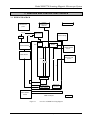
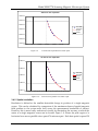
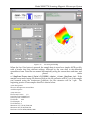
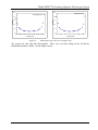
1
Scanning Magnetic
Microscope
Model SMM-770
Operating Guide
TRISTAN TECHNOLOGIES, Inc
San Diego, California
USA
www.tristantech.com
Model SMM 770 Scanning Magnetic Microscope System
Operating Guide
for
Single channel HTS SQUID microscope
Model SMM-770
Option 77P, 77V, 77T, 77B, 77HR, MS-770
TRISTAN TECHNOLOGIES, Inc
San Diego, California
USA
© Copyright 2005
www.tristantech.com
TRISTAN TECHNOLOGIES --Page 2
Model SMM 770 Scanning Magnetic Microscope System
Revision Record
Date
Revision
Description
May 6, 2005
Version 1.
Initial Release
May 11, 2005
Version 1.1
actual data included
May 31, 2005
Version 1.2
more data
© 2005 by Tristan Technologies
All rights reserved. No part of this manual may be reproduced, stored in a retrieval
system, or transmitted in any form or by any means, electronic, mechanical,
photocopying, recording, or otherwise, without prior written permission of
TRISTAN TECHNOLOGIES Inc.
TRISTAN TECHNOLOGIES Inc. reserves the right to change the functions, features,
or specifications of its products at any time, without notice.
Any questions or comments in regard to this product and other products from
Tristan Technologies, please contact or see http://www.tristantech.com:
Tristan Technologies
6191 Cornerstone Ct. East., Suite 107
San Diego, CA 92121
U. S. A.
Technical Support: (858) 550 – 2700
FAX: (858) 550 – 2799
[email protected]
http://www.tristantech.com
TRISTAN TECHNOLOGIES --Page 3
Model SMM 770 Scanning Magnetic Microscope System
WARRANTY
TRISTAN TECHNOLOGIES Limited Warranty
TRISTAN TECHNOLOGIES Inc. warrants this product for a period of twelve (12)
months from date of original shipment to the customer. Any part found to be
defective in material or workmanship during the warranty period will be repaired
or replaced without charge to the owner. Prior to returning the instrument for
repair, authorization must be obtained from TRISTAN TECHNOLOGIES Inc. or an
authorized TRISTAN TECHNOLOGIES service agent. All repairs will be warranted
for only the unexpired portion of the original warranty, plus the time between
receipt of the instrument at TRISTAN TECHNOLOGIES and its return to the owner.
This warranty is limited to TRISTAN TECHNOLOGIES’ products that are
purchased directly from TRISTAN TECHNOLOGIES, its OEM suppliers, or its
authorized sales representatives. It does not apply to damage caused by accident,
misuse, fire, flood or acts of God, or from failure to properly install, operate, or
maintain the product in accordance with the printed instructions provided.
This Warranty Is In Lieu Of Any Other Warranties, Expressed Or Implied,
Including Merchantability Or Fitness For Purpose, Which Are Expressly
Excluded. The Owner Agrees That TRISTAN TECHNOLOGIES’ Liability
With Respect To This Product Shall Be As Set Forth In This Warranty, and
Incidental Or Consequential Damages Are Expressly Excluded.
TRISTAN TECHNOLOGIES --Page 4
Model SMM 770 Scanning Magnetic Microscope System
Table of Contents
1.
SYSTEM OVERVIEW
10
1.1
INITIAL INSPECTION
12
1.2
REPACKING FOR RETURN SHIPMENT
12
1.3
RETURN FROM CUSTOMERS OUTSIDE THE U.S.A.
12
1.4
SYSTEM COMPONENTS
12
2.
CRYOGENIC SYSTEM
16
2.1
LIQUID NITROGEN MAINTENANCE
17
2.2
NORMAL INSTALLATION AND OPERATION
17
2.3
SAFETY PRECAUTIONS FOR HANDLING CRYOGENIC LIQUIDS
17
2.4
GENERAL INFORMATION
19
3.
SENSORS AND CONTROL ELECTRONICS
20
3.1
WIRING DIAGRAM
20
3.2
SENSOR INITIAL PERFORMANCE TEST
22
3.3
MEASURED PARAMETERS AND FACTORY TEST DATA
22
3.4
SENSOR CALIBRATION
25
4.
SYSTEM SOFTWARE
31
4.1
START
31
4.2
EXCITATION
33
4.3
TRACK CONSTRUCTION
33
4.4
MOVE MANUALLY
35
4.5
SQUID PARAMETERS
36
4.6
ACQUISITION PARAMETERS
37
4.7
FILTER PARAMETERS
38
4.8
SCAN CONTROLS
39
4.9
OUTPUT FILE
46
4.10
VIDEO CAMERA
50
TRISTAN TECHNOLOGIES --Page 5
Model SMM 770 Scanning Magnetic Microscope System
5.
SERVICE AND MAINTENANCE
51
5.1
INSPECTION AND CLEANING
51
5.2
CRYOGENIC DEWAR MAINTENANCE
51
5.3
GANTRY MAINTENANCE
53
5.4
VIBRATION ISOLATION EQUIPMENT
53
6.
OPTIONAL EQUIPMENT
53
6.1
OPTION 77P PNEUMATIC VIBRATION ISOLATION
53
6.2
OPTION 77V ACTIVE VIBRATION CANCELLATION
53
6.3
OPTION 77T TILT TIP FRAME
54
6.4
OPTION 77B FIXED FIELD CAPABILITY
55
6.5
OPTION 77HR 12.5 MICRON STEP CAPACITY
59
6.6
OPTION MS-770 TWO LAYER MU METAL MAGNETIC SHIELD
59
7.
APPENDICES
62
7.1
THERMOMETRY AND COOLDOWN
62
7.2
CALCULATING B-FIELDS
63
7.3
WIRING AND HARDWARE CONFIGURATION
66
7.4
BACKGROUND SUBTRACTION
73
7.5
STAGE CALIBRATION
74
8.
HTS SQUID HANDLING CAUTIONS
75
Table of Figures
Figure 1-1: SQUID System.................................................................................................. 10
Figure 1-2: Microscope Setup.............................................................................................. 11
Figure 2-1: Cryogenic Dewar .............................................................................................. 16
Figure 3-1 Overview of SMM770 wiring diagram.............................................................. 20
Figure 3-2 SMM770 detailed wiring diagram ..................................................................... 21
Figure 3-3: Manufacturer-supplied noise spectrum and tune specs..................................... 22
Figure 3-4 SQUID noise spectrum at (0-200Hz), 81.8 Kelvin, gain=x100......................... 24
Figure 3-5 SQUID noise spectrum at (0-3.2 kHz), 81.8 Kelvin, gain=x100....................... 24
TRISTAN TECHNOLOGIES --Page 6
Model SMM 770 Scanning Magnetic Microscope System
Figure 3-6 SQUID noise spectrum at (0-12.8 kHz), 81.8 Kelvin, gain=x100..................... 25
Figure 3-7: Horizontal Scan of parallel wires 0.003" apart. ................................................ 26
Figure 3-8: Horizontal scans at increasing liftoffs to parallel wires 0.009” apart. .............. 26
Figure 3-9: Vertical Scan of parallel wires 0.009" apart ..................................................... 27
Figure 3-10:
Vertical scan of parallel wires 0.003" apart.................................................... 27
Figure 3-11: Central peak of a horizontal scan across 76 micron separation meander coil 28
Figure 3-12: US Dollar bill .................................................................................................. 29
Figure 3-13: Image showing what a small amount of processing can accomplish.............. 29
Figure 3-14:: The following output of data is a 3D post processing of the image shown in
Figure 3-11...................................................................................................................... 30
Figure 4-1: Selection of Initialization file............................................................................ 31
Figure 4-2: Main panel after system start ............................................................................ 32
Figure 4-3: Track parameters............................................................................................... 33
Figure 4-4: Preview of track to be used for discrete scanning............................................. 34
Figure 4-5: Preview of QuikScan (continuous) track .......................................................... 35
Figure 4-6: Manual control of the sample stage .................................................................. 36
Figure 4-7: iMAG SQUID parameters................................................................................. 37
Figure 4-8: Data acquisition parameters .............................................................................. 38
Figure 4-9: Filter parameters ............................................................................................... 39
Figure 4-10: Scan controls ................................................................................................... 40
Figure 4-11: View of scan track........................................................................................... 41
Figure 4-12: Generation of stripchart and 3D graph............................................................ 42
Figure 4-13: Contour lines in the 3D plot............................................................................ 43
Figure 4-14: Cursor usage in the 3D plot............................................................................. 44
Figure 4-15 Illumination control in the 3D plot................................................................... 45
Figure 4-16: Z-axis control .................................................................................................. 46
Figure 4-17: File saving and loading ................................................................................... 47
Figure 4-18 Example data file.............................................................................................. 48
Figure 4-19: Deletion of data files ....................................................................................... 49
Figure 4-20 Gap between the windown and sample as seen by the video camera .............. 50
Figure 5-1 Measured boil off rate ........................................................................................ 52
Figure 6-1 Passive Newport table vs. Newport table with the Activator installed in a
horizontal configuration.................................................................................................. 54
TRISTAN TECHNOLOGIES --Page 7
Model SMM 770 Scanning Magnetic Microscope System
Figure 6-2 Passive Newport table vs. Newport table with activator and without activators
and load of 500lbs........................................................................................................... 54
Figure 6-3 Magnet test ring (option 77B) ............................................................................ 56
Figure 6-4 Field profile along Z axis of magnetic ring........................................................ 56
Figure 6-5 Field profile along y and x axis of magnetic ring .............................................. 57
Figure 6-6:
x100
Noise spectra of SMM-770 system with 5.5 Gauss external applied field, Gain
58
Figure 6-7:
x20
Noise spectra of SMM-770 system with 5.5 Gauss external applied field, Gain
59
Figure 6-8: MS770 mu-metal shield engineering drawings. ............................................... 60
Figure 6-9 Option MS-770 two layer mu metal shield ........................................................ 61
Figure 7-1 Elapsed time for SQUID tip coldwon to operating temperatures ...................... 62
Figure 7-2 Unsubtracted background................................................................................... 73
Figure 7-3 Subtracted background....................................................................................... 74
Table of Tables
Table 1
System Parameters ..................................................................................................... 23
Table 3-2:
SQUID performance tested with iMAG® ................................................................. 23
Table 3-3:
Iterative Calibration Results for SMM-770............................................................... 25
Table 4
Pt thermometer calibration ......................................................................................... 62
TRISTAN TECHNOLOGIES --Page 8
Model SMM 770 Scanning Magnetic Microscope System
DIRECTIVE 97/23/EC OF THE EUROPEAN PARLIAMENT AND OF THE COUNCIL of 29
May 1997 on the approximation of the laws of the Member States concerning pressure equipment
states that “Whereas equipment subject to a pressure of not more than 0,5 bar does not pose a
significant hazard due to pressure; whereas there should not therefore be any obstacle to its free
movement within the Community; whereas this Directive applies to equipment subject to a maximum
allowable pressure exceeding 0,5 bar”. Since the maximum allowable design pressure is less than
0.5 bar, Tristan’s model SM770 is in conformity with DIRECTIVE 97/23/EC.
TRISTAN TECHNOLOGIES --Page 9
Model SMM 770 Scanning Magnetic Microscope System
1. SYSTEM OVERVIEW
The Model SMM-770 Scanning Magnetic Microscope System is a close spaced HTS SQUID
magnetometer system (Figure 1-1) that has been designed and built by Tristan to detect Bz
magnetic field. The main system consists of:
Liquid nitrogen dewar with HTS SQUID Sensor
Scanning stage
X-y tilt stage
Vibration Isolation table
iMAG® SQUID control electronics1, motion control hardware and software, data
acquisition hardware and software and a computer system.
Flux-Locked Loop
iMC-303
SQUID Controller
iFL-301-H
Liquid
Nitrogen
Dewar
Interface Card(s)
Control Computer
Software
data acquisition,
analysis,
motion control
HTS
SQUID
dc magnet (BZ)
Sample
HItachi Camera
Non-Magnetic
Stand-off
X-Y Scanning Table
Z
X
Y
note: some optional items shown
Figure 1-1:
1
SQUID System
iMAG® is a registered trademark of Tristan Technologies, Inc.
TRISTAN TECHNOLOGIES --Page 10
Model SMM 770 Scanning Magnetic Microscope System
770 MICROSCOPE SCANNING STATION
Z
X
DEWAR
FIBER OPTIC LIGHT
Y
VIDEO MICROSCOPE
SQUID
WINDOW
SPACER PLATE
(OPTIONAL)
X-Y MICRO TILT STAGE
Z-AXIS, MANUAL
Z-AXIS DIGITAL
READOUT
X-Y AXIS, MOTORIZED
VIBRATION TABLE TOP
ACTIVE
DAMPING
(3 UNITS)
VIBRATION
ISOLATION
AIR TABLE
Figure 1-2:
Microscope Setup
TRISTAN TECHNOLOGIES --Page 11
Model SMM 770 Scanning Magnetic Microscope System
1.1 INITIAL INSPECTION
All Tristan Technologies instruments and equipment are carefully inspected and packaged
at Tristan Technologies prior to shipment. However, if a unit is received mechanically
damaged, notify the carrier and the nearest Tristan Technologies representative, or the
factory in San Diego, California. Keep the shipping container and packing material for the
carrier and insurance inspections.
WARNING
USE CARE WHEN HANDLING THE DEWAR. THE SQUID SENSOR IS MOUNTED ON A SPECIAL
MECHANISM TO ACCURATELY PLACE THE TIP CLOSE TO THE WINDOW. MISHANDLING MAY
CAUSE MISALIGNMENT OF THE SQUID MOUNTING.
If the unit does not appear to be damaged but does not operate to specifications, contact the
nearest Tristan Technologies representative or the Tristan Technologies factory and
describe the problem in detail. Please be prepared to discuss all surrounding
circumstances, including installation and connection details. After obtaining authorization
from the Tristan Technologies representative, return the unit for repair along with a tag on
it identifying yourself as the owner. Please enclose a letter describing the problem in as
much detail as possible.
1.2 REPACKING FOR RETURN SHIPMENT
If it is necessary to return the system, you should repack the unit in its original container (if
available). For this reason, it is advisable to save the original crate sent by Tristan
Technologies. However, if this is not possible, use the following instructions for
repackaging:
1. Wrap the unit in either bubble wrap or foam rubber
2. Cover the bottom of a sturdy container with at least 3 inches of Styrofoam pellets or
shredded paper.
3. Set the unit down onto the packing material and fill the rest of the container with
Styrofoam or shredded paper. The unit must be completely protected by at least 3
inches of packing material on all sides.
1.3 RETURN FROM CUSTOMERS OUTSIDE THE U.S.A.
To avoid delays in Customs clearance of equipment being returned, contact the Tristan
Technologies representative in your area, or the Tristan Technologies factory in San Diego,
California, for complete shipping information and necessary customs requirements. Failure
to do so can result in significant delays.
1.4 SYSTEM COMPONENTS
•
Dewar NLD-770
s/n 153
•
Option 77P
Pneumatic vibration isolation non magnetic microscope table
•
Option 77V
Active vibration cancellation
TRISTAN TECHNOLOGIES --Page 12
Model SMM 770 Scanning Magnetic Microscope System
•
Option 77T
Tilt tip frame
•
Option 77B
Fixed field capability
•
Option 77HR
12.5 micron step capability
•
Option MS-770
Two layer Mu metal shield
•
Electronics Rack
BNC Adapter: National Instruments, model BNC-2090, s/n 112FBE6
Controller iMAG®, model iMC-303, s/n 001063
GPIB cable
Flux-Locked loop (FLL), iFL-301-H s/n2020
Composite fiber optic cable CC-60
Cable SP-7 , HTS Cryogenic Cables
(1) Special Cable: Single-Ended BNC to Differential Dual BNC.
Motor drive , model MID-7602, s/n E006A6
Motion Controller cable
•
Computer control and data acquisition system and software
Dell Precision PWS370 with dual Pentium 4 2.8 GHz CPUs and 1.00 GB RAM, model
WHM, s/n 49YGZ61, service code 9309223945
Windows XP Pro, product key J8C4W-PTCW4-X7JPR-WTBWV-XXYTB
GPIB card, National Instruments, model PCI-GPIB, s/n 110EF26
DAQ card, National Instruments, model PCI-6036E, s/n 10ECA27
Motion Control card, National Instruments, model PCI-7342, s/n DF5E03
Video card, National Instruments, model PCI-1405, s/n 111FD9C
LabView custom software ,MagScan version 3.0
•
Video monitors, 19”: Dell, model DP/N-0G6566,
s/n CN-0G6566-47804-51U-U3V2
s/n CN-0G6566-47804-51U-U3MU
•
Compressor: Jun-Air, model 3-04, s/n 582261
TRISTAN TECHNOLOGIES --Page 13
Model SMM 770 Scanning Magnetic Microscope System
•
Microscope & Dewar Assembly
SQUID sensor mod HTM-0 s/n Mi 10803
7B Fixed DC capability
•
Microscope gantry:
Dewar Vacuum gauge Dunaway DST-531
Laser Ranger; Banner LG10A65PI
Readout; Red Lion PAXP-1/8
Omron DC power supply S8VS-06024A
Tip/Tilt stage 2071-S00
Motorized room temperature XY stage with controller consistinf of:
o X motorized stage, 12.5 micron step
Parker Daedel, model 106006BTMSD4L3C2M1E1, s/n 052316401A
Parker Daedel, model ZETA57-51-MO, s/n 05033000124
o Y motorized stage, 12.5 micron step
Parker Daedel, model 106008BTMSD4L3C2M1E1, s/n 052316401A
Parker Daedel, model ZETA57-51-MO, s/n 05033000125
Manual room temperature Z stage, 1 micron step,
•
Newport 16660 / Oriel 14000
Microscope/telescope with illuminator and high resolution video camera
CCD camera: Hitachi, model KP-D20BU, s/n 4087307
Camera lens: Computar
Fiber optic light source: Dolan-Jenner, model DC-950AK, s/n 0504031224
•
Passive Damping System: TMC Micro-g, model 63-531, s/n 3349301
•
Active Damping System: Newport Optics Activator AD500, model NVX-1206-1,
s/n 4076-0059
s/n 4076-0072
s/n 4076-0076
•
Oriel micrometer 1400
•
Battery for oriel micrometer, SR44, 1.5V, No 938882
•
MuMetal Shield 2104-S100-4
•
Accessory pack
Dewar o-ring 166, Qty. 2
TRISTAN TECHNOLOGIES --Page 14
Model SMM 770 Scanning Magnetic Microscope System
Nose piece o-ring 030, Qty 2
Nose piece screws 4-40-1/4” SHCS, Brass
Spacer plate Screws ¼”-20 SHCS
•
System Manuals
SMM-770 system manual
Dell Precision workstation 370 Quick Reference Guide
Dell Desktop Computers Product Information Guide
Dell Drivers and User Documentation E193FP LCD Monitor CD (2)
Windows XP professional SP2
Dell Precision Workstation ResourceCD
Parker Compumotor Zeta Drive Installation Guide
Daedal Linear and Rotary Positioners Operating Manual (2)
Zeta Drive User Guide
Parker Optical L/H Switch Cable Pigtail Ended Cable drawing (2)
Parker Quality Assurance Data Sheet (2)
Newport Isostation vibration isolated workstation
Newport Active vibration Damping system
DC950 Illuminator Operation Manual
Duniway digital gauge Instruction Manual
Hammond Enclosures RCHS-Series Assembly Instructions
NI-DAQ 7 DAQ Quick Start Guide
NI-488.2 CD, NI Montion control CD, NI-IMAQ 3.1 Getting Started CD
NI GPIB Installation guide
MID-7604/7602 Power Drive User Guide (2)
DAQ BNC-2090 User Manual
IMAQ 1405 User Manual
MID-7604/7602 Power Drive Front Panel DIP Switch Settings Sheet
NI Registration Cards (4), Global Service Support Booklet (4)
Jun-Air compressor
OMRON Sensor
Banner L-GAGE Laser sensor
Red Lion Analog input panel meter
Imag Manuals (2)
TRISTAN TECHNOLOGIES --Page 15
Model SMM 770 Scanning Magnetic Microscope System
2. CRYOGENIC SYSTEM
The cryogenic system includes a fiberglass liquid nitrogen dewar (Figure 2-1). The sensorwindow gap is pre-set at the factory and does not need adjustment.
Figure 2-1:
Cryogenic Dewar
TRISTAN TECHNOLOGIES --Page 16
Model SMM 770 Scanning Magnetic Microscope System
2.1 LIQUID NITROGEN MAINTENANCE
The system nitrogen level can be measured with a small fiberglass stick (supplied)
2.2 NORMAL INSTALLATION AND OPERATION
2.2.1 PROBE INSTALLATION (WARM DEWAR)
•
Inspect dewar and verify internal cavity is free from ice, moisture, or other
contaminants. CONTAMINANTS COULD RESULT IN INCORRECT
MEASUREMENTS OR PROBE DAMAGE!
2.2.2 NITROGEN TRANSFER
•
Verify Nitrogen level in Microscope dewar.
•
Open transfer port at top of dewar.
•
Begin Nitrogen transfer observing all standard cryogenic handling and safety
procedures.
•
Observe transfer levels. * Note: Dewar capacity is 2.29 liters.
•
Plug transfer port after allowing sufficient vent time.
•
Verify all connectors at top of probe are free from moisture.
2.3 SAFETY PRECAUTIONS FOR HANDLING CRYOGENIC LIQUIDS
The potential hazards of handling liquid nitrogen stem mainly from the following
properties:
WARNING
1. THE LIQUID IS EXTREMELY COLD
2. THE ULTRA-LOW TEMPERATURE OF LIQUID NITROGEN CAN CONDENSE AND SOLIDIFY AIR.
3. VERY
GAS.
SMALL AMOUNTS OF LIQUID
NITROGEN
ARE CONVERTED INTO LARGE VOLUMES OF
4. NITROGEN IS NOT LIFE SUPPORTING.
2.3.1 EXTREME COLD-- COVER EYES AND EXPOSED SKIN
Accidental contact of liquid nitrogen or the cold gas that results from its rapid evaporation
may cause a freezing injury similar to a burn. Protect your eyes and cover the skin where
the possibility of contact exists. Eye protection should always be worn when transferring
liquid nitrogen.
2.3.2 KEEP AIR AND OTHER GASES AWAY FROM LIQUID NITROGEN
The low temperature of liquid nitrogen, vacuum pumped liquid nitrogen, or cold gaseous
nitrogen can solidify another gas. Solidified gasses and liquid, particularly solidified air,
can plug pressure-relief passages and foul relief valves. Plugged passages are hazardous
because of the continual need to vent the nitrogen gas that evolves as the liquid
continuously evaporates. Therefore, always store and handle liquid nitrogen under positive
TRISTAN TECHNOLOGIES --Page 17
Model SMM 770 Scanning Magnetic Microscope System
pressure and in closed systems to prevent the infiltration and solidification of air or other
gases. Do not permit condensed air on transfer tubes to run down into the container
opening.
2.3.3 KEEP EXTERIOR SURFACES CLEAN TO PREVENT COMBUSTION
Atmospheric air will condense on exposed nitrogen-cooled piping. Nitrogen, having a
lower boiling point than oxygen, will evaporate first from condensed air, leaving an
oxygen-enriched liquid that may drip or flow to nearby surfaces. Areas and surfaces upon
which oxygen-enriched liquid can form, or come in contact with, must be cleaned to
oxygen-clean standards to prevent possible ignition of grease, oil, or other combustible
substances. Leak-testing solutions should be selected carefully to avoid mixtures that can
leave a residue that is combustible. When combustible type foam insulations are used, they
should be carefully applied to reduce the possibility of exposure to oxygen-enriched liquid
that could, upon impact, cause explosive burning of the foam.
2.3.4 Pressure-Relief Devices Must Be Adequately Sized.
Liquid nitrogen evaporates very rapidly when heat is introduced or when liquid nitrogen is
first transferred into warm or partially cooled equipment. Even minor deterioration of the
vacuum in the nitrogen container can result in significant evaporation. Pressure relief
devices for liquid nitrogen equipment must, therefore, be of adequate capacity to release
nitrogen vapor resulting from such heat inputs, and thus, prevent hazard due to excessive
pressure. This system has been designed to safely vent the evolving nitrogen gas in the
event of any reasonable failure mode. If transfer lines can be closed off at both ends so that
a cryogenic liquid or the related cold gas can become trapped between the closed ends, a
pressure-relief device must be provided in that line to prevent excessive pressure build-up.
WARNING
DO NOT MAKE ANY MODIFICATIONS TO THIS SYSTEM WHICH MIGHT
AFFECT ITS ABILITY TO VENT NITROGEN GAS IN THE EVENT OF AN
EMERGENCY, SUCH AS LOSS OF VACUUM IN THE DEWAR VACUUM SPACE
2.3.5 KEEP EQUIPMENT AREA WELL VENTILATED
Although nitrogen is nontoxic, it can cause asphyxiation in a confined area without
adequate ventilation. Any atmosphere which does not contain enough oxygen for
breathing can cause dizziness, unconsciousness, or even death. Nitrogen, being colorless,
odorless, and tasteless cannot be detected by the human senses and will be inhaled
normally as if it were air. Without adequate ventilation, the expanding nitrogen can
displace air and result in an atmosphere that is not life supporting. The cloudy vapor that
appears when liquid nitrogen is exposed to the air is condensed moisture, not the gas itself.
The issuing nitrogen gas is invisible. Liquid containers should be stored in large, wellventilated areas.
If a person becomes groggy or loses consciousness when working around nitrogen, get
them to a well-ventilated area immediately. If breathing has stopped, apply artificial
respiration. If a person loses consciousness, summon a physician immediately.
TRISTAN TECHNOLOGIES --Page 18
Model SMM 770 Scanning Magnetic Microscope System
2.4 GENERAL INFORMATION
•
Use caution when handling delicate fiber-optic cables. Do not bend smaller than a 10 cm
radius.
•
Do not fill with liquid nitrogen unless dewar vacuum is < 50 microns.
•
Transfer nitrogen with fill tube inserted to the dewar bottom.
•
Do not touch sapphire window at any time ever!
TRISTAN TECHNOLOGIES --Page 19
Model SMM 770 Scanning Magnetic Microscope System
3. SENSORS AND CONTROL ELECTRONICS
3.1 WIRING DIAGRAM
Cryocon
2 therm and 1
heater Pt
220-230 V AC
power
Manual Level
Analog Out
IMag control
NI BNC
Panel
Pressure Gauge
GPIB
FLL
CPU and Monitors
Pressure
Sensor
Sheild
Z Optical Readout
Omron
Motor Drivers
SQUID Cryocable
Fiber-Lite Light
source
F/O
2 Pt Thermo
and 1 heater
Shield
F/O
DC Magnet
SQUID
CCD Camera
X, Y stage motion
and Z manual
Stratford LCD
Micrometer
Anti Vibration Table
Active Vibration
Control
SMM-770 Naples
Figure 3-1
Overview of SMM770 wiring diagram
TRISTAN TECHNOLOGIES --Page 20
Compressor
Model SMM 770 Scanning Magnetic Microscope System
Figure 3-2
SMM770 detailed wiring diagram
TRISTAN TECHNOLOGIES --Page 21
Model SMM 770 Scanning Magnetic Microscope System
3.2 SENSOR INITIAL PERFORMANCE TEST
3.3 MEASURED PARAMETERS AND FACTORY TEST DATA
The following parameters were measured at Tristan prior to shipment. Performance data is
the result of testing done at Tristan.
3.3.1 SQUID sensors
The HTM-0 SQUID sensor was initially tested in a HTS superconducting shield to
eliminate all external environmental noise contributions. Testing was done using Tristan
iMAG® 300 SQUID electronics. The flux transfer function (nT/φo) and noise (pT/√Hz) was
determined. The test results of the supplied SQUID sensors are shown in Figure 3-3 as a
function of frequency, and are listed along with sensitivity (informational purposes only).
Figure 3-3:
Manufacturer-supplied noise spectrum and tune specs
3.3.2 Measurements in a non-superconducting shield
Additional measurements were made at Tristan in a non-superconducting mu-metal
shield. These tests determine the bandwidth and voltage transfer function for the device.
Operation at various gains and ranges are verified using the customer’s electronics. Testing
is done using Hewlett-Packard (34401 A DMM, 35665A dynamic signal analyzer) and other
appropriate test equipment.
TRISTAN TECHNOLOGIES --Page 22
Model SMM 770 Scanning Magnetic Microscope System
Because of the local environmental noise (even inside a mu-metal shield), these
measurements show different noise levels than the results shown in Figure 3-3. It should be
noted that HTS SQUID sensors typically show a 10 - 30% variation from device to device.
Table 3-2 and Table 3-2 summarize the main system’s parameters and SQUID performance
using a Tristan Technologies, Inc. iMC-303 iMAG® SQUID Controller.
Table 1 System Parameters
IFL-301-H
S/N 2020
SQUID
Mi10803
Channel
1
Gain
X100
Slew
Normal
Heat Time (1k R)
2 sec
Cool Time
99 sec
Triangle Amplitude
1.65 Vp-p
Bias%
35
Bias%
35
Mod%
12
Skew
18
Table 3-2:
SQUID performance tested with iMAG®
Gain
Slew
X100
Normal
x100
Slow
X50
Normal
Units
V/Φo
0.95
Noise @5kHz
9.15
pT/√Hz
Bandwidth
22
kHz
Loop Locked
yes
0.95
yes
yes
Note – gains 1-50 are done with one feedback resistor and gains 100-500 are done with a different
feedback resistor. The Gain X100 and X50 have the same analog gain so that will have to be
manually accounted for when measuring data not through the software.
3.3.3 System Noise
The following spectra were taken of the complete SMM 770 system.
TRISTAN TECHNOLOGIES --Page 23
Model SMM 770 Scanning Magnetic Microscope System
Figure 3-4
SQUID noise spectrum at (0-200Hz), 81.8 Kelvin, gain=x100
Figure 3-5
SQUID noise spectrum at (0-3.2 kHz), 81.8 Kelvin, gain=x100
TRISTAN TECHNOLOGIES --Page 24
Model SMM 770 Scanning Magnetic Microscope System
Figure 3-6
SQUID noise spectrum at (0-12.8 kHz), 81.8 Kelvin, gain=x100
3.4 SENSOR CALIBRATION
The SQUID microscope sensor position is calibrated to determine a number of parameters.
Using a combination of parallel wires at various liftoff values, the SQUID-to-window
spacing is determined. The SQUID sensitivity is determined and the system resolution is
calculated. The following charts and graphs show the data for this unit.
The system calibration for a typical SQUID is calibrated as follows:
Table 3-3:
Calibration Parameters for SMM-770
Close Wire Spacing
Wide Wire Spacing
SQUID to Wire Lift-off
0.003” (76 µm)
0.009” (229 µm)
0.0016” (400 µm)
3.4.1 Lift-off: SQUID – to- Source spacing
Determination SQUID–to–Source spacing requires comparison horizontal scans of parallel
wire loops carrying current in opposite directions at various heights. Theoretical values
may be calculated from Appendix 6.1.3 (Field Above a Straight Wire) and assuming infinite
length with height d. By changing d to r = (d2 + x2)½ for lateral displacement x, and defining
the polar angle θ = tan-1(x/d), the vertical field varies as sinθ*Br. Note that the magnetic
field components of each wire pass constructively interfere, and thus the widely spaced
wire loop produces greater field at close lift-off distances.
TRISTAN TECHNOLOGIES --Page 25
Model SMM 770 Scanning Magnetic Microscope System
8.E-06
76 micron wire spacing
400 micron liftoff
7.E-06
Induction field (nT)
7.E-06
6.E-06
Scan Data
Theoretical
6.E-06
5.E-06
5.E-06
-2
-1.5
-1
-0.5
0
0.5
1
1.5
2
Horizontal scan (mm)
Figure 3-7:
Horizontal Scan of parallel wires 0.003" apart.
The above theoretical line is derived from a best fit to the gap between the SQUID and the
wire source. This result is verified by successive horizontal scans at increasing liftoffs, and
a pair of vertical scans of two sets of parallel wires. The vertical scan plots illustrate the
position of the SQUID probe tip and the gap to the Sapphire window.
229 micron wire spacing
6.00E-06 T
5.00E-06 T
4.00E-06 T
25um data
25um calculated
100um data
100um calculated
3.00E-06 T
2.00E-06 T
200um data
200um calculated
400um data
400um calculated
1.00E-06 T
0.00E+00 T
-1.00E-06 T
-2.00E-06 T
-2 mm
Figure 3-8:
-1.5 mm
-1 mm
-0.5 mm
0 mm
0.5 mm
1 mm
1.5 mm
2 mm
Horizontal scans at increasing liftoffs to parallel wires 0.009” apart.
TRISTAN TECHNOLOGIES --Page 26
Model SMM 770 Scanning Magnetic Microscope System
229 micron wire separation
7.00E-06 T
6.00E-06 T
5.00E-06 T
Scan Data
Theoretical
4.00E-06 T
Window
SQUID
3.00E-06 T
2.00E-06 T
1.00E-06 T
-0.2 mm
0 mm
0.2 mm
Figure 3-9:
0.4 mm
0.6 mm
0.8 mm
1 mm
Vertical Scan of parallel wires 0.009" apart
76 micron wire separation
2.50E-06 T
2.00E-06 T
Scan Data
Theoretical
1.50E-06 T
Window
SQUID
1.00E-06 T
5.00E-07 T
0 mm
0 mm 0.1 mm 0.2 mm 0.3 mm 0.4 mm 0.5 mm 0.6 mm 0.7 mm 0.8 mm 0.9 mm
Figure 3-10:
Vertical scan of parallel wires 0.003" apart
3.4.2 Spatial resolution
Resolution is defined as the smallest detectable change in position of a single magnetic
source. This can be calculated by comparison of the maximum observed spatial magnetic
field gradient to the system noise level across the measurement bandwidth of interest.
However, this is more easily understood visually by illustrating the accuracy at which the
center of a single magnetic source can be located. Figure 3-11 shows the peak region of a
horizontal scan across parallel wires spaced 76 microns apart. Each data point is spaced 50
TRISTAN TECHNOLOGIES --Page 27
Model SMM 770 Scanning Magnetic Microscope System
microns apart, and it is evident from inspection that the center of the source is between the
two central data points. Therefore, the system spatial resolution is better than 50 microns.
76 micron wire separation meander peak
7.40E-06 T
7.20E-06 T
7.00E-06 T
6.80E-06 T
Scan Data
Theoretical
6.60E-06 T
6.40E-06 T
6.20E-06 T
6.00E-06 T
-0.3 mm
Figure 3-11:
-0.2 mm
-0.1 mm
0 mm
0.1 mm
0.2 mm
0.3 mm
Central peak of a horizontal scan across 76 micron separation meander coil
TRISTAN TECHNOLOGIES --Page 28
Model SMM 770 Scanning Magnetic Microscope System
3.4.3 Example data
The first plot is the entire subject: a US Dollar bill. The outputs are at resolution of 10
micron step size of the middle three letters in the word “STATES” on the front above the
bust of Washington.
Figure 3-12:
Figure 3-13:
US Dollar bill
Image showing what a small amount of processing can accomplish
TRISTAN TECHNOLOGIES --Page 29
Model SMM 770 Scanning Magnetic Microscope System
Figure 3-14::
The following output of data is a 3D post processing of the image shown in Figure 3-13.
TRISTAN TECHNOLOGIES --Page 30
Model SMM 770 Scanning Magnetic Microscope System
4. SYSTEM SOFTWARE
4.1 START
Ensure that the iMAG iMC-303 unit and the Motor Driver MID-7602 unit are powered on.
Click the “Mag Scan” icon on the desktop. This will start the magnetic scanning
application. The user will be presented with a dialog box, asking the user to select an
initialization file. The selected initialization file controls a variety of system settings. These
files are located under c:\MagScan\Configuration\*.ini. It is possible for the user to
manually edit these files, but Tristan recommends caution when doing so. Tristan strongly
recommends that the user do not edit the Default.ini file, but rather make copies of it
and edit those. All *.ini files are presented in the dialog box for user selection.
Additionally, there is a Current.ini file, which contains the settings of the last time the
software was run.
Figure 4-1:
Selection of Initialization file
Typically, the user will select the Default.ini file, and proceed by pressing the OK
button. The user will then see a progress bar indicating the configuration of the iMAG unit.
Note that the iMAG unit must have its GPIB address = 15, which can be set from the front
panel. After the iMAG unit is configured, the user will then see a progress bar indicating
configuration and homing of the sample stage.
TRISTAN TECHNOLOGIES --Page 31
Model SMM 770 Scanning Magnetic Microscope System
CAUTION
The user must ensure that the stage is clear of obstacles throughout the entire working
region (154 mm x 154 mm) before homing starts. Improperly placed samples could cause
permanent damage to the sample and/or dewar. The Tristan warranty does not cover
resulting damage
Homing typically takes about 60 seconds. After this is completed, the user has access to the
main screen of the software, shown below:
Figure 4-2:
Main panel after system start
In the upper right corner, a text box shows system messages. These messages are
automatically
logged
to
a
text
file
under
c:\MagScan\System_Log\<YYYYMMM>\<date>_<time>.log.
These are simple
ASCII files, and are named based on the current date and time. The user can insert
comments into these files using the User Notes button.
The left side of the main panel shows two sets of tabs. The upper tab set controls the
acquisition of data, and the lower one controls the display of the results.
Pressing <control>-H will pop-up a help window for any item that the mouse cursor is
over. This help window provides a description of what each control and indicator does.
TRISTAN TECHNOLOGIES --Page 32
Model SMM 770 Scanning Magnetic Microscope System
In general, user interface items that are colored white are controls that the user can adjust.
User interface items that are colored gray are indicators. User interface items that are
grayed-out have been disabled.
4.2 EXCITATION
The Excitation tab controls the application of a signal to an excitation coil or sample. This
option is disabled for this SMM-770 delivery. Assorted controls related to excitation, such
as phase shift and its real and imaginary components, are disabled throughout the user
interface.
4.3 TRACK CONSTRUCTION
The Track tab controls the movement of the sample stage.
Figure 4-3:
Track parameters
The parameters in the table specify a serpentine track over which to move the stage by
discrete points. The stage will move to each of these points, stop, wait, collect a SQUID
measurement, and then proceed on to the next point. The speed at which the stage moves is
set in the *.ini file. The resulting track can be previewed by pressing the Preview button.
TRISTAN TECHNOLOGIES --Page 33
Model SMM 770 Scanning Magnetic Microscope System
Figure 4-4:
Preview of track to be used for discrete scanning.
Collecting data in this discrete manner can be time-consuming. Alternatively, the user can
perform a “Quik-Scan” (discussed below) whereby the stage is moved continuously while
data is collected at uncontrolled points along the track; this is useful for a quick sample
survey to identify regions of interest for more precise study by discrete scanning. The
continuous track can be displayed by pressing the button on the right from SloScan to
QuikScan. Because of the nature of the hardware, the full set of coordinates over which to
continuously move the stage must be uploaded to the stage motion controller.
Acceleration, deceleration, and interpolated points must be constructed from the defined
track, with care taken to avoid the memory and speed limitations of the hardware. This
construction and limitation-checking is automated. If the QuikScan parameters are
improperly set, an error message will be clearly displayed and the graph grayed out. The
user will be offered suggestions as to how to change the parameters to create a valid
QuikScan track.
TRISTAN TECHNOLOGIES --Page 34
Model SMM 770 Scanning Magnetic Microscope System
Figure 4-5:
Preview of QuikScan (continuous) track
4.4 MOVE MANUALLY
The Move tab manually controls the stage. This is useful for moving the stage to a position
where it is easier to change samples.
TRISTAN TECHNOLOGIES --Page 35
Model SMM 770 Scanning Magnetic Microscope System
Figure 4-6:
Manual control of the sample stage
CAUTION
The user must ensure that the stage is clear of obstacles throughout the entire working
region (154 mm x 154 mm) before homing starts. Improperly placed samples could cause
permanent damage to the sample and/or dewar. The Tristan warranty does not cover
resulting damage.
The HALT button will immediately terminate movement. The HOME button will force the
stage to home; this is theoretically unnecessary, but this utility is provided in the event of a
hardware error.
4.5 SQUID PARAMETERS
The SQUID tab controls the iMAG settings. Tristan strongly recommends making
adjustments to the iMAG by using these controls rather than the physical front panel of the
instrument. Changes made from the physical front panel may not be registered in the
software, and thus the data displays and files would be inaccurate. This is of special
concern with the gain setting.
The data output of the iMAG can be viewed in the Raw Data tab. This is especially useful
during a manual tune of the iMAG.
TRISTAN TECHNOLOGIES --Page 36
Model SMM 770 Scanning Magnetic Microscope System
Figure 4-7:
iMAG SQUID parameters
If the Tune procedure takes longer than 45 seconds, you may see a pop-up window
indicating that there is a problem with iMAG communications. This message can be
ignored. It is due to the fact that the iMAG does not support GPIB communications during
an auto-tune procedure.
4.6 ACQUISITION PARAMETERS
The Acquire tab controls the data acquisition parameters. The Sample Rate can be adjusted
from 10 Hz to 10 kHz. The Settle Time defines the period of time to wait after the stage has
moved to a discrete position and before collection of a block of data. The Measure Time
defines the duration of a block of data to collect. The resulting block of data is processed to
calculate DC and AC components that provide a measure of the magnetic field strength at
the particular sample location.
TRISTAN TECHNOLOGIES --Page 37
Model SMM 770 Scanning Magnetic Microscope System
Figure 4-8:
Data acquisition parameters
4.7 FILTER PARAMETERS
The Filter tab controls the software filtering of raw SQUID data. This is not to be confused
with the hardware filtering of raw SQUID data that can be selected using the the iMAG
HiPass and LoPass filters.
TRISTAN TECHNOLOGIES --Page 38
Model SMM 770 Scanning Magnetic Microscope System
Figure 4-9:
Filter parameters
In general, these filter settings are only useful for minimizing the Mains noise seen under
the Raw Data tab. They will not have much affect on the calculation of DC and AC
components.
4.8 SCAN CONTROLS
The Scan tab controls collection of magnetic field data from a sample.
TRISTAN TECHNOLOGIES --Page 39
Model SMM 770 Scanning Magnetic Microscope System
Figure 4-10:
Scan controls
The Start Scan button will initiate discrete scanning using the track defined under the Track
tab. This scanning can be stopped by pressing the button again. The progress of the
scanning can be viewed by using the Map button. This button shows the progress of the
scanning.
TRISTAN TECHNOLOGIES --Page 40
Model SMM 770 Scanning Magnetic Microscope System
Figure 4-11:
View of scan track
After the track has completed, the track count will decrement, and another track
performed, until the track count reaches zero. Data at each track point is averaged. If the
track count is set to -1, the track is repeated endlessly.
Pressing the QuikScan button will perform a continuous scan of the sample approximately
along the defined track. This will be performed only once.
As scanning proceeds, the data will be plotted as a stripchart under the Mag & Phase tab,
and plotted in the 3D graph on the right, as shown below:
TRISTAN TECHNOLOGIES --Page 41
Model SMM 770 Scanning Magnetic Microscope System
Figure 4-12:
Generation of stripchart and 3D graph
The stripchart can be paused, panned, and zoomed using the palette controls. The 3D
graph can be rotated by <control>-rightclick and dragging the mouse across the graph,
panned by <shift>-rightclick, and zoomed by <alt>-rightclick. The current location of the
scan stage is indicated by a red dot.
Settings under the Surface tab allow the user to add various features such as contour lines, a
cursor, or illumination control, as shown in the figures below:
TRISTAN TECHNOLOGIES --Page 42
Model SMM 770 Scanning Magnetic Microscope System
Figure 4-13:
Contour lines in the 3D plot
TRISTAN TECHNOLOGIES --Page 43
Model SMM 770 Scanning Magnetic Microscope System
Figure 4-14:
Cursor usage in the 3D plot
TRISTAN TECHNOLOGIES --Page 44
Model SMM 770 Scanning Magnetic Microscope System
Figure 4-15
Illumination control in the 3D plot
The X and Y values from the cursor can be exported to the track parameters under the Track
tab by pressing the small round button next to the track parameters table. This is useful for
selecting a smaller scan region for more detailed study.
The Z-axis tab controls scaling and trimming of the Z-axis.
TRISTAN TECHNOLOGIES --Page 45
Model SMM 770 Scanning Magnetic Microscope System
Figure 4-16:
Z-axis control
Customized color mapping of the Z-axis is available under the Z Grad selector under the
Surface tab. The particular custom color map is specified from an ASCII file by the Custom
Colors button under the Surface tab. These files are located under
c:\MagScan\Color_Map\*.txt.
The difference between two scans can be plotted by using the Sample vs Ref selection
under the Surface tab. However, to do this, the user must have data loaded as Sample and
data loaded as Reference, using the utilities provided under the File tab. Only data collected
during discrete scans can be subtracted, and the two files must have a matching scan track.
Pressing the camera button will capture an image and save it with an automaticallygenerated name, consisting of the date and time. It will be stored under
c:\MagScan\Experiment_Data\<YYYYMMM>\<date>_<time>_3DImage.bmp.
4.9 OUTPUT FILE
The File tab controls saving and loading of the scan data.
TRISTAN TECHNOLOGIES --Page 46
Model SMM 770 Scanning Magnetic Microscope System
Figure 4-17:
File saving and loading
When the Save Data button is pressed, the sample data is stored as a simple ASCII text file,
with a header that lists relevant settings, followed by the scan data in tab-delimited
spreadsheet form. Data files are named automatically using the current date and time, and
are
placed
under
c:\MagScan\Experiment_Data\<YYYYMMM>\<date>_<time>_MagScan.txt. If the
sample was scanned using the discrete SloScan, the file extension will be *.txt; if the sample
was scanned using the continuous QuikScan, the file extension will be *.quk. The
beginning portion of an example data file is shown below:
<start description>
The user description is entered here.
<end description>
<start miscvar>
File Format Version
1.0
Scan Start Time 3198091711.3
Scan Stop Time 3198093717.5
Total Scans
0.00
Total Samples 0
Total Channels 1
Minimum XYZ -50.00,-50.00,0.00
Maximum XYZ 50.00,50.00,0.00
Resolution XYZ 10.0000,10.0000,1.0000
TRISTAN TECHNOLOGIES --Page 47
Model SMM 770 Scanning Magnetic Microscope System
Orientation
Standard
<end miscvar>
<start header>
X position [mm]
[deg]
<end header>
<start databody>
-50.000 -50.000 0.000
-40.000 -50.000 0.000
-30.000 -50.000 0.000
-20.000 -50.000 0.000
-10.000 -50.000 0.000
0.000 -50.000 0.000
10.000 -50.000 0.000
20.000 -50.000 0.000
30.000 -50.000 0.000
40.000 -50.000 0.000
50.000 -50.000 0.000
50.000 -40.000 0.000
40.000 -40.000 0.000
30.000 -40.000 0.000
20.000 -40.000 0.000
10.000 -40.000 0.000
0.000 -40.000 0.000
-10.000 -40.000 0.000
-20.000 -40.000 0.000
-30.000 -40.000 0.000
-40.000 -40.000 0.000
-50.000 -40.000 0.000
-50.000 -30.000 0.000
-40.000 -30.000 0.000
-30.000 -30.000 0.000
-20.000 -30.000 0.000
-10.000 -30.000 0.000
Y position [mm]
1.11680E-7
1.05115E-7
1.11305E-7
1.15239E-7
1.17729E-7
1.18715E-7
1.16748E-7
1.13808E-7
1.11195E-7
1.10079E-7
1.13069E-7
1.09179E-7
1.08981E-7
1.09549E-7
1.10870E-7
1.11470E-7
1.14418E-7
1.16637E-7
1.16594E-7
1.12890E-7
1.10677E-7
1.09514E-7
1.06294E-7
1.09226E-7
1.12193E-7
1.13648E-7
1.12666E-7
Z position [mm]
Ch1_Amplitude [T]
Ch1_Phase
0.00000E+0
0.00000E+0
0.00000E+0
0.00000E+0
0.00000E+0
0.00000E+0
0.00000E+0
0.00000E+0
0.00000E+0
0.00000E+0
0.00000E+0
0.00000E+0
0.00000E+0
0.00000E+0
0.00000E+0
0.00000E+0
0.00000E+0
0.00000E+0
0.00000E+0
0.00000E+0
0.00000E+0
0.00000E+0
0.00000E+0
0.00000E+0
0.00000E+0
0.00000E+0
0.00000E+0
etc.
Figure 4-18
Example data file
Stored *.txt (discrete SloScan data) data files can be retrieved as either a Sample or a
Reference, and displayed using the selector under the Surface tab. If the user wishes to
display Sample versus Reference, the two files must have identical tracks. Stored *.quk
(continuous QuikScan data) files can not be retrieved for display.
Data files can be deleted by pressing the Delete button. This will pop-up a panel for editing
the data files, as shown below:
TRISTAN TECHNOLOGIES --Page 48
Model SMM 770 Scanning Magnetic Microscope System
Figure 4-19:
Deletion of data files
TRISTAN TECHNOLOGIES --Page 49
Model SMM 770 Scanning Magnetic Microscope System
4.10 VIDEO CAMERA
The Hitachi video camera monitors the gap between the sample and the bottom of the
dewar. It is mounted behind the dewar, facing to the front, and the gap between the dewar
and the sample is back-lit using the Dolan-Jenner fiber-optic illuminator.
The software for viewing the camera image is independent of the MagScan application. It is
started by clicking on the Camera icon on the desktop. This will open up a front panel on
the right-hand monitor that shows the live camera image.
The image can be panned and zoomed using the controls to the left of the image. Also,
there are controls for drawing fixed shapes on top of the image.
Pressing the camera button will capture an image and save it with an automaticallygenerated name, consisting of the date and time. It will be stored along with the scan data
files
under
c:\MagScan\Experiment_Data\<YYYYMMM>\<date>_<time>_Camera.bmp.
Figure 4-20
Gap between the windown and sample as seen by the video camera
TRISTAN TECHNOLOGIES --Page 50
Model SMM 770 Scanning Magnetic Microscope System
5. Service and Maintenance
5.1 Inspection and Cleaning
The most important feature in the care of the system is regularly filling the system with liquid
Nitrogen. This must be done to keep the SQUID detection circuitry operational. In addition to this,
the system must be kept magnetically clean near the sensor region. Always use gloves and special
care in handling the dewar.
5.2 Cryogenic Dewar Maintenance
5.2.1 Vacuum
In normal operation the Dewar should not need pumping since the Dewar has activated charcoal
getter inside on the cold surfaces. When the Dewar is warm, the static pressure at the start (with any
vacuum pump removed) should be <50 micron. Upon cool down, the vacuum should quickly drop to
zero (<5 microns). In the event that the Dewar vacuum must be renewed, a turbo molecular pump or
liquid nitrogen trapped diffusion pump should be used to reduce the vacuum <4 x 10⁻⁵ Torr. Do not
allow rapid gas flow in or out in order to avoid destroying the delicate superinsulation throughout
the Dewar. Use a throttle valve or a short pulsing motion on the Cryolab plug valve to keep the flow
rate low especially in the beginning when the bulk of the air is being removed.
5.2.2 Liquid nitrogen filling.
Liquid nitrogen may be refilled with a funnel and a small supply Dewar or Styrofoam coffee cup, or
with a flexible metal hose and dip tube, which can reach near the bottom of the Dewar. The reservoir
is 6” deep and the neck is 6” long, so a fill tube should be about 12”-1/4” long to reach through the
Mu metal shielding port. Initial cool-down and filling the 2.4 liter reservoir takes about 25 minutes
with a pressurized supply Dewar or 3 – 4 hours with a funnel and hand pouring. Refilling a nearly
empty Dewar should take about 5 minutes with a pressurized supply Dewar and about 20 minutes
with a funnel and cup. Be sure that attention is paid during and after filling so that water moisture
does not accumulate on the Dewar top and drip into the neck. Before shipping the measured boil off
rate (Figure 5-1) showed a 5 day interval between required refills.
WARNING
Do not allow water to drip into the neck! This will quickly seal the dewar creating a
serious explosion hazard.
Remove blockages at once with a hot metal rod or compressed nitrogen flowing
through a tube!
TRISTAN TECHNOLOGIES --Page 51
Model SMM 770 Scanning Magnetic Microscope System
Boil 0ff SMM770#3
run#1- 5/20/05
Depth=15.3 cm
2.29 liter volume
Level (cm)
15.00
10.00
5.00
0.00
0
1
2
3
4
5
6
7
8
Days
Figure 5-1
Measured boil off rate
5.2.3 Sensor Maintenance
The sensor window is .002” (51 microns) Sapphire.
WARNING
The Sapphire widow is very fragile! It must never be touched with the sample or
anything else. It is not covered by any warranties.
If the window is broken and vacuum is lost while the Dewar is cold, several things will happen:
1.
There will be a rapid loss of liquid nitrogen from the reservoir. This could form a
violent stream of Liquid Nitrogen, which could be hazardous. Use caution. When the
Dewar has emptied itself, and the danger of liquid nitrogen “burns “ has passed, the
window must be sealed and the Dewar evacuated as soon as possible.
2.
To reduce the chance that the SQUID sensor itself is not damaged by moisture,
temporarily seal the window with tape or other appropriate plug. Attach a vacuum pump
hose to the Cryolab valve operator and open the valve with a clockwise twisting motion.
Keep pumping until the Dewar is at room temperature (many hours).
3.
The cold inner parts will condense moisture from the available air. This moisture, if
allowed to collect, will destroy the superinsulation and the SQUID sensor itself. If the
Dewar is warm when the vacuum is lost, the available moisture in air is enough to ruin
the SQUID within ~20 minutes. If an emergency seal and vacuum pump cannot be
applied immediately, an airtight bag with desiccant packets can be used to store the
TRISTAN TECHNOLOGIES --Page 52
Model SMM 770 Scanning Magnetic Microscope System
Dewar until repairs can be made. It may be possible to save the SQUID sensor itself with
quick action.
4.
In any case, once the Dewar is warm, “back-fill” the Dewar with dry nitrogen and attach
a solid seal (glue on a small coin), and re evacuate the Dewar for safety in handling. A
new window must be installed at the factory.
5.
It is also likely that the super insulation is disturbed from rapid gas (air) flow and the
unit will have to be returned to the factory for service. Keep in mind that the only thing
that holds the Dewar top and bottom together is the vacuum in the Dewar.
6.
Do not attempt to move the Dewar from its gantry housing until you are certain
that the Dewar is holding a good vacuum. If this is not possible, carefully wrap the
Dewar end joints with strong tape and then carefully remove it from the gantry. If tape
needs to be wrapped around the Dewar, the front of the gantry may be opened. Remove
the laser mounted in front and remove the 6 small screws from the sides of the cover.
The Dewar is held in with 3 PVC bolts. Care must be taken to disconnect all the wiring
including the small connector at the base of the Dewar for the AC calibration coil. Add
more tape, pack it in a LOW g SHIPPING CONTAINER and return it to Tristan
Technology for service.
Keep alcohol and solvents off the Sensor window edges at ALL times. IT WILL DISSOLVE the
adhesive causing loss of vacuum.
5.3 Gantry Maintenance
Oil and grease the x-y stages per Parker-Hanafin instructions. Do not use excessive spray oils as
they make it difficult to keep the interior of the cabinet clean. Keep dust and contaminations out of
the work area and keep the cabinet closed when possible. Keep external surfaces clean with
household window cleaner.
Video Camera, Optics, and Fiber Optic light units should be maintained in accordance with the
appropriate Hitachi, Computar, and Stocker-Yale manuals.
5.4 Vibration Isolation equipment
The Air Suspension table and Active Damping isolation units are described in the Newport Optics
Manual. Special attention needs to be given for the Jun-Air compressor and air filters and regulators
to maintain proper operation. Check fluid level in the compressor according to the manual.
6. Optional Equipment
6.1 Option 77P Pneumatic Vibration Isolation
An IsoStation Vibration Isolated Workstation air suspension table is used in combination with
option 77V. This table is designed for high load applications and has been tested at 500 lb and is
designed for >5Hz isolation.
6.2 Option 77V Active Vibration Cancellation
The SMM770, IsoStation Vibration Isolated Workstation air suspension table is equipped with an
active damping system from Newport. The Optics Activator AD500, model NVX-1206-1, improves
low frequency performance providing a 12db gain at 1Hz and 500 lb.
TRISTAN TECHNOLOGIES --Page 53
Model SMM 770 Scanning Magnetic Microscope System
Figure 6-1
Passive Newport table vs. Newport table with the Activator installed in a horizontal configuration
Figure 6-2
Passive Newport table vs. Newport table with activator and without activators and load of 500lbs.
6.3 Option 77T Tilt Tip Frame
¼-20 thread 3” base 15 degree plane and knob has 360 degree of marks. e.g. - one turn of knob is 17
micron at the left edge.
TRISTAN TECHNOLOGIES --Page 54
Model SMM 770 Scanning Magnetic Microscope System
X tilt:
Full 360 deg turn of knob, across 150 mm of stage surface, yields left +17 mil, right -5 mil, which is
0.1165 deg of tilt.
Y tilt:
Full 360 deg turn of knob, across 150 mm of stage surface, yields back +17 mil, front -5 mil, which
is 0.1165 deg of tilt.
A 1 deg turn of the tilt adjustment thus causes 150 mm*tan(0.1165deg/360deg) = 0.847 uM height
change across the stage.
6.4 Option 77B Fixed Field Capability
The system was tested with a ring of magnets attached to the bottom of the system. The ring is
shown in Figure 6-3.
TRISTAN TECHNOLOGIES --Page 55
Model SMM 770 Scanning Magnetic Microscope System
Figure 6-3
Magnet test ring (option 77B)
The field profile for the ring for an unshielded laboratory measurement utilizing a Hall probe is
shown in Figure 6-4 and Figure 6-5.
6
Field along Z-axis
5
4
Field(G)
3
2
1
0
-1
-2
-3
-55 -45 -35 -25 -15
-5
5
15
25
35
45
55
Distance (mm)
Figure 6-4
Field profile along Z axis of magnetic ring
TRISTAN TECHNOLOGIES --Page 56
Model SMM 770 Scanning Magnetic Microscope System
50
50
Field along X-axis
40
40
30
30
Field(G)
Field(G)
Field along Y-axis
20
20
10
10
0
0
-32
-22
-12
-2
8
18
28
-32
-22
Figure 6-5
-12
-2
8
18
28
Distance (mm)
Distance (mm)
Field profile along y and x axis of magnetic ring
The system ran well with this field applied. There was very little change in the fft and the
bandwidth remained >10Khz. See the Figures below:
TRISTAN TECHNOLOGIES --Page 57
Model SMM 770 Scanning Magnetic Microscope System
Figure 6-6:
Noise spectra of SMM-770 system with 5.5 Gauss external applied field, Gain x100
TRISTAN TECHNOLOGIES --Page 58
Model SMM 770 Scanning Magnetic Microscope System
Figure 6-7:
Noise spectra of SMM-770 system with 5.5 Gauss external applied field, Gain x20
6.5 Option 77HR 12.5 Micron Step Capacity
The stage was supplied with a nominal step resolution of 1/2000 mm. The maximum recommended
speed is 240 rpm, and manufacture’s stated repeatability is +/- 3µm.
In additional, a vertical translator, with an associated digital micrometer, provides up to 1 inch travel
length with a 1 µm repeatability. A laser ranger and associate display allows for a relative
position reading within a 10 µm accuracy. The factory set calibration can be lost if the laser
sensor is repositioned. The calibration procedure must be repeated following the
instructions described in the Laser Ranger; Banner LG10A65Piand the Readout; Red Lion
PAXP-1/8 manuals.
6.6 Option MS-770 Two Layer Mu Metal Magnetic Shield
The optional MS770 two layer mu-metal shield provides attenuation of external magnetic fields.
Figure 6-8 gives nominal dimensions (in inches).
TRISTAN TECHNOLOGIES --Page 59
Model SMM 770 Scanning Magnetic Microscope System
Figure 6-8:
MS770 mu-metal shield engineering drawings.
The Mu metal shield is sensitive to mechanical shock and bending. The high nickel content material
is very soft and must be handled with care. This is especially true when lifting of and removing the
top cylinder for instrument setup and service. Normal sample set-up access is available through the
inspection door and LN2 can be added to the dewar through the access port in the top. Lifting of the
top cylinder should be avoided as much as much as possible to reduce risk of damage.
The door should be opened and closed slowly to avoid bending the hinges of damaging the twist
lock fasteners.
One note about opening and closing the dual-layer door. When closing the door, grasp the black
door handle and unlatch the bottom latch first.this helps take the weight of the doors off the hinge.
When opening the latches, OK to start at the top while supporting the door by grasping the door
handle
In the event miss-handling or accident causes the interior magnetic shielding is degraded, contact
Tristan for instructions on degaussing the shield.
Shielding Tested –May 2005
50Hz testing with APS 520A Fluxgate Magnetometer and SR770 Spectrum Analyzer.
TRISTAN TECHNOLOGIES --Page 60
Model SMM 770 Scanning Magnetic Microscope System
50Hz Transverse (at the door): 1587:1 (64 dB).
50Hz Axial: 2857:1 (69 dB).
Also DC with MEDA FM300 in vertical axis: At center of shield, 689:1 (56 dB).
6.6.1 Instrument specifications
1. sensor located midway up shield and 4" from centerline (towards front).
2. microscope magnetic sensitivity = 10 pT/rtHz
6.6.2 Shield Inside dimensions:
1. 33.0" diam +/- 1/4" (fits over oddly shaped footprint to be provided)
2. 32.1" inside height +/- 1/4" (1/4" clearance on instrument top).
3 "lid on bottom" geometry so that heavy and delicate microscope is undisturbed once set up.
6.6.3 Access holes:
1. front door: 1/4 circumference x 12" - 14" high, with top of opening 16" from base (inside dims). hinges on right side
at ~ "4:00 o'clock" ( door is not centered). This is the primary access for sample set up and change and has easy finger
operation closing fasteners.
2. top port: LN2 funnel access: 1" id, in line with dewar top (1.3" from centerline of 33" can, towards the front "6:00
o'clock, for weekly access.
3. bottom port: positioned at ~ "10:45 o'clock": 3" Id big enough for 64 pin d-connector and the AC and DC power and
all the separate signal cables, and fiber optic light source; provided with a cap with slot and clearance for the cable
bundle. One time set up only.
4. Rear port: 5" id x 6" length, center is 15" from lower edge and at "12:00 o'clock”: access for video camera and
adjustments; cap with 1/2" diam cable feedthrough access (these cables could also be lead through the #3 port as well, if
it is a shielding compromise.
Figure 6-9
Option MS-770 two layer mu metal shield
TRISTAN TECHNOLOGIES --Page 61
Model SMM 770 Scanning Magnetic Microscope System
7. Appendices
7.1 Thermometry and Cooldown
Table 4
Pt thermometer calibration
Cryocon Model 62 Calibration for SMM 770 Naples
Room
Voltage
Bias
Temperature
Channel
(K)
Liquid
Liquid
Nitrogen (K)
Nitrogen (Ω)
Sensor
Installed Location
10µV
A
293.883
77.46 (+/- 0.005)
19.79 (+/− 0.005)
Pt100/385
Wire Bundle base
10µV
B
293.874
77.51 (+/- 0.03)
19.81 (+/− 0.02)
Pt100/385
Probe Tip
33µV
A
77.406 (+/- 0.001) 19.773 (+/− 0.002)
Pt100/385
Wire Bundle base
33µV
B
19.78 (+/− 0.005)
Pt100/385
Probe Tip
77.42 (+/- 0.01)
95.00
Temperature (K)
SMM770 Cooldown
90.00
85.00
SQUID Tip
80.00
Cu bundle
75.00
0:00
1:12
2:24
3:36
4:48
Time (Hours)
Figure 7-1
Elapsed time for SQUID tip coldwon to operating temperatures
TRISTAN TECHNOLOGIES --Page 62
Model SMM 770 Scanning Magnetic Microscope System
7.2 Calculating B-fields
This section is for assistance in calculating various magnetic fields for different geometries. Units
are cgs.
7.2.1 Dipole:
TRISTAN TECHNOLOGIES --Page 63
Model SMM 770 Scanning Magnetic Microscope System
7.2.2 Field at center of loop:
7.2.3 Field above straight wire.
TRISTAN TECHNOLOGIES --Page 64
Model SMM 770 Scanning Magnetic Microscope System
7.2.4 Meander Field
Field in nT is given by:
d
d
⎤
⎡
x−
⎥
⎢ −x− 2
4
2
Bm = ⎢
+
⎥ 2 x10 x ( I )
d
d
⎢ h2 + ( x + ) h 2 + ( x − )2 ⎥
2
2 ⎦
⎣
Where:
d = Separation between wires (0.00762 cm)
x = horizontal distance with respect to meander center (cm)
h = Vertical liftoff from meander center to SQUID (cm)
I = Current flowing the meander given in amperes
TRISTAN TECHNOLOGIES --Page 65
Model SMM 770 Scanning Magnetic Microscope System
7.3 Wiring and Hardware Configuration
MOTORS
Parker ZETA57-51-MO, phase coils wired in series
Motor red wire (A+) (phase A) --> Pin 1 of Stepper Motors terminal block of MID-7602
Motor yellow wire (phase A center tap 1) --> Motor blue wire (phase A center tap 2)
Motor black wire (A-) (phase A inverse) --> Pin 2 of Stepper Motors terminal block of MID-7602
Motor bare wire (case ground) --> Pin 3 of Stepper Motors terminal block of MID-7602
Motor white wire (B+) (phase B) --> Pin 4 of Stepper Motors terminal block of MID-7602
Motor orange wire (phase B center tap 1) --> Motor brown wire (phase B center tap 2)
Motor green wire (B-) (phase B inverse) --> Pin 5 of Stepper Motors terminal block of MID-7602
Axis 1 = X Axis (left-right)
Axis 2 = Y axis (forward-back)
LIMIT SWITCHES
Limit switch yellow wire (CCW limit NO) --> Pin 1 of Limits terminal block of MID-7602
Limit switch green wire (home NO) --> Pin 2 of Limits terminal block of MID-7602
Limit switch white wire (CW limit NO) --> Pin 3 of Limits terminal block of MID-7602
Limit switch black wire (dc ground) --> Pin 6 of Limits terminal block of MID-7602
Limit switch red wire (+5V dc) --> Pin 7 of Encoders terminal block of MID-7602
DIP SWITCHES
NI-MID-7602 Axis Configuration on front panel, behind cover plate
0111001100, 0111001100
(if pin 5 = 1, there is an occasional humming sound from Axis 1 stepper)
ANALOG DATA
iMAG: channel 1 -> BNC-2090: ACH0 / ACH8 (mode DIFF)
On back of BNC-2090, switch set to MIO-16
7.3.1 Ini file description
[Comm]
FLL_RS232_Resource=ASRL1::INSTR
If RS-232 communication is used for the iMAG unit, this specifies the RS-232 port to use.
The available ports are listed under NI-MAX.
FLL_GPIB_Resource=15
TRISTAN TECHNOLOGIES --Page 66
Model SMM 770 Scanning Magnetic Microscope System
If GPIB communication is used for the iMAG unit, this specifies the GPIB port to use. This
number must match the iMAG unit setting.
FLL_Comm_Type=GPIB
This specifies the type of iMAG unit communication to use. Either "GPIB" or "RS232".
[Motion]
MotorEnable=TRUE
Enables or disables movement of the stage. Either "TRUE" or "FALSE".
MotorConv_(usteps/mm)=-2000.0,2000.0,2000.0
Linear scaling of the stage motion for the X, Y, and Z axes.
MotorSet_(usteps/rev)=2000,2000,2000
Rotational scaling of the stage motion for the X, Y, and Z axes.
MotorVelocity_(RPM)=240.0
Stage motor velocity, same for all three axes.
MotorAccel_(RPS/s)=20.0
Stage motor acceleration, same for all three axes.
HomePosition_(mm)=0,0,-75
Offset of stage home position for the X, Y, and Z axes.
InitPos_(mm)=0.0,0.0,-75.0
Initial position of stage after homing for the X, Y, and Z axes.
PosLimit_(mm)=77,77,38
Maximum movement allowed by software for the stage for the X, Y,and Z axes.
NegLimit_(mm)=-77,-77,-75
Minimum movement allowed by software for the stage for the X, Y,and Z axes.
Track_Min_(mm)=-50,-50,0
Default track minimum for X, Y, and Z axes.
Track_Max_(mm)=50,50,0
Default track maximum for X, Y, and Z axes.
Track_Res_(mm)=10,10,1
Default track step size for X, Y, and Z axes.
Z_Active=FALSE
Specifies whether there is a motor for control of the Z axis. Either "TRUE" or "FALSE".
Scan_Count_(N)=10
TRISTAN TECHNOLOGIES --Page 67
Model SMM 770 Scanning Magnetic Microscope System
Default number of discrete SloScans to perform.
QuikScan_Speed=30
Default interval (ms) between points for a continuous QuikScan.
QuikScan_InterpolationFactor=100
Default number of points to interpolate when generating a set of points for a QuikScan.
QuikScan_SmoothingFactor=50
Default smoothing of QuikScan points, to avoid sharp turns during a serpentine track.
QuikScan_AccelDecelPoints=32
Default duration of acceleration/deceleration of a continuous QuikScan.
QuikScan_AccelDecelCurve=0.4
Default magnitude of acceleration/deceleration of a continuous QuikScan.
[Excitation]
AO_Port=Dev1/ao0
Specifies port to be used for output of an excitation signal. The available ports are listed
under NI-MAX.
Channel=1
Specifies the channel number to assign to the excitation channel. This number must be
NumSQUIDs <= n <= (NumChans-1), and it must be after the
Calibration_(T/V)=8.47e-8
Specifies the calibration of the excitation coil. If = 0, then the coil parameters listed below
are used to calculate an approximate calibration.
Frequency_(Hz)=15.00
Default frequency of the excitation signal. Allowed values are in the range 0 to 1000 Hz.
Amplitude_(T)=1e-9
Default amplitude of the excitation signal.
Samples_Per_Buffer_(N)=250
Points in the excitation buffer.
Cycles_Per_Buffer_(N)=5
Periodic cycles in the excitation buffer.
Offset_(V)=0.00
Default amplitude of the excitation signal. Allowed values are in the range -10 to 10 V.
Type=SineWave
TRISTAN TECHNOLOGIES --Page 68
Model SMM 770 Scanning Magnetic Microscope System
Default type of the excitation signal. Either "SineWave", "TriangleWave", "SquareWave", or
"SawtoothWave".
Coil_Diameter_(m)=0.1
Diameter of the excitation coil.
Coil_Turns_(N)=1
Turns of the excitation coil.
Coil_Resistor_(ohms)=66800
Resistance in series with the excitation coil.
Active=FALSE
Default enabling/disabling of excitation signal. Either "TRUE" or "FALSE".
Enabled=FALSE
Specifies whether or not the excitation hardware exists. Either "TRUE" or "FALSE".
[DAQ]
AI_Port=Dev1/ai0
Specifies port(s) to be used for all analog input(s), such as SQUID, excitation sensing, and
misc. sensing. The available ports are listed under NI-MAX. For example, ten ports would
be specified as "Dev1/ai0:9"
NumChans=1
Specifies the number of channels specified under "AI_Port". For example, if
AI_Port=Dev1/ai0:9, then NumChans=10.
NumSQUIDs=1
Specifies the number of SQUID channels. This number must be <= NumChans. For multichannel systems, SQUIDs are always the first channels, followed by other channels such as
excitation sensing.
Xcoords_(mm)=0
X coordinate frame of the SQUID array. For example, a set of nine SQUIDs could be "3,0,3,-3,0,3,-3,0,3".
Ycoords_(mm)=0
X coordinate frame of the SQUID array. For example, a set of nine SQUIDs could be
"2,3,4,-1,0,1,-4,-3,-2".
Coil_Calibration_(T/V)=2.45e-5, 2.45e-5, 2.45e-5, 2.45e-5, 2.45e-5, 2.45e-5, 2.45e-5, 2.45e-5,
2.45e-5
Calibration of the SQUID array for gains of x1 thru x500. Calibrations for differet gains are
delimited by commas, and sets for each channel are delimited by semicolons. For example, a set of
nine SQUIDs could have
“2.45e-5, 2.45e-5, 2.45e-5, 2.45e-5, 2.45e-5, 2.45e-5, 2.45e-5, 2.45e-5, 2.45e-5;
TRISTAN TECHNOLOGIES --Page 69
Model SMM 770 Scanning Magnetic Microscope System
2.45e-5, 2.45e-5, 2.45e-5, 2.45e-5, 2.45e-5, 2.45e-5, 2.45e-5, 2.45e-5, 2.45e-5;
2.45e-5, 2.45e-5, 2.45e-5, 2.45e-5, 2.45e-5, 2.45e-5, 2.45e-5, 2.45e-5, 2.45e-5;
2.45e-5, 2.45e-5, 2.45e-5, 2.45e-5, 2.45e-5, 2.45e-5, 2.45e-5, 2.45e-5, 2.45e-5;
2.45e-5, 2.45e-5, 2.45e-5, 2.45e-5, 2.45e-5, 2.45e-5, 2.45e-5, 2.45e-5, 2.45e-5;
2.45e-5, 2.45e-5, 2.45e-5, 2.45e-5, 2.45e-5, 2.45e-5, 2.45e-5, 2.45e-5, 2.45e-5;
2.45e-5, 2.45e-5, 2.45e-5, 2.45e-5, 2.45e-5, 2.45e-5, 2.45e-5, 2.45e-5, 2.45e-5;
2.45e-5, 2.45e-5, 2.45e-5, 2.45e-5, 2.45e-5, 2.45e-5, 2.45e-5, 2.45e-5, 2.45e-5;
2.45e-5, 2.45e-5, 2.45e-5, 2.45e-5, 2.45e-5, 2.45e-5, 2.45e-5, 2.45e-5, 2.45e-5;”
Measure_Time_(s)=1.0
Duration over which to measure SQUID signal. This data block is then used to calculate DC
offset, AC amplitude, and AC phase relative the the excitation sensing.
Settle_Time_(s)=0.100
Duration to wait after a discrete SloScan stage movement, and before acquisition of a block
of data for analysis.
Sample_Rate_(Hz)=1000
Data acquisition rate. Allowed values are in the range of 10 to 50000 Hz.
HighPassFilter=DC
Default iMAG high-pass filtering. Either "DC" or "0.3Hz".
LowPassFilter=off
Default iMAG low-pass filtering. Either "off", "5Hz", "500Hz", or "5kHz".
Gain=x1
Default iMAG gain. Either "x1", "x2", "x5", "x10", "x20", "x50", "x100", "x200", or "x500".
Offset=0
Default iMAG offset. Integer in the range of 0 to 100.
Slew=normal
Default iMAG slew. Either "normal" or "slow".
[Filter]
Active=FALSE
Default enable/disable of the software filtering. Either "TRUE" or "FALSE".
Topology=IIR_Butterworth
Default software filter topology. Either "IIR_Butterworth", "IIR_Chebyshev",
"IIR_InverseChebyshev", "IIR_Elliptic", "IIR_Bessel", or "FIR_Window".
Type=Lowpass
Default software filter type. Either "Lowpass", "Highpass", "Bandpass", or "Bandstop".
FIR_Window=None
TRISTAN TECHNOLOGIES --Page 70
Model SMM 770 Scanning Magnetic Microscope System
Default software filter FIR windowing. Either "None", "Hanning", "Hamming",
"Triangular", "Blackman", "ExactBlackman", "BlackmanHarris", "KaiserBessel", or
"FlatTop"
Hi_Cutoff_(Hz)=37
Default software filter high-frequency cutoff.
Lo_Cutoff_(Hz)=25
Default software filter low-frequency cutoff.
PB_Ripple_(dB)=1
Default software filter pass-band ripple.
SB_Attenuation_(dB)=90
Default software filter side-band attenuation.
IIR_Order=3
Default software filter IIR order. Allowed range is 0 to 3.
FIR_Taps=10
Default software filter FIR taps. Allowed range is 0 to 100.
Zero_Phase=FALSE
Default software filter zero-phase enable/disable. Either "TRUE" or "FALSE".
[Display]
Amp_Units=Pk-Pk
Either "Pk-Pk" or "RMS".
Angle_Units=Degrees
Either "Degrees" or "Radians".
FFT_Averaging=None
Either "None", "Vector", "RMS", or "PeakHold".
FFT_Weighting=Exponential
Either "Linear" or "Exponential".
FFT_AvgNum=10
Number of running averages for the FFT display.
FFT_Window=None
Either "None", "Hanning", "Hamming", "BlackmanHarris", "ExactBlackman", "Blackman",
"FlatTop", "4TermBHarris", "7TermBHarris", or "LowSidelobe".
Type=Sample
Default 3D display type. Either "Sample", Reference", or "SampleVsReference".
TRISTAN TECHNOLOGIES --Page 71
Model SMM 770 Scanning Magnetic Microscope System
Mode=Magnitude
Default 3D display mode. Either "Magnitude", "Phase", "Mag&Phase", "Real", "Imaginary",
"Real&Imag", "Arbitrary", "Arb+90deg", or "Arb&Arb+90deg".
Arb_Angle_(deg)=0
Default 3D display arbitrary angle.
Trim_Z_Max_(T)=1000
Default Z-axis trim maximum.
Trim_Z_Min_(T)=-1000
Default Z-axis trim minimum.
Trim_Active=FALSE
Default Z-axis trim enable/disable. Either "TRUE" or "FALSE".
Illumination_Active=FALSE
Default 3D display illumination enable/disble. Eithe "TRUE" or "FALSE".
Illumination_Color=16777215
Default 3D display illumination color.
Illumination_Latitude=45
Default 3D display illumination latitude.
Illumination_Longitude=45
Default 3D display illumination longitude.
Transparency_A=0
Default 3D display transparency for Sheet A.
Transparency_B=0
Default 3D display transparency for Sheet B.
Photo=BMP
File format for screen captures. Either "JPG", "PNG", or "BMP".
[Misc]
Code=39
Reserved
Language=English.txt
Reserved
TRISTAN TECHNOLOGIES --Page 72
Model SMM 770 Scanning Magnetic Microscope System
7.4 Background subtraction
The following two figures show the capability of scan subtraction. The first Figure shows a
background scan in an unshielded environment. The second Figure shows the result of subtracting
two consecutive background scans.
Figure 7-2
Unsubtracted background
TRISTAN TECHNOLOGIES --Page 73
Model SMM 770 Scanning Magnetic Microscope System
Figure 7-3
Subtracted background
7.5 Stage calibration/test
X axis:
Minimum travel distance = -77 mm
Maximum travel distance = +77 mm
13 May 05
13 May 05
CGA
CGA
Backlash = 8 microns
27 May 05
CGA
Movement resolution < 10 micron
27 May 05
Nominal 500 micron movement = 19.5 mil 27 May 05
Nominal 50 mm movement = 50.02 mm
27 May 05
CGA
CGA
CGA
Y axis:
Minimum travel distance = -77 mm
Maximum travel distance = +77 mm
13 May 05
13 May 05
CGA
CGA
Backlash = 8 microns
27 May 05
CGA
Movement resolution < 10 micron
27 May 05
Nominal 500 micron movement = 19.5 mil 27 May 05
Nominal 50 mm movement = 49.99 mm
27 May 05
CGA
CGA
CGA
Z axis:
Minimum travel distance = 0 mm
Maximum travel distance = 25.4 mm
27 May 05
27 May 05
CGA
CGA
Backlash < 2 microns
27 May 05
CGA
27 May 05
CGA
27 May 05
CGA
X tilt:
Full 360 deg turn of knob, across 150 mm
left +17 mil, right -5 mil, 0.1165 deg
Y tilt:
Full 360 deg turn of knob, across 150 mm
back +17 mil, front -5 mil, 0.1165 deg
TRISTAN TECHNOLOGIES --Page 74
Model SMM 770 Scanning Magnetic Microscope System
8. HTS SQUID Handling Cautions
SQUIDS ARE ELECTROSTATIC SENSITIVE AND HYDROSCOPIC –Use Care.
•
When turning the system off remove power cable from the FLL and then turn off AC power.
•
Do NOT touch the bare SQUID room temperature connections when the SQUID is cold or
disconnect the powered FLL amplifier at the dewar connector (DO remove the fiberoptic
power cable first from the FLL- if necessary and then the cryocable connector). Static
discharge through the leads will destroy the squid junctions when @77K.
•
When testing the SQUID/cables at room temperature limit the current by using a manual (1K
or higher) range on a low current DVM.
•
Store the device in a dry atmosphere when not in use. Do NOT let sit in a sealed dewar with
water condensed on the bottom.
•
Do not heat the sensor without additional limits on the heater current if the sensor is mounted
in vacuum.
•
Do not heat the SQUID when sitting at room temperature in a dewar.
Avoid static electricity by handling the sensors - SQUID is an electrostatic sensitive device; Do
NOT touch the bare SQUID room temperature connections when the SQUID is cold or
disconnect the powered FLL amplifier at the dewar connector (DO remove the fiber-optic
power cable from the FLL first and then the cryo-cable connector). Static discharge through
the leads will destroy the squid junctions when @77K.
When testing the SQUID/cables at room temperature limit the current by using a manual (1K or
higher) range on a low current DVM.
Do not heat the sensor without additional limits on the heater current if the sensor is mounted in
vacuum.
To make measurements in a magnetically unshielded environment use grounded tight RF-shielding.
1. Cool down the SQUID for 5 minutes with TRISTAN electronics not connected.
( LONGER IS ALWAYS BETTER IF TIME ALOTS)
2. Switch on the TRISTAN electronics; go to SETUP; go to MANUAL TUNING; ENTER;
3. Reduce BIAS, MOD, SKEW - all to 0 %.
4. Leave the electronics warming at least for about 10 minutes.
5. Connect 10-pos. LEMO connectors between the electronics and the cryocable.
6. Connect the 5-pos. LEMO plug to the FLL last, all other connectors should be connected.
7. Press TUNE.
8. Heat the SQUID for about 2 sec in low field area.
9. Leave SQUID cooling down after heating for about 2 min. Then run TUNE again.
When turning the system off DO remove the fiber-optic power cable first from the FLL and then
turn off AC power.
TRISTAN TECHNOLOGIES --Page 75
Model SMM 770 Scanning Magnetic Microscope System
Store the device in a dry atmosphere when not in use. Do NOT let sit in a sealed dewar with water
condensed on the bottom.
Do not heat the SQUID when sitting at room temperature in a dewar
Handling for Shipping and/or Storing at Room Temperature
•
SQUIDs are electro-static discharge (ESD) sensitive devices. Always store them in appropriate
ESD safe packaging.
•
Use appropriate static sensitive handling equipment such as ESD mats and wrist straps when
handling and connecting High Temperature SQUIDs.
•
Connect the shunting plug at the top connector when the SQUID is not in use for extended
periods and always for shipment.
Be sure to install shunting plugs on the connector of the SQUID when it is not in use for extended
periods or during shipping.
•
•
Store the SQUID sensor in a moisture free environment by using desiccant in conjunction with a
closed container. This is especially important in humid or damp environments.
Typically desiccant (Silica Gel) appears blue when active and red or pink when saturated with
moisture.
When warming the SQUID sensor, immediately place the SQUID sensor in a plastic bag when it
is removed from the cryogen to minimize the condensation of water vapor on the cold surfaces.
After the SQUID sensor reaches room temperature, dry the body of the SQUID sensor / Probe
(observing ESD precautions) and then store with desiccant.
•
When testing the SQUID cables at room temperature limit the current by using a manual range
on a low current digital multi-meter. Do not use the auto-range function of the digital multimeter. Select the “K Ω range”.
•
Do not heat the SQUID (using HEAT button on the Tristan model iMC-303 SQUID Control
Electronics or equivalent heating circuit) at when the SQUID sensor is at room temperature.
•
Ship the SQUID sensor with the shunting plug attached and in a closed container with desiccant
inside.
•
NEVER allow the SQUID to sit in a sealed dewar with water condensed on the bottom.
TRISTAN TECHNOLOGIES --Page 76