1
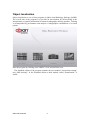
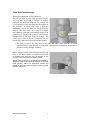
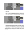
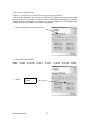
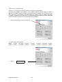
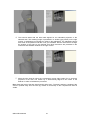
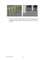
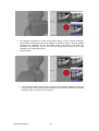
4. The exercise starts with the tube head aligned for an orthoradial projection of the
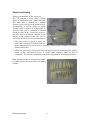
selected area. The resulting image is presented in a window (gray frame) on the right
monitor. A radiopaque round object is visible in the radiograph. The radiopaque object
also exists in the patient model but is not yet visualised. Use the 3D-mouse to change
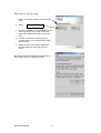
the position of the scene to any desired view. Brief instructions are presented in the
instruction and feedback window on the right monitor.
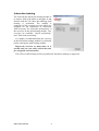
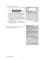
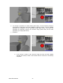
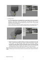
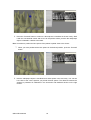
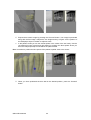
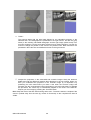
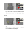
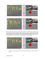
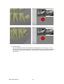
5. Move the tube head by using the conventional mouse (right button) for an eccentric
projection of the area. The central ray is automatically aligned to the centre of the
detector in order to facilitate the procedure.
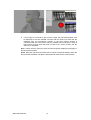
Hint: Grasp the back of the tube head housing and move it. The task is easier to complete if the
scene in pushed away from the user (by means of 3D-mouse) so the complete tube head is
visible.
Qbion AB© 2009-06
21