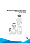
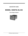
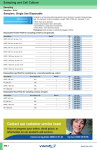
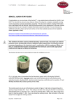
1
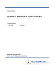
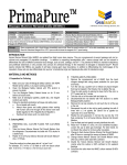
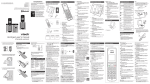
Other Specialty Cell Products
from Global Cell Solutions
Culturing Cells
on GEM™ using
the BioLevitator ™
• 3D Stem SFM
• 3D CryoShield AOF
• 3-DMEM
• MEM-3D
• RPMI 1640-3D
• 3-DMEM/F12
For more information on these products and other services from
GCS, please visit our website: www.globalcellsolutions.com
Global Cell Solutions, Inc.
Hamilton Company, USA
770 Harris Street, Ste 104
Charlottesville, VA 22903 USA
P.O. Box 10030 , Reno, NV 89520, USA
+1 (800) 648-5950 or +1 (775) 858-3000
+1 (434) 975-4271
Hamilton Bonaduz AG
www.globalcellsolutions.com
Via Crusch 8, CH-7402 Bonaduz, GR, Switzerland
+41 (81) 660 60 60
www.hamiltoncompany.com
3-D Cell Culture Simplified.™
Table of Contents
Table of Contents
1. INTRODUCTION
1.1 The GEM™ ....................................................................................2
1.2 GEM™ Cultures ............................................................................4
1.2.1 Inoculation ..........................................................................4
1.2.2 Expansion ............................................................................6
1.2.3 Collection ............................................................................7
2. MATERIALS
2.1 Required Materials ........................................................................8
2.2 Supply Reordering Information ......................................................9
3. WORKING WITH THE GEM™
3.1 Preparing the GEM™ ..................................................................10
3.2 Harvesting the Cells ....................................................................12
3.3 Inoculation ..................................................................................14
3.3.1 Preparing for Inoculation....................................................14
3.3.2 Starting the BioLevitator™ ................................................16
3.4 Maintaining BioLevitator™ Cultures ............................................17
3.4.1 Notes on Maintaining BioLevitator™ Cultures ..................17
3.4.2 Feeding the BioLevitator™ Cultures ..................................18
3.4.3 Imaging and Counting BioLevitator™ Cultures ..................20
3.5 Harvesting BioLevitator™ Cultures..............................................22
3.6 Freezing Cells on GEM™ ............................................................24
3.7 Thawing Cells on GEM™..............................................................25
4. VISUAL GUIDES
4.1 Visual Guide to Inoculation..........................................................26
4.2 Troubleshooting Visual Guide ......................................................27
4.2.1 Troubleshooting the Culture................................................27
4.2.2 Troubleshooting Cell Behavior ............................................28
5. TERMS AND CONDITIONS ................................................................29
6. REFERENCES........................................................................................30
7. NOTES............................................................................................31-32
8. CONTACT INFORMATION ..................................................Back Cover
© 2009 Global Cell Solutions, Inc., All rights reserved.
CULTURING CELLS ON GEM™ USING THE BIOLEVITATOR™
V1.05.09
1
1. Introduction
1. Introduction
1.1 The GEM™
The GEM™ (Global
Eukaryotic Microcarrier)
is a pipette-able, magnetic,
alginate, microcarrier that
supports the culture of
adhesion-dependent cell
lines. Composed of an
alginate core embedded with
magnetic particles and coated
with covalently bound
gelatin, the GEM™ is also an
ideal matrix for threedimensional (3-D) biology.
Complimenting the GEM™ is
a digital bench-top incubator
and bioreactor, the
BioLevitator™.
Figure 1. A 70µm confocal Z-projection
of CHO cells stained with DAPI (blue),
WGA (green), and Phalloidin (red).
Figure 2. The GEM™ consists of an alginate core
with magnetic particles and a protein coat.
Protein Coat
Alginate
Commonly used for dental impressions and as a food additive, alginate was first
used for cell culture applications as an encapsulate for storage (Pilwat G, 1980) or
to immuno-isolate cells for transplantation (Lim F, 1980). Alginate is an unbranched
polysaccharide collected primarily from seaweed composed of repeating guluronic
(G) and mannuronic (M) acid residues. In the presence of a divalent cation, such as
calcium, alginate gels. The GEM™ is composed of a dense alginate hydrogel that
allows for the exchange of ions and small molecules much like the polysaccharide
components of the extra cellular matrix.
The alginate GEM™ core is also optically clear and non-autofluorescent.
Absorbance, luminescence and fluorescence assays are easily performed with cells
still attached. Simply pipette from the culture vessel into assay or immunolabel the
cells for confocal microscopy as in Figure 1. Working with cells on the GEM™
ensures the proper 3-D context is maintained and avoids the use of trypsin.
2
GLOBAL CELL SOLUTIONS
A thin covalently bound coat of gelatin surrounds the alginate core of the GEM™
and promotes cell adhesion. This gelatin coat can also be modified with the
addition of reconstituted basement membrane or laminin. When coated with
gelatin or purified proteins, the GEM™ is a unique 3-D culture substrate possessing
both the protein and polysaccharide components of extracellular matrix.
Magnetics
Dispersed in the alginate gel are small sub-micron magnetic particles that serve to
simplify culture manipulations and allow for automated culture. Media changes or
assay washes using the GEM™ simplifies handling, saves time and reduces errors.
The high density of the GEM™ culture also allows for a single media change to
maintain up to 100 million cells. Digital spatial control using magnets is possible
within automated culture systems. The BioLevitator™ possesses four independent
magnets capable of holding the GEM™ stationary during automated processes
occurring in the robotic CellHOST™.
CULTURING CELLS ON GEM™ USING THE BIOLEVITATOR™
3
1. Introduction
1. Introduction
1.2 GEM™ Cultures
GEM™ culture, as with all cultures, consists of three phases: inoculation, expansion
and collection. The same principles dictating traditional 2-D culture also apply to
GEM™ culture. This section outlines the critical success factors for GEM™ culture
and provides a guide for translating current culture methods into successful GEM™
cultures.
Figure 3. Growth kinetics change with increased inoculation count.
1.2.1 Inoculation
A successful inoculation will produce an even distribution of adhered cells across
the entire GEM™ population ensuring all the surface area available is utilized for
expansion. Creating a successful inoculation begins by understanding plating
efficiency. Plating efficiency is best correlated to the split ratio of 2-D flask culture.
Cells able to be inoculated at low numbers to large surface areas posses a high
plating density and high split ratio (i.e. the population of a single flask can be split
into many flasks at passage). Immortalized cells types such as HEK293 or HeLa
cells often posses high plating efficiencies in contrast to primary cells types such as
HUVEC that require more cells per unit area. Plating efficiency will directly impact
the three critical variables for loading the GEM™ in the BioLevitator™: 1) cell-toGEM™ ratio, 2) media type and volume and 3) agitation during loading.
4
GLOBAL CELL SOLUTIONS
Cell-to-GEM™ Ratio
A direct correlate of the plating efficiency, the cell-to-GEM™ ratio
represents the minimum number of cells required to successfully inoculate
a given volume of GEM™. The simplest method for determining the cellto-GEM™ ratio is a quick titration experiment. In short, a serial dilution
of cell number is administered to a fixed number of GEM™ aliquoted in
a multi-well plate.
Once the cell-to-GEM™ ratio is determined then any ratio higher may
be used to initiate a culture. The benefit of this is illustrated in Fig. 3, on
page 4, with increasing numbers of cells being added to a fixed number
of GEM™. Note that the cultures will return the same number of cells,
but the growth kinetics change. This may influence how you plan your
experiments and the time line from inoculation to end point.
Media Volume and Type
Adding too much media is the most common cause of culture failure
when working with the GEM™. Plating efficiency also dictates a minimum
cell concentration per volume. Inoculate in small volumes of 10mL in the
BioLevitator™ and slowly increase this volume as the culture expands.
Depending on the growth rate of the cells the first media addition can
range from within 24 hours to days.
Media formulation and supplements can lower the required cells per
volume and increase the percent of cells that load. The use of higher
serum concentrations, richer media, and conditioned media can increase
cell viability during inoculation and the adhesiveness of the GEM™.
Ensure the media is not expired and supplement with additional Lglutamine as it quickly decomposes. Also consider the use of richer media
such as DMEM/F12.
Agitation During Loading
Agitation will ensure a homogenous inoculation and prevent clumping.
The BioLevitator™ provides a precise digital control of the agitation
period, interval, and intensity. Agitation period is the total time committed
to the inoculation phase and ranges from less than an hour to days.
Observing the rate at which cells adhere to the GEM™ and therefore the
agitation period can be done in a multi-well plate. Ideally this occurs
concurrent to testing the cell-to-GEM™ ratio. Most cell types will load in
under 4 hours. Decreasing agitation interval and increasing intensity will
prevent clumping during inoculation. Use the “Visual Guide to
Inoculation” in this manual to diagnose clumping and adjust your settings
CULTURING CELLS ON GEM™ USING THE BIOLEVITATOR™
5
1. Introduction
1. Introduction
appropriately.
1.2.2 Expansion
1.2.3 Collection
Expanding the culture is easily accomplished following a successful inoculation.
As with traditional culture, the media and its components must be maintained to
ensure steady growth and good yields. Use an indicator such as phenol red to
monitor culture pH and richer media such as DMEM/F12 to support rapid growth.
Concurrently, stirring speed may be increased to maintain the GEM™ in suspension
and reduce clumping. A successful expansion requires incremental additions of
media, regular media changes, and monitoring for clumping. Maintaining the
cultures is discussed in further detail in section 3.4.
Because the alginate core uses calcium ions to gel, in the presence of
trypsin/EDTA the GEM™ dissolves. Left behind are the cells in suspension and
a small number of magnetic particles which are easily removed with the
CubeMagnet™. The high density of the GEM™ culture is deceiving and
successful dissolution will require more trypsin than one may first estimate. For
example, 50 million CHO cells on GEM™ will require a minimum of 10mL of
trypsin solution to account for the large number of cells and the GEM™’s
gelatin coating. 3-Dissolution™ is a trypsin alternative that can be used in
excess and generally provides superior cell viability after collection.
Keeping the cells on the GEM™ is the ideal method however for most
applications including immunohistochemistry, cell-based assays,
cryopreservation and patch clamp recording to list a few. The optically clear
and non-autofluorescent GEM™ keeps cells adhered to a 3-D matrix allowing
for assays to be run directly from culture. Simply dispense the pipette-able
GEM™ into an assay plate and begin. Viral transduction, electroporation and
chemical transfection are also all simply performed with large cell populations
using the GEM™.
Increasing Media Volume
Expanding from the inoculation volume must be done incrementally. The ideal
schedule for media addition is determined by observation and experience. Keep
in mind the culture will posses a higher osmolarity than the media being added.
Large additions of media can put the cells into osmotic shock which retards
growth, causes cells to fall from the GEM™, and may destroy the culture. In
general once the inoculation media with phenol red has become yellow, then a
volume of no more than 50% of the current volume may be added. Please see
the cell specific technical sheets for media addition schedules.
Changing Media
Media changes will increase in frequency as the cell number rises. A good
estimate is that 10 million cells will consume approximately 10mL of media in
24 hours. This number can change however depending on cell and media type.
Monitor the pH with phenol red and exchange no more than 50% of the media
volume at one time. With optimization larger exchanges can be made. Also keep
in mind that growth may posses a limiting reagent in the media that when
properly supplemented can extend media lifespan. Finally, monitor cell numbers
beginning 48 hours after inoculation for growth rate to optimize media changes.
Observation
Monitoring the culture is simply done by taking a small aliquot for observation
using a microscope. The majority of GEM™ should posses cells and any clumps
contain only 3 or 4 GEM™ at most. Hoechst stain is suggested because the
optical clarity of the GEM™ can make it difficult to distinguish some cell types.
Methods for Hoechst staining and taking cell counts are available in this manual.
6
GLOBAL CELL SOLUTIONS
CULTURING CELLS ON GEM™ USING THE BIOLEVITATOR™
7
2. Materials
2. Materials
2.2 Supply Reordering Information
2.1 Required Materials
The following is a list of the supplies needed to begin cell culture in the
BioLevitator™.
Product
Description
Catalog Number
3D Cell Culture Kit
16 BioLevitator™ tubes
+ 4 x 2mL GEM™ (50%)
GKT-3000
(1 month supply)
Serological pipets
CubeMagnet™
1 Small cube shaped magnet
GMM-1002
45mL BioLevitator™
Tubes
BioMagnet™
1 PTFE-coated magnet
GMM-1001
3-Dissolution™
Trypsin-alternative cell
dissociation reagent
GSP-0100
500mL bottle
GSP-0302
Equipment
Reagents
Consumables
BioLevitator™
GEM™ substrate
Pipettor tips
CubeMagnet™
Culture media
BioMagnet™
3-Dissolution™
Hemocytometer
(or cell dissociation
reagent of your choice)
(or cell counting system)
Cryogenic vials
Light microscope
Phosphate Buffered
Saline
(Ca2+and Mg2+-free)
(appropriate magnification
for viewing cells; 10-20X
objective; DAPI filter)
Trypan Blue (if you are
using a hemocytometer)
GEM™
Global Eukaryotic Microcarriers, 4x2mL
GEM™-3034
OQ Kit
(Operational Quality Kit)
2mL GEM™ + 2x 2mL of 3D-validated
cell line + 500mL DMEM/F12
+ 4 BioLevitator™ tubes
GKT-2000
Cells of your choice
48 tubes for the BioLevitator™
GTB-1000
Pipettor
37ºC, 5% CO2
incubator
Multi-well plate
Ice
Fetal Bovine Serum (FBS)
BioLevitator™ Tubes
A CO2 source for the
BioLevitator™
Figure 4.
CubeMagnet™
3-DPBS
Ca2+and Mg2+ -free
Figure 5.
BioMagnet™
Figure 6.
BioLevitator™Tube
WARNING: CubeMagnet™ and BioMagnet™ contain a
STRONG MAGNETIC DEVICE! The magnet should be stored in a safe
place and handled with care to avoid injury or damage. Avoid bringing
the CubeMagnet™ and BioMagnet™ within close proximity of each other. Keep at
least 18 inches away from recording devices, computers, and metal objects.
8
GLOBAL CELL SOLUTIONS
CULTURING CELLS ON GEM™ USING THE BIOLEVITATOR™
9
3. Working with the Gem™
3. Working with the Gem™
3.1 Preparing the GEM™
Materials Needed
Method:
1 vial of GEM™ substrate: 2mL of 50% packed slurry
Culture Media: Use complete media that the cells are normally grown or
passaged in. Warm the media to at least room temperature.
CubeMagnet™
Pipettor and Tips
1. While holding one vial (2mL)
of GEM™ substrate over the
CubeMagnet™ to immobilize
the GEM™, carefully aspirate
off the storage buffer. Fig. 8.
2. Add 1mL of the media. Fig.7.
3. Cap the vial and invert to mix.
4. Repeat steps 1-3 for a total
of 4 washes.
These wash steps are necessary to allow
the GEM™ substrate to absorb the
media components and to remove the
storage buffer. Omitting these wash
steps may lead to sub-optimal loading
of cells on the GEM™ substrate.
Aspirate /
wash
5. Aspirate off the final wash and add
1mL of the media. The GEM™ have
been washed and acclimated in the
medium.
1mL
complete
medium
Figure 8.
Hold the vial over the CubeMagnet™
4X
Magnet
Figure 7: Wash and acclimate the GEM™.
10
GLOBAL CELL SOLUTIONS
CULTURING CELLS ON GEM™ USING THE BIOLEVITATOR™
11
3. Working with the Gem™
3. Working with the Gem™
3.2 Harvesting the Cells
Materials Needed:
T75 flask of cells1 : Use a flask that has reached 80-90%
confluency.
3-Dissolution™2
(GCS catalog # GSP-0100)
Phosphate Buffered Saline (PBS): Ca2+/Mg2+ -free
Culture Media: Use complete media that the cells are
normally grown or passaged in. Warm the media to at least
room temperature.
Hemocytometer or counting system:
You may need Trypan Blue if using a hemocytometer.
10 million cells
Conditioned
media
Pipettor and tips
Serological pipets
Figure 9: Harvest cells from a flask and save the conditioned media..
Light microscope: Make sure you have the appropriate
magnification objective for viewing cells (a minimum of
10X objective).
Method:
1. Remove and SAVE the culture medium in a 15mL or larger sterile conical tube.
NOTE: This is conditioned media and contains essential growth factors that
stimulate growth and adhesion.
2. Rinse the cells gently with 5.0mL of Ca2+/Mg2+ free PBS then remove. Add 5mL
of 3-Dissolution™ to the flask and place in 37°C incubator. Observe and
monitor the cells every 1-2 minutes until the cell layer is dispersed (time varies
with cell type).
3. Add 5mL of the pre-warmed media to stop the reaction and suspend the cells by
gently pipetting up and down.
4. Transfer the cell suspension to a sterile 15mL tube using a serological pipette.
1
As with other cell culture protocols, perform all manipulations of cells and cells on GEM™ described here
in a biosafety hood.
2 The following protocol describes the harvesting of cells using 3-Dissolution™, a trypsin-free cell
dissociation reagent (catalog # GSP-0100-101) that can be purchased from Global Cell Solutions. However,
if you are using a Trypsin/EDTA-like product of your choice follow the manufacturers’ recommended
protocol. This protocol assumes that the user is harvesting cells from a plastic tissue culture-treated T75
flask that is 80-90% confluent. Adjust the volumes of 3-Dissolution™ or other dissociation solution
proportionately for different size flasks harvested.
12
GLOBAL CELL SOLUTIONS
5. Observe the cells with microscope to ensure a single cell suspension.
6. Determine the number of harvested cells/mL utilizing standard Trypan Blue
exclusion and a hemocytometer or other counting method.
NOTE: Do not agitate cells by hitting or shaking the flask. This action may
cause certain cell types to clump together. A mono-dispersed culture assists
in obtaining ideal loading onto the GEM™ substrate.
CULTURING CELLS ON GEM™ USING THE BIOLEVITATOR™
13
3. Working with the Gem™
3. Working with the Gem™
3.3 Inoculation
Aspirate /
wash
Materials Needed:
5mL
complete
medium
Conditioned media: This is the media you saved in the previous
‘Harvesting protocol.’
Culture Media: Use complete media that the cells are normally grown or
passaged in. Warm the media to at least room temperature.
1 BioLevitator™ Tube: Label the tube with the date, the cell line, and any other
experimental information.
Magnet
Prepare
the GEM™
Pipettor and Tips
2-6 million
cells
Serological Pipets
Conditioned
media
1 vial of GEM™: Make sure the GEM™ have been washed as described
previously in the protocol for ‘Preparing the GEM™’.
GEM™
Single Cell Suspension: This is the 15mL tube of cells you collected in the
previous ‘Harvesting’ protocol.
2x 15mL conical tubes
BioLevitator™: Make sure it is properly set up and connected to a CO2 source.
See the BioLevitator™ manual provided with the instrument by Hamilton
Company.
BioLevitator™
3.31 Preparing for Inoculation
Method:
Figure 10: Combine the washed GEM and the prepared cells to start the inoculation.
1. Turn the BioLevitator™ on and pre-warm it. Adjust the carbon dioxide on the
BioLevitator™ to the appropriate levels and verify that the levels are accurate
(see BioLevitator™ User Manual provided by Hamilton).
2. Add 5mL of the conditioned media to the tube.
3. Add 5mL of fresh complete culture media to the tube.
4. Invert the vial of washed GEM™ as needed to re-suspend the GEM™ substrate.
Quickly draw up 0.5mL of re-suspended GEM™ slurry in a pipette and dispense
this into the BioLevitator tube.
5. Add an appropriate volume of cell suspension containing approximately
2 to 6 million cells to the tube1. Minimize the volume of cell suspension
added. Swirl slightly to mix cells and GEM™ substrate. Seeding density will
vary according to the type of cells being used and downstream application.
Note: Order of addition is important! Always add cells last!
6. Put the cap on the tube and place in the BioLevitator™.
1
This is the range that is most commonly used. The example given in this manual describes maintaining a 6
million CHO cell inoculation and culture. Feel free to adjust this inoculation cell count. Contact Global Cell
Solutions customer service for any questions or guidance for different inoculation counts and cell types.
14
GLOBAL CELL SOLUTIONS
CULTURING CELLS ON GEM™ USING THE BIOLEVITATOR™
15
3. Working with the Gem™
3. Working with the Gem™
3.3 Inoculation (continued)
3.4 Maintaining the BioLevitator™ Cultures
3.4.1 Notes on Maintaining the Cultures:
BioLevitator™
Figure 11: Put the cells and GEM™ in the BioLevitator™ and begin the inoculation protocol.
3.3.2 Starting the BioLevitator™
In order to have successful growth of cell cultures, it’s important to monitor the pH of
the culture environment. Most cell lines grow well at pH 7.4; however some specific
cell lines may vary slightly between pH 7.0 and pH 7.7. Phenol red is commonly
used a pH indicator in most culture medias. It is red at pH 7.4, orange at pH 7.0, and
becomes yellow at pH 6.5, lemon yellow below pH 6.5, pink at pH 7.6 and purple at
pH 7.8. The assessment of color is highly subjective, so it may be useful to make up a
set of media standards.
The general guide to maintaining successful cultures is to visually monitor the
culture and make sure it remains in the correct pH range. When the media is ‘spent’
or seems more yellow, then it is time to replenish the culture with fresh media.
However, it’s important that the cells are able to retain some of the secreted growth
factors and signaling molecules that are in the ‘spent’ media. For this reason it’s
important not to shock the cells or drown them by adding too much fresh media or
removing too much old media at once. Never add more than 50% of the culture
volume at any given time. The maximum amount of culture in one tube is 40mL, so it
is fine to gradually add in media as needed until this volume is reached, and then
begin to switch out old media with fresh after that point.
As the cell numbers increase, so does the rate at which the media components
are consumed. Fresh media may need to be added at a higher frequency. The
following formula is useful in deciding when to change media:
10 million cells will use 10mL of media in a 24 hour period in normal growth.
Method:
1. On the BioLevitator™ touch screen, select the tube that you are setting up for
culture. (See the BioLevitator™ User Manual provided by Hamilton Company.)
2. Select the ‘Experiment’ you would like to run. If needed, adjust the
inoculation, culture, or harvest parameters as described in the BioLevitator™
User Manual provided by Hamilton Company.
3. Select whether you want to run any or all of the Inoculation, Culture, or
Harvest protocols for your tube (usually the Inoculation and Culture
protocols are selected).
Total Number of Cells in Tube/ (10 x 106) X 10 = Amount of Fresh Media to Add to Culture (ml)
When the cell density becomes so high, that very frequent media changes are
needed, it is then possible to split the culture into multiple tubes, or some cells may
be removed, saved, or set aside to serve any desired purpose.
4. Select Start. Loading times may vary from 2 to 36 hours depending on type of
cell being used, the passage number, whether the cells are fresh or newly
thawed, and whether they’ve been over-trypsinized or not.
16
GLOBAL CELL SOLUTIONS
CULTURING CELLS ON GEM™ USING THE BIOLEVITATOR™
17
3. Working with the Gem™
3. Working with the Gem™
3.4.2 Feeding the BioLevitator Cultures
Materials Needed
Culture Media: Use complete media that the cells are normally grown or
passaged in and make sure it contains 15mM HEPES. Warm the media to at
least room temperature.
Serological Pipets
Following is a sample protocol for maintaining a
culture of CHO K1 cells (Chinese Hamster Ovary)
in the BioLevitator™. The methods are to be used
as a general guideline, and in no way represent how
every cell culture should be maintained. For more
information on a particular cell type, or for any
additional guidance, please visit the Global Cell
Solutions website or contact our Technical Support1.
Example of Methods for Maintaining
CHO K1 BioLevitator™ Culture:
Note: The first day of inoculation is referred to as
Day 0. The CHO K1 culture protocol described
starts at a speed of 60 RPM on day 0, increases to
68 RPM on Day 1, and to 76 RPM on Day 3. Follow
the instructions in the BioLevitator™ User Manual
provided by Hamilton Company to ensure that your
protocol is set to these parameters, or to adjust your
protocol as such.
Figure 12.
20X fluorescent overlaid image
stack of CHO K1 cells growing
on GEM™ substrate stained
with 0.1mg/ml Hoechst Stain
Figure 13. A typical growth curve generated from
CHO K1 cells in a BioLevitator™.
1. Day 1 of culture:
Visually monitor culture. Add 5mL of fresh media to the tube.
2. Day 2 of culture:
Visually monitor culture. Add 10mL of fresh media to the tube.
3. Day 3 of culture:
Visually monitor culture. Take a sample for cell count starting on this day. Adjust
your media accordingly. Typically, remove 5mL of ‘spent’ media and add 20mL
of fresh media to the tube.
4. Day 4 of culture:
Visually monitor culture. Take samples for cell counts as often as needed,
and in order to accurately adjust media feedings. Use the guidelines
described, and replenish media as necessary. Typically you will need to
do 2 feedings on this day, about 8 hours apart, replacing 20mL of ‘spent’
media with 20mL of fresh during both feedings.
5. Day 5 and onwards:
Increase the frequency of feedings to maintain optimal growth. You may
need to split the cultures. Always use cell counts and the pH indicator to
maintain your culture.
______________________________
1
www.globalcellsolutions.com
18
GLOBAL CELL SOLUTIONS
CULTURING CELLS ON GEM™ USING THE BIOLEVITATOR™
19
3. Working with the Gem™
3. Working with the Gem™
3.4.3 Imaging and Counting BioLevitator™ Cultures
Materials Needed
Imaging Method:
450µL of culture: This sample should be taken from the BioLevitator™ culture
tube. 50uL will be used for imaging the cells and 400uL will be used for
counting.
1. Put 50µL of the cell culture sample (cells on GEM™) in one of the wells
in the 96-well plate.
96-well culture plate: Make sure this is optically clear.
2. Add 100µL of the Hoechst 33342 solution to the sample in the well.
1:2000 Hoechst 33342 solution: Dilute 0.5µL of Hoechst 33342 (10mg/mL in
water) in 1mL room temperature culture media.
3. Incubate the well at room temperature for 15-20 minutes.
Light microscope: Brightfield with UV channel and DAPI filter; 10X objective.
4. Capture images of the sample under UV light and DAPI filter
using the microscope.
CubeMagnet™
Pipettor and tips
Phosphate Buffered Saline (PBS): Ca2+/Mg2+ -free. Make sure this is
at room temperature.
3-Dissolution™ : warm this to 37ºC
(GCS catalog # GSP-0100-101)
Culture media: Warm the media to at least room temperature.
Hemocytometer: Make sure it’s cleaned and dried after every use.
Hemocytometer coverglass: Cleaned and dried after every use.
Microcentrifuge tube
Cell Counting Method:
5. Use the CubeMagnet™ to draw down the remaining 400µL sample of cells on
the GEM™.
6. Use the pipettor to aspirate off the media above the GEM™ culture sample.
7. Add 1mL of PBS to the culture sample.
8. Use the CubeMagnet™ again to draw down the cells on GEM™ and aspirate the
PBS off this time.
9. Add 200µL 3-Dissolution™ to the sample. (More 3-Dissolution™ may be
required if the cells are more confluent.)
Trypan Blue: 0.4% w/v in PBS.
10. Incubate the culture sample and
3-Dissolution™ for 10-15 minutes at 37ºC. Ensure that the GEM are dissolved
by observing under a microscope.
11. Neutralize the sample by adding 400µL of culture media. Use more media to
neutralize if you used more 3-Dissolution™.
12. Place the coverslip evenly on the middle of the hemocytometer.
13. Add 50µL of Trypan Blue and 50µL of the cell culture sample to a
microcentrifuge tube. Tap the tube to mix.
14. Remove 10µL from the cell/Trypan Blue mix with a pipettor and add 10µL to
each side of the coverslip by allowing a drop held at the end of the tip to be
taken under the slide by capillary action.
15. Using the hemocytometer to count the viable and non viable cells.
20
GLOBAL CELL SOLUTIONS
CULTURING CELLS ON GEM™ USING THE BIOLEVITATOR™
21
3. Working with the Gem™
3. Working with the Gem™
3.5 Harvesting BioLevitator™ Cultures
Materials Needed:
3-Dissolution™ (GCS catalog
#GSP-0100-101) Make sure the
3-Dissolution™ is prewarmed to
37°C.
Phosphate Buffered Saline (PBS):
Ca2+/Mg2+ -free. Make sure this is at
room temperature.
Culture Media: Use complete media
that the cells are normally grown or
passaged in. Warm the media to at
least room temperature.
Tube from BioLevitator™ with cells:
This is the tube containing cells on
GEM™ that have reached the end of
the culture phase.
Aspirate
Count
Aliquot
Hemocytometer or counting system:
You may need Trypan Blue if using a
hemocytometer.
Pipettor and tips
Serological pipets
Light microscope: Make sure you
have the appropriate magnification
of objectives for viewing cells, at
least a 10X objective.
CubeMagnet™
Method:
1. Remove the tube from the BioLevitator™ and place in a tissue culture hood.
2. Using the CubeMagnet™, pull the confluent GEM™ substrate to the bottom of
the tube. Aspirate and discard the media without disturbing the pellet.
3. Gently wash the GEM™ substrate in 20mL of Ca2+/Mg2+ -free PBS. Using the
CubeMagnet™, pull the confluent GEM™ substrate to the bottom of the tube.
Aspirate and discard the PBS wash.
4. Add 10mL 3-Dissolution™.1 Gently mix solution up and down with a pipette.
Continue gentle titration until GEM™ substrate dissolves. Incubation with
mixing at 37ºC will help speed up the dissolution of the GEM™. You may return
the tube to the BioLevitator™ and use the Harvest protocol for this step.
Note: Visually monitor progress with a light microscope to make sure the GEM™
have dissolved. Additional 3-Dissolution™ may be necessary to dissolve the
GEM™ entirely, especially if they are very confluent.
Note: This process may take 15-20 minutes, depending upon how tightly the cells
are packed on the GEM™.
20mL
Ca+2/Mg+2
free PBS
5. Add 10mL of culture media to the tube to inactivate the 3-Dissolution™.2
Gently mix.
37°C
Gentle Agitation
6. Using the CubeMagnet™, pull the remnants of the GEM™ substrate (little black
particles) to the bottom of the tube and transfer the cell solution to a new 50mL
tube.
Aspirate &
PBS wash 3-Dissolution™
7. Take an aliquot of the cell suspension and obtain a cell count. Cells are ready for
downstream assays or other applications.
Transfer cell
suspension
Magnet
Assay
Magnetic
Particles
Magnet
Downstream
applications
Figure 14: Harvest the BioLevitator culture from the GEM.
22
GLOBAL CELL SOLUTIONS
1
Keep in mind that 0.5ml of GEM™ substrate is equivalent in surface area to a T75 flask (75cm2).
Increase/decrease the amount of dissociation solution accordingly depending upon the starting volume of
GEM™ used in your culture.
2
If you are using a Trypsin/EDTA-like solution, add an equal volume of media.
CULTURING CELLS ON GEM™ USING THE BIOLEVITATOR™
23
3. Working with the Gem™
3. Working with the Gem™
3.6 Freezing Cells on GEM™
3.7 Thawing Frozen Cells on GEM™
Materials Needed:
DMSO1: Dimethyl Sulfoxide
Culture Media: This can be
antibiotic free and does not need
to be at room temperature.
Fetal Bovine Serum (FBS)
Cryogenic Vials: You will need
4x 2mL pre-labeled vials..
BioMagnet™
Serological pipettes
Ice: It’s easier to follow this protocol
if you have the ice sitting in a flat
container or bucket for easy
placement of tubes and vials. It is
recommended that most steps of this
protocol be executed over ice.
Cells: These are the cells on GEM™
from the BioLevitator™ culture.
Note: DMSO will cause the cells to detach from the GEM™ substrate when it’s at room
temperature. Keep all cells on GEM™, freezing media, and wash solutions ICE cold.
Method:
1. Prepare 10mL of ‘DMSO Freezing
Media Wash’ (5% DMSO, 20% FBS):
a. Combine 0.5mL DMSO, 2mL of
FBS, and 7.5mL of culture media.
b. Place on ice.
2. Prepare 5mL ‘Freezing Media Wash’
(20% FBS):
a. Combine 1mL of FBS and 4mL
of culture media.
b. Place on ice.
3. Remove the tube of cells from the
BioLevitator™ and place in a tissue
culture hood.
4. Using the BioMagnet™, pull the
confluent GEM™ to the bottom of
the tube. Aspirate all media, taking
care not to disturb the GEM™ layer.
5. Gently add in 3.6mL of the ‘DMSO
Freezing Media Wash’. Mix gently.
6. Using the BioMagnet™, pull the
confluent GEM™ to the bottom of
the tube. Aspirate the ‘DMSO
Freezing Media Wash’, taking care
not to disturb the GEM™ layer.
7. Repeat step 6. Aspirate the second
freezing media wash, taking care not
to disturb the GEM™ layer.
8. Gently add 3.6mL of ‘Freezing Media
Wash’.
9. With a 1mL pipette, draw up 400ul
DMSO. Add 1 drop of DMSO into the
tube and swirl. Then add in 2 drops
and swirl. Add in 4 drops and swirl.
Continue in this fashion (doubling the
number of drops used) until all 400 µl
of DMSO are used. (The final
concentration of DMSO in the tube
will be 10%.)
Materials:
20% FBS Culture Media: Make 1mL
cell culture media that contains 20%
FBS and has been warmed to no
more than room temperature.
Frozen Cells on GEM™: This is one
of the cryogenic vials previously
frozen containing cells on GEM™.
1 BioLevitator™ Tube
CubeMagnet™
Serological pipettes
BioLevitator™: Make sure the
BioLevitator™ is pre-warmed and
CO2 adjusted.
Method:
1. Add 10mL of the 20% FBS culture media to the BioLevitator tube.
2. Thaw a cryovial of frozen cells on GEM™ in a 37°C water bath until cells are
just thawed.
3. Place the cryovial on the CubeMagnet™ and pull the cells on GEM™ to the
bottom of the vial.
4. Gently aspirate off the freezing media. Re-suspend the cells on GEM™ in 1mL of
the room temperature culture media that contains 20% FBS and transfer to the
BioLevitator tube.
5. Place the tube in the pre warmed BioLevitator™.
6. Change media as needed1.
10. Gently swirl the tube and aliquot
1mL of the cell-media-DMSO
mixture into pre-labeled 2mL
cryogenic vials.
11. Freeze according to ATCC Technical
Bulletin No. 3, Cryogenic
Preservation of Animal Cells.2
___________________________________________________________________________________________
1
The DMSO we have been successful using was purchased from Sigma-Aldrich (Catalog No. D2650).
2
http://www.atcc.org/common/technicalInfo/TechLit.cfm (viewed on December 3, 2005)
___________________________________________________________________________________________
1
For further expansion of thawed cells on GEM, additional GEM may need to be added. For more
information contact Customer Support.
24
CULTURING CELLS ON GEM™ USING THE BIOLEVITATOR™
GLOBAL CELL SOLUTIONS
25
4. Visual Guides
4. Visual Guides
4.1 Visual Guide to Inoculation
4.2 Troubleshooting Visual Guide
Cells attach to the GEM™ in four phases. Visually monitor cell attachment onto the
GEM™ at the end of the inoculation, prior to the culture phase, and identify the
various stages of attachment.
4.2.1 Troubleshooting the Culture
Absorption
Observation:
Moderate Clumping
The cell is rounded and has made contact
with the GEM™ and is touching it.
Observation:
Doublets and triplets of GEM™ are formed
with cells growing in between.
Explanation:
Attachment
These small groups are not detrimental to
the culture but avoid collections of 5 or more
GEM™ to ensure the total available surface
area for expansion is being utilized.
Observation:
Solution:
The cell has flattened on the side of contact
with the GEM™, and appears like a rounded
column attached to the GEM™ at the base of
the column.
Increase agitation by decreasing the agitation
interval. If clumping persists, increase the
agitation intensity also.
Spreading
Heavy Clumping
Observation:
The cell continues to flatten onto the GEM™
and is more "bell shaped" with a wider base.
Observation:
Large groups of GEM™ or the entire population
are forming large masses in the BioLevitator™
tube. Fibroblasts for example are very adhesive
and will seek adhesion on multiple surfaces.
Solution:
Settling
Observation:
The cell has completely flattened down on the
GEM™ and has started normal cellular processes.
26
GLOBAL CELL SOLUTIONS
Increase agitation by decreasing the agitation
interval. If clumping persists, increase the agitation
intensity also. Inoculate under constant agitation.
Reduce the serum concentration.
CULTURING CELLS ON GEM™ USING THE BIOLEVITATOR™
27
4. Visual Guides
5. Terms and Conditions
Section Heading
4.2.2 Troubleshooting Cell Behavior
Sheeting
Bridging
Terms and Conditions
Shedding
Quality
All Global Cell Solutions products are
manufactured and tested to meet rigorous
quality standards. Every lot of GEM™
substrate is cell culture tested. Please refer to
Certificate of Analysis for further information.
Product Use
Observation:
Observation:
Observation:
Confluent GEM™
present as well as
GEM™ with large bare
spots and loosely
attached sheets of cells
Cells are attached and
stretched between two
or more GEM™.
Confluent GEM present
in culture as well as viable
loose cells.
Explanation:
Explanation:
Some cell types are more
prone to this behavior
(ex. Fibroblasts) and link
GEM™ together to
increase the size of their
settled footprint. Other
cell types attach to
multiple GEM™
because of insufficient
agitation/mixing.
Some cell types exhibit
this behavior when they
reach confluency on the
GEM™. GEM™ cultures
continue to divide (unlike
the halting of growth due
to contact inhibition in
a 2D flask) and one of the
daughter cells remains
attached to the GEM™
while the other becomes
free floating.
Explanation:
Osmotic shock to cells
can be caused when
too much media is
added or exchanged at
one time, and this can
cause sheeting.
Solution:
Do not add or
exchange more than
50% of the total
volume in media. For
example, a 10mL
culture should receive
a maximum of 10mL of
fresh media while a
40mL culture should
receive a maximum of
20mL of media
exchange.
28
Solution:
Increase the agitation
speed, especially during
the inoculation phase, to
reduce the frequency of
attached cells coming in
contact with another
GEM™ OR reduce the
agitation pause during the
inoculation phase.
Solution:
Add additional GEM™
substrate to the culture
at the first sign of
shedding so the culture
can continue to expand.
You may also harvest the
shed cells for downstream
applications while
maintaining the
BioLevitator™ culture as a
miniature cell factory.
GLOBAL CELL SOLUTIONS
You may use the Materials1 solely for internal
research purposes in your laboratory, and for
no other purpose whatsoever. Without
limitation, you may not attempt to reverse
engineer or design around the Materials,
generate analogs of or derivatives based on
the Materials, expose the Materials to
humans or use the Materials in humans,
chemically analyze or chemically modify the
structure of the Materials, develop
formulations of the Materials or use the
Materials for any commercial purpose.
You may not sell, transfer, disclose or
otherwise provide access to the Materials to
any person, entity or location without the
prior written consent of GCS, except that you
may allow access to the Materials to
individuals performing research under these
Terms; provided, that such individuals are
bound by written agreement to use the
Materials only in the manner permitted
under these Terms.
Handling of Materials. You understand and
agree that the Materials may have
unpredictable and unknown biological
and/or chemical properties, that they are to
be handled and used with caution, and that
they are not to be used for testing in or
treatment of humans. The Purchaser will
handle and use the Materials in compliance
with all applicable laws and regulations.
1
Disclaimer of Warranties. The materials are
supplied to you with no warranties of any
kind, express or implied, including any
warranty of merchantability or fitness for a
particular purpose or that they are free from
the rightful claim of any third party, by way
of infringement or the like. GCS makes no
representations that the use of the materials
will not infringe any patent or proprietary
rights of any third parties.
Limitation of Liability. In no event will GCS
be liable for any use by the purchaser of the
materials or any loss, claim, damage or
liability of any kind which may arise from or
in connection with the use, handling or
storage of the materials by or through
purchaser. The purchaser hereby agrees to
indemnify, defend and hold harmless GCS,
its employees, independent contractors from
damages, costs or expenses for any loss,
claim, injury or liability of whatsoever kind
or nature, including without limitation any
resulting from any illness or injury to persons
or property, whatever the cause may be,
which may arise from the purchaser’s use,
handling or storage of the materials, even if
GCS has been advised of the possibility of
such damages. Purchaser agrees that the
foregoing limitations of liability shall apply
even if a limited remedy provided hereunder
fails of its essential purpose.
Trademarks: GEM™, CubeMagnet™,
BioMagnet™, 3-Dissolution™ (Global Cell
Solutions); Sigma-Aldrich® (Sigma-Aldrich).
Tangible materials to be provided to you by Global Cell Solutions.
CULTURING CELLS ON GEM™ USING THE BIOLEVITATOR™
29
6. References
7. Notes
References
Lim F, S. A. (1980). Microencapsulated islets as bioartificial endocrine pancreas.
Science., 210(4472):908-10.
Pilwat G, W. P. (1980). Immobilization of human red blood cells. Z Naturforsch,
35(3-4):352-6.
30
GLOBAL CELL SOLUTIONS
CULTURING CELLS ON GEM™ USING THE BIOLEVITATOR™
31
7. Notes
32
GLOBAL CELL SOLUTIONS
Contact
Contact
Dear Participant,
Thank you for choosing automated 3D Cell Culture. We appreciate your efforts and
are ready to assist you and your organization through this process. Please use the
following information to reach us with questions or comments.
Corporate
Global Cell Solutions, Inc.
770 Harris Street
Suite 104
Charlottesville, VA 22903
+1 (434) 975-4271 (main)
+1 (434) 975-4216 (fax)
Scientific
Attn: Automated 3D Cell Culture
[email protected]
www.globalcellsolutions.com