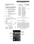
1
CardioView
User Manual
Caution: The computerized interpretation provided by CardioView is only valid when
used in conjunction with clinical findings. All computer generated tracings and
interpretations must be confirmed by a qualified physician.
Copyright Notice
Copyright © 2003 by QRS Diagnostic, LLC. All rights reserved.
No part of the documentation may be reproduced, stored in a retrieval system or
transmitted in any form, or by any means, electronic, mechanical, photocopy,
recording or otherwise, without the prior consent of QRS Diagnostic, LLC.
Information in this manual is subject to change without notice and does not
represent a commitment on the part of QRS Diagnostic, LLC.
License Agreement
The software described in this manual is supplied under a license agreement and
may only be used in accordance with the terms of that agreement.
Trademarks
All brands and product names are trademarks or registered trademarks of their
respective holders and are hereby acknowledged trademarks.
European Representative:
Advena Ltd.
PO Box 30, Leominster
Herefordshire HR6 OZQ
UK
CAUTION: Federal law restricts this device to the sale by or on order of a physician
YEAR 2000: This software is Year 2000 compliant, provided it is run on a
computer system that is itself Year 2000 compliant. It is recommended that 4
digits be entered for all year values. If a two digit year is entered it is assumed to
be in the 20 th century and is stored as a 4 digit year value. Dates of birth occurring
in the year 2000 or later are required to be entered with a 4 digit year.
6000-4153 Rev C
WARRANTY
All instruments sold and supplied by QRS Diagnostic, LLC are guaranteed to be
free from defects in material and workmanship for a period of 3 years from date of
purchase. All supplies and accessories carry a 90-day limited warranty. This
includes oximetry fingertip sensors and ECG lead wires. If in the judgement of
QRS Diagnostic the instrument is proven to be defective during the warranty
period it will be repaired or replaced with no charge for parts or labor. An
extended hardware warranty is available at additional cost.
This warranty does not cover any instrument that has been damaged by accident,
misuse, abuse or has been altered or repaired by anyone other than an
authorized QRS Diagnostic agent. This warranty also does not cover any unit that
has had a serial number removed, defaced or rendered illegible.
This warranty is in lieu of all other warranties expressed or implied. Including
warranties of merchantability and fitness, and is hereby limited to repair or
replacement of instruments found defective during the warranty period.
ALL REPAIRS MUST BE MADE BY AN AUTHORIZED QRS DIAGNOSTIC
AGENT. THE INSTRUMENT MUST BE RETURNED FOR REPAIRS AT THE
EXPENSE OF THE PURCHASER. IN-WARRANTY REPAIRED UNITS WILL
BE RETURNED AT THE EXPENSE OF QRS DIAGNOSTIC OR ITS
AUTHORIZED AGENT. INSTRUMENTS SENT BY MAIL OR COMMON
CARRIER SHOULD BE INSURED AGAINST LOSS OR DAMAGES, AS THEY
ARE NOT COVERED BY THIS WARRANTY.
SERVICE
For service, please contact the QRS Diagnostic, LLC Service Department at (763)
559-8492. Service Representatives are available by phone Monday through
Friday, from 8 a.m. to 5 p.m. Central Standard Time. You can also contact
Technical Support by email at [email protected]. In the event that
your problem can not be fixed over the phone or via email you will be issued a
Returned Merchandize Authorized number (RMA#). Please have the serial
number of your unit available. Please re-package your unit in the original packing
materials if possible and ship it to:
QRS Diagnostic
14755 27 th Ave N
Plymouth, MN 55447
This document contains proprietary information, which is protected by copyright.
All rights are reserved. No part of this document may be photocopied,
reproduced, or translated to another language without the written consent of
QRS Diagnostic, LLC. The information contained in this document is subject to
change without notice.
PRECAUTIONS
Intended use
CardioView is intended for use with an IEC 60950 compliant computer. The system
includes the CardioView Software and a 6 or 12 lead Universal ECG that combine to
allow you to connect your patient directly to the computer to monitor their
Electrocardiograph.
Warnings
This equipment is not IEC60601-1 compliant unless it is supplied from a
computer or power supply that complies with IEC60950 or equivalent.
Note: All CE marked devices will have been tested to this standard.
The following precautions need to be taken when a defibrillator is used on a
patient:
§ Do not touch the patient during defibrillation.
§ Do not touch the defibrillator’s paddle-electrode surface when discharging the
defibrillator.
§ Keep defibrillator electrodes well clear of other electrodes or metal parts in
contact with the patient.
§ If a person is touching the patient, bed, or any conductive material in contact
with the patient during defibrillation, the delivered energy may be partially
discharged through that person. Make sure persons stand away from the
patient, bed, and other conductive material before discharging the
defibrillator.
CAUTION:
Conductive parts of electrodes and associated connectors, including the neutral
electrode, should not contact other conductive parts including earth.
Ensure that the electrodes are connected only to the patient and not to any
other conductive parts or equipment to prevent hazards to the patient from
leakage currents.
The Universal ECG should be used with Ag/AgCl ECG “skin” electrodes. These
should be used in accordance with manufacturers instructions.
There are no hazards associated with using this device with a pacemaker.
The Universal ECG is not intended to be sterilized or used in a sterile
environment and therefore is not suitable for direct cardiac application.
The Universal ECG is not suitable for use with flammable anaesthetics and
offers no water ingress protection.
Inspect the Universal ECG electrocardiograph patient cable regularly for signs of
mechanical damage, particularly around patient leads. To verify correct
operation of the electrocardiograph, connect all patient leads to a patient or an
ECG simulator and check that there is a valid ECG signal on every lead by
viewing the signal through CardioView.
Use during defibrillation or with large input signals
The design of the Real Time monitor is such that the traces can cross in the presence of
large signals. This is to allow for the convenience of viewing all signals simultaneously
while still allowing maximum input dynamic range.
It is recommended that a three lead view be selected for use during defibrillation to
ensure that the signals are clearly separated following electrode polarization.
Symbols used on the Universal ECG
Attention: Consult accompanying documents.
Type BF defibrillator proof equipment.
TABLE OF CONTENTS
CHAPTER 1 - INTRODUCTION……………………………………………………..1
W HAT IS CARDIOVIEW …………………………………………………………………...1
SYSTEM REQUIREMENTS…………………………………………………………………1
I NSTALLATION…………………………………………………………………………….2
CHAPTER 2 - CARDIOVIEW………………………………………………………...3
ECG RECORDING………………………………………………………………………..4
CHAPTER 3 - SETTING UP CARDIOVIEW……………………...………...…...…6
CHAPTER 4 - WORKING WITH CARDIOVIEW………………………...……….10
DOWNLOADING AN ECG………………………………………………………………..10
PATIENTLIST………...………………………………………………………….………16
ECG LISTS……………………………………………………………………………..17
VIEWING AN ECG………………………………………………………………………18
PRINTING AN ECG………………………………………………………………….…..23
BATCH PROCESSING………………………………………………………………..….24
EXPORTING ECGS……………………………………………………………………..26
I MPORTING ECGS………………………………………………………………………27
CHAPTER 5 - REFERENCE………………………………………………………..28
MENU COMMANDS……………………………………………………………………...28
CHAPTER 6 - ECG ANALYSIS PROGRAM………………………………......…30
ANALYSIS & I NTERPRETATION………………………………………………………….31
STATISTICAL PERFORMANCE OF THE ECG ANALYSIS PROGRAM………………………31
CHAPTER 7 - MAINTENANCE…………………………………………………….33
CHAPTER 8 - TROUBLESHOOTING……………………………………………..34
APPENDIX: SYSTEM SPECIFICATIONS``………………………………………38
1
CHAPTER 1 - Introduction
CHAPTER 1 - I NTRODUCTION
What is CardioView
CardioView is a Windows based application that allows the acquisition, analysis,
review, and printing of ECGs.
System Requirements
Requirements for the CardioView software are:
n
n
n
n
n
n
n
n
n
n
n
n
Pentium II 400 MHz IBM compatible computer.
32MB RAM.
SVGA display.
One spare serial or USB port.
PS/2 mouse or keyboard port (recommended)
1.44MB floppy disk drive.
CD Rom Drive.
At least 30MB of available hard disk space for the program, plus additional
space to store the ECG data (approximately 90kBytes per 12 lead ECG).
Windows 98, 2000, ME, XP or Windows NT.
Any pointing device supported by Windows.
A Windows supported printer.
A MAPI compliant electronic mail system must be installed on your computer
(e.g. Outlook Express).
Requirements for email support:
n
A MAPI compliant electronic mail system must be installed on your computer.
2
CHAPTER 1 - Introduction
Installation
Installing CardioView
The program on the installation disk is used to install CardioView onto your
computer.
u
To install CardioView:
1. Start Microsoft Windows
2. Insert the CardioView installation CD into drive D (CD ROM drive).
3. Open the ‘Start’ menu and select ‘Run’.
4. Type D:\ and press ENTER (‘D:’ represents the allocated letter for the CD Rom
drive and may vary between computers).
5. A window will open up showing the files on the CD. Double click the one whose
name contains the word “setup”.
6. Follow the setup instructions on the screen. The setup program leads you
through the installation process and prompts you to provide information and
make several choices.
Networking CardioView
CardioView can run on a stand-alone computer or in a multi-user network
environment, where ECG files are stored in a common location and accessed from
multiple workstations.
The license for CardioView is for installation and use on a single computer only, as
described in the End User License Agreement. Alternatively, you can purchase a
copy of CardioView Reader version that will allow you to view, add comments, and
email ECG’s on up to 25 computers. Please contact your local distributor for pricing
and availability.
1
u
CHAPTER 1 - Introduction
CardioView workstation installation:
1. Setup and configure the appropriate file and directory access permission’s for
each CardioView user and/or workstation. CardioView must have read and write
access to a shared directory on the network where you wish to store all your
ECG files. (See your Network administrator)
2. On each workstation, map a drive letter to the shared network drive or directory
where you wish to store all your ECG files.
3. For each workstation
§ Install CardioView as described in the previous section.
§ Run CardioView.
§ From the Options menu select Settings, then click the ‘Data Directory’ tab.
§ Select the shared network drive and directory, then click OK.
Note:
CardioView does not support Universal Naming Convention (UNC) paths or
long filenames. To access a network drive you must first map a drive letter to
the shared network drive or directory .
2
CHAPTER 1 - Introduction
What is the Universal ECG
QRS Diagnostic has developed the world’s smallest ECG module. This simple to use
device collects ECG data and transmits it to a PC, Laptop, or Pocket PC to be displayed
using either of QRS Diagnostic’s software packages. The Universal ECG is available in
both a 6 lead and a 12 lead format.
Connecting the 6 or 12 Lead Universal ECG
To use the CardioView software, you need to connect the 6 or 12 Lead Universal
ECG to the PC.
u
To install the Universal ECG:
1. Connect the Universal ECG to the serial port of the PC. If the PC has only USB
ports, you can use a QRS Diagnostic recommended USB serial adapter. To
install the USB adapter, please follow the USB serial adapter set-up instructions.
2.
3.
4.
Remove the Mouse or Keyboard plug from the PS/2 port at the rear of the PC
and insert the in-line PS/2 power adapter plug found on the power cable
supplied (MMI part # 800-000-032).
Reconnect the Mouse or Keyboard plug into the in-line PS/2 power adapter plug.
Insert the DC power plug found at the other end of the PS/2 power adapter cable
into the socket found in the DB9 connector.
NOTE: It is recommended that the supplementary power adapter plug be
used on all occasions. This includes when being used with a USB adapter.
The PS/2 power adapter ensures that the power supplied to the Universal
ECG is consistent. Disruptions to the power supply may cause the Universal
ECG to reset, which may result in data loss.
3
CHAPTER 2 - CardioView
CHAPTER 2 - CARDIOVIEW
CardioView Main Screen
When you start CardioView, it will initially display a blank workspace. You can then
open ECG files, which are displayed in a window in the workspace. The title bar of
the active ECG window shows the patients name and date that the ECG was
recorded.
You can customise the workspace to show or hide the Toolbar, Status Bar, Control
Panel, or Measurements Panel, and change the size and position of the ECG
window.
The menu bar, the horizontal list of menu names, is at the top of CardioView. Use
the mouse or keyboard (Alt+ underlined letter) to choose an item from the menu.
Some menu commands have a shortcut key listed next to them on the menu. Use
the shortcut key method to choose the menu item directly.
Below the menu bar is the toolbar, a row of command buttons that give you quick
access to common CardioView commands. You can position your mouse cursor
over each toolbar button and hold it there for a couple of seconds to pop up an
explanation of what the button is for.
The Measurements panel shows the general measurements of the active ECG file,
if it has been analyzed.
The Status Bar displays information about each toolbar button as they are
highlighted with the mouse. It also, shows transtelephonic status information and
other visual indicators.
4
CHAPTER 2 - CardioView
ECG Recording
When you select new ECG from the main screen the Recording ECG screen will
appear. It will initially display a blank workspace, however once a patient is
connected the ECG will be displayed on the screen in real time.
The Heart Rate box displays the active Heart Rate of the patient.
The Calibration pulse provides a visual indication of the combined sensitivity and
speed.
The sensitivity of the ECG represents the number of millimeters that represent one
millivolt. This is represented by the width of the Calibration pulse. The available
options are 5mm/mV, 10mm/mV, and 20mm/mV.
The speed of ECG represents the number of millimeters that are passed in one
second. This is represented by the height of the Calibration pulse. The available
options are 12.5mm/s, 25mm/s and 50mm/s.
The lead display provides the user with the option to view 3, 6 or 12 leads on the
screen at any one time. The ability to select between the Limb leads and the V
leads is also available when viewing 3 or 6 leads at a time. The view select arrows
found at the top and bottom of the left hand side of the screen allow the user to
scroll through the different sets of leads when you view the 3 or 6 lead sets.
The Lead Identifiers found on the left hand side of the screen identify each of the 12
leads. If a lead is disconnected, a red circle with a diagonal line through it is placed
over the lead label.
The filter controls allow the user to turn the Muscle and the Mains (50 or 60Hz)
filters on and off. The frequency of the mains filter is selected under the options
menu of CardioView.
5
CHAPTER 2 - CardioView
The status bar represents whether 10 seconds of valid ECG data has been
received. When the status bar is full, the save button is activated and data can now
be recorded.
The pause button allows you to view the past fifteen minutes of ECG. The user can
then select the 10 seconds of ECG they wish to analyze and save by pressing the
save button. The record button is only available when in the pause mode and
allows the user to continue recording the ECG.
The Print button allows you to print all or a selected portion of the paused ECG.
The save option allows the user to select whether they wish to save all 12 leads or
the limb leads only.
6
CHAPTER 3 - Setting up CardioView
CHAPTER 3 - SETTING UP C ARDIOVIEW
The Settings dialog allows you to change general program settings, select the
desired input device, setup the connection to the Universal ECG or Biolog 3000i,
and change the data directory where ECG files are stored.
u
To display the Program Settings:
1. From the Options menu select Settings. The Settings dialog is displayed.
2. Click the General, Connection, or Data Directory tab. (Use the SHIFT and
keys to select a tab using the keyboard )
TAB
General Settings
Amplitude units
Sets the unit in which the amplitudes of the ECG are measured, to microvolts or
millimeters.
Power Filter
Sets the frequency of the mains power filter. This will vary depending on the
country of use.
Measurement system
Sets the measurement system for the patients’ height and weight to metric or
imperial units.
7
CHAPTER 3 - Setting up CardioView
Action after receiving an ECG
n
Analyze ECG
If the Analyze ECG option is checked, ECGs will automatically be analyzed after
they are saved.
Language:
Sets the language to be used by CardioView. Changing the language will not take
affect until you click the OK button to accept the changes.
ECG analysis
n
Include Interpretation
If checked, CardioView will generate an interpretation when an ECG is analyzed.
n
Suppress incomplete patient details warning
If checked, CardioView will not generate an ‘incomplete patient details’ warning
when an ECG is analyzed. The patient’s date of birth, weight, height, and sex,
can affect the interpretation. If you leave this option unchecked the interpretation
will include a warning if any of these details have not been entered.
Report title:
The report title will appear on the top right of all printed reports. You may wish to
enter the name of your practice, company name, or leave this box empty if you do
not want a report title.
Connection Settings
Device:
The ECG device which connects to CardioView. CardioView offers two options for
downloading ECG’s, the Universal ECG or Biolog 3000i connection.
8
CHAPTER 3 - Setting up CardioView
Com Port:
The communications port that the cable or the Biolog 3000i is connected to.
Baud Rate:
The maximum transmission speed between the Biolog 3000i and CardioView. The
default setting is 57600. You can try selecting a higher baud rate, which may
reduce the time to download ECGs. However, some computer systems may not be
able to support higher baud rates. If you have any communication errors or
problems connecting to the Biolog 3000i, you may have to set the baud rate to a
lower value.
The baud rate for the Universal ECG is constant and cannot be changed.
ECG Recording
The selection of filters to be used while recording an ECG from a 6 or 12 Lead
Universal ECG. A 50 or 60Hz mains filter and a muscle filter is available. The
frequency of the 50/60Hz filter is selected in the general section of these settings.
These can also be controlled in the main window and while recording the ECG.
You can also turn the QRS beep and the Audible ‘Leads Off’ indicator on or off.
Some PC’s may not function correctly using normal sound in which case the MIDI
sound should be selected.
Two color schemes are available for the ECG recording. Green trace on a black
background and a black trace on a white background.
Data Directory Settings
Directory:
Selects the main database directory into which ECG files will be stored.
9
CHAPTER 3 - Setting up CardioView
Drive:
Selects the drive that contains the data directory.
10
CHAPTER 4 - Working with CardioView
CHAPTER 4 - W ORKING WITH C ARDIOVIEW
Downloading an ECG
u
To download an ECG from the Universal ECG:
1. Connect the
Universal ECG to
the serial port on
the PC.
2. Connect the
power cable from
the Cable to the
PS/2 plug.
Note: Once the cable
is connected
correctly, a green
light will blink slowly
on the body of the
cable.
3. From the
File menu,
select New ECG,
or click the New
ECG button on
the toolbar.
11
CHAPTER 4 - Working with CardioView
4. For an existing
patient, select the
patient from the
list and click the
OK button.
For a new patient
click the New
button. Enter the
patient details and
then click the OK
button.
5. Apply the ECG electrodes to the patient, using correct lead
placement.
Anatomical Positioning of the 6 or 12 lead Universal ECG Electrodes
12
CHAPTER 4 - Working with CardioView
6. The patient’s ECG will appear on the screen.
7. After 10 seconds of ECG has been received, the status bar will be filled. This
means that sufficient data has been received for CardioView to analyze.
8. Press the ‘Save’ button to transfer the
most recent 10 seconds of ECG to
CardioView. This can be done multiple
times (32 maximum) with the number of
saves appearing above the ‘Save’
button. The most recent 10 seconds will
be
displayed
in CardioView.
or
Press the ‘PAUSE’ button to review up
to 15 minutes of ECG.
9. Use the scroll bar at the bottom of the
screen to identify the 10 seconds of
ECG
you wish to transfer to CardioView.
If a
suitable ECG is not found you can
return to the ECG recording by pressing
the
‘RECORD’ button. This will start a
fresh
recording. Pressing the ‘Save’
button will
transfer the selected 10
seconds to
CardioView.
10. If you wish to print more than 10
seconds
of the ECG, press the Print
button. Selecting ‘All’ will print the entire
recording,
selecting ‘Pages’ allows you to identify
which pages you wish to print with page 1
being from the start of the recording, and
selecting ‘Selection’ will print the current
view.
11.
To exit and view the ECG, press the button located in the top r
corner of the screen.
Note: The ECG will only be analyzed if the Analyze ECG option is check
General Options.
13
CHAPTER 4 - Working with CardioView
The following hot keys have been allocated for switching between the views:
Hot key
View
‘F2’
‘F3’
‘F4’
Switches to 3 lead view
Switches to 6 lead view
Switches to 12 lead view
Toggle through the
variations of each view
‘+’ & ‘-‘
u
To download an ECG from the Biolog 3000i:
In the options menu, the device must be set to Biolog 3000i.
1. Connect the Biolog 3000i to the
serial port on the PC using the
direct download cable.
2. Switch on the Biolog 3000i.
3. From the File menu, select New
ECG, or click the New ECG
button on the toolbar.
4. For an existing patient, select the
patient from the list and click the OK
button.
For a new patient click the New
button. Enter the patient details and
then click the OK button.
14
CHAPTER 4 - Working with CardioView
5. CardioView will then download and
analyze the ECG from the
Biolog 3000i and display it on the
screen.
Note: The ECG will only be
analyzed if the Analyze ECG option
is checked in the General Options.
15
CHAPTER 4 - Working with CardioView
Patient List
The Patient list is a list of all patients in the data directory, and is displayed in the
‘Select Patient’ dialog before downloading a new ECG, or when assigning a patient
to an ECG. When you select a patient, their full details are displayed to the right of
the list. The patient list contains the following columns:
n
n
Name - The patients name.
Number - Patient number or ID.
Sorting the patient list
The patient list is usually sorted by the patient’s name. You can change the order of
the columns, with the Sort By button. You can also sort the ECG list by a particular
column by clicking that column’s header, click the column’s header a second time to
change the sort order from descending to ascending and vice versa.
Resizing the patient list and columns
You can resize the window containing the list of patients to display more items by
using the mouse to drag a corner or side of the window. You can also resize the
width of each column by clicking and dragging the separator between each column
header.
Searching
To search for a patient you can type the name of the patient (or number depending
on the current column order) in the search box. The Patient list will automatically
scroll to the desired patient. You can also use the scroll bar, PAGE UP, PAGE
DOWN, and ARROW keys to locate the patient you want.
16
CHAPTER 4 - Working with CardioView
ECG Lists
There are several dialogs that display a list of ECGs. The ‘Open ECG’ and ‘Batch
Process’ dialog boxes, show a list of all ECGs in the data directory. The ‘Received
ECGs’ dialog displays a list of all ECGs in the receive directory. The ‘Import ECGs’
dialog displays a list of all ECGs in a directory you select. Each list contains the
following columns:
n
n
n
n
n
n
n
- Type of ECG, single lead, 6 lead or 12 lead.
Name - The patients name.
Number - Patient number or ID.
Recorded - Date and time the ECG was recorded.
Device - Device and serial number of the ECG recorder.
Received - Date and time the ECG was received by CardioView.
Filename - Name of the ECG file.
Sorting the ECG list
The ECG list is usually sorted in the order of the columns, usually by patient name,
then the date that the ECG was recorded. You can change the order of the
columns, with the Sort By button. You can also sort the ECG list by a particular
column by clicking that column’s header, click the column’s header a second time to
change the sort order from ascending to descending and vice versa. By default,
dates are sorted in descending order, so that the most recent ECGs are on top.
Selecting multiple ECGs
To select multiple ECGs hold down the CTRL key, and then click each ECG you
want to select. To select a group of ECGs that are next to each other, click the first
one then hold down the SHIFT key and click the last one in the group.
17
CHAPTER 4 - Working with CardioView
Resizing the ECG list and columns
You can resize the window containing the ECG list to display more items by using
the mouse to drag a corner or side of the window. You can also resize the width of
each column by clicking and dragging the separator between each column header.
Searching
To search for an ECG you can type the name of the patient (or other details
depending on the current column order) in the search box. The ECG list will
automatically scroll to the desired patient. You can also use the scroll bar, PAGE
UP, PAGE DOWN, and ARROW keys to locate the ECG you want.
Viewing an ECG
CardioView can work with single lead, six lead, and twelve lead ECGs. When you
open an ECG, CardioView will display it in a window with the patient’s name and
the date of the ECG displayed in the title bar. You can then switch between different
views of the ECG, zoom in, make measurements, etc. You can also view, enter,
and edit comments and an interpretation.
u
To review an ECG:
1. Select Open ECG from the File menu, or click the Open ECG
toolbar.
2. Select the ECG you wish to view.
3. Click the Open button.
button on the
18
u
CHAPTER 4 - Working with CardioView
To compare multiple ECGs:
1. Select Open ECG from the File menu.
2. Hold down the CTRL key, and then click each ECG you want to select. Up to
four ECGs may be viewed at a time.
3. Click the Open button.
For all ECG views you can
n
n
n
n
n
n
Select Zoom In from the View menu, or click the Zoom In button
toolbar to enlarge the ECG.
on the
Select Zoom Out from the View menu, or click the Zoom Out button
toolbar to reduce the size of the ECG.
on the
Click the Callipers button
on the toolbar to select the callipers cursor, then
click and drag the mouse in any of the ECG views to make measurements.
Click the Zoom button
on the toolbar to select the zoom cursor, then click the
mouse in any ECG view to zoom in on a section of ECG. You can also click the
right mouse button to zoom out.
Select Scale or Sensitivity from the ECG menu, to change the horizontal or
vertical scale.
Select Show Grids from the View menu, or click the Grids button
on the
toolbar to show or hide the grid lines. Note: For single lead ECGs grids are only
displayed in the Zoom view.
n
Click the muscle button
on to activate the muscle filter.
n
Click the 50 or 60 Hz mains filter
on to activate the mains filter.
For 12 lead and 6 lead ECG views you can:
n
Select Previous Lead or Next Lead from the View menu, or click the Previous
Lead
or Next Lead
button on the toolbar to show different leads.
n
Click in the caption of a single lead to display a zoomed view of the complex or
lead.
n
Select the Show Measurements option in the View menu, or click the
Measurements button
on the toolbar. This will toggle the showing of markers
(and lead specific measurements in the zoomed view) in the averaged
complexes on or off.
n
In the 12 Lead or Zoom view, click the Average On/Off button
on the toolbar
to switch between showing the averaged complexes or 10 second lead strip.
For single lead ECG views you can:
n
Click the Invert ECG button
to invert the polarity of the ECG.
19
n
CHAPTER 4 - Working with CardioView
Click the Strips button
the window.
to change the number of ECG strips shown in
Following are some of the views that are available for 12 lead ECGs:
12 Lead
This view shows you 12 leads of the averaged dominant complexes, and a rhythm
strip.
20
CHAPTER 4 - Working with CardioView
ECG Strips
This view shows up to 10 seconds of three leads.
Zoom
This view shows a single lead strip or averaged complex. Lead-specific
measurements can be displayed in the upper left corner of the Window. The P, PR,
QRS and QT intervals are also shown in the lead itself.
21
CHAPTER 4 - Working with CardioView
Details
The details view shows the interpretation, comments and measurements of the ECG. You
can edit the interpretation and comments.
Interpretation
If you have the Include Interpretation option enabled in the General Settings,
CardioView will generate an interpretation when an ECG is analyzed, otherwise the
interpretation field will be blank. The interpretation is initially marked unconfirmed.
You can confirm the interpretation by selecting the ‘Interpretation (Unconfirmed)’
checkbox. From the Edit menu you can cut, copy, and paste text from and to the
Windows clipboard.
Important: ECG interpretation programs are not and can never be intended to
replace sound medical reasoning by a trained professional.
Comments
You can type any comments for the ECG. The comments will be saved together
with the ECG recording. From the Edit menu you can cut, copy, and paste text from
and to the Windows clipboard.
Measurements
A summary of the measurements and a matrix of all measurements that are
computed by CardioView are displayed if the ECG has been analyzed. The
measurements cannot be edited.
22
CHAPTER 4 - Working with CardioView
Printing an ECG
CardioView allows you to print a number of different ECG reports, and select
whether to include the interpretation, comments, and measurements, and to select
the scale, sensitivity, and type of minor grids.
u
To print an ECG that you are viewing:
1. Select Print from the File menu, or click the Print button
on the toolbar.
2. Select the reports and print options you want, then click the OK button.
Note: To print multiple ECGs, refer to the section on Batch Processing.
Print what
Reports
Zoomed
view
Prints one or more of the selected reports in the ‘Reports to
print’ group.
Prints the ECG as displayed in the zoom view of the active
ECG. This option is enabled only if the active file is in the
Zoom view.
Reports to print
Allows you to select one or more report formats.
Include
Allows you to select whether to include measurements, interpretation, and
comments in the report(s).
23
CHAPTER 4 - Working with CardioView
Scale
Allows you to select the speed (12.5, 25, or 50mm/s) and sensitivity (5, 10, or 20
mm/mV), of the ECG reports.
Minor grid:
You can select whether to print lines, dots, or no minor grid lines on the ECG. If
printing to a low resolution printer or fax, you may need to select dots or no grid
lines for a clear printout.
Batch Processing
The Batch Processing command allows you to perform actions on more than one
ECG. You can print, delete, assign a patient, analyze, export, or email multiple
ECGs.
u
To process multiple ECGs:
1. From the File menu, select Batch Process. The Batch Process dialog is
displayed.
2. Select the ECGs you wish to process.
3. Click one of the buttons labelled Print, Delete, Assign Patient, Analyze, Export,
Send Mail, or Open to perform the indicated action.
Note: The Batch Process dialog will remain open until you click the close button.
This allows you to perform a series of actions on a group of selected ECGs.
Print…
Prints the selected ECGs.
24
CHAPTER 4 - Working with CardioView
Delete…
Deletes the selected ECGs. You will be prompted before the ECGs are deleted.
Note that if you delete all the ECGs of a patient, CardioView will not retain the
patient’s details and they will not be displayed in the patient list.
Assign Patient…
Assigns a patient to the selected ECGs. You can select an existing patient or create
a new one. This enables you to correct a mistake if the ECG was allocated to the
wrong patient, or to select the patient for an ECG that has not been assigned to a
patient.
Analyze
Analyzes the selected 12 or 6 lead ECGs. If the ECG has already been analyzed it
will be re-analyzed, and if the Include Interpretation option in the General Options is
checked, the current interpretation will be overwritten.
Export…
Copies the selected ECGs to disk.
Send Mail…
Sends the selected ECGs to another person as an attachment to an electronic mail
message. You must have a MAPI compatible e-mail system installed to enable this
command.
Open
Opens and displays the selected ECGs. CardioView will only open four ECGs at a
time. If you have selected more than four ECGs, only the first four will be opened.
25
CHAPTER 4 - Working with CardioView
Exporting ECGs
u
u
You can copy (or export) ECG files to disk so you can transfer them to another
CardioView system, or to backup your data. Each ECG file contains all ECG data
and patient information.
There are several ways you can export ECGs. You can export them from the Batch
Process or Open ECG dialogs, or if you are viewing an ECG you can export it by
selecting Export from the File menu.
To export an ECG that you are viewing:
1. While viewing an ECG, select Export from the File menu.
2. Select the drive and directory you wish to export the ECG file to, then click OK.
To export ECGs to disk:
1. From the File menu, select Batch Process.
2. Select the ECGs you wish to export.
3. Click the Export button.
4. Select the drive and directory where you wish
to export the ECGs to, then click OK.
26
CHAPTER 4 - Working with CardioView
Importing ECGs
You can import ECG files from disk or another directory into CardioView. You may
wish to import ECGs from another CardioView system, or retrieve an ECG that you
have archived to disk or another directory. When you import an ECG file, it will be
copied into the CardioView data directory, but will be given a different filename.
Each ECG file contains all ECG data and patient information.
u
To import ECGs:
1. From the File menu, select Import ECGs From.
2. Select the drive and directory
where the ECGs are stored that
you wish to import, then click the
OK button.
3. Select the ECGs you wish to
import, then click the OK button.
Note: You can import an ECG that you are viewing directly, by selecting Import from
the File menu, if the ECG file is not located in the CardioView data directory. The file
will then be copied to the CardioView data directory.
27
CHAPTER 5 – Reference
CHAPTER 5 – REFERENCE
Menu Commands
File
New ECG…
(F2)
Open ECG...
(F3)
Close
(Ctrl+F4)
Export…
Import…
Delete...
Patient details...
Import ECGs From…
Batch process…
Printer setup...
Print...
(F5)
Send mail…
Exit
Initiates RT ECG or if the receiving device is set to Biolog 3000i it will download a stored ECG
from the Biolog 3000i.
Opens an existing ECG stored in the database.
Closes the active ECG. (The active ECG is the one highlighted on the screen.)
Copies the active ECG file to disk.
Copies the active ECG file to the database.
Removes the active ECG file from the database.
Displays patient details of the active ECG.
Copies ECGs from disk to the database.
Provides the ability to print, delete, assign a patient, analyze, export or email multiple ECGs.
Allows you to select a printer and change its settings.
Displays printing options and prints the active ECG.
Sends the active ECG file as an attachment to an email.
Exits CardioView.
Edit
(The Edit menu is only enabled when editing the comments or interpretation)
Undo
(Ctrl+Z)
Cut
Copy
Paste
Delete
Select All
(Ctrl+X)
(Ctrl+C)
(Ctrl+V)
(Del)
(Ctrl+A)
When editing the interpretation or comments, it allows you to reverse the most recent editing
operation(s).
Copies the selected text to the clipboard and removes it.
Copies the selected text to the clipboard.
Pastes text from the clipboard. The text will be pasted at the current cursor position.
Deletes the selected text.
Automatically selects the entire interpretation or comment field.
View
12 Lead
Selects the 12 lead view of the active ECG.
ECG Strips
Selects the three lead strip view of the active ECG.
Zoom
Selects the zoom view of the active ECG.
Details
Selects the details view of the active ECG which shows the interpretation,
measurements and comments.
Zoom In
(Ctrl+)
Zoom in on the current view.
Zoom Out
(Ctrl -)
Zoom out on the current view.
Next Lead
(PgDn)
Selects the next lead.
Previous Lead
(PgUp)
Selects the previous lead.
Show
Measurements
(Ctrl+M)
Displays or removes the measurements from all open ECGs.
Show Grids
(Ctrl+G)
Displays or removes the grid lines from all open ECGs. (Grid lines are not shown for
Single lead ECGs, except in the zoom view)
Show
Averaged
Complexes
(Ctrl+K)
If enabled, averaged complexes will be displayed in the 12 or 6 lead view if the ECG
has been analyzed.
28
Show Toolbar
Displays or removes the toolbar from the top of the screen.
Show Measurements
Panel
Displays or removes the measurements panel from the bottom of the screen.
Show Control Panel
Displays or removes the control panel from the right hand side of the screen.
Show Status Bar
Displays or removes the Status Bar from the bottom of the screen.
ECG
Speed
Selects the horizontal scale of the active ECG.
Sensitivity
Selects the vertical scale of the active ECG.
Details…
Displays the date recorded, date received, the date last modified, the device that
recorded the ECG, and file name.
Remove Analysis
Removes the computer analysis of the active 12 or 6 lead ECG.
Analyze
Analyzes an active 12 or 6 lead ECG.
Options
General…
Defines general program settings.
Window
Cascade
Displays open ECGs overlapping each other from top left to bottom right of the
screen.
Tile Horizontally
(Shift+F4)
Displays open ECGs horizontally across the screen.
Tile Vertically
(Shift+F5)
Displays open ECGs vertically across the screen.
Close All
Closes all open ECGs.
Help
About...
Displays general information about CardioView.
29
CHAPTER 6 - ECG Analysis Program
CHAPTER 6 - ECG ANALYSIS P ROGRAM
Analysis & Interpretation
CardioView provides analysis and interpretation of 12 lead ECGs. The analysis and
interpretation is based on algorithms developed by the Belgian company,
Cardionics S.A., which have been incorporated into the QRS Diagnostic
CardioView software.
The ECG Analysis/Interpretation Program uses 10 seconds of 12 simultaneously
recorded leads. The software produces accurate and consistent ECG
measurements, averaged beats, and rhythm and morphology statements.
Why use an interpretive cardiograph?
While a computer-interpreted ECG report is not a substitute for interpretation by a
qualified physician, computerized interpretation can improve physician and staff
productivity. The program’s measurements and interpretation can help the
physician save time when interpreting reports.
The ECG Analysis Program is highly effective at screening normal ECGs. It makes
quick and consistent measurements over the entire ECG, providing more data for a
more accurate interpretation. The program’s computerized interpretation can help
the physician determine a final interpretation. The program can help identify
problem areas, thereby saving time for the physician or editing technician who may
only need to add, delete, or modify statements.
Those who read ECGs infrequently may find the interpreted reports to be a useful
training tool.
What to expect from the analysis program
The ECG Analysis Program provides an analysis of the amplitudes, durations, and
morphologies of the ECG waveform. The analysis is based upon standards of
interpretation of these parameters as well as calculations of the electrical axis and
relationship between leads.
The interpreted ECG is a tool to assist the physician in making a clinical diagnosis.
It is best used in conjunction with the physician’s knowledge of the patient, the
patient’s history, the results of the physical examination, the ECG tracing, and other
findings.
30
CHAPTER 6 - ECG Analysis Program
Statistical Performance of the ECG Analysis Program
The performance of the interpretation has been evaluated using two databases:
The ECG analysis program was tested using the CSE database, which contains
ECGs from 1220 adults. The performance of the analysis software was assessed
according to the clinical diagnosis derived from ECG-independent evidence and it
was compared with that of other ECG and VCG programs, which had been
evaluated on the same database.
The ECG analysis program was also tested using 1463 ECGs collected by
Cardionics, and interpreted by an ECG expert at a University Hospital (Université
Catholique de Louvain). This database includes pathologies that are different from
those found in the CSE database. The results from the ECG expert were compared
with the program’s interpretations.
The sensitivity and specificity were calculated according to the following standard
formulas, in which TP represents a true positive result, FN a false negative result,
TN a true negative result, and FP a false positive result:
Sensitivity = TP / (TP + FN)
Specificity = TN / (TN + FP)
Results
Cardiac Disorder
Normal
Population
382
Sensitivity
90.1
Specificity
82.6
Left Ventricular Hypertrophy
183
53.0
97.3
Right Ventricular Hypertrophy
55
39.7
98.6
Biventricular Hypertrophy
53
34.0
99.4
Anterior Myocardial Infarction
170
82.0
94.5
Inferior Myocardial Infarction
273
72.9
97.4
Combined Myocardial Infarction
73
68.0
98.3
Combined Infarction and Hypertrophy
31
52.0
100.0
Total Hypertrophy
291
50.0
95.9
Combined 547
76.8
87.2
Total Myocardial Infarction
Infarction and Hypertrophy)
(Includes
Cardiac Disorder
Population
Sensitivity
Specificity
Normal
300
87.3
85.2
LBBB complete
71
55.1
95.2
RBBB complete
95
47.3
96.7
Left Anterior Fascicular Block
103
41.5
94.5
Intraventricular conduction disturbance
52
60.3
90.8
Ischemia
86
65.2
95.4
31
CHAPTER 6 - ECG Analysis Program
Note:
1. Modifications may be made to this interpretive program from time to time that
could affect these results.
2. Though false positive errors (indicating features that do not exist) will
intentionally outnumber false negative errors (missing features that do exist),
both will occur, thus the necessity for over reading by a qualified physician of any
computer-interpreted ECG. The computer interpretation indicates features of the
ECG, it does not produce a definitive diagnosis.
3.
ECG simulators should not be used to test the program’s interpretation.
Simulators generally produce waveforms that emulate how certain ECG patterns
look, which do not represent the full information content of genuine ECGs. A
simulated ECG is not an adequate substitute for human ECG.
32
CHAPTER 8- Troubleshooting
CHAPTER 7 - MAINTENANCE
1. Cleaning and Disinfection
Universal ECG can be cleaned and disinfected very simply, as follows:
To clean the Universal ECG plastic case, wipe the surfaces of the case with a clean
cloth moistened with water only.
NOTE: Do not wipe the case with alcohol. Alcohol will damage plastic surfaces over
time, and may cause them to crack.
To disinfect the Universal ECG, wipe the device with a hospital grade disinfectant.
NOTE: Do not saturate the Universal ECG with any liquid because excess fluid could
enter the case and damage the electronics.
33
CHAPTER 8- Troubleshooting
CHAPTER 8 - TROUBLESHOOTING
Symptom, Likely Problem and Solution
Please review the suggestions for troubleshooting some minor difficulties presented
below. If you cannot resolve the difficulty after following these procedures, please
contact your supplier, or call QRS Diagnostic.
Cannot Connect to Device or “Com” Port.
1. Ensure that the serial port “COM” number is set correctly for the port which you are
using. You may have a message such as “Unable to Open Com Port. ECG Capture
Will Abort”- this means that you are using a COM number which doesn’t exist or is
being used for something else. Check that your use of the COM port does not clash
with the configuration of other serial port applications such as ActiveSync, HotSync,
or fax and modem applications.
2. If the message is “Unable to Find Cable,” then the COM number is valid and free,
but may be for a different port, or the cable may not be connected properly. Consider
plugging the cable into a different port.
3. Your operating system control panel has a “System” icon, which gives access to
“Device Manager” under the “Hardware” tab (this varies slightly between versions of
windows- check your operating system instructions). The Device Manager has a list
of expandable icons. The one marked “Ports” will show you the COM numbers
available. Many commonly available PCs do not have clear indications of which
physical ports match the COM numbers, so you may have to try each one, or get
assistance from someone with experience in PC support.
4. If using the Universal ECG, ensure that supplementary power is supplied to the
socket at the back of the Universal ECG DB9 plug, as described in “Connecting the 6
or 12 Lead Universal ECG.”
34
CHAPTER 8- Troubleshooting
Leads Off Indication Displayed During Monitoring
The patient leads are not connecting to the patient. Ensure that the electrodes are
making contact with the patient skin and that all hair and surface grime has been
removed. If this fails please attempt the following:
1. Identify which lead is off
2. Check that the electrode is connected properly
3. If the electrode is connected correctly, remove the snap or Biotab adapter and swap it
with an adapter from a lead that is working.
4. Identify which lead is now off.
5. If the same lead is still off then there is a fault with the cable. Contact QRS
Diagnostic for repair.
6. If the new lead is now off then there is a problem with the snap or Biotab adapter.
7. Twist the adapter around the banana plug several times in both directions.
8. Replace the adapter and check the signal.
9. If the lead is still off contact QRS Diagnostic for new adapters.
Real Time ECG Display Doesn’t Fit on the Screen
1. The resolution of the display is not set to meet the minimum requirements of
CardioView.
2. Within the control panel, change the display setting to a minimum of 800 x 600
pixels.
Excessive Noise on Traces
1. Noise on ECG traces is often caused by poor electrode contact. Perform checks as
described for “Leads Off” problems.
2. Nearby equipment which is “electrically noisy” can cause noise on the traces,
particularly if it is coupled to the patient or the patient leads. Some laptop computer
power supplies can introduce excessive noise, and this may be improved by operating
the laptop from its batteries. The filters can also help, note that if using the mains
filter it needs to be set to 50 or 60 Hz (under Options->Settings->General) depending
on the normal frequency of the mains supply in your country.
35
CHAPTER 8- Troubleshooting
Traces are Interrupted Periodically
1. Check that supplementary power is being delivered to the socket in the back of the
DB9 plug of the Universal ECG, as described in “Connecting the 6 or 12 Lead
Universal ECG.”
2. Try shutting down all other applications which are running at the same time in other
windows. In some cases computers are set up to perform background tasks, such as
looking for Internet updates for other applications, and performing power saving
tasks. If the trace is being interrupted regularly, check for such background tasks
using the “Scheduled Tasks” icon in the Windows 2000 Control Panel or the
“Scheduled Tasks” program in Windows 98 (the method for this varies in different
versions of Windows. Check your operating system instructions for details).
CardioView Will Not Print
If you are printing to a Networked printer it is recommended that you capture a printer
port (LPT 2-9) for use with the printer. Please refer to Windows Help for directions on
how to capture a printer port.
Incomplete Download From Biolog to CardioView
1. If you are attempting to download to CardioView from the Biolog and the
download status bar reads less than 100% and fails you may need to change
the batteries in the Biolog or turn off the backlight.
2. This symptom can also occur when reaching the upper limit of the number of
ECGs which can be stored in one directory by CardioView. If there are more
than 1000 ECGs, move some of them to a different directory. They can still
be accessed by changing the directory under “Options->Settings->Data
Directory.”
36
APPENDIX
ELECTROMAGNETIC COMPATIBILITY NOTICE
This equipment has been tested and found to comply with the limits for a class B digital
device, pursuant to part 15 of the FCC Rules, AS/NZS3200.1.2:1995 and IEC601–1–
2:1993. These limits are designed to provide reasonable protection against harmful
interference in a residential installation. This equipment generates, uses and can
radiate radio frequency energy and, if not installed and used in accordance with the
instructions, may cause harmful interference to radio telecommunications. In addition,
radio frequency transmitters such as CB radios and wireless or cellular mobile phones
may adversely affect this device.
There is no guarantee that interference will not occur in a particular installation. If this
equipment does cause harmful interference to radio or television reception, or is itself
affected by equipment off and on, the user is encouraged to try to correct the
interference by one or more of the following antenna:
• Reorient or relocate the receiving antenna.
• Increase the separation between the interfering devices.
• Connect the equipment into an outlet on a circuit different from that to which
the other equipment is connected.
• Consult the dealer or an experienced radio/TV technician for help.
37
APPENDIX
APPENDIX: SYSTEM SPECIFICATIONS ``
Universal ECG, Amplifier & Controller
Filters
Gain/Sensitivity:
5,10,20mm/mV
Permanent:
Input Range:
Acquisition
Sample Rate:
+/- 6mV
500 Samples/Second
Frequency
Response:
0.05 to 175Hz +/- 3dB
Defibrillator
protection:
Patient leads are isolated from system
and operator, with 4kV protection
Common Mode
Rejection:
Safety
Standards:
-80dB (minimum)
Leads Off
Indicators:
Power Source:
Supply Voltage:
Supply Current:
High Pass:
Baseline Wander:
0.05Hz 1st order
Baseline reset by
adaptive zeroing
algorithm
User Selectable:
Notch Filters (Mains Power
Noise Rejection):
50/60Hz
Low pass (Muscle Artifact
Filter):
Report Capabilities
35Hz
Connection status for each lead is
shown on "Recording ECG" screen
Display
(Recommended
minimum screen size)
Can be powered by the PC Serial port
control lines in most cases, depending
on the PC being used.
Can draw extra power if necessary
from a PC PS/2 port or an external
mains adapter.
Note: This equipment is not IEC606011 compliant unless it is supplied from a
computer or power supply that
complies with IEC60950 or equivalent
Environmental Conditions
Low
High
Operating Temperature
0°C
40°C
Storage Temperature
-20°C
70°C
Humidity:
5%
85%
Complies with: AAMI EC11; IEC601-1;
IEC601-1-1; IEC601-1-2; IEC601-2-25
User selectable
Report formats
4-16V DC
< 18mA DC
Physical Specifications
Size:
91 mm x 85 mm x 20 mm (excluding
cables)
6 Lead Cable:
4 patient leads
Patient
Leads:
1 meter
12 Lead Cable:
10 patient leads
PC
Connection:
3 meter Cable, DB9 Male
Electrode Connections:
4mm "Banana
plug" or "SnapOn" adapters
Weight:
280 - 380 grams (depending on cable
options)
ABS Plastic
Electrode Labelling:
Matt Black
Display & Operating Console
Abbreviations
and colors to
comply with
either IEC or
AAMI standards
Dependent on
PC (supplied by
user)
Case
material:
Color: