1
FOR RESEARCH USE ONLY!
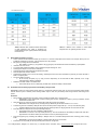
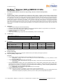
Table 1: Minimum time needed to extract various DNA
or RNA fragments from native or denatured 4%
polyacrylamide gel (29:1 polyacrylamide: bisacrylamide)
at 100-150 volt.
X.
XI.
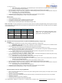
Table 2: Minimum time needed to extract DNA
fragments from 1% agarose gel at 80-110 volt.
DNA or RNA precipitation procedure
•
Add 0.1 volumes of 3 M Potassium acetate, pH-5.2 and 0.7-1 volumes of isopropanol to the solution. For example, add 70 µl of 3 M
Potassium acetate pH-5.2 and 500 – 700 µl isopropanol to a 700 µl sample.
•
Mix gently by inverting the tube several times.
Note: addition of carrier (e.g. 20 µg tRNA or 20 µg glycogen) to the solution will increase the efficiency of precipitation.
•
Incubate at -20°C for 10 min.
•
To increase DNA or RNA precipitation yield incubate the samples overnight at –20°C.
•
Centrifuge the sample at 4°C for 30 min at 20,000 g.
•
Carefully discard the supernatant without disturbing the pellet.
•
Wash the pellet with cold 70% ethanol.
•
Centrifuge the sample at 4°C for 30 min at 20,000 g. Centrifuge the tube in the same orientation as previously to recover the DNA or
RNA in a compact pellet.
•
Air-dry the pellet for 5-20 min.
Do not over-dry the pellet (e.g., by using a vacuum evaporator), as this will make the DNA, especially if it is of high
molecular weight, difficult to redissolve.
•
Redissolve the DNA or RNA in a suitable buffer.
Use a buffer with pH >8.0 for redissolving, as DNA does not dissolve readily in acidic buffers.
Protein Extraction from Polyacrylamide Gel with DiaEasy™ Dialyzer tubes
IMPORTANT: Fixation of proteins before electro elution (e.g. fixation with methanol, acetic acid, etc) is not recommended. Fixation greatly
reduces extraction yield. A sensitive protein staining solution may be used, as it permanently stains the gel without undue fixing of the
protein.
Procedure:
•
•
•
•
•
•
Fill the DiaEasy™ Dialyzer tube with 0.8 ml of dH2O. Incubate for at least 5 min. Empty the tube.
•
IMPORTANT: Check carefully that no dH2O is leaking from the tube. Absorbance of water, by the dry membrane, causes
the decrease in water level.
After staining the gel, excise the gel slice containing the protein with a clean, sharp scalpel.
•
Minimize the size of the gel slice by removing extra gel. Maximum gel slice size should be 1 cm x 0.5 cm.
Transfer the gel slice to a DiaEasy™ Dialyzer tube. Fill the tube with protein-running buffer (0.7 – 0.8 ml). Close the tube gently.
•
Avoid air bubbles in the tube. Do not fill the tube with several gel slices, for larger gel slices use more than one tube.
Place the DiaEasy™ Dialyzer tube in the provided supporting tray.
•
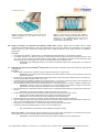
The supporting tray can hold 1-4 DiaEasy™ Dialyzer tubes
•
IMPORTANT: The arrow on the cap should point face up. The two membranes of the DiaEasy™ Dialyzer tube must be in
perpendicular to the electric field to permit the electric current to pass through the tube.
Place the supporting tray containing the DiaEasy™ Dialyzer tubes in a horizontal electrophoresis tank containing protein running
buffer
•
IMPORTANT: Immerse fully the DiaEasy™ Dialyzer tubes with the tray in the buffer.
Pass electric current (usually at 100 volt) until the protein exits from the gel slice.
155 S. Milpitas Blvd., Milpitas, CA 95035 USA | T: (408)493-1800 F: (408)493-1801 | www.biovision.com | [email protected]