1
FOR RESEARCH USE ONLY!
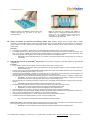
Electro-elution time is to be adjusted for each individual sample. It takes at least 85 min for BSA protein to be electro-eluted
from a 10% SDS-PAGE slice (see Table 3).
Reverse the polarity of the electric current for 120 seconds. This step will release the protein from the membrane.
Open the DiaEasy™ Dialyzer tubes gently, pipetting the protein-containing solution up and down carefully (at least 5 times) and
transfer the solution to a clean 1.5 ml microcentrifuge tube.
•
Do the pipetting on the inner side of the membrane.
Centrifuge the microcentrifuge tube for 1 min at maximum speed. This step will remove gel residues.
Transfer the protein-containing solution to a clean 1.5-ml microcentrifuge tube.
•
•
•
•
•
Important Notes:
•
Use the extracted protein directly.
•
Concentrate the extracted protein by standard concentration methods.
•
Precipitate the extracted protein by standard precipitation protocols.
•
Dialyze directly the extracted protein with a clean DiaEasy™ Dialyzer tubes.
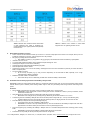
Elution Time Table: The elution time depends on the size of the protein molecule to be eluted, the applied voltage, the size of gel slice, the ratio
of the polyacrylamide : bisacrylamide and the percentage of the polyacrylamide gel. Electro-elution time at the elution step was to be adjusted
for each individual sample.
Protein (kDa)
Time (Min)
Protein (kDa)
Time (min)
14
35-45
50
75-85
19-26
45-55
66
85-95
29
55-65
81
105-115
40
60-70
116
120-130
45
65-75
128
140-150
Table 3: Minimum time needed to extract different-sized
proteins from 10% SDS-polyacrylamide gel (29:1
polyacrylamide : bisacrylamide) at 100 V.
XII. Protein Extraction from polyacrylamide Gel compatible with MALDI-MS by DiaEasy™ Dialyzer Tubes
Procedure
Fill the DiaEasy™ Dialyzer tube with 0.8 ml of dH2O. Incubate for at least 5 min. Empty the tube.
•
IMPORTANT: Check carefully that no dH2O is leaking from the tube. Absorbance of water by the dry membrane causes
decrease in the water level.
•
After staining the gel, excise the gel slice containing the protein with a clean, sharp scalpel.
•
Minimize the size of the gel slice by removing extra gel. Maximum gel slice size should be 1 cm x 0.5 cm.
•
Transfer the gel slice to a DiaEasy™ Dialyzer Tubes. Fill the tube (0.7 to 0.8 ml) with protein running buffer containing 250 mM Tricine
pH 8.5, 0.025% SDS and 25 mM Tris-Base. Close the tube gently.
•
Avoid air bubbles in the tube. Do not fill the tube with several gel slices, for large gel slices use more than one tube.
•
Place the DiaEasy™ Dialyzer Tubes in the provided supporting tray.
•
The supporting tray can hold 1-4 DiaEasy™ Dialyzer Tubes
•
Place the supporting tray containing the DiaEasy™ Dialyzer Tubes in a horizontal electrophoresis tank filled with protein-running
buffer: 250 mM Tricine pH 8.5, 0.025% SDS and 25 mM Tris-Base.
•
IMPORTANT: Immerse fully the DiaEasy™ Dialyzer Tubes with the tray in the buffer.
•
Pass electric current at 150 volt until the protein exits from the gel slice.
•
The electro-elution time is to be adjusted for each individual sample. It takes at least 2 hours for BSA protein to be electroeluted from a 10% SDS-PAGE gel slice.
•
For other proteins from BSA, increase electro elution time presented in Table 3, by 30%.
•
Reverse the polarity of the electric current for 120 seconds. This step will release the protein from the membrane.
•
Open the DiaEasy™ Dialyzer Tubes gently, pipetting the protein-containing solution up and down carefully (at least 5 times) and
transfer the solution to a clean 1.5 ml microcentrifuge tube.
•
Do the pipetting on the inner side surface of the membrane.
•
Centrifuge the microcentrifuge tube for 1 min at maximum speed. This step will remove gel residues.
•
Transfer the protein-containing solution to a clean 1.5-ml microcentrifuge tube.
•
Protein precipitation protocol for analysis by MALDI-MS
•
•
•
•
•
•
Add 1:10 by volume of MS buffer of your choice to the protein containing solution and mix properly. For example, add 70 µl of MS
buffer to a 700 µl sample.
Incubate for 15 min at room temperature.
Add 1: 5 by volume of 50% Trichloroacetic acid (TCA) and mix properly. For example, add 154 µl of 50% TCA to a 770 µl sample.
Incubate for 1 hour at 4°C.
Centrifuge the sample at 4°C for 30 min at 20,000 g.
Carefully descent the supernatant without disturbing the pellet.
155 S. Milpitas Blvd., Milpitas, CA 95035 USA | T: (408)493-1800 F: (408)493-1801 | www.biovision.com | [email protected]