1
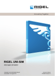
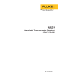
INSTRUCTION MANUAL
Rigel 333
Multi Parameter Patient Simulator
Version 2.0
Rigel Medical
Seaward Group
18 Bracken Hill
Peterlee, County Durham • SR8 2SW
England
www.rigelmedical.com
Dear User:
We appreciate your purchase of the Rigel 333 MultiParameter Patient Simulator. Properly used, the
instrument will deliver years of high performance and
accuracy.
This instruction manual has been designed as a tool to
assist you in getting the most from your Rigel 333. In order
to properly use and to maintain this instrument, please
read the manual carefully.
Rigel Medical, part of the Seaward Group is recognised as
an innovative designer and manufacturer of advanced
biomedical and industrial test instruments. Seaward
Electronic Ltd is ISO 9001-2000 Registered and fully
committed to a continuous improvement process. Further,
we guarantee absolute satisfaction with our products.
Your business is important to us and we are dedicated to
providing you with the best customer and technical service
possible. Please contact us should you have any
questions or concerns regarding your instrument.
We hope you will consider us again when you have a
requirement for accurate, reliable, affordable test
instruments.
Sincerely,
RIGEL MEDICAL
2
Copyright
Copyright © 2003 by Rigel Medical. All rights
reserved. No part of this publication may be
reproduced or transmitted in any form other than for
the purchaser’s personal use without written
permission from Rigel Medical .
Quality Assurance
Seaward Electronic Ltd is ISO 9001-2000
Registered. This instrument was thoroughly tested
and inspected according to Seaward’s ISO 90012000 quality standards and test procedures, and
was found to meet those specifications when it was
shipped from the factory.
Warranty
Rigel Medical warranties the Rigel 333 MultiParameter Patient Simulator against defects in
materials and workmanship for one year from the
date of original purchase.
During the warranty period, we will repair or, at our
option, replace at no charge a product that proves to
be defective, provided you return the product
shipping prepaid to Rigel Medical. This warranty
does not apply if the product has been damaged by
accident or misuse, or as the result of service or
3
modification by other than Rigel Medical, or if its
serial number is defaced or removed.
Rigel Medical reserves the right to discontinue the
Rigel 333 at any time, and change its specifications,
price, or design without notice and without incurring
any obligation. Rigel Medical guarantees availability
of service parts for 5 years after the manufacture of
the unit is discontinued.
The warranty is void if you elect to have the unit
serviced and / or calibrated by someone other than
Rigel Medical.
The purchaser assumes all liability for any damages
or bodily injury that may result from the use or
misuse of the unit by the purchaser, his employees,
agents or customers.
In no event shall Rigel Medical or Seaward
Electronic be liable for consequential damages
Trademarks
Rigel Medical and Rigel 333 are trademarks of
Seaward Electronic Ltd. Any other trademark names
used in this manual are only for editorial purposes
and the benefit of the respective trademark owner,
with no intention of improperly using that trademark.
4
TABLE OF CONTENTS
General Description
6
Controls and Indicators
7
Operating Instructions
8
Baseline ECG Waveform
9
Performance Waveforms
15
Arrhythmia Waveforms
17
Automatic Testing Sequences
21
Respiration Waveforms/ Apnea
27
Blood Pressure waveforms
28
Temperature
29
Calibration
30
Specifications
30
Accessories
34
Appendix
35
5
GENERAL DESCRIPTION
The Rigel 333 Multi-Parameter Patient
Simulator is an advanced micro controller
based instrument. It is designed to
simulate patient signals of ECG,
arrhythmia, invasive blood pressure,
respiration, and temperature. The device
also simulates square, sine, triangle, and
pulse performance waveforms.
The easy to operate Rigel 333 is menu
driven via eight tactile feel soft keys. All
functions are displayed on a two line
sixteen character LCD display.
The small hand held instrument is powered
by one 9 Volt battery or an optional AC
adapter. The Rigel 333 is CE marked and
shipped with a Certificate of Calibration
traceable to the NIST.
The Rigel 333 is a rugged instrument that
performs its simulations quickly,
accurately, and with ease.
6
CONTROLS AND INDICATORS
1.
2.
3.
4.
5.
6.
7.
8.
9.
On-Off Key
ECG Lead Snaps
LCD Display
Temperature Connector
Blood Pressure Connector
Hi-Level ECG Output
Function Keys: ‘F1’, ‘F2’, ‘F3’, ‘F4’.
Arrow Keys: ‘<’ and ‘>’.
Menu Key
4
5
6
7
3
8
1
2
9
7
OPERATING INSTRUCTIONS
The operating menu of the Rigel 333 is arranged in
a tree structure. When turned on, the micro
controller initialises a test routine and displays the
model and software version numbers and then
switches to the Main Menu.
During operation, the ‘MENU’ key returns the
currently displayed menu to the previous menu.
The ‘F1’, ‘F2’, ‘F3’, and ‘F4’ keys select the
parameter for simulation, the specific type of
parameter, and the functional characteristics of the
parameter.
The ‘<’ and ‘>’ arrow keys move through the
available choices under the selected
parameter.
Basic Operating Instructions:
1.
2.
3.
Connect the Rigel 333 to a patient monitor
using the ECG lead snaps, correct blood
pressure interface cable, and correct
temperature interface cable.
Turn the Rigel 333 On.
Select the parameter for simulation,
‘ECG’, ’ Resp’, ‘ BP’, or ‘Temp’.
8
4.
Continue to select choices under the
parameter to be simulated until the
functional characteristics have been
chosen.
5. Each Parameter has default settings.
The following are the menu sequence displays, their
abbreviations, definitions, and default settings.
Ecg
SIMULATOR MENU
Resp
Bp
Temp
Ecg: ECG
Default Setting: Normal Sinus Rhythm (NSR) at 80
BPM with Amplitude of 1 mV
Resp: Respiration
Default Setting: 30 BPM, Impedance 500 Ohms,
Delta R 1.0 Ohm, and Apnea Off
Bp: Blood Pressure
Default Setting: 120/80 Dynamic Setting
0 Static Setting
Temp: Temperature
Default Setting: YSI 700 series set to 25°C
YSI 400 series set to 25°C
9
Base
Base:
Perf:
Arth:
Aut:
Aut
Baseline ECG
Performance Waveforms
Arrhythmia Waveforms
Automatic Test Sequences
NSR
NSR:
PCR:
ST:
RWD:
ECG Menu
Perf
Arth
Baseline ECG
PCR
ST
RWD
Normal Sinus Rhythm
Pacer Waveforms
ST Segment Analysis Waveforms
R Wave Detection
Rate
NSR/
Ampl
Rate and Amplitude selections under NSR are:
Rate:
Ampl:
30 60 70 80
90 100 120 150
180 210 240 270
300 350
.15 0.3 0.5 1.0
10
2.0 3.0 4.0 5.0
Atr
Pacer Waves
Vent
Selecting Atr branches to:
Atr_Pcr Waves
+ve AP PCR ASP
+ve: Changes pacer pulse from positive (+ve) to
negative ( – ve).
AP: Atrial Pacer – Normal Paced Rhythm
QRS with rate of 60 BPM and Pacemaker pulses
with amplitudes of + 2 mV and –2 mV with duration
of 0.1 ms or 2 ms with a normally paced QRS T
(QRS amplitude of 1 mV, duration of 100 ms, T
wave amplitude of 0.2 mV, duration of 180 ms, and
Q-T interval of 350ms)
PCR: Pacer Pulses Alone. Amplitude default
setting of 1.0 mV and width of 1.0 ms.
Widt: width selections
0.1 0.2 0.5 1.0
1.5 2.0
Ampl: amplitude selections
.15 0.3 0.5 1.0
2.0 3.0 4.0 5.0
ASP: Asynchronous Pacer – Ineffective pacing.
11
A non-synchronized waveform that combines QRS
waves at 30 BPM and other specifications as in AP
above with pacer waves with a 0.1 ms or 2 ms width
and amplitude of ± 2 mV at 80 BPM.
Note: 0.1 ms pacer width for AP and ASP can only
be selected by going to ‘PCR’. A selection other
than 0.1 ms produces 2 ms wide pulses.
Selecting Vent branches to:
Vtr_Pcr Waves
VP AVP
VP:
AVP:
Ventricular Pacer at 70 BPM
Atrial Ventricular Pacer at 70 BPM
Selecting ST branches to:
ST-SEGMENT
ele dep MI TalT
ST segment analysis waveforms are divided into
four classes:
ele:
ST Elevation
dep:
ST Depression
MI:
Myocardial Infarction
TalT:
Tall T Wave Rejection
ST _ELVATION
7% 13% 20% Flat
12
ST Elevation: 7, 13, and 20% DC levels of ST
Elevation may be selected. Example: at a QRS
amplitude of 1 mV, ST segments are produced at
positive DC levels of 70, 130, and 200 micro volts.
Waveforms may be selected with a ‘Flat’, a positive
‘+sl’, or a negative ‘-sl’ slope. The ST segment is
proportional to the ECG amplitude setting.
ST _DPRESION
7% 13% 20% Flat
ST Depression: Identical to ST Elevation except
the ST segments are now depressed.
Selecting MI Myocardial Infarction
branches to:
Myocard_Inf
Isc
Inj
Inf
Iinf
Isc:
Inj:
Inf:
Iinf:
Ischemia
Injury
Infarction
Inferior Infarction
Isc: Ischemia : Normal Sinus Rhythm (NSR) with
fully inverted T waves. This is a condition of
13
reduced blood supply to the heart in a normal
patient.
INJ: Injury. A waveform with ST elevation of 20%
with a negative slope and inverted T wave.
Inf: Infarction. Normal Sinus Rhythm (NSR) with a
large Q wave with the amplitude increased six times
and the width increased three times compared to
normal.
Iinf: Inferior Infarction. A waveform with the Q wave
modified as in Infarction and the ST segment
elevated 7% as in ST Elevation.
Selecting TalT branches to:
TalT/Rate = 80
Rate Ampl
An 80 BPM QRS test signal of 1 mV amplitude and
100 ms duration is generated with a T wave
duration of 180 ms and Q-T interval of 350 ms.
The T wave amplitude may be varied from 0 to 1.2
mV in steps of 0.1 mV .
Rate: 80 BPM
Amplitude:
0.0 0.1 0.2 0.3
0.4 0.5 0.6 0.7
14
0.8 0.9 1.0 1.1
1.2
Selecting RWD branches to:
RWD/
Ampl
Widt
RWD: R Wave Detection. A QRS waveform is
generated at 70 BPM with selectable width and
amplitude changes.
Widt: Width default setting 100 msec
70 80 90 100
110 120
10 40 50 60
Ampl: Amplitude default setting 1.0 mV
.15 0.3 0.5 1.0
2.0 3.0 4.0 5.0
SIN
SIN:
SQR:
TRI:
PLS:
Perf. Waves
SQR
TRI
Sine Wave
Square Wave
Triangle Wave
Pulse Wave
Freq
SIN/
Ampl
15
PLS
Freq
SQR/
Ampl
Freq
TRI/
Ampl
The frequency and amplitude default settings and
choices are the same for the sine, square and
triangle performance waveforms.
Freq: Frequency default setting 1.0 Hz
0.9 1.0 2.0 3.0
4.0 5.0 6.0 7.0
8.0 9.0 10 20
30 40 50 60
70 80 90 100
0.1 0.2 0.3 0.4
0.5 0.6 0.7 0.8
Ampl: Amplitude default setting 2.0 mV
2.0 3.0 4.0 5.0
0.1 0.2 0.5 1.0
Pulse wave default setting: A pulse wave is
generated at 4 second intervals with an amplitude of
1mV and width of 20 ms.
16
Selecting Arr branches to:
Atr
AC
Arrhythmia Menu
Vent
Arrhythmias are divided into Atrial, Atrial
Conduction, and Ventricular waveforms. Normal
Sinus Rhythm (NSR) at 80 BPM is the default
waveform in this menu. Function keys select the
desired arrhythmia and the MENU key clears the
arrhythmia to NSR.
The following are the arrhythmia definitions:
Atrial: Atrial Arrhythmias
SA: Sinus Arrhythmia: The ECG rate uniformly
increases and decreases continuously. The pattern
is cyclic with rates changing in the following order:
60, 70, 80, 90, 100, 90, 80, 70, 60 BPM.
M80: Missing Beat: Normal Sinus Rhythm is
th
generated at 80 BPM with every 10 beat missing.
AFLT: Atrial Flutter: Varying ECG rates with 12
cycles at 60 BPM for 12 seconds, 9 cycles at 90
BPM for 6 seconds, 15 cycles at 150 BPM for 6
17
seconds repeating with large P waves at 300 BPM.
This corresponds to ventricular responses of 5:1 for
12 seconds, 3:1 for 6 seconds, and 2:1 for 6
seconds.
AFB: Atrial Fibrillation: Irregular QRS complexes
with no P waves and constantly changing R-R
intervals are generated. The rate varies in a cyclic
fashion at 30, 60, 70, 80 and 30 BPM with low
amplitude oscillations on the baseline.
PAT: Paroxysmal Atrial Tachycardia: NSR is
generated at 180 BPM with inverted P waves.
NODL: Junctional Premature Contraction: NSR
is generated at 80 BPM with a short PR interval.
The QRS starts immediately following the P wave.
AC: Atrial Conduction Arrhythmias
AB1: First Degree AV Block: The QRS is
generated at 80 BPM, the P wave precedes the
QRS by a fixed but prolonged PR interval > 0.2
seconds ( PR interval = 0.26 seconds).
MB1: Second Degree AV Block: Mobitz I:
Wenckebach: The QRS is generated at 80 BPM.
There is a progressive lengthening of the PR
interval with intermittent dropped beats. The PR
intervals are 170, 230, and 310 ms.
18
MB2: Second Degree AV Block: Mobitz II: The
th
QRS is generated at 80 BPM with every 4 QRS
missing. The PR interval is constant at 170 ms.
AB3: Third degree AV Block: The P wave and
QRS are independent of each other. The P wave is
generated at 80 BPM and the QRS is generated at
50 BPM.
RBB: Right Bundle Branch Block: A prolonged
QRS (>0.12 sec) is generated at 80 BPM. The
resulting QRS looks like the letter “M”.
LBB: Left Bundle Branch Block: A widened QRS
is generated at 80 BPM with a large wide S wave.
LAH: Left Anterior Hemiblock: A QRS is
generated at 80 BPM with an S wave larger than the
R wave.
Ventricular: Ventricular Arrhythmias
PV1: Premature Ventricular Contraction 1: NSR
is generated at 80 BPM. Each time the ‘F1’ function
key is pressed one PVC is generated.
19
PV3: Premature Ventricular Contraction 3: NSR
is generated at 80 BPM. Each time the ‘F2’ function
key is pressed 3 PVCs are generated.
PV6: Premature Ventricular Contraction 6: NSR
is generated at 80 BPM. Each time the ‘F3’ function
key is pressed 6 PVCs are generated.
PV12: Premature Ventricular Contraction 12:
NSR is generated at 80 BPM. Each time the ‘F4’
function key is pressed 12 PVCs are generated at
different intervals.
PV24: Premature Ventricular Contraction 24:
NSR is generated at 80 BPM. Each time the ‘F1’
function key is pressed 24 PVCs are generated at
different intervals.
BGY: Bigeminy: NSR is generated at 80 BPM with
every other beat as a PVC.
TGY: Trigeminy: NSR is generated at 80 BPM
with every third beat as a PVC.
PVC: Premature Ventricular Contraction:
Continuous PVCs are generated at 80 BPM.
VFLT: Ventricular Flutter: Sine waves at 240 BPM
are generated with irregular amplitudes.
20
VFB: Ventricular Fibrillation: A totally irregular
waveform is generated with chaotic undulations of
the baseline.
VTC: Ventricular Tachycardia: A fast moving
series of PVCs is generated at 210 BPM.
PVR: Right Focal PVC: NSR is generated at 80
th
BPM with every 10 beat a right focal PVC.
Selecting Aut branches to:
<Auto Sequence>
RWD
TalT
PPR
TAC
The Automatic Test Sequence generates test
waveforms in sequences according to AAMI
requirements eliminating the need for the user to
make numerous manual selections.
The automatic test sequences are:
RWD:
TalT:
PPR:
TAC:
R Wave Detection
Tall T Wave Rejection
Pacemaker Pulse Rejection
Time for Alarm for Tachycardia
The automatic sequence is initiated when the test is
selected and continues until all of the test patterns
21
have been generated. During the test sequence the
values generated are displayed on the LCD. At the
completion of the automatic test, the display will
return to its steady mode.
RWD: R Wave Detection. R waves are
generated with three varying parameters of
amplitude, width, and rate. The three parameter
values are displayed on the LCD while generated.
Each test waveform is displayed for 20 seconds.
The complete waveform test sequence is performed
in three separate sets.
Set 1: Variable Parameters
Amplitude: 0.5, 2, 5 mV
Width:
100, 70, 120 ms
Rate:
80, 30, 210 BPM
Set 1 Complete Test Sequence
Amplitude
(mV)
0.5
2.0
Width
(ms)
100
70
120
Rate
Time
(BPM)
(sec)
80, 30, 210 20
80, 30, 210 20
80, 30, 210 20
100
70
120
80, 30, 210
80, 30, 210
80, 30, 210
22
20
20
20
5.0
100
70
120
80, 30, 210
80, 30, 210
80, 30, 210
20
20
20
As the test progresses, the indicated heart rate
displayed on the patient monitor should be within
± 10% or ± 5 BPM whichever is greater of the
applied rate.
Set 2: Variable Parameters
Amplitude: 0.15 mV
Width:
70, 120 ms
Rate:
30, 210 BPM
Set 2 Complete Test Sequence
Amplitude
(mV)
0.15
0.15
Width
(ms)
70
120
Rate
(BPM)
30, 210
30, 210
Time
(sec)
20
20
The monitor will not respond to the waveforms in
this sequence Set.
Set 3: Variable Parameters
Amplitude: 1.0 mV
Width:
10 ms
Rate:
30, 210 BPM
Set 3 Complete Test Sequence
23
Amplitude Width
Rate
Time
(mV)
(ms)
(BPM)
(sec)
1.0
10
30, 210
20
The patient monitor will not respond to the
waveforms in the Set 3 test sequence.
TalT: Tall T Wave Rejection. QRS and T
waves are generated with the following values:
QRS: Rate
80 BPM
Amplitude 1 mV
Width
100 ms
T Wave Duration 180 ms
QT Interval
350 ms
T Wave Amplitude 0.0, 0.2, 0.4, 0.6, 0.8,
1.0, and 1.2 mV .
In the automatic test sequence the T Wave
Amplitude steps through the changes at one minute
intervals. The display indicates the T Wave
Amplitude and the QRS Rate.
As the T wave amplitude increases, the first value at
which the patient monitor counts the T wave at 80 ±
8 BPM should be noted. This value should match
the patient monitor manufacturer’s specification.
PPR: Pacemaker Pulse Rejection. The
test sequence cycles through normal paced rhythm
24
(AP), ineffective pacing (Asynchronous Pacing
ASP), and pacemaker pulses alone (PCR).
For normal pacing (AP),QRS and pacer waves are
generated with the following values:
QRS: Amplitude
Width
T wave: Amplitude
Duration
Q-T Interval
R-R Interval
Pacer: Amplitude
Width
1 mV
100 ms
0.2 mV
180 ms
350 ms
1 Sec
2 mV, -2 mV
2 ms, 0.1 ms
For ineffective pacing (ASP), the values of the QRS
and pacer waves are the same as normal pacing
except for the QRS rate that becomes 30 BPM and
the pacer rate that becomes 80 BPM.
During the ASP and AP test sequences the display
will show the pacer amplitude, the pacer width, and
the QRS rate.
For pacemaker pulses alone (PCR) the values
generated are:
Pacer: Rate
60 BPM
Width
2.0 ms, 0.1 ms
Amplitude 2 mV, -2 mV
25
During the PCR test sequence the display will show
the pacer amplitude, the pacer width, and the QRS
rate.
Each set of values in the test sequence is generated
and displayed for 20 seconds.
TAC: Time to Alarm for Tachycardia.
The TAC test is designed to measure the time it
takes for the patient monitor to alarm after the onset
of ventricular tachycardia. The low and high alarms
on the patient monitor should be set at 60 BPM 100
BPM before starting the test.
This auto test sequence generates a QRS wave
form at the rate of 80 BPM alternating with a
ventricular tachycardia waveform with rates of 206
and 195 BPM and amplitudes of 1.0, 0.5, 2.0, and
4.0 mV.
Each waveform is generated for 20 seconds and the
display will show the amplitude and the rate of the
waveform being generated.
The following is the sequence of waveforms:
QRS
Ventricular Tachycardia
Rate
Amp
Width Rate
Amp
(BPM) (mV)
(ms)
(BPM) (mV)
80
1
100
206
1.0
80
1
100
206
0.5
80
1
100
206
2.0
26
80
1
100
195
2.0
80
1
100
195
1.0
80
1
100
195
4.0
RESPIRATION:
Select Resp in the Simulator Menu to access the
Respiration selections.
Rate
Resp Menu
Imp
dR
Apne
Respiration waveforms are generated with four
selectable rates, baseline impedances, and delta
impedance variations.
The Respiration default settings are:
Respiration Rate 30 BPM
Impedance
500 Ohms
Delta Impedance 1.0 Ohm
Apnea
Off
The value changes that may be made are:
Rate: 15, 30, 60, 120 BPM.
Impedance: 250, 500, 750, and 1000 Ohms.
Delta Impedance: 0.1, 0.5, 1.0, and 1.5 Ohms.
Apnea:
Off
Resp/Apne=Off
Cont
12s
27
32s
Off: Apnea is absent. Normal respiration waveforms
are generated.
Cont: Continuous Apnea. No respiration waveforms
are generated.
12s: No respiration waveform is generated for 12
seconds.
32s: No respiration waveform is generated for 32
seconds.
BLOOD PRESSURE:
Select Bp in the Simulator Menu to access Blood
Pressure selections.
Dyna
BP1 Menu
Stat
Two blood pressure waveforms are generated with
selections of 12 static and 6 dynamic values. BP1
values are selected from the listed choices and BP2
values are ½ of those selected for BP1.
The Dynamic pressure waveforms track the Normal
Sinus Rhythm rates.
The default settings for pressure values are:
BP1
Dynamic: 120/80 Static: 0
BP2
Dynamic: 60/40 Static: 0
The Dynamic pressure value selections are:
100/60 120/80
28
50/10
70/30
60/20
80/40
The Static pressure value selections are:
0
5 10 20
25 30 40 50
100 150 200 300
TEMPERATURE:
Select Temp in the Simulator Menu to access
Temperature value selections.
YSI400
Temp Menu
YSI700
Temperature simulation is provided for both YSI 400
and YSI 700 standards. The temperature default
setting for both is 25 degrees Centigrade.
The Temperature value selections are:
25 C
37 C
for both YSI 400 and YSI 700.
CALIBRATION
The Rigel 333 is shipped from the factory fully
calibrated with a Certificate of Calibration traceable
to the NIST. Annual calibration is recommended.
Contact the Customer Service Department for
29
instructions for returning the instrument to Rigel
Medical for either calibration or repair.
SPECIFICATIONS
ECG: 12 leads with independent outputs
referenced to RL.
NORMAL SINUS RHYTHM (NSR)
ECG Rates: 30, 60, 70, 80, 90, 100, 120, 150, 180,
210, 240, 270, 300, and 350 BPM. Accuracy 0.5%.
Amplitudes: 0.15, 0.3, 0.5, 1.0, 2.0, 3.0, 4.0, and
5.0 mV on Lead II. Lead 1 is 0.6 X Lead II, Lead III
is 0.4 X Lead II, and Lead V is the same as Lead II.
High Level: 500 X low level output on Lead II.
Accuracy: 2% (1-5mV).
PERFORMANCE WAVEFORMS (PERF)
Sine, Square, Triangle, Pulse
Frequencies: 0.1 to 0.9 in 0.1 Hz steps.
1.0 to 9.0 in 1.0 Hz steps.
10 to 100 in 10 Hz steps.
Accuracy: 1%
Amplitudes: 0.1, 0.2, 0.5, 1.0, 2.0, 3.0, 4.0, and 5.0
mV on Lead II.
30
Pulse: 20 ms pulse of 1mV amplitude repeated at 4
second intervals.
R WAVE DETECTION (RWD)
Widths: 10, 40, 50, 60, 70, 80, 90, 100, 110, and
120 ms.
Amplitudes: 0.15, 0.3, 0.5, 1.0, 2.0, 3.0, 4.0, 5.0
mV on Lead II.
PACER (PCR)
AP: Atrial Pacer set at 70 BPM.
ASP: Asynchronous Pacer – Ineffective Pacing.
Pacer Amplitudes: + 2 mV and –2mV.
QRS Amplitudes: 1 mV
PCR: Pacer pulses alone.
Pacer Widths: 0.1, 0.2, 0.5, 1.0, 1.5, and 2.0 ms.
Amplitudes: 0.15, 0.3, 0.5, 1.0, 2.0, 3.0, 4.0, and
5.0 mV.
VP: Ventricular Pacer set at 70 BPM.
AVP: Atrial Ventricular Pacer set at 70 BPM.
QRS Amplitude: 1 mV.
Pacer Amplitude: -2mV.
RESPIRATION
Baseline Impedance: 250, 500, 750, and 1000
Ohms.
Delta Impedance: 0.1, 0.5, 1.0, and 1.5 Ohms.
Accuracy: 10%.
Rates: 15, 30, 60, and 120 BPM.
31
Accuracy: 1%
Apnea: Off, continuous, 12 seconds and 32
seconds.
Lead Configuration: Leads I and II.
BLOOD PRESSURE
Impedance: 350 Ohms.
Excitation: 2 to 16 Volts.
Sensitivity: 5µV/V/mmHg.
Static: 0, 5, 10, 20, 25, 30, 40, 50, 100, 150, 200,
and 300 mmHg.
Dynamic: 50/10, 60/20, 70/30, 80/40, 100/60, and
120/80 mmHg. Dynamic waveforms track all NSR
rates.
TEMPERATURE
Compatibility: YSI 400 and 700 Series.
Temperature: 25 and 37 degrees Centigrade.
Accuracy: 2% of setting
POWER REQUIRMENTS:
One 9 Volt alkaline battery or optional AC adapter.
PHYSICAL CHARACTERISTICS:
Dimensions: 13.9 X 8.9 X 3.8 cm.
Weight: 0.3 kg
TEMPERATURE REQUIREMENTS:
Operating: 15 to 35°C.
Storage: 0 to 55°C.
32
CALIBRATED DC OUTPUTS:
10 Calibrated DC outputs from 0.04 to 2 mV.
OUTPUT CONNECTIONS:
Part # 303, Open Ended Pressure cable:
Pressure:
Pin 1
Pin 4
Pin 2
Pin 5
Single Pressure
+ Excitation
+ Signal
- Signal
- Excitation
Pressure:
Pin 3
Pin 2
Dual Pressure
+ Signal
- Signal
Part # 304, Open Ended temperature Cable:
3.5 mm Stereo Jack
YSI 400
YSI 700
Tip, Ring, Shield.
Tip, Ring.
High Level Output: 3.5 mm Stereo Jack
Signal Positive
Signal Ground
Input/Unused
Tip
Shield
Ring
33
Note: Specifications are subject to change without
notice.
ACCESSORIES
Description
Part Number
User Manual
OPTIONAL ACCESSORIES
Description
Part Number
Soft Carrying Case
110 VAC Adapter
220 VAC Adapter
ECG Snap Studs
Open Ended Temperature Cable
Pressure Cables:
Open Ended
Space Labs Dual
Space Labs Single
Datascope Dual
Datascope Single
Hewlett Packard Dual
Hewlett Packard Single
Fukuda Denshi Dual
Bard Dual
Bard Single
Mennen Medical Single
Ivy Biomedical Single
34
301
302
302-220
1000
304
303
305
305-S
306
310
307
308
309
311
311-S
312
314
APPENDIX
35
Designing and Manufacturing Innovative
Biomedical and Industrial Test Instruments in
the United Kingdom Since 1982.
•
•
•
•
•
•
Electrical Safety Analysers
Pacemaker Analysers
SPO2 Analysers
NIBP Simulators
Defibrillator Testers
Patient Simulators
For a full list of products, please order a free copy of our
product catalogue online at www.rigelmedical.com
Rigel Medical
Seaward Group
18 Bracken Hill, Peterlee, County Durham
SR8 2SW, England
Phone: +44 191 5863511 • Fax: +44 191 5860227
www.rigelmedical.com
36
Rigel 333 Manual Rev 2.0
37