1
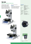
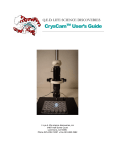
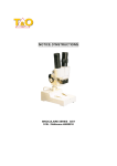
National Optical & Scientific Instruments Inc.
11113 Landmark 35 Drive
San Antonio, Texas 78233
Phone (210) 590-9010 Fax (210) 590-1104
INSTRUCTIONS FOR
MODEL DC3-420TH
STEREOSCOPIC ZOOM MICROSCOPE
WITH DIGITAL CAMERA
(For microscope operation only. Camera operation covered in separate
Motic Image 2000 manual included with your microscope)
HOW TO USE YOUR MICROSCOPE SERIAL NUMBERS
1. Microscope serial number: This number (etched on the post base) is the
number under which your warranty is registered.
2. Microscope DM number: This number (found on a white sticker on the bottom of
the microscope) is used for logging on the Motic web site, which gives you the
ability to download free software upgrades.
3. Motic CD DM number: This number is to be used to register the software when
loaded on the computer for the first time.
Copyright © 1/2/01
National Optical & Scientific Instrument Inc.
About the Digital Microscope
The manual for your new digital microscope is in two parts. This first part describes the basic nomenclature
and functions of the microscope, which can be used as a fully functional microscope, independent of the
camera. The second part is the Motic Images 2000 User’s Manual, which provides detailed documentation
for installation and operation of the Motic Images 2000 software. In order to achieve optimum results, it is
important that you carefully read both this and the documentation manuals before operating your microscope or
camera.
Stereoscopic microscopes are used for viewing 3-dimensional objects, inspection or assembly of small parts,
and for dissection of biological specimen. They provide an upright, unreversed image which permits easy
manipulation of the object being viewed while looking through the microscope. They are designed for viewing
solid objects at low magnification, but they will also permit viewing of some transparent specimen slides.
UNPACKING
1. Your microscope is packed with the following components, all of which have been checked at the factory.
Carefully remove all components and check against this list. Retain styrofoam container in case
microscope must be transported or returned to factory for any reason.
Make certain not to touch any of the lens surfaces while handling the microscope. Dust, dirt or fingerprints
can damage the delicate lens surfaces or adversely affect image quality.
A.
B.
C.
D.
E.
F.
G.
H.
I.
J.
K.
L.
M.
N.
O.
P.
Microscope, with WF10x eyepieces (pair)
Two instruction manuals (this and separate software documentation)
CD Motic Images 2000
Calibration slide
12VDC switching power supply, operates on 100v-240v, 50H/60H
Power cord
USB cable (for connecting to computer)
S cable (for connecting to high resolution video monitor)
RCA cable (for connecting to standard resolution video or projector)
Rubber eyecups (pair)
Frosted glass stage plate
Frosted blue filter
Plastic black/white contrast plate (inserted in microscope stage)
“L” hex wrench (for changing stage plates)
Dustcover
Warranty card
2. Examine packing material before you discard it. Retain the styrofoam container in case you need to
transport, store, or return the microscope for service. If it becomes necessary to ship the microscope for
any reason, pack it in the styrofoam container, and then pack the styrofoam in another corrugated shipping
container for optimum protection. Use of the styrofoam alone will not provide adequate protection in
transit, and will void your warranty.
ASSEMBLY
The microscope is packed full assembled, except minor parts.
1. Install the rubber eyecups over the top edge of the eyepieces.
2. Attach female cable connector to male connector located on top of post.
A. Align the key-way slot located on the 7-pin female cable connector to the key located in the male
connector on the post.
3
B. Lock connector into position by pressing down on the black strain relief until the connector snaps into
locked position.
3. Use “L” hex key wrench to loosen locking set screw below front edge of stage, to permit removal of plastic
stage plate.
4. At this point, you may insert either the plastic contrast plate, or the frosted glass stage plate (see Notice
under operation instructions below), following instructions listed under “operation” below.
OPERATION
Your microscope is fully functional as a standard microscope. The following instructions apply only to operation
of the microscope. Refer to the Motic Images 2000 User’s Manual for instructions for installation of the
software and operation of the camera. Some steps for microscope operation are altered slightly in the software
documentation, in order to utilize some of the unique features provided by the digital camera and software.
1. ILLUMINATION
A. Before operating microscope, adjust intensity control located on side of base to the minimum position.
This should be done prior to each time light is turned on or off, in order to extend bulb life.
B. Supplied is a 12VDC switching power converter and separate power cord. Note that the switching
power converter will operate on either 120v or 240v current, 50hertz or 60 hertz.
Plug one end of power cord into 12VDC power converter and other end into power outlet. Plug 12VDC
power converter into power jack on base of microscope.
C. The microscope is furnished with two stage plates. The frosted glass plate is used when viewing
transparent specimen slides or for viewing some specimen thin enough through which light can pass
(insect wings, etc.) The plastic black/white contrast plate can be used when viewing opaque objects or
for dissecting. Choose side of plate providing best contrast with specimen.
D. There are three rocker type light controls located on top surface of microscope base.
MAIN
“I”
“T”
= Turns power on and off
= Turns incidental light on (top illumination)
= Turns transmitted light on (substage illumination)
NOTE: USE TRANSMITTED ILLUMINATION ONLY WITH FROSTED GLASS STAGE PLATE AND
BLUE FILTER IN PLACE. HEAT GENERATED IN BASE FROM BOTTOM LIGHT WILL WARP OR
DAMAGE THE PLASTIC BLACK/WHITE PLATE. SUCH DAMAGE WILL NOT BE COVERED BY
WARRANTY.
Remove black/white stage plate by loosening locking set screw located on front of base with supplied
“L” wrench. Insert daylight blue filter into machined groove provided in center of base. Install frosted
glass stage plate. Tighten locking set screw.
Incidental illumination can be used with either frosted glass plate or black/white plastic stage plate. The
top light has an intensity control located on side of base. When using incidental light, always set
intensity control at its lowest level before turning lamp on or off. This measure will extend bulb life.
The top light can also be centered on specimen by using the top light beam adjustment screw. This
allows user to select the best spot illumination required for specimen being viewed.
Transmitted and incidental illumination combined can provide extra illumination for certain objects
where additional top illumination will enhance the object being viewed.
4
E. Camera is turned on by separate on/off switch located on back of microscope base. Always remember
that for the digital microscope to function, the camera must be turned on, it addition to turning on
microscope illumination.
2. INTERPUPILLARY ADJUSTMENT
This permits each user to adjust spacing between eyepieces in order to accommodate distance
between their eyes. While looking through the microscope eyepieces with both eyes, grasp eyepiece
tube housings with both hands and rotate them on their axis, moving eyepieces apart or together until a
full field of view is observed and images blend into one. Interpupillary distance is now corrected for
your own inter-ocular distance and does not require further adjustment later unless another user
changes this adjustment.
3. FOCUSING
A. Adjust zoom control knobs (located on both sides of head) so that the lowest magnification number “1”
is positioned at the black index dot on head. Lower magnifications have larger fields of view, making it
easier to position and locate area to be viewed.
B. Place a flat object or specimen slide (cover glass up), on stage plate.
C. Position focusing knobs in the center of focusing range.
D. Viewing head is mounted on a post. The height of viewing head can be adjusted up or down on the
post in order to focus on different sized objects. Loosen the locking knob located on the locking
support collar, allowing the support collar to slide down to bottom of post. While firmly holding viewing
head with one hand, loosen locking knob located on back of focusing assembly so that head can move
freely up or down on post.
E. While looking through microscope, move viewing head up or down on post until object can be seen in
approximate focus. Tighten focusing assembly locking knob. Position the support collar under the
focusing block and tighten locking knob on support collar. It is not necessary to make this adjustment
every time you change objects to be viewed, so long as the different objects are of similar thickness or
height.
F. Both eyepieces have knurled diopter adjustment rings.
clockwise direction to the lowest position.
Rotate both left and right diopters in a
G. Adjust zoom control to the highest magnification by aligning the number “4” on knob to the black index
dot on head.
H. While looking through right eyepiece with one eye, rotate focusing control knob until specimen comes
into sharp focus through right eyepiece.
I.
Adjust zoom control knob to the lowest magnification.
J.
Adjust the right diopter until the image is sharp. Do not change the focusing knob position.
K. Without changing the position of the focusing knob, adjust the left eyepiece diopter until you obtain a
sharp image in left eyepiece. the image should now be sharp throughout the zoom power range.
5
SPECIFICATIONS
Use of eyepieces other that WF10x will affect microscope magnification according to the chart below.
However, the digital camera uses a constant eyepiece factor of 10x. Therefore, regardless of eyepieces used
in microscope, use of auxiliary objectives will affect camera image on computer or video monitor only to the
extent listed in the second segment below, that following “WF10 eyepieces.” Any reticle installed in eyepiece
will be visible only when viewing through microscope, and will not be visible on computer or video monitor.
Specification Chart
Eyepieces
Zoom
Objective
Position
WF5X
Field No.22
(Optional)
(No Reticle Holder)
Interpupillary
Distance
59~83
WF10X
Field No. 20
(Supplied)
Accepts Reticle
22.8mm O.D.
Interpupillary
Distance
54~78
WF15X
Field No. 13
(optional)
(No Reticle Holder)
Interpupillary
Distance
50.5~74.5
WF20X
Field No. 10
(optional)
(No Reticle Holder)
Interpupillary
Distance
52~76
Model
DC3-420T
Standard Objective
1X
(supplied)
Auxiliary Objective
0.5X
(Optional)
Working Distance – 79mm
Working Distance - 137mm
Auxiliary Objective
0.75X
(Optional)
Working Distance –
90mm
Total
Field
Magnification
Size
Auxiliary Objective
1.5X
(Optional)
Working Distance 34mm
Total
Field
Magnification
Size
Total
Magnification
Field
Size
Total
Magnification
Field
Size
1X
5X
22mm
2.5X
44mm
3.75X
29.3mm
7.5X
14.7mm
2X
10X
11mm
5X
22mm
7.5X
14.6mm
15X
7.3mm
3X
15X
7.3mm
7.5X
14.7mm
11.25X
9.8mm
22.5X
4.9mm
4x
20X
5.5mm
10X
11mm
15X
7.3mm
30X
3.7mm
1X
10X
20mm
5X
40mm
7.5X
26.7mm
15X
13.3mm
2X
20X
10mm
10X
20mm
15X
13.3mm
30X
6.7mm
3X
30X
6.7mm
15X
13.3mm
22.5X
8.9mm
45X
4.4mm
4x
40X
5mm
20X
10mm
30X
6.7mm
60X
3.3mm
1X
15X
13mm
7.5X
26mm
11.25X
17.3mm
22.5X
8.7mm
2X
30X
6.5mm
15X
13mm
22.5X
8.7mm
45X
4.3mm
3X
45X
4.3mm
22.5X
8.7mm
33.75X
5.8mm
67.5X
2.9mm
4x
60X
3.25mm
30X
6.5mm
45X
4.3mm
90X
2.2mm
1X
20X
10mm
10X
20mm
15X
13.3mm
30X
6.7mm
2X
40X
5mm
20X
10mm
30X
6.7mm
60X
3.3mm
3X
60X
3.3mm
30X
6.7mm
45X
4.4mm
90X
2.2mm
4x
80X
2.5mm
40X
5mm
60X
3.3mm
120X
1.7mm
Specimen Height
Minimum
Maximum
0.0mm
93mm
Specimen Height
Minimum
Maximum
0.0mm
19mm
Specimen Height
Minimum
Maximum
0.0mm
66mm
Specimen Height
Minimum
Maximum
0.0mm
120mm
MAINTENANCE
WARNING: For your own safety, turn switch off and remove plug from power source before
maintaining your microscope. If the power cord is worn, cut or damaged in any way, have it replaced
immediately to avoid shock or fire hazard.
1. OPTICAL MAINTENANCE
A. Do not attempt to disassemble any lens components. Consult a microscope service technician when
any repairs not covered by instructions are needed.
B. Prior to cleaning any lens surface, brush dust or lint off lens surface using a camel hair brush. You can
also use an ear syringe or canned compressed air, such as that sold by most computer stores.
6
C. To clean eyepiece lenses, do not remove from eyepiece tube. Clean only the outer lens surface.
Breath on lens to dampen surface, then wipe with lens paper or tissue or use a cotton swab moistened
with distilled water. Wipe lenses with a circular motion, applying as little pressure as possible. Avoid
wiping dry lens surface as lenses are scratched easily. If excessive dirt or grease gets on lens
surfaces, a small amount of xylene can be used on a cotton swab or lens tissue. To clean objective
lenses, do not remove objectives from microscope. Clean front lens element only, following same
procedure.
2. MECHANICAL MAINTENANCE
The only mechanical adjustment the microscope might require is the tension of the focusing
mechanism. This has been adjusted at the factory, but over the course of time it may loosen and
cause the head of the microscope to slip downward on the focusing block.
The tension adjustment collar is located between arm and focus knob on left side of microscope. With
a jewelers type screwdriver, loosen slotted set screw located on knurled surface of the tension
adjustment collar. Turn collar clockwise to tighten tension, counter-clockwise to loosen tension. After
adjusting, tighten the set screw to lock collar in place.
NOTE: It is recommended that you leave the tension as loose as possible for ease of focusing, yet not
so loose that it permits the head of microscope to drift downward from its own weight and cause the
microscope to “drift” out of focus.
3. ELECTRICAL MAINTENANCE
The extent of electrical maintenance, by other than a qualified technician, should be bulb replacement. BE
CERTAIN TO TURN SWITCHES OFF AND REMOVE PLUG FROM POWER SOURCE OUTLET
BEFORE CHANGING BULBS.
A. To replace top bulb (factory #800-422 12v 15 watt halogen), remove light shade by rotating in a
counter-clockwise direction. Remove light housing by rotating in a counter-clockwise direction.
Remove light bulb by firmly grasping and pulling straight out from bi-pin socket. Note that this socket
holds bulb securely, so you might have to pull rather firmly. Using a cloth, hold new bulb and gently
push new bulb into bi-pin socket. Replace light housing and light shade.
B. To replace bottom bulb (factory #800-170) .... remove cover plate located on bottom of base by
removing four rubber feet that secure cover to base. Holding lamp with a cloth, gently pull lamp
straight out from socket. Push new lamp into place in same manner and replace cover plate.
7
TROUBLESHOOTING
PROBLEM
Light fails to operate.
Image does not remain in focus
Poor resolution
(image not sharp)
REASON FOR PROBLEM
Outlet inoperative.
SOLUTION
Have qualified service technician
repair outlet.
AC power cord not connected.
Plug into outlet.
Lamp burned out.
Head of microscope drops from
its own weight.
Objective lenses dirty.
Replace lamp.
Adjust tension control.
Clean objective lenses.
Eyepiece lens dirty.
Clean eyepiece lenses.
Spots in field of view.
Eyepiece lens dirty.
Clean eyepiece lenses. ***
***Spots in field of view can also result from dirt on inside of eyepiece. It is recommended that you have
service technician clean inside of lens.
OPTIONAL ACCESSORIES AND PARTS:
#605-400
#615-400
#620-400
#705-420
#775-420
#715-420
#800-170
#800-422
#931-420
#965-400-05
#965-400-10
WF5X Eyepieces
WF15X Eyepieces
WF20X Eyepieces
Auxiliary 0.5x objective lens
Auxiliary 0.75x objective lens
Auxiliary 1.5x objective lens
Replacement bulb, bottom light, 12v 10 watt halogen bi-pin
Replacement bulb, top light, 12v 15 watt halogen bi-pin
Ring light adapter, O.D. 54.5mm (permits mounting auxiliary ring light on objective pod)
Eyepiece reticle, 5mm/100 divisions, O.D. 22.8mm (for use only with WF10x eyepieces)
Eyepiece reticle, 10mm/100 divisions, O.D. 22.8mm (for use only with WF10x eyepieces)
WARRANTY
Microscope, 5-year limited warranty: Manufacturer warrants this instrument to be free from defects in
material and workmanship under normal use and service for 5 years from date of purchase. It does not cover
damage resulting from abuse or misuse, repairs or alterations performed by other than authorized repair
technicians or damage occurring in transit. Warranty does not cover bulbs or fuses.
Camera, 1-year limited warranty: Manufacturer warrants camera to be free from defects in material and
workmanship under normal use and service for 1 year from date of purchase. It does not cover damage
resulting from abuse or misuse, repairs or alterations performed by other than the manufacturer, or damage
occurring in transit.
Software, 90-day limited warranty: Manufacturer warrants software to be free from defects in material and
workmanship under normal use and service for 90 days from date of purchase.
For warranty service, instrument should be well packed to avoid damage in transit, including a description of
the difficulty, and shipped postage prepaid to National at the address on front. National will repair or replace at
no charge and return postage prepaid. If failure was caused by misuse, alterations, accident , or abnormal
conditions of operation, an estimate for repairs will be submitted for your approval prior to work being
performed.
If you have questions concerning this product or warranty, contact dealer from which it was purchased. Or
contact National, asking for warranty assistance.
(7/6/01)
8