1
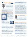
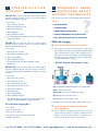
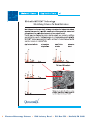
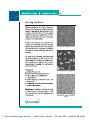
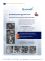
INTRODUCTION Electron Microscopy Sciences and QuantomiX have joined forces here in The United States to market and sell the breakthrough solutions of QuantomiX’s proprietary WETSEMTM Technology. This technology enables scanning electron microscopes (SEM) to image and analyze wet samples such as cells, tissue biopsies, foods and ink, in their native environment. Eliminating the need for time-consuming preparation procedures, the WETSEMTM Technology ensures that sample integrity is not compromised by artifacts. QuantomiX innovative technology opens new opportunities for application specific tools to improve drug discovery, and advances treatment and diagnostic solutions for the medical and pharmaceutical markets. W E T S E MT M TECHNOLOGY Electron Microscopy (EM) is a prime tool for high-resolution imaging, which has been the cornerstone of our understanding of living organisms and our material environment. Because EM requires samples to be placed in a vacuum, it does not lend itself for use with wet samples. In the 50 years since its development, this drawback has been a major impediment in the use of EM for biomedical research, and for many clinical and industrial applications. Light Microscopy, on the other hand, requires only minimal sample preparation. However, the resolution of light microscopy is limited to a few hundred nanometers. The revolutionary technology of QuantomiX solves the problem of preparing wet samples for high-resolution imaging. It closes the resolution gap between conventional electron microscopy and light microscopy and offers the convenient sample preparation of light microscopy. For the first time, rapid and routine EM imaging of biological samples in a wet environment is now possible without the artifacts normally associated with sample preparation. CONCEPT The New QX capsule completely isolates wet samples from the vacuum in the microscope chamber. This makes possible the imaging of fully hydrated samples - including food, cosmetics, ink, human, animal, plant, and microbial cells, tissues, and fluids-at resolutions unachievable with light microscopy. The QX capsule fits the standard SEM specimen stage. The capsule combines the function of a specimen holder, cell culture dish, or a tissue specimen holder with an electron transparent, vacuum tight window. This unique receptacle permits electron microscopy of samples held in water or any other liquid medium at atmospheric pressure. Imaging of samples in the QX capsule can be accomplished with backscattered electron detection, x-ray detection, or light detection, to reveal structure as well as material composition. A D V A N TA G E S Artifacts associated with sample preparation techniques are eliminated. Sample preparation time drastically reduced or completely eliminated. Direct imaging of wet samples (food, cosmetics, inks, cells, tissues). Compositional analysis of wet samples by X-ray microanalysis. Wide spectrum of staining and labeling capabilities for cells and tissues. Ability to image unstained or unfixed cells and tissues. Imaging of both adherent and non-adherent cells. High resolution histopathology. Intracellular imaging in a scanning EM. Imaging the entire cell surface. Excellent preservation and imaging of lipid structures. Easy-to-automate sample processing and imaging. Ability to work with a variety of sample consistencies (pastes, foam, creams, emulsions, etc.). F E AT U R E S Direct imaging of all types of wet samples, including suspensions, emulsions, creams, cells, and tissues. Rapid and simplified sample preparation. Compositional analysis of wet samples by X-Ray analysis. Excellent preservation and imaging of lipid structures. Easy to automate sample processing and imaging. Utilizes SEM backscattered electron imaging based on atomic number difference. Compatible with light microscopy for comparative studies. Single use. A P P L I C AT I O N S Lipid Imaging and Analysis. Imaging lipid bodies in fully-wet cells and tissues. Airborne Particles. EDS of wet samples with QX capsules. Imaging particles in their fully-wet environment. Experimental Biology. Subcellular organelles, cytoskeleton and motility, cell contacts, receptor distribution, extracellular matrix, tissue analysis. Industrial applications: food, oils, dyes, pharmaceuticals. Pathogen characterization. Clinical diagnosis: Histopathology, cytology, oncology. Tissue engineering, implants and prostheses. Quality Control/Quality Assurance. Life Sciences and Medicine: Cultured and primary cells/ Tissue histology/ Nerve cells and myelin imaging/ Microbiology/Viruses/ and Plants. Environmental and toxicological applications. Nanotechnology and Bio nanotechnology. Industrial and R & D: Emulsions / Suspensions / Foods / Personal care goods / Cosmetics / Inks. Electron Microscopy Sciences 1560 Industry Road P.O. Box 550 Hatfield, PA 19440 1 W E T S E M TM P R O D U C T LISTINGS QX-102 CAPSULES The QX-102 Capsules are used for imaging various wet materials and biological samples, such as liquid samples (foods, cosmetics, oils, paints, etc.), particles in solutions, adherent and non-adherent human and animal cells, and microorganisms. The samples can be visualized either directly or by following appropriate contrast enhancement staining or labeling procedures. The capsule serves as a cell culture dish, specifically designed for the SEM. No coating or embedding of the sample are required, enabling electron microscopy imaging with easy sample preparation comparable to light microscopy. The QX-102 Capsules are supplied sterile for single use in boxes of 24. ACCESSORIES FOR THE QX-102 CAPSULES: MP-10 Multi-well Plate The Multi-well Plate is designed to enable parallel handling of multiple QX-102 Capsules. It serves as a well plate for holding the capsules during various manipulations, for culturing cells in QX-102 Capsules, and for inspection in an inverted light microscope. The Multi-well Plate is specially designed to maintain humidity of the samples during incubation, and is compatible with standard laboratory equipment. The Multi-well Plates are supplied sterile for single use in boxes of 2. MA-4 Multi-well AspiratorThe Multi-well Aspirator system is designed to safely and conveniently aspirate liquids from the QX-102 capsules, and is required for applications that need liquid exchange in capsules during sample preparation or cell culturing. The Multi-well aspirator drains liquids simultaneously from up to four capsules placed in a row in the MP-10 Multi-well Plate. QX-102 Calibration CapsuleThe QX-102 Calibration Capsule is a QX-102 Capsule that has been specially designed to assist first time users in finding optimal imaging conditions for WETSEMTM imaging in their SEM. The Calibration Capsule contains nanoparticles of two different sizes; 40 and 500 nm. The particles are easily visualized in an SEM and provide a convenient means to calibrate the parameters for optimal wet imaging conditions. PI-24 Particle Imaging KitWhen imaging particles with WETSEMTM Technology, image quality is dependent on the proximity of the particles to the QX-capsule membrane. The closer the particles are, the higher the resolution. Coating the membrane with a suitable charged polymer will attract particles to the membrane and improve the resulting image. The particle imaging kit provides the WETSEMTM Technology user with the tools and reagents necessary to coat the QX-capsule membrane. Poly-L-Lysine is a positively charged polymer which will attract negatively charged particles. Poly [sodium 4-sulfonate] (PSS) is a negatively charged polymer which will attract positively charged particles. Aspirator tips are designed to safely aspirate the QX-102 capsules, without damaging the capsule membrane. Aspirator tip should be used with a standard calibrated pipette to aspirate liquids from the QX capsule liquid dish. To apply liquids into the liquid dish use conventional tips. The supplied distilled water is used to wash the membrane from residual reagents. QX-302 CAPSULES The QX-302 Capsules are used for imaging various thick, non-adherent samples, such as tissue biopsies, plants and material specimens, in their natural wet state.The capsule is suitable for variable sample sizes, with a maximum diameter of 3mm and thickness of up to 1mm. No coating or embedding of the sample are required, enabling electron microscopy imaging with easy sample preparation comparable to light microscopy. The QX-302 Capsules are supplied sterile for single use in boxes of 6. ACCESSORIES FOR THE QX-302 CAPSULES MP-12 Multi-capsule PlateThe Multi-capsule Plate is designed to enable parallel handling of a number of individual QX-302 Capsules. It serves for holding the capsules during specimen preparation, and for storage. The Multi-capsule Plates are supplied sterile and intended for single use. QX Aspirator TipsThe QX Aspirator Tips are used to safely and conveniently aspirate liquids from an individual QX-102 capsule with a pipette. The tips fit on any standard pipette, and are designed to ensure safe liquid removal. In cases where multiple capsules will be used or multiple liquid exchanges are required, it is recommended to use the MA-4 Multi-well Aspirator, which drains liquids simultaneously from multiple capsules. QX-302 Imaging BufferQX-302 Imaging Buffer is a solution optimized for imaging samples in an SEM with QX-302 capsules and is formulated to minimize damage to the samples by the electron beam. It is applied on samples in the QX-302 capsules prior to SEM imaging. The QX-302 Imaging Buffer includes spacers that are used during sample preparation. The QX-302 Imaging Buffer is supplied sterile and lyophilized. QX Imaging BufferQX Imaging Buffer is a solution optimized for imaging samples in an SEM with QX-102 capsules and is formulated to minimize damage to the samples by the electron beam. It is applied on samples in the QX-102 capsules prior to SEM imaging. The QX Imaging Buffer is supplied sterile and lyophilized. 2 QX-302 Calibration CapsuleThe QX-302 Calibration Capsule is a QX-302 Capsule that has been specially designed to assist first time users in finding optimal imaging conditions for WETSEMTM imaging in their SEM. The Calibration Capsule contains nanoparticles of two different sizes; 40 and 500 nm. The particles are easily visualized in an SEM and provide a convenient means to calibrate the parameters for optimal wet imaging conditions. Electron Microscopy Sciences 1560 Industry Road P.O. Box 550 Hatfield, PA 19440 S TA R T E R K I T S F O R W E T S E M TM SK-102-24The F R E Q U E N T LY A S K E D QUESTIONS ABOUT W E T S E M TM T E C H N O L O G Y SK-102-24 Starter Kit includes QX-102 Capsules and all the necessary accessories to get started with the QuantomiX WETSEMTM technology. This section covers the most frequently asked questions. The kit contains: Topics: 1. QX-102 Capsules (24 units) SEM & IMAGING 2. MP-10 Multi-well Plates (2 units) APPLICATIONS 3. MA-4 Multi-well Aspirator 4. QX Imaging Buffer HANDLING THE CAPSULES ? 5. QX-102 Calibration Capsule SAMPLE TREATMENT IN QX CAPSULES 6. User Manual, Applications Manual, Instructional Training Movie, Imaging Quick Guide CELL BIOLOGY WITH QX-102 CAPSULES SK-302-12The SEM and Imaging The kit contains: Q A SK-302-12 Starter Kit includes QX-302 Capsules and all the necessary accessories to get started with the QuantomiX WETSEMTM technology. 1. QX-302 Capsules (12 units) 2. MP-12 Multi-capsule Plates (2 units) 3. QX-302 Imaging Buffer 4. QX-302 Calibration Capsule 5. User Manual, Graphic User Guide,WETSEMTM Imaging Quick Guide WILL THE QX CAPSULES FIT MY SEM? The QX capsules are designed to fit into the specimen stage of most types of SEMs. The QX capsule works with SEMs equipped with BSE (Back Scattered Electrons) detectors. Either variable pressure or high vacuum mode can be used. Adapters are available for different stub holder dimensions. Following is a partial list of SEM models compatible with WETSEMTM technology: QX-102 Capsule (dimensions in mm) SK-102-302The SK-102-302 Starter Kit includes QX-102 Capsules and QX-302 Capsules, and all the necessary accessories to get started with the QuantomiX WETSEMTM technology for all types of samples. The kit contains: 1. QX-102 Capsules (12 units) 2. QX-302 Capsules (6 units) 3. MP-10 Multi-well Plates (2 units) 4. MP-12 Multi-capsule Plates (2 units) FEI ( XL30ESEM, XL30ESEM FEG, Quanta series, Quanta FEG) 5. MA-4 Multi-well Aspirator JEOL (5600, 5900, 6000 series) 6. QX Imaging Buffer 7. QX-302 Imaging Buffer ZEISS / LEO (1450, 1500 Series, EVO, Supra, Ultra) 8. QX-102 Calibration Capsule Hitachi (2600, 3000 series, 4000 series) 9. User Manuals, Applications Manual, Instructional Training Movie, Graphic User Guide,WETSEMTM Imaging Quick Guide Camscan (CS3000 range, MV2300 range) For suitability of other models and availability of Adapters, please contact us. PI-24 Particle Imaging Kit The kit contains: 1. Aspirator Tips(24 units) 2. Poly-L-Lysine 1% WT(1ml) 3. Poly [sodium 4-styrenesulfonate] 0.3%WT(1ml) 4. Distilled water(4ml) Q A CAN THE CAPSULES BE USED IN HIGH VACUUM MODE? The QX capsules are compatible with high vacuum as well as low vacuum mode. The capsules are designed to withstand a pressure difference of up to 1 atmosphere, therefore no restrictions exist on vacuum levels, and they can also be used at 1x10 -6 Torr. tel: 215.412.8400 fax: 215.412.8450 email: [email protected] web: www.emsdiasum.com 3 Q A Q A Q A Q A Q A Q A 4 WILL THE SAMPLE IN A QX CAPSULE REMAIN FULLY WET DURING IMAGING IN A SEM? Q A Yes. The membrane of the QX capsule is impermeable to water and the capsule completely isolates the sample from the vacuum. No drying of the sample occurs during imaging or storage. WHY IS IT NECESSARY TO DEHYDRATE THE SAMPLE IN CONVENTIONAL SEM IMAGING, AND WHY IS NO DRYING NEEDED WITH WETSEMTM TECHNOLOGY? In traditional SEM, the sample is placed directly in high vacuum. A wet sample exposed to vacuum will lose its water in an uncontrolled dehydration process, which often distorts or destroys the structure of the sample.To preserve the structure in vacuum, the sample must be dehydrated in a controlled manner. The sample in the QX capsule is completely isolated from the vacuum and remains at atmospheric pressure. Therefore, there is no loss of water and no drying of the sample is needed. Q A Q A WHAT IS THE RESOLUTION WITH WETSEMTM TECHNOLOGY? In general the resolution is about the same as dry SEM samples, depending on the specific sample and the SEM model used. (see: Thiberge et al. PNAS Vol.101, No.10, March 9, 2004. 3346-3351 and Review of Scientific Instruments,Vol. 75 No. 7, July 2004) Q HOW DEEP BELOW THE MEMBRANE CAN IMAGING BE PERFORMED? A The beam penetration depth depends on the acceleration voltage. For a sample in a water-based medium imaged with a 30 kV beam, information is retrieved from a depth of approximately 2-4 micrometer. At 10 kV this depth is reduced to a few hundred nanometers. By varying the acceleration voltage it is possible to obtain unique 3D information from your sample. Q DOES THE MEMBRANE INTERFERE WITH IMAGING? The capsule membrane is very thin and flat. It is made of material transparent to electrons with energies above a few keV. Thus, the membrane induces minimal scattering of the beam and of the back scattered electrons detected by the BSE detector. The membrane itself is flat and hence does not interfere with the imaging process. In X-ray mode, the membrane produces a constant background that can be subtracted. DOES THE ELECTRON BEAM CAUSE DAMAGE TO MY SAMPLE? Fluids are very good conductors of heat. Therefore, the damage by the electron beam is very small. The QX-102 Imaging Buffer is specially formulated to minimize the damage caused by the beam and should be used whenever applicable. Also, to minimize the damage it is recommended to work with lower beam currents (i.e. reduced spot size). A Q A ARE THERE ANY RESTRICTIONS TO THE ELECTRON BEAM CURRENT? There are some considerations to the choice of beam parameters and the recommended parameters can be found in the QX-102 User Manual. Generally, higher beam currents result in stronger signals, but increase the possibility of damage to the sample and affect the resolution. Lower beam currents minimize the damage and result in better resolution at high magnifications, but may give a signal that is not satisfactory. The optimal beam current depends on the sample and is determined empirically. IS THERE A PROBLEM OF CHARGING WITH QX CAPSULES? No. Since the sample inside the QX-102 capsule is wet, and the capsule itself is conductive, there is no charging. ARE THE QX-102 CAPSULES COMPATIBLE WITH LIGHT MICROSCOPES? Yes. The membrane of the QX-102 capsule, as well as the MP-10 multi-well plate, are transparent to light and allow imaging of samples with a light microscope. The imaging should be done only with the QX-102 capsules properly placed in the MP-10 multi-well plate, which is especially designed for holding the capsules during various manipulations. The plate fits into a microscope stage for standard multi-well plates. CAN I PERFORM EDS WITH WETSEMTM TECHNOLOGY? Yes. QX capsules can be used for EDS (energy dispersive spectroscopy) analysis of samples using a SEM equipped with an EDS system. In fact, the QX-technology uniquely suits EDS of wet samples. Note, the presence of the metal grid and polymer membrane will be taken into account in the analysis. WHAT IMAGING PARAMETERS SHOULD BE USED IN WETSEMTM TECHNOLOGY? SEM imaging with QX capsules differs from standard SEM imaging in some aspects. Also, the factors that affect imaging vary among applications. Recommendation for parameters and guidelines for optimization are found in the QX-102 User Manual. Optimization of the imaging conditions is best done using the Calibration Capsule (cat no. RT-56). CAN THE LIQUID SAMPLE LEAK FROM THE CAPSULE AND CONTAMINATE THE DETECTOR OF MY SEM? The membrane of the QX capsule is vacuum-tight and impermeable to liquids and gases. Thus, when the capsule is handled correctly there is no leakage of the sample and no contamination. Correct handling relates to both physical handling during sample preparation stages and to the parameters used during imaging. All care must be taken to prevent physical contact with the membrane. Any contact (e.g. with the tip of the pipette or with the hand) can potentially tear the membrane. Electron Microscopy Sciences 1560 Industry Road P.O. Box 550 Hatfield, PA 19440 Imaging of capsules must begin with a low probe current (small spot size). Initial probe current should be no higher than 200 pA. If the obtained signal is not sufficiently strong, probe current can gradually be increased until the desired image is obtained. Probe current should not exceed 1nA. Initial magnification should also be low. Magnification should be increased gradually until the desired image is obtained. Probe current should not exceed 1nA. FAQ / Applications Q A Q A CAN I IMAGE ANY LIQUID WITH THE CAPSULES? ? Many lipid structures are visualized without any enhancement. If staining is required, Osmium tetroxide is well suited for lipids; for detailed protocols, see the QX-102 User Manual. Q A Q A QX capsules are suitable for imaging various types of wet samples and liquids, including cells, tissues, bacteria, emulsions, oils, food samples, cosmetics, inks, particles in solutions, etc. For solvents that should be avoided, please consult the QX-102 User Manual. ARE THE QX CAPSULES COMPATIBLE WITH ACIDIC AND BASIC SOLUTIONS? The QX capsules have been validated for compatibility with commonly used organic and inorganic acids and bases (5 hour exposure). Results are shown in the following table: SOLUTION CONCENTRATION PH COMPATIBILITY Q A Q NaOH 0.1M- 0.1mM 13 - 10 positive NaOH >0.1M >13 negative Tris (hydroxymethyl) aminomethane base 0.05M 10-7 (adjusted by HCl) positive A Acetic Acid <0.01M >3.5 positive Acetic Acid >0.01M <3.5 negative HCl 1M- 0.1mM 0 - 4 positive Q A Q A CAN I IMAGE LIVE CELLS WITH QX-102 CAPSULES? Cells can be introduced live into the SEM and short time imaging of live cells may be possible. However, the radiation absorbed by the cells during image scanning is expected to cause structural changes and to affect the viability of the cells. Also, the contrast between different constituents of native cells may be too low for high-resolution imaging, and some staining or labeling may be required. Live cells or microorganisms can be imaged either in their growth medium or in PBS. CAN I IMAGE LIPID RICH STRUCTURES WITH WETSEMTM TECHNOLOGY? WETSEMTM technology allows analysis of lipid structures in an unperturbed state. The ability to image wet samples with SEM avoids the problem of lipid extraction that occurs due to dehydration with organic solvents during lipid imaging with conventional techniques. CAN I IMAGE FOAMS WITH WETSEMTM TECHNOLOGY? Yes. QX-102 capsules can be used for imaging cosmetic, food or other foam samples. I WOULD LIKE TO IMAGE UNTREATED FOOD SAMPLES. WHAT DETAILS CAN I SEE WITH QX-102 CAPSULES? The imaging contrast in QX-102 capsules is created from variations in atomic numbers of sample constituents. Thus, constituents and structures that have a significant difference in atomic numbers can be visualized without any enhancement. The contrast between water and fat is especially well visualized in QX-technology, enabling analysis of fat structure and content in food, cosmetics and other samples. I WANT TO LOOK AT COSMETIC CREAM, WHICH HAS A PASTE-LIKE STRUCTURE. CAN I DO THIS WITH QX-102 CAPSULES? Yes. Notice that the field you image is that closest to the capsule membrane. Therefore, the sample has to be in direct contact with the membrane. IF I HAVE A SAMPLE THAT RELEASES GAS INSIDE THE CAPSULE, WILL THE MEMBRANE OF THE CAPSULE WITHSTAND THE PRESSURE? As long as the internal pressure does not increase significantly above 1 atmosphere, the membrane will withstand the pressure. ? FAQ / Handling The Capsules Q A Q A I CAN HEAR NOISE OF LOOSE PARTS INSIDE AN UNOPENED PACKAGE OF CAPSULES. DOES THIS MEAN THE CAPSULES HAVE BEEN DAMAGED DURING THE SHIPMENT? No. The packaging of the capsules is designed to prevent the capsules from moving around and to protect them from damage during shipment. To keep the capsules in a dry environment, the package contains two cylinders of desiccants, which can cause this noise. CAN I USE ONLY PART OF THE CAPSULES FROM THE PACKAGE AND STORE THE REMAINING UNUSED CAPSULES? Yes. In order to maintain sterility of the capsules, open the package in a sterile environment, such as laminar flow hood. Take out the desired number of capsules, close the box and seal the top cover with tape. Store the package in a clean, dry place. tel: 215.412.8400 fax: 215.412.8450 email: [email protected] web: www.emsdiasum.com 5 FAQ / Sample Treatment In QX Capsules Q A Q A Q A CAN I STORE MY SAMPLES IN QX CAPSULES? Yes. Samples can be stored in sealed QX capsules and they remain wet. The samples can also be stored in open capsules (liquid dishes), when properly sealed in the MP-10 multi-well plate. For sealing of liquid dishes and for storage of biological samples, please refer to the QX-102 User Manual. A Q A Q A 6 Q A HOW DO I APPLY LIQUIDS TO THE QX CAPSULES? Liquids and samples are applied onto the liquid dishes of the capsules using standard lab pipettes. When treating multiple samples, a repetitive dispensing pipette is most convenient. It is important to apply the liquid carefully, not to touch the capsule membrane with the pipette tips. Q A HOW DO I REMOVE LIQUIDS FROM THE QX CAPSULES? The liquids are removed using the MA-4 multi-well aspirator designed for safe aspiration of liquids from the capsules. Other means should not be used, since they may lead to rupturing of the capsule membrane. For detailed instructions of working with the MA-4 multi-well aspirator, refer to the instructions accompanying the product. See also the QX-102 User Manual for recommendations for proper liquid handling during staining and labeling procedures. FAQ / Cell Biology With QX-102 Capsules Q ? best attachment and growth will depend on the cell type. Fibronectin has been found to support growth of many types of cells, and thus in many cases will be the recommended first choice. Please consult QX-102 User Manual for coating protocols. WILL MY CELLS GROW ON THE QX-102 CAPSULE MEMBRANE? ? The QX-102 capsule is designed to be compatible with growth of cells, including established cell lines, as well as primary cultures. Our recommendation is to coat the membrane with a suitable attachment factor prior to cell seeding. CAN CELLS BE GROWN IN THE QX-102 CAPSULES FOR MORE THAN ONE DAY? Yes. The MP-10 multi-well plate holder for the capsules is designed to maintain CO 2 and humidity levels so that the capsules can be used as standard cell culture dishes for long-term growth of cells. During long incubations, ensure that the wells on the sides of the MP-10 plate stay filled with water. Also, it is recommended to change the growth medium of the cells to fresh medium every 2 days. Q A CAN I USE QX CAPSULES FOR NON-ADHERENT CELLS? Yes. Cells in suspension, such as blood cells, bacteria and protozoa, can be attached to the capsule membrane coated with attachment factor such as poly-L-lysine or gelatin. The cells can be attached to the coated membrane by incubating or by centrifuging. For detailed protocols, see the QX-102 User Manual. ARE THERE SPECIFIC STAINS FOR DIFFERENT CELLULAR COMPARTMENTS? Since the imaging contrast in WETSEMTM technology is based on variations in atomic numbers, heavy metal stains give the best contrast when imaging biological samples. Due to different affinities of heavy metals to various molecules, some cellular structures stain more strongly and can be visualized. For example, osmium tetroxide has a high affinity for lipids, and can be used for staining lipid vesicles. Most of the heavy metals stain the nuclei, and some of them, such as gold chloride, can be used for staining chromosomes. However, since the heavy metal stains are usually quite non-specific, for detailed localization studies immunogold labeling may be required. CAN I CARRY OUT INTRACELLULAR IMMUNOLABELING WITH WETSEMTM TECHNOLOGY? Yes. Colloidal gold particles are well visualized with WETSEMTM technology and intracellular antigens can be labeled on fixed, permeabilized cells using commercially available gold conjugates. Generally, smaller gold particles (less than 10 nm) are better suited for intracellular labeling. Particles less than 10 nm need to be Silver enhanced for imaging. Q HOW DO I CHOOSE THE FIXATION PROTOCOL FOR IMMUNOGOLD LABELING? A The fixation protocols generally used in immunocytochemistry are also applicable to WETSEM TM technology . The correct choice, optimal concentration and time of fixation depend on the nature of the antigen and the antibody. Optimal conditions may be established based on your prior experience with the application or by performing preliminary experiments using immuno-fluorescence labeling. WHAT ATTACHMENT FACTOR SHOULD I USE FOR MY CELL LINE? The QX-102 capsule membrane can be coated with various attachment factors such as Fibronectin, Collagen, Laminin, Gelatin, poly-L-lysine or with a combination of them. The factors that provide Electron Microscopy Sciences 1560 Industry Road P.O. Box 550 Hatfield, PA 19440 A P P L I C AT I O N N OT E S # 1 tel: 215.412.8400 fax: 215.412.8450 email: [email protected] web: www.emsdiasum.com 7 8 Electron Microscopy Sciences 1560 Industry Road P.O. Box 550 Hatfield, PA 19440 A P P L I C AT I O N N OT E S # 2 tel: 215.412.8400 fax: 215.412.8450 email: [email protected] web: www.emsdiasum.com 9 10 Electron Microscopy Sciences 1560 Industry Road P.O. Box 550 Hatfield, PA 19440 A P P L I C AT I O N N OT E S # 3 tel: 215.412.8400 fax: 215.412.8450 email: [email protected] web: www.emsdiasum.com 11 12 Electron Microscopy Sciences 1560 Industry Road P.O. Box 550 Hatfield, PA 19440 A P P L I C AT I O N Life Science N OT E S # 4 Application Note Imaging Wet Cells Bridging The Gap Between Light and Electron Microscopy The need to dehydrate and prepare samples before imaging in an electron microscope has long been a serious limitation for cell biology research. Biological specimens are hydrated in their natural state, but current methods of imaging these specimens under high resolution require dehydration and extensive preparation. On the other hand, samples can be viewed in their native state in light microscopes, but those are limited to low resolution viewing. QuantomiX proprietary WETSEM™ technology brings together the immediate viewing capabilities of light microscopes and the high resolution capacity of electron microscopy, and provides an ideal solution for high resolution imaging of fully wet cells. Viewing Wet Samples in Their Native State with WETSEM™ Technology The solution offered by WETSEM™ technology enables high-resolution imaging of samples without lengthy preparation and with no dehydration artifacts. The QX capsules, based on WETSEM™ technology, are used for holding biological samples in the electron microscope. The samples are kept in a sealed, vacuumresistant capsule during the imaging process. This method has been used successfully for many types of biological samples, including nonadherent cells, and has enabled the collection of valuable information. Mitochondria visualization in C2C12 cells that were fixed and stained with 2% PTA. Mitochondria are easily identified. Their pleomorphic forms and structural variations are clearly seen. tel: 215.412.8400 fax: 215.412.8450 email: [email protected] web: www.emsdiasum.com 13 Life Science Application Note QX Capsules – Empowering Imaging Capabilities Fig 1. HeLa cells cultured in the QX capsule in growth medium, were fixed with paraformaldehyde and stained with uranyl acetate. Cell-cell contact between two neighboring cells as well as fillopodia are clearly visible. Clear marking of fine intracellular cytoskeletal structures is also seen. Fig 1: HeLa Cells Fig 2. Wet mast cells were fixed with 4% paraformaldhyde and 0.125% glutaraldehyde and stained with uranyl acetate. High resolution imaging with WETSEMTM enables visualization of individual secretory granules which appear as dark holes. Some granules show high electron density. Dr. Y. Satoh, Dept. of Cell Biology and Functional Morphology, Iwate Medical University, School of medicine, Uchimaro, Japan. Fig 2: Mast Cells Fig 3. Epidermal growth factor receptor (EGFR) labeling of A431 cell. Anti-EGFR antibodies were visualized with 40nm colloidal gold particles. Note the very precise visualization of individual gold beads on the cell surface. This application is suitable for a variety of intracellular and extracellular immunogold labeling. Fig 3: A431 Cells Dr. J.Schlessinger, Dept. of Pharmacology, Yale University School of Medicine, New Heaven, CT. Fig 4. Lipid bodies in human white blood cells. After separation, cells were plated on the QX Capsule membrane. Efficient attachment and spreading of the population of interest were achieved. Fixation and consequent uranyl acetate and osmium staining enhanced visualization of cellular organelles, and highlighted the lipid bodies. Fig 4: White Blood Cells Advantages G High resolution imaging of wet samples G Visualization of intra- and extracellular structures G Imaging of adherent and non-adherent cells G G G Molecular immunolabeling Minimal sample preparation Artifact-free w w w. q u a n t o m i x . c o m 14 G [email protected] Electron Microscopy Sciences 1560 Industry Road P.O. Box 550 Hatfield, PA 19440 A P P L I C AT I O N N OT E S # 5 tel: 215.412.8400 fax: 215.412.8450 email: [email protected] web: www.emsdiasum.com 15 16 Electron Microscopy Sciences 1560 Industry Road P.O. Box 550 Hatfield, PA 19440 IMAGES Cross section of mouse spinal cord, uranyl acetate Porcine skeletal muscle, uranyl acetate Arabidopsis thaliana stigma, osmium tetroxide Deodorant, EDS Mitochondria gold-immunolabeled in HeLa cells Trypanosoma brucei, uranyl acetate NIH-3T3 cell, osmium tetroxide Maternal Milk 1 2 1/2Epidermal growth factor receptors immuno-labeled with 40 nm gold nanoparticles on A431 cells (imaged at 30 kv). The distribution of the receptors on the cell membrane is shown by counterstaining with uranyl acetate. In collaboration with Professor Joseph Schlessinger, Department of Pharmacology, Yale University School of Medicine. 3 4 3/4Cross section of rabbit aorta counterstained with uranyl acetate. Elastin in the tissue is seen in black (imaged at 30 kv). In collaboration with Professor Elazar Edelman, Harvard-MIT, Biomedical Engineering Center 5 5Ruffled borders in macrophage A-21 cells, counterstained with uranyl acetate (imaged at 20 kv). In collaboration with Professor Paul Matsudaira, Whitehead Institute For Biomedical Research, MIT. tel: 215.412.8400 fax: 215.412.8450 email: [email protected] web: www.emsdiasum.com 17 IMAGES (A,B)Hematoxylin and eosin stain of normal colon (A) and IBD (B), x200. (C,D) Using wet tissue for SEM: (C) “en face” SEM photograph of normal colon; (D) “en face” SEM photographs of colon affected by Crohns Disease show marked irregularity of the cell borders with elongation of the crypts’ orifices, x3200. 18 Electron Microscopy Sciences 1560 Industry Road P.O. Box 550 Hatfield, PA 19440 QUICK USE GUIDE FOR IMAGING IMPORTANT NOTES WETSEMTM images can be obtained with various detectors. With most SEM configurations, best images are obtained with a BSE detector positioned at the end of the SEM column (located under the pole piece). High probe currents, especially when scanning at high magnifications, may damage the QX capsule membrane and sample. To prevent damage, avoid scanning the same area at high magnifications for prolonged periods. See Table 1 for the maximum recommended probe current for your SEM configuration. TABLE 1: SUITABLE PARAMETER RANGE FOR WETSEMTM IMAGING PARAMETER RECOMMENDED RANGE COMMENTS Acceleration Voltage 15 - 30 kV Not lower than 10 kV Tungsten filament 0.4 - 1.0 nA Not higher than 1.0 nA FEG 0.1 - 0.5 nA Not higher than 0.5 nA Semiconductor (BSE) 6 - 10 mm Acceptable: 4 - 15 mm Robinson (BSE) 10 - 20 mm Better efficiency at high keV Scintillator (BSE) 6 - 10 mm Acceptable: 6 - 10 mm Everhart-Thornley (SE) 8 - 12 mm Acceptable: 6 - 15 mm In-lens / Through the lens (all detectors) 2 - 4 mm Manufacturer dependent Probe Current (based on source type) Working Distance (based on detector type) CALIBRATION CAPSULE USE Figure 1 Figure 2 For first time imaging, always use the Calibration Capsule. 01Reconstitute the Imaging Buffer with 1ml of double distilled water. 02Place the Calibration Capsule in the Multi-well Plate, and remove the sealing stub. 03Apply 15l of Imaging Buffer into the Calibration Capsule, and seal with the stub. 04Place the hydrated Calibration Capsule in the SEM. 05Set the acceleration voltage to 30kV. 06Set the spot size at 35% of the spot size range. 07See Table 1 for a working distance suitable for your SEM configuration. 08Initially, use the SE detector and focus on the metal support grid (see Figure 1). 09Once focused, change to the BSE detector. 10It is recommended to start sample imaging with a slow scan speed (a few seconds per frame). 11Increase contrast drastically. The grid should give a strong, white signal (see Figure 2). 12Move from the grid to one of the windows. 13Increase magnification until the 500nm silica beads are visible (see Figure 3). Figure 2 Figure 4 14If no beads are visible, increase the contrast further. 15If no beads are visible yet, gradually increase the spot size. (Note that spot size correlates to probe current. See Table 1 for the maximum probe current that should be used for WETSEMTM with your SEM configuration). 16Once the silica beads are visible, improve the focus and zoom in. 17Identify the 40nm gold beads, focus and correct astigmatism. The gold beads should appear perfectly round after the correction (see Figure 4). 18Obtain a slow-scan image and save. SAMPLE IMAGING 01Apply steps 1-12 in the above procedure to your sample. 02Adjust contrast and brightness until features of interest are visible. 03If no features are visible, gradually increase spot size. 04Once features are identified, focus, adjust contrast and brightness, and obtain the required images. 05Zoom in as required, adjusting focus and correcting astigmatism. tel: 215.412.8400 fax: 215.412.8450 email: [email protected] web: www.emsdiasum.com 19 PRICING QX-102 PRODUCTS PART NUMBER DESCRIPTION QTY PER PACKAGE PRICE ($) SK-102-24 QX-102 Starter Kit1 1 2200 SP-102-24 QX-102 Starter Package 2 (w/o aspirator) 1 1500 QX-102-24 Cell biology and general liquid sample Capsules 24 1400 MP-10-2 Multi-well Plate 2 55 MA-4 Multi-well Aspirator 1 860 AT-60 QX Aspirator Tips 60 90 PI-24 Particle Imaging Kit 1 99 IB-64 QX-102 Imaging Buffer 1 70 RT-56 QX-102 Calibration Capsule 1 145 QTY PER PACKAGE PRICE ($) QX-302 PRODUCTS PART NUMBER DESCRIPTION SK-302-12 QX-302 Starter Kit 3 1 2200 QX-302-6 Tissue Capsules 6 1100 MP-12-2 Multi-capsule Plate 2 55 IB-74 QX-302 Imaging Buffer 1 85 RT-58 QX-302 Calibration Capsule 1 249 QTY PER PACKAGE PRICE ($) 1 2500 COMBINATION PRODUCTS PART NUMBER SK-102-302 20 DESCRIPTION QX-102 & QX-302 Combined Starter Kit 4 Electron Microscopy Sciences 1560 Industry Road P.O. Box 550 Hatfield, PA 19440