1
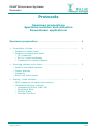
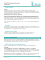
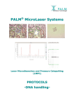
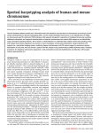
®
PALM®
MicroLaser Systems
Red blood cells
Cut areas from a
histological slide
after catapulting
found in the
collection device
Chromosome metaphase
Selection of one chromosome
PROTOCOLS
-- ffo
nss
on
attiio
arra
pa
ep
prre
np
en
me
ecciim
pe
orr ssp
-- ssp
n
on
eccttiio
olllle
d cco
nd
an
na
on
attiio
olla
n iisso
en
me
ecciim
pe
-- d
nss
on
attiio
plliicca
pp
ap
ma
am
ea
nssttrre
wn
ow
do
PALM® MicroLaser Systems
Protocols
Protocols
Specimen preparation
Specimen isolation and collection
Downstream applications
Specimen preparation..................................................
4
Preparation of slides................................................................
4
• Samples on glass slides
• Samples on membrane slides
o UV treatment
o Poly-L-Lysine treatment
o Treatment to remove RNases
Mounting sections onto slides ...................................................
•
•
•
•
5
Paraffin embedded sections
Frozen sections
Cytospins
Blood and tissue smear
Staining of the sections ...........................................................
6
• DEPC treatment of water and solutions
• Histological staining methods
o Hematoxylin/Eosin (H&E, HE)
o Methylene Blue
o Methyl Green
o Nuclear Fast Red
Protocols_EN_0403.doc
2/14
PALM® MicroLaser Systems
Protocols
Specimen isolation and collection................................
8
Laser cutting (microdissection) of the samples ...........................
8
• Tricks for improvement of the morphology sight of sections
o Ethanol
o Glycerol in PBS
Laser Pressure Catapulting of the samples .................................
9
Looking into the cap to see the catapulted samples ....................
9
Getting the collected cells from the cap into the tip of the tube.....
9
Downstream Applications ............................................ 10
DNA ..........................................................................................
10
Preparation of DNA from catapulted samples..............................
10
RNA ..........................................................................................
11
Some special tips for working with RNA .....................................
11
Preparation of frozen sections ..................................................
11
Preparation of RNA from catapulted samples ..............................
12
o
Proteinase K digestion
Chromosome preparation on request........................................
14
Isolation of living cells on request............................................
14
Protocols_EN_0403.doc
3/14
PALM® MicroLaser Systems
Protocols
Specimen preparation
Preparation of slides
When working with low magnifying objectives, including 40x and 63x long distance objectives,
regular 1 mm thick glass slides can be used. Due to the short working distance of the 100x
magnifying objectives 0.17 mm thin cover glass slides have to be used.
• Samples on glass slides
With the PALM® MicroBeam almost every kind of biological material can be microdissected and
catapulted directly from glass slides. Even archival pathological sections can be used after
removing the cover slip and the mounting medium.
To facilitate easy catapulting additional adhesive substances or „Superfrost + charged slides“
should only be applied when absolutely necessary for the adhesion of special material (e.g.
brain sections).
• Samples on membrane slides (PALM® MembraneSlides)
MembraneSlides are special slides covered with a membrane. The membrane is easily cut
together with the sample and acts like a stabilizing backbone during catapulting. Therefore
even large areas are catapulted by a single laser pulse without affecting the morphological
integrity. The membrane facilitates catapulting of even large areas with one single laser pulse.
This feature is especially important for isolated single cells, chromosomes and also living cells
or small organisms.
P.A.L.M. offers slides with PEN membrane. This polyethylene naphthalate membrane is
1.35 µm thick and is highly absorptive in the UV-A range, which facilitates laser cutting. The
PEN-membrane can be used for all applications.
There are also MembraneSlides available covered with POL (polyester) membrane, which
is 0.9 µm thick and less sensitive to the UV laser, e.g. a higher laser energy is required for
cutting. Thus, it is possible to perform laser ablation of unwanted specimen with moderate
laser energy without immediate cutting the membrane. After "cleaning" of the surrounding a
higher laser energy is required to circumcise and catapult the selected specimen. These slides
are only available on request.
o UV treatment
To overcome the hydrophobic nature of the membrane it is advisable to irradiate with UV light
at 254 nm for 30 minutes. The membrane gets more hydrophilic, therefore the sections
(paraffine and cyrosections) will obtain a better adherence. Positive side effects are
sterilization and destruction of potentially contaminating nucleic acids.
Protocols_EN_0403.doc
4/14
PALM® MicroLaser Systems
Protocols
o Poly-L-Lysine treatment
Additional coating with poly-L-Lysine (0.1 % w/v) will be only necessary for special materials
(e.g. brain sections) and should be performed by distributing a drop of the solution on the
membrane. Let it air dry at room temperature for 30 minutes. Avoid any leakage underneath
the membrane, as this might result in impairment of Laser Pressure Catapulting.
o Treatment to remove RNases
To ensure RNase-free membrane-slides, we dip them for a few seconds into RNase-ZAP
(Ambion, #9780, 250 ml), followed by two separate washings in DEPC-treated water (see page
6) and drying at 37 °C for 30 minutes up to 2 hours. Subsequently the standard UV-treatment
(see above) is performed as usual.
NOTE: Find special tips for work with RNA at page 11 (Applications: RNA).
Mounting sections onto slides
Sections are mounted onto MembraneSlides the same way as routinely done with glass slides.
For cutting and catapulting a coverslip or standard mounting medium must not be applied.
• Paraffin embedded sections
Mount the sections onto the slide as routinely done. Afterwards let dry at 37 °C up to 56 °C
overnight in a drying oven.
Deparaffinization
Paraffin will reduce the efficiency of laser cutting; sometimes it will make it impossible to cut
and catapult. If working with unstained sections it is therefore very important not to forget
removing the paraffin before laser cutting and laser pressure catapulting. If applying staining
procedures deparaffinization is routinely included in any protocols.
Procedure:
Xylene
Ethanol 100 %
Ethanol 96 %
Ethanol 70 %
rinse with water
2
1
1
1
times for 2 minutes
minute
minute
minute
Prolonged Xylene treatment could resolve membrane fixation of POL slides; Xylene treatment
should therefore be kept as short as possible. With the PALM® MembraneSlides (#1440-1000)
it is possible to extend the time of Xylene treatment up to 15 minutes.
Protocols_EN_0403.doc
5/14
PALM® MicroLaser Systems
Protocols
• Frozen sections
Fixation
After mounting the sections there are many possibilities to fix the material. If RNA
preparations are planned with these frozen sections we at PALM recommend ethanol fixation.
This is performed after 20 seconds of air drying on ice by dipping the mounted sections for 1-5
minutes into icecold (-20 °C) 70 % ethanol.
Removing the freeze supporting substance
If OCT or another tissue freezing medium is used it is important to get rid of the media on the
slide before Laser Microdissection, because these media will interfere with laser efficiency.
Removing of the medium is very easily done by gently washing the slide for about 1 minute in
water. If the sections are stained, the supporting substance is removed „automatically“ in the
aqueous staining solutions or the diluted ethanols.
Frozen sections should always be allowed to dry for 5 up to 30 minutes at room temperature
before use.
• Cytospins
Cytospins can be prepared on glass slides or on MembraneSlides. After centrifugation with a
cytocentrifuge let the cells air-dry over night. Then fix for 5 minutes in 100 % methanol. Allow
the cytospins to dry at room temperature before staining.
• Blood and tissue smear
Distribute a drop of (peripheral) blood or material of a swab smear over the slide.
Let smears shortly air-dry and fix them for 2 up to 5 minutes in 70 % ethanol.
Staining of the sections
Using frozen tissue one has to be aware of endogeneous RNases that may still be active after
short fixation steps. Therefore it is recommended to keep all incubation steps of histochemistry
as short as possible. Please use RNase-free water and solutions.
• DEPC treatment of water and solutions
RNases in aqueous liquids can be destroyed with 0.1 % DEPC (diethylpyrocarbonate).
CAUTION!
DEPC is highly toxic and should be handled under a fume hood. Wear gloves!
1 L of bidestilled H2O (or solution) + 1 ml DEPC (e.g. Roth, #K028.1) are stirred for 6-8 hours
at room temperature; then let incubate overnight in a fume hood. The next day residual DEPC
is removed by autoclaving. Store treated solutions at room temperature.
NOTE: All chemical substances containing amino groups (e.g. TRIS, MOPS, EDTA, HEPES, etc.)
cannot be treated directly with DEPC. Prepare these solutions in DEPC-treated H2O.
Protocols_EN_0403.doc
6/14
PALM® MicroLaser Systems
Protocols
• Histological staining methods
o Hematoxylin/Eosin (H&E, HE)
HE-staining is used routinely in most histological laboratories and does not interfere with DNA
and RNA preparation. The nuclei are stained blue, the cytoplasm pink/red.
Procedure:
directly from distilled water
10 minutes Mayer’s hematoxylin solution (e.g. SIGMA, #MHS-32)
10 minutes rinsing in running tap water
5 minutes Eosin Y (e.g. SIGMA, #HT110-2-32)
increasing ethanol series
let air-dry
HE-staining of Cryosections for RNA preparation:
directly from distilled water
3 minutes Mayer’s Hematoxylin solution (e.g. SIGMA, #MHS-32)
3 minutes rinsing in RNase-free tap water or blueing solution
30 seconds up to 3 minutes Eosin Y (e.g. SIGMA, #HT110-2-32)
quick increasing ethanol series
let air-dry at room temperature
o Methylene Blue
The nuclei are stained dark blue. Not recommended for RNA extraction!
Procedure:
directly from distilled water
5-10 minutes Methylene Blue solution (0.05 % in water; SIGMA, #31911-2)
rinsing in Aqua dest
let air-dry at room temperature
o Methyl Green
The nuclei are stained dark green, the cytoplasm light green.
Procedure:
directly from distilled water
5 minutes Methyl Green solution (DAKO, #S1962)
rinsing in Aqua dest
let air-dry at room temperature
o Nuclear Fast Red
The nuclei are stained dark red, the cytoplasm lighter red.
Procedure:
directly from distilled water
5 to 10 minutes Nuclear fast red solution (DAKO, #S1963)
rinse in A. dest
let air-dry at room temperature
Protocols_EN_0403.doc
7/14
PALM® MicroLaser Systems
Protocols
Specimen isolation and collection
Laser cutting (microdissection) of the samples
Please have a look also in the PALM® MicroBeam User Manual
• Tricks for improvement of the morphology sight of sections
o Ethanol
Go forward to search an interesting area on the section and pipet about 5 µl of ethanol onto
this area. You may use 70 % or 100 % ethanol. When using absolute ethanol a little destaining
of the section may happen, but the drying is much quicker.
Observe the area at the screen. The depiction of the cells is improved immediately after having
contact with the ethanol. Now mark the cells or cell area with the software tool. Ethanol is
evaporating rapidly; now catapult the marked cells or cell areas.
o Glycerol in PBS
The same effect is produced by 1 % glycerol in 1x PBS (phospate buffered saline). The time
period until drying of the section is longer than with ethanol. Handling of pipetting, drying,
marking and cutting is the same as shown with ethanol above.
Protocols_EN_0403.doc
8/14
PALM® MicroLaser Systems
Protocols
Laser Pressure Catapulting of the samples
Catapulting into the cap
Please have a look also in the PALM® MicroBeam User Manual
Pipette 3 to 5 µl bidistilled water, buffer or light weight mineral oil (PCR oil) into the inner ring
of the cap. Be aware that aquaeus solutions will dry out after a while. The catapulted cells or
cell areas will stick onto the wet inner surface of the cap and will not fall down after the
catapulting procedure. For single cells or very small areas spinning down from oil may be
difficult and therefore aquaeus solutions should be preferred.
When using membrane mounted samples the dissected membrane acts as a backbone for the
selected area/cell and can therefore be catapulted with a single laser shot from a remaining
„bridge“ at the border. The morphological integrity is completely preserved with this
procedure.
When using glass mounted samples it may be advisory to put more liquid (up to 20 µl) into the
cap since the smaller „flakes“ produced by multiple LPC points cannot be catapulted so straight
to the centre of the cap as areas on membrane.
Looking into the cap to see the catapulted samples
To control the efficiency of catapulting it is possible to look into the collection device (e.g. PCRcap) with the 5x, 10x, 40x and 63x objectives. By using the software function „go to
checkpoint“ the slide is moved out of the light path and the cap can be lowered further towards
the objectives for looking inside. Normally most catapulted areas/cells can be found within the
small inner ring of the caps.
Getting the collected cells from the cap into the tip of the tube
After microdissection, the fluid is shortly spun down in a bench centrifuge (5-10 minutes,
13000 rpm) and samples can then be stored for later use. For RNA extraction first appropriate
lysis buffer is added to the PCR tube and after closure mixed by inversion. Then the lysate is
spun down as above (for future RNA isolation/analysis the tube is placed on ice or stored at
–80 °C in a freezer).
Protocols_EN_0403.doc
9/14
PALM® MicroLaser Systems
Protocols
Downstream Applications
DNA
Preparation of DNA from catapulted samples
For DNA-prep from paraffin sections we usually use a Proteinase K containing catapult buffer.
This step is not necessary for frozen sections.
Catapult Buffer
0.5 M EDTA pH 8.0
1 M Tris pH 8.0
Igepal CA-630 (SIGMA #I-3021)
(Proteinase K 20 mg/ml)
ddH2O
Proteinase K Lyophilizate
(Roche, Mannheim, Germany; Catalog #1413783)
or
(Qiagen GmbH, Hilden, Germany; Catalog #19131)
Proteinase K Solution 20 mg/ml
20 µl
200 µl
50 µl
(100 µl)
fill up to 10 ml
Always prepare a fresh mixture of Catapult Buffer and Proteinase K.
1. Take an autoclaved cap or tube
2. Pipet 3-5 µl of Catapult Buffer, PCR oil or DNase-free water in the middle of the cap
3. Put the cap/tube into the cap/tube holder
4. Perform laser microdissection and laser pressure catapulting of selected cells or cell areas
5. Remove the cap from the cap holder and put it onto a new 0.5 ml microfuge tube (or just
close the used tube with the attached cap)
6. Centrifuge the tube at full speed for 5-10 minutes
7. Add 10-15 µl Catapult Buffer containing Proteinase K onto the cells (which are now in the tip
of the tube)
8. Proteinase K digestion
a) Vortex gently. Digest for 2 - 18 hours at 55 °C(*) followed by a heating step at 90 °C for
10 min to inactivate Proteinase K.
Optional (and recommendable for AutoLPC): Catapult directly into 20 µl Catapult Buffer
containing Proteinase K. Digest upside down in the cap for 2 - 18 hours at 55 °C(*) in an
incubator. After centrifugation inactivate Proteinase K for 10 minutes at 90 °C.
b) At best use a thermal cycler with a heating lid for the standard digestion. If not going on
immediately, store the samples in the fridge at 4 °C.
(*)
The time necessary for complete digestion depends on the kind and on the number of
catapulted cells.
Protocols_EN_0403.doc
10/14
PALM® MicroLaser Systems
Protocols
RNA
Some special tips for working with RNA
Working with RNA is more demanding than working with DNA, because of the chemical
instability of the RNA and the ubiquitous presence of RNAses.
• It makes sense to designate a special area for RNA work only.
• Clean benches with 100 % ethanol or a special cleaning solution e.g. RNaseZap (Ambion,
#9780) or similar.
• Always wear gloves. After putting on gloves, do not touch surfaces or equipment to avoid
reintroduction of RNases to decontaminated material.
• Use sterile, disposable plasticware.
• Use filtered pipetter tips.
• Glassware should be baked at 180 °C for 4 hours. (RNases can maintain activity even after
prolonged boiling or autoclaving!)
• Purchase reagents that are RNAse-free.
• All solutions should be made with DEPC-(diethylpyrocarbonate) treated H2O.
• Treat all used material with DEPC.
• For best results use either fresh samples or samples that have been quickly frozen in liquid
nitrogen or at –80 °C. (This procedure minimizes degradation of RNA by limiting the activity
of endogeneous RNAses.) All required reagents should be kept on ice.
• Store RNA, aliquoted in ethanol or RNA buffer, at –80 °C. Most RNA is relatively stable at
this temperature. Store prepared slides also at –80 °C.
• RNA is not stable at elevated temperatures, therefore avoid high temperatures (>65 °C)
since these affect the integrity of RNA.
DEPC treatment of water and solutions see at page 6.
Preparation of frozen sections
Prepare your sections onto the slide as you do routinely. Let 20 seconds air dry on ice. Fix in
70 % ice cold ethanol for 1-5 minutes, dip in RNase-free water, perform your preferred
staining (e.g. hematoxylin), dip into 70 %, 96 % and 100 % ethanol, respectively.
Subsequently let air dry for 5 - 15 minutes. The slides can now be used at once (even if they
are somewhat wet) or deep frozen at –80 °C.
Procedure
For cryosections we perform catapulting of the cells into a buffer without Proteinase K. If using
paraffin sections for microdissection, please use a buffer containing Proteinase K like
mentioned in the DNA protocol (also see below).
Best use freshly cut and stained specimens.
Protocols_EN_0403.doc
11/14
PALM® MicroLaser Systems
Protocols
Preparation of RNA from catapulted samples
To ensure RNase-free membrane-slides, we at P.A.L.M. dip them for a few seconds into RNaseZAP (Ambion, #9780, 250 ml), followed by two separate washings in DEPC-treated water and
drying at 37 up to 55 °C for 30 minutes up to 2 hours. Afterwards the standard UV-treatment
(see page 4) is performed as usual shortly before use.
To reduce the chance of contamination with exogenous RNases, we use only special reagents
and solutions for RNA isolation, reverse transcription and RT-PCR. All used solutions and tubes
are prepared with DEPC treated water or are purchased as guaranteed RNase free. We also
recommend the use of filter tips.
Best results are obtained using freshly prepared cryosections.
Catapult Buffer:
0.5 M EDTA pH 8.0
1 M Tris pH 8.0
Igepal CA-630 (SIGMA# I-3021)
(optional: Proteinase K 20 mg/ml)
ddH2O DEPC treated, autoclaved
20 µl
200 µl
50 µl
(100 µl)
fill up to 10 ml
Proteinase K solution 20 mg/ml
(only necessary for paraffin sections)
(Qiagen GmbH, Hilden, Germany; Catalog #19131)
Always prepare a fresh mixture of Proteinase K with Catapult Buffer when necessary.
Laser microdissection and pressure catapulting:
1. Take an autoclaved cap/tube
2. Pipet 3-5 µl of Catapult Buffer into the middle of the cap (optional method see below)
3. Put the cap/tube into the cap/tube holder
4. Perform laser microdissection and laser pressure catapulting of selected cells or cell areas
into the cap
NOTE: for frozen sections you can go on directly with #5. For formalin-fixed material a
Proteinase K digestion is necessary at that point (see below)
5. Remove the cap/tube from the cap/tube holder and put it onto a 0.5 ml microfuge tube
containing lysis buffer or respectively pipet lysis buffer into the tube with the attached cap
6. Mix and lyse the cells by inversion
7. Centrifuge the sample at full speed for 5 minutes
If using cryosections you can now go on straight forward with RNA extraction by using kits
like the PALM® RNA-ExtractionKit (#4600-0100, 50 rct.), RNeasyMini (Qiagen, #74104),
Absolutely RNA Nanoprep kit (Stratagene, #400753), High Pure RNA Tissue kit (Roche,
#2033674), Purescript RNA isolation kit (Gentra, Distrib.: Biozym #212010) or your extraction
procedure of choice. The PALM® RNA-ExtractionKit is optimized for work with Laser
Microdissection and Presure Catapulting (LMPC).
If not going on directly, store the samples in lysis buffer at –80 °C.
If using paraffin sections it is necessary to perform a Proteinase K digestion step
before starting the RNA extraction (see below).
Protocols_EN_0403.doc
12/14
PALM® MicroLaser Systems
Protocols
o Proteinase K digestion
a) After centrifugation (#4 from above) pipet 20 µl of Catapult Buffer containing Proteinase
K onto the catapulted cells (which are now in the tip of the tube).
b) Vortex gently. Digest for 2 - 18 hours at 55 °C(*). At best use a thermal cycler with a
heating lid for the standard digestion.
c) Centrifuge the sample at full speed for 5 minutes and then pipet lysis buffer into the
tube. Mix and lyse the cells by inversion.
d) Incubate at 42 °C for 30 min to improve the lysis.
e) Centrifuge the sample at full speed for 5 minutes. Now proceed with the RNA-extraction
(see above)
Optional method (also recommendable for AutoLPC):
a) Instead of step 2 from above, catapult directly into 20 µl of Catapult Buffer containing
Proteinase K.
b) Digest upside down in the cap for 2 - 18 hours at 55 °C(*) in an incubator.
c) Centrifuge the sample at full speed for 5 minutes and then pipet lysis buffer into the
tube. Mix and lyse the cells by inversion.
d) Incubate at 42 °C for 30 min to improve the lysis.
e) Centrifuge the sample at full speed for 5 minutes. Now proceed with the RNA-extraction
using kits or your extraction procedure (see above).
If not going on immediately, store the samples at –80 °C in lysis buffer.
P.A.L.M. Procedure
For best RNA-quality we use frozen sections on PALM® MembraneSlides and perform
catapulting of the cells directly into RNase-free water containing 0.5 % Igepal or into Catapult
Buffer without Proteinase K. After catapulting the samples are mixed with lysis buffer as soon
as possible to protect the RNA.
NOTE: If using paraffin sections for catapulting, please use a buffer containing Proteinase K.
Best use freshly cut specimens. Frozen sections should not be stored for more than a few days
at –80 °C. Freezing should be performed after ethanol fixation or after staining and drying.
It is difficult to estimate the amount of RNA that is to be expected after extraction since many
factors like species, cell/tissue-type, degradation, fixation, staining, fragmentation, extraction
procedure and others will influence the outcome.
From mouse liver frozen sections we can roughly retrieve 5-20 pg of RNA per cell (calculated
from extractions of 1000 cells and analysis on an Agilent Bioanalyzer;
(see also Agilent Application Note 5988-EN on our homepage or at www.chem.agilent.com).
(*)
The time necessary for complete digestion depends on the kind and on the number of
catapulted cells.
Protocols_EN_0403.doc
13/14
PALM® MicroLaser Systems
Protocols
Chromosome preparation
Protocols on request
Isolation of living cells
Protocols on request
Please note: There is also a review brochure available dealing with live cell laser
micromanipulation (PALM Scientific Edition No. 11, ISBN No. 3-9808893-0-0).
For questions, remarks or protocol requests please contact
P.A.L.M.’s Service & ApplicationLaboratory
Email: [email protected]
Service Line: +49 (0) 81 58 99 71-300
Protocols_EN_0403.doc
14/14