1
When many spins add together, as do those of the hydrogen nuclei in a sample of water, the net
magnetization is along B0. A collection of N spins at temperature T will, at equilibrium, have net
magnetization M:
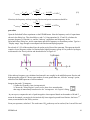
M = N γ ℏ ⟨ m⟩
(3)
where <m>, the average z-component spin quantum number, is given by statistical thermodynamics.
m=+1 /2
∑
⟨ m⟩ =
m=−1/ 2
m=+1 /2
∑
−m γ ℏ B0 /(k B T )
me
−m γ ℏ B 0 /(k B T )
(4)
e
m=−1/ 2
Magnetization is so weak (because both γ and B0 are small) that M simplifies to the following:
2 2
N γ ℏ B0
(5)
M=
2 kB T
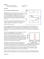
For this experiment, it is worth noting that M is proportional to γ 2 , so proton spectra should be more
intense than fluorine spectra, for the same number of nuclei. In fact, the intensity ratio is γ 3 rather
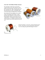
than just γ 2 because detection relies on voltage induced in the B1 coil, and that voltage (called Vout
below) is proportional to γ .
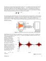
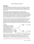
When taking a spectrum, the sample's spin magnetic moment M (which is the net sum of all the
precessing nuclear magnetic moments) is first enhanced with a "polarizing" field Bp created by the
outer magnet. The Bp pulse also tips M from the z axis onto the y axis. Magnetization M is initially
directed along the Earth's magnetic field, which defines the z direction. Polarization field is along the x
axis, which is the longitudinal axis of the instrument. Recall that, by the right-hand rule, z crossed into
x generates torque in the y direction. The polarizing pulse is long enough and strong enough that it both
increases the amplitude of M and reorients M to y.
After the polarizing pulse, M begins to relax in two ways: Mz returns slowly to its equilibrium value
along the z axis, and (Mx,My) begin to "dephase". Dephasing refers to the individual nuclear magnetic
moments, which were
temporarily aligned along M,
spreading out. The time constant
for that spread is T2. The time
constant for re-growth of Mz, the
relaxation known as spin-lattice
relaxation, is T1. Often, T1 is
greater than T2. Component Mz
is not detected by our
instrument, so T1 is not directly
Figure 1: The polarizing field Bp enhances M and tips it 90o.
observed. The component of M
Dephasing then spreads M with time constant T2*.
in the (x,y) plane is detected by
the B1 coil. The sketches in
Figure 1 are like those shown by Engel and Reid in their Section 28.13.2
EFNMR.odt
2