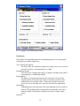
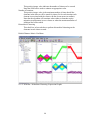
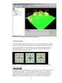
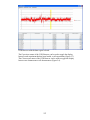
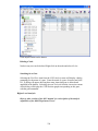
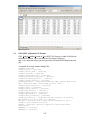
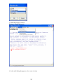
1
MeV
MultiExperiment Viewer
Version 4.0
July 06, 2006
Table of Contents
1. General Information............................................................... 6
2. TM4 Software Overview ........................................................ 7
3. Starting MultiExperiment Viewer ........................................ 8
4. Loading Expression Data ....................................................... 9
4.1.
Loading MeV (.mev) Format Files...................................................................... 9
4.2.
Loading TIGR Array Viewer (.tav) Files .......................................................... 10
4.3.
Loading Tab Delimited, Multiple Sample (.txt) Files (TDMS Format) ............ 11
4.4.
Loading Affymetrix Data (.txt, .TXT) Files ....................................................... 12
4.6.
Loading GenePix (.gpr) Data Files .................................................................. 13
4.7.
Loading Agilent Files........................................................................................ 14
4.13.
Initial View of the Loaded Data, Main Expression Image............................ 20
4.14.
Result Navigation Tree ................................................................................. 21
4.15.
The History Node and Log............................................................................ 21
5. Adjusting the Data................................................................ 23
5.1.
Adjustment / Filter Overview ............................................................................ 23
5.2.
Replicate Analysis............................................................................................. 24
5.3.
Data Transformations....................................................................................... 25
5.4.
Data Filters (Data Quality and Variance Based Filters) ................................. 27
5.5.
Data Source Selection....................................................................................... 32
6. Display Options..................................................................... 34
6.1.
Sample Annotation ............................................................................................ 34
6.2.
Selecting Gene Annotation................................................................................ 36
6.3.
Color Scheme Selection .................................................................................... 36
6.4.
Setting Color Scale Limits ................................................................................ 37
6.5.
Element Appearance ......................................................................................... 39
7. Viewer Descriptions.............................................................. 40
7.1.
Overview ........................................................................................................... 40
7.2.
Expression Images ............................................................................................ 40
7.3.
Expression Graphs............................................................................................ 43
7.4.
Centroid Graphs ............................................................................................... 45
2
7.5.
Table Views....................................................................................................... 47
7.6.
Common Viewer Activities................................................................................ 49
8. Working with Clusters ......................................................... 51
8.1.
Storing Clusters and Using the Cluster Manager ............................................ 51
9. Utilities Menu ........................................................................ 58
10. Creating Output.................................................................... 63
10.1.
Saving the Analysis ....................................................................................... 63
10.2.
Saving the Expression Matrix ....................................................................... 63
10.3.
Saving Viewer Images................................................................................... 64
10.4.
Saving Cluster Data...................................................................................... 64
11. Modules.................................................................................. 65
Description Conventions and General Pointers ........................................................... 65
11.1.
HCL: Hierarchical clustering....................................................................... 67
11.2.
TEASE: Tree-EASE....................................................................................... 72
11.3.
ST: Support Trees ......................................................................................... 78
11.4.
SOTA: Self Organizing Tree Algorithm........................................................ 81
11.5.
RN: Relevance Networks............................................................................... 86
11.6.
KMC: K-Means/K-Medians Clustering ........................................................ 89
11.7.
KMS: K-Means / K-Medians Support ........................................................... 91
11.8.
CAST: Clustering Affinity Search Technique ............................................... 93
11.9.
QTC: QT CLUST .......................................................................................... 95
11.10.
SOM: Self Organizing Maps......................................................................... 97
11.11.
GSH: Gene Shaving .................................................................................... 100
11.12.
FOM: Figures of Merit ............................................................................... 102
11.13.
PTM: Template Matching ........................................................................... 106
11.14.
TTEST: T-tests ............................................................................................ 109
11.15.
SAM: Significance Analysis of Microarrays............................................... 117
11.16.
ANOVA: Analysis of Variance .................................................................... 121
11.17.
TFA: Two-factor ANOVA ........................................................................... 124
11.18.
SVM: Support Vector Machines.................................................................. 126
11.19.
KNNC: K-Nearest-Neighbor Classification ............................................... 133
11.20.
DAM: Discriminant Analysis Module......................................................... 137
11.21.
LEM: Linear Expression Maps................................................................... 142
3
11.22.
GDM: Gene Distance Matrix...................................................................... 161
11.23.
COA: Correspondence Analysis ................................................................. 168
11.24.
PCA: Principal Components Analysis ........................................................ 170
11.25.
TRN: Expression Terrain Maps .................................................................. 172
11.26.
EASE: Expression Analysis Systematic Explorer ....................................... 178
11.27.
FOM: Figure of Merit................................................................................. 192
11.28.
BRIDGE: Bayesian Robust Inference for Differential Gene Expression ... 194
11.29.
USC: Uncorrelated Shrunken Centroids .................................................... 196
12. Scripting .............................................................................. 205
Creating a New Script................................................................................................. 205
The Script Tree Viewer: Script Construction.............................................................. 206
Adding an Algorithm................................................................................................... 207
Script XML Viewer...................................................................................................... 214
Loading a Script.......................................................................................................... 216
Running a Script ......................................................................................................... 216
13. Comparitive Genomic Hybridization Viewer .................. 219
14. Working with the Single Array Viewer ............................ 235
15. Appendix: File Format Descriptions................................. 238
15.1.
TAV Files .................................................................................................... 238
15.2.
Tab Delimited, Multiple Sample Files (TDMS files) .................................. 239
15.3.
GenePix Files.............................................................................................. 240
15.4.
MEV Files ................................................................................................... 241
15.5.
Annotation Files (.ann) ............................................................................... 244
15.6.
Bioconductor (MAS5) Files ........................................................................ 245
15.7.
Affymetrix GCOS (Pivot Data) File............................................................ 246
15.8.
GEO SOFT Affymetrix File Format............................................................ 247
15.9.
GEO SOFT two channel file format............................................................ 248
15.10.
dChip or DFCI core file format .................................................................. 249
15.11.
SAM group or class loading file format...................................................... 250
15.12.
TTEST group of class loading file format................................................... 251
15.13.
ANOVA group or class loading file format................................................. 251
16. Appendix: Preferences Files .............................................. 252
4
17. Appendix: Distance Metrics............................................... 254
18. Appendix: MeV Script DTD .............................................. 257
19. Appendix: R Package Installation..................................... 260
Installing under OS X (Precompiled Binary Version)........... 261
Updating under Windows ........................................................ 269
Installing under OS X............................................................... 269
Installing under Windows........................................................ 270
Running under OS X ................................................................ 270
Running under Windows ......................................................... 270
20. License ................................................................................. 271
21. Contributors........................................................................ 272
References.................................................................................. 274
5
1. General Information
1.1.
Obtaining MeV
http://mev.tm4.org
Maintainer / Contact Information
TIGR MeV Team – [email protected]
Platform / System Requirements
Java Runtime Environment (JRE) 1.4 or later
Java3D v1.3 or later required for PCA, TRN, DAM and COA functions
1.2.
Referencing MeV
Users of this program should cite:
Saeed AI, Sharov V, White J, Li J, Liang W, Bhagabati N, Braisted J, Klapa M,
Currier T, Thiagarajan M, Sturn A, Snuffin M, Rezantsev A, Popov D, Ryltsov A,
Kostukovich E, Borisovsky I, Liu Z, Vinsavich A, Trush V, Quackenbush J.
TM4: a free, open-source system for microarray data management and analysis.
Biotechniques. 2003 Feb;34(2):374-8.
http://www.tigr.org/software/tm4/menu/TM4_Biotechniques_2003.pdf
1.3.
A note on non-Windows operating systems
The majority of our MeV development and testing was performed on Windows
operating systems. Although MeV will run under other operating systems, there
may be some incompatibilities or bugs revealed in this manner. Please report any
such issues to [email protected].
MacOSX users can simulate the ‘right-click’ by using <control> click.
1.4.
For more help
A link to the online copy of this manual can be found in Help Æ MeV Manual in
the main MultipleExperiment Viewer toolbar. For more help beyond the scope of
this manual, or to submit a bug report, see the MeV website, at
http://mev.tm4.org.
6
2. TM4 Software Overview
MultiExperiment Viewer is one member of a suite of microarray data
management and analysis applications originally developed at The Institute for
Genomic Research (TIGR). Within the suite, known as TM4, there are four
programs: MADAM, Spotfinder, MIDAS and MeV. Together, they provide
functions for managing microarray experimental conditions and data, converting
scanned slide images into numerical data, normalizing the data and finally
analyzing that normalized data. These tools are all OSI certified (see section 12)
open-source and are freely available through the TM4 website,
http://www.tm4.org.
The Microarray Data Manager (MADAM) is a data management tool used to
upload, download, and display a plethora of microarray data to and in a database
management system (MySQL). An interface to MySQL, Madam allows scientists
and researchers to electronically record, capture, and administrate annotated gene
expression and experiment data to be shared with and ultimately used by others
within the scientific community.
TIGR Spotfinder is image-processing software created for analysis of the image
files generated in microarray expression studies. TIGR Spotfinder uses a fast and
reproducible algorithm to identify the spots in the array and provide quantification
of expression levels.
Microarray Data Analysis System (MIDAS) is an application that allows the
user to perform normalization and data analysis by applying statistical means and
trim the raw experimental data, and create output for MeV.
MultiExperiment Viewer (MeV) is an application that allows the viewing of
processed microarray slide representations and the identification of genes and
expression patterns of interest. Slides can be viewed one at a time in detail or in
groups for comparison purposes. A variety of normalization algorithms and
clustering analyses allow the user flexibility in creating meaningful views of the
expression data.
7
3. Starting MultiExperiment Viewer
3.1.
If using Windows, run the TMEV batch file (TMEV.bat) to start the program.
This batch file invokes the Java interpreter and stores input parameters. Similarly,
if using Linux or Unix, run the tmev.sh file. Macintosh users should double-click
on the application file named TMEV_MacOSX_4_0.
3.2.
A main menu bar will appear, with four menus: File, Display, Window and
References. Use the File menu in the main menu bar to open a new Single or
Multiple Array Viewer, load a new preferences file or log in to a database. MeV
will continue to run while this menu bar is present. To exit the entire application,
select Quit from the File menu.
3.3.
Expression data can be viewed from within either a Single or a Multiple Array
Viewer. However, the former can open only one set of expression data at a time.
The Multiple Array Viewer can display many samples together. The real power
of MeV is in the program’s analysis modules, found only in the Multiple Array
Viewer. That is where the clustering and visualization of data can take place.
Therefore, the remainder of this manual will focus on the Multiple Array Viewer.
Please see section 13 for details regarding the Single Array Viewer.
8
4. Loading Expression Data
MeV can interpret files of several types, including the MultiExperiment Viewer
format (.mev), the TIGR ArrayViewer format (.tav), the TDMS file format (Tab
Delimited, Multiple Sample format), the Affymetrix file format, and GenePix file
format (.gpr). See section 15 for details regarding the different file formats.
In addition to being formatted correctly, the input data should already be
normalized. Using normalized data as input will result in more statistically valid
output. MIDAS, a member of the TM4 software suite, is one program that can do
this normalization.
The maximum number of samples that can be loaded into a Multiple Array
Viewer at one time depends on the available RAM in the computer running MeV
and the number of expression values from the samples.
4.1. Loading MeV (.mev) Format Files
Select Load File from the File menu to launch the file loading dialog. At the top
of this dialog, use the drop-down menu to select the type of expression files to
load. On the left-hand side of the dialog is a file browser. Use this browser to
locate the files to be loaded.
The default file type to load is the .mev file. This file type is an update of the
older .tav file format. Details about this file format can be found in the appendix
(15.4). In the section of the loader labeled “MeV Expression Files (*.mev),” the
contents of the folder selected in the file browser will be displayed in the box
labeled “available”. Select the .mev files to load and click the “add” button to add
them to the list of files to be loaded. Similarly, select the file(s) containing the
appropriate annotation information in the section labeled “MeV Annotation Files
(*.ann, *.dat).”
The Load Integrated Intensities/Load Median Intensities options specify that
MeV should load either integrated intensities (default) or Median intensities.
Note that care should be taken to select the intensity measurements, integrated or
median, that have been previously normalized in MIDAS so that the loaded data
will have been normalized and possibly trimmed.
The Load Auxiliary Spot Information option will load spot background, spot pixel
count and other spot specific items. Loading this information will not impact the
analysis results but will allow you to view this data when clicking on an
expression element. The default is not to load this spot information since it
consumes significant system memory and can severely limit the number of mev
files that can be loaded due to memory constraints.
The Use Annotation Contained in MeV File option overrides the use of an
annotation file by loading annotation that is contained within the MeV file. This
is a specialized case and for ease of annotation updates it is suggested that you
adhere to using a separate annotation file even if the mev file contains annotation.
9
This option is off by default.
The Remove Annotation Quotes option removes quotations from annotation fields
where the annotation entries start and end with quotation marks. This option is
provided to counteract the behavior of some popular ‘spread sheet’ programs
where cells containing text with a delimiter such as a comma are automatically
enclosed by quotation marks. If MeV stalls on loading an annotation file, try
loading it with this option selected.
4.1.1. The Expression File Loader (MeV Files)
4.2. Loading TIGR Array Viewer (.tav) Files
To load .tav-formatted files, use the drop-down menu to select the TIGR
ArrayViewer (*.tav) option. This loader is very similar to the .mev loader. Use
the file browser to select the .tav files to load. Instead of selecting annotation files
to load alongside the data files, however, you must select a preferences file. This
preferences file contains information that MeV uses to determine what type of
.tav file is being loaded. See the Appendix (section 15.13) for more details on
preferences files (15.13).
10
4.2.1. The Expression File Loader (.tav Files)
4.3. Loading Tab Delimited, Multiple Sample (.txt) Files (TDMS Format)
Select the Tab Delimited, Multiple Sample Files (TDMS) (*.*) option from the
drop-down menu to load TDMS format files. Navigate to the folder containing
the file and select the desired file in this format. The file will be displayed in a
tabular format in the file loader preview table. Click the cell in the table which
contains the upper-leftmost expression value in the file. The header labels for the
annotation fields will be displayed at the bottom of the dialog. Check that the
correct fields are listed before clicking Load.
11
4.3.1. Loading Tab Delimited, Multiple Sample Files (TDMS Format)
4.4. Loading Affymetrix Data (.txt, .TXT) Files
Selecting Affymetrix Data Files (*.txt) from the drop-down menu allows the
loading of Affymetrix files. Select the Affymetrix files to be loaded as you would
when loading .mev files, using the file browser.
These files contain a single intensity value per spot, instead of the usual two that
MeV requires. The values loaded from these files will be use as a Cy5 value, that
is, the numerator in the calculation of the ratio of intensities. Therefore there are
several options for simulating a second intensity value (the denominator). Select
from the radio button options to choose a method. If Absolute is selected, the
denominator is given a value of 1 for all ratio calculations. If Mean Intensity is
selected, the average of all intensity values for that gene across all loaded
Affymetrix files is used as the denominator for that spot. Similarly, if Median
Intensity is selected, the median of all intensity values for that spot is used. If
Reference is selected, a reference Affymetrix file, selected in the file selector at
the bottom of the dialog, is used. The intensity value of each record in the
Affymetrix file is used as the denominator of the ratio calculation for the
corresponding spot in each of the loaded data files.
12
4.4.1. The Expression File Loader (Affymetrix Files)
4.5. Automatically Setting Color Scale Limits
When data are loaded in MeV, it automatically generates expression image.
Expression images convey expression levels by converting the numeric
expression value (log2(A/B) or absolute expression value) as a color that is
extracted from the color gradient. For Affymetrix data and two channels data,
each has different data range in the data set. Now MeV has new function to
automatically setting color scale limits according to different data set. It follows
the rule as it is.
For Affymetrix data the low end is set to 0 and midpoint is set as median and high
end is set to the value so that 80% of the data fall below this value.
For two color array data the low end is set to -3 and midpoint is set to 0 and high
end is set to 3.
Users can change color scale limits after loading as instructed in the following
section.
4.6. Loading GenePix (.gpr) Data Files
GenePix (.gpr) files can be loaded by selecting the GenePix file loader option
from the list of available file formats to load. Use the file system navigation tree
13
on the left to move the directories containing files to load. Files appearing in the
Available file list can be added to the Selected file list using the Add or Add All
buttons.
4.6.1. The Expression File Loader (GenPix Files)
4.7. Loading Agilent Files
Agilent format text files can be loaded by selecting the Agilent file loader option
from the list of available file formats to load. Use the file system navigation tree
on the left to move the directories containing files to load. Files appearing in the
Available file list can be added to the Selected file list using the Add or Add All
buttons. The upper file selection area is for the selection of Agilent Oligo Feature
Extraction text files and lower file selection area is for the text version of the
pattern file that corresponds to your slide.
14
4.7.1. Agilent File Loader
4.8. Loading GEO Simple Omnibus Format in Text (SOFT) Affymatrix format File
After Expression File Loader dialog is launched, SOFT Affymatrix format file
can be loaded by selecting the GEO SOFT Affymatrix file loader option from the
list of available file formats to load.
On the left-hand side of the dialog is a file browser. Use this browser to locate the
files to be loaded. The desired format file can be selected from the list on the top
of right-hand side of the dialog. The file will be displayed in a tabular format in
the file loader preview table. Since SOFT format is relatively flexible. Columns
may appear in any order after the first column (ID column). So users have to
follow the instructions (in red) given under the table to reorder their table by
using mouse to drag the column to the position required. Then click the cell in the
table which contains the upper-leftmost expression value in the file. The platform
format file can be selected from the list on the bottom of right-hand side of the
dialog. Users can choose to load the platform. The default value is not loading.
Then click button Load to load file.
15
4.8.1. GEO SOFT File Loader
4.9. Loading GEO Simple Omnibus Format in Text (SOFT) two channel format File
SOFT two channel format file can be loaded by selecting the GEO SOFT two
channel file loader option from the list of available file formats to load.
On the left-hand side of the dialog is a file browser. Use this browser to locate the
files to be loaded. The desired format file can be selected from the list on the top
of right-hand side of the dialog. The file will be displayed in a tabular format in
the file loader preview table. Since SOFT format is relatively flexible. Columns
may appear in any order after the first column (ID column). So users have to
follow the instruction (in red color) given under the table to reorder their table by
using mouse to drag the column to the position required. Then click the cell in the
table which contains the upper-leftmost expression value in the file. The platform
format file can be selected from the list on the bottom of right-hand side of the
dialog. Users can choose to load the platform. The default value is not loading.
Then click button Load to load file.
16
4.9.1. GEO SOFT two-channel file loader
4.10. Loading Bioconductor(MAS5) Format files
Bioconductor (MAS5) format file can be loaded by selecting the Bioconductor
(MAS5) file loader option from the list of available file formats to load.
On the left-hand side of the dialog is a file browser. Use this browser to locate the
files to be loaded. The desired format file can be selected from the list on the top
of right-hand side of the dialog. The file will be displayed in a tabular format in
the file loader preview table. Click the cell in the table which contains the upperleftmost expression value in the file. The call format file generated by
Bioconductor can be selected from the list on the bottom of right-hand side of the
dialog. Then click button Load to load file.
17
4.10.1. Bioconductor (MAS5) Format file loader
4.11.
Affymatrix GCOS Format files
Affymatrix GCOS (Gene chip operating software) output (pivot data) files can be
loaded by selecting the Affymatrix GCOS file loader option from the list of
available file formats to load.
This file loader can actually load three similar file formats by choosing data
option radio buttons.
Only Intensity-----only containing signal intensity for every experiment
Intensity with Detection -----containing detection for every experiment
Intensity with Detection and p-value-----containing detection and P-value
for every experiment
On the left-hand side of the dialog is a file browser. Use this browser to locate the
files to be loaded. The desired format file can be selected from the list in the
middle of the dialog.
.
The file will be displayed in a tabular format in the file loader preview table.
Choose the data options by clicking proper radio button. Then click the cell in the
table which contains the upper-leftmost expression value in the file. Then click
button Load to load file.
18
4.11.1. Affymatrix GCOS Format file loader
4.12.
Loading dChip Output files
Selecting dChip/DFCI_core Format Files (*.*) from the drop-down menu allows
the loading of dChip or DFCI_core output files. Select the data files to be loaded
as you would when loading .mev files, using the file browser.
These files contain a single intensity value per spot, instead of the usual two that
MeV requires. The values loaded from these files will be use as a Cy5 value, that
is, the numerator in the calculation of the ratio of intensities. Therefore there are
several options for simulating a second intensity value (the denominator). Select
from the radio button options to choose a method. If Absolute is selected, the
denominator is given a value of 1 for all ratio calculations. If Mean Intensity is
selected, the average of all intensity values for that gene across all loaded
Affymetrix files is used as the denominator for that spot. If Reference is selected,
a reference Affymetrix file, selected in the file selector at the bottom of the dialog,
is used. The intensity value of each record in the Affymetrix file is used as the
denominator of the ratio calculation for the corresponding spot in each of the
loaded data files.
19
4.12.1. dChip File Loader
4.13.
Initial View of the Loaded Data, Main Expression Image
For each set of expression values loaded, a column is added to the main display
(4.13.1). This display is an Expression Image viewer, in which each column
represents a single sample and each row represents a gene. The names of the
samples are displayed vertically above each column, and any annotation field of
interest from the input files can be displayed to the right of each row. MeV
expects that each sample loaded will have the same number of elements, in the
same order, and that each gene (spot) is aligned with that element in every other
sample loaded. For example, using that rule, all input files will have data for gene
x in row y. Clicking on a spot displays a dialog with detailed information about
that spot. For more detail regarding the Expression Images viewer, see section
7.2.
20
4.13.1. Main View in MeV. (Borders drawn on image.)
4.14.
Result Navigation Tree
The left side of the main interface is a navigation tree. At any time, clicking on the
Main View node will return to the main display. Output from any MeV module will
be added as a new subtree under the Analysis node. In general, clicking on a node
in the tree will navigate to the associated result or data view and that view will be
displayed in the right panel. The initial tree will also include a Cluster Manager
node to manage clusters stored from analysis results. The Script Manager
manages activities related to analysis script creation, loading, modification, and
execution. The Cluster Manager and the Script Manager will be covered in detail
in is sections 7.2 and 10 respectively.
4.15.
The History Node and Log
The History Node contains a log of most activities. For each log entry a date and
time is recorded. Major events such as file loading, analysis loading, script
loading, algorithm execution, and cluster storage events are logged to the History
Log. If the analysis is stored to a file, then the History is retained and restored so
that new events can be logged. The history log can be stored to a text file by right
clicking in the viewer and selecting the Save History to File menu option.
21
4.15.1. The History Log
22
5. Adjusting the Data
5.1. Adjustment / Filter Overview
Prior to starting an analysis, certain data adjustments can be done using the items
in the Adjust Data menu. These include normalization for genes/rows, log
transformations, and various filters.
5.1.1. Adjust Data Menu
Adjustments may not necessarily affect the main display or the values displayed
when elements are clicked on the matrix displays, but will influence the
calculation of the expression matrix, the foundation of all analyses. Adjustments
will also be reflected when the entire matrix or individual clusters are saved as
text files, although the original data files are not overwritten. Furthermore, with
the exception of three options: “Set Lower Cutoffs”, “Set Percentage Cutoffs” and
“Adjust Intensities of Zero”, all the changes made to an expression matrix are
irreversible for the current MeV session. Different types of adjustments may be
applied on top of one another in any sequence, and the same type of adjustment
may be applied repeatedly to the matrix, although this may not make sense from
the point of view of analysis.
Because of the above features, sometimes it might not be a good idea to apply
data transformations halfway through an analysis (except for perhaps the “Set
Lower Cutoffs”, “Set Percentage Cutoffs” and “Adjust Intensities of Zero”
options), as the post-transformation analyses and displays might not be entirely
consistent with the pre-transformation analyses. A good way to use these options
might be to apply any required adjustments to the data set, save the entire adjusted
matrix as a tab delimited, multiple sample (TDMS) formatted text file (using the
“Save Matrix” option under the “File” Menu), and then load this new file in a new
23
MeV session, during which no further data adjustments will be made. This will
ensure consistency throughout the MeV session. Adjustment options are described
below:
5.2. Replicate Analysis
RAMA: Robust Analysis of MicroArrays
Robust estimation of cDNA microarray intensities with replicates. The package
uses a Bayesian hierarchical model for the robust estimation. Outliers are
modeled explicitly using a t-distribution, and the model also addresses classical
issues such as design effects, normalization, transformation, and nonconstant
variance.
The initialization dialog shown below allows the user to denote the dye labeling
scheme used in the experiment.
RAMA Initialization Dialog
Rserve Connection
RAMA is a package written in the R programming language and requires a
connection to a computer running RServe to function. See Section 19 for details
on installing R and RServe.
By default, Bridge will look on the local machine for an Rserve server. However,
since Rserve is a TCP/IP server, theoretically it could be running anywhere. The
user need only enter an IP address and port number separated by a : in the Text
Field “Enter a new location”. By clicking “Add”, the new location will populate
the pull down menu. It will be saved to the user’s config file and be available for
later use.
24
Rama Results
Sample output from this module is shown below (11.26.2). Rama estimates
intensities based on the data input. The new set of intensities replaces the loaded
dataset and is available for further analysis. A table of the results is also made
available.
RAMA Results: Table of results
5.3. Data Transformations
Log2 Transform
This is fairly self-evident, just taking the log2 transform of every element in the
matrix. Note that this adjustment should not usually be necessary. When .tav or
.mev files are loaded into MeV, the program will automatically compute the log2
ratio of the two intensities and use them in the expression matrix. TDMS files also
often contain pre-calculated log2 ratios.
Normalize Genes/Rows
This will transform values using the mean and the standard deviation of the row
of the matrix to which the value belongs, using the following formula:
Value = [(Value) – Mean(Row)]/[Standard deviation(Row)]
Divide Genes/Rows by RMS
25
This will divide the value by the root mean square of the current row, where
root mean square = square root [∑(xi)2/(n-1)], where xi is the ith element in the
row consisting of n elements.
Divide Genes/Rows by SD
This will divide each value by the standard deviation of the row it belongs to.
Mean Center Genes/Rows
This will replace each value by [value – Mean(row that value belongs to)].
Median Center Genes/Rows
This will replace each value by [value – Median(row that value belongs to)].
Digital Genes/Rows
This will divide up the interval between the minimum and the maximum values in
a row into a number of equal-sized “bins”. Each value is now replaced by an
integer value of zero or greater, denoting which bin it belongs to (e.g., the
minimum value is assigned to bin “zero”, indicating it belongs to the lowest bin;
the maximum value is assigned to the highest bin, and the rest of the values fall in
the intermediate bins).
Sample/Column Adjustmenst
These function in the same way as their corresponding options on genes/rows,
except that the current column values, rather than the current row values, are used
in the computation.
Log10 to Log2
This assumes that the current data are log 10 transformed, and transforms them to
log base 2, i.e., it assumes that the input data is in the form log10x, and it outputs
log2x.
Log2 to Log10
This assumes that the current data are log 2 transformed, and transforms them to
log base 10, i.e., it assumes that the input data is in the form log2x, and it outputs
log10x.
Unlog2 transformation
26
This assumes that the current data are log2 transformed, and removes the log2
transformation.
5.4. Data Filters (Data Quality and Variance Based Filters)
Lower Cutoffs
Select Use Lower Cutoffs to exclude from analysis any genes for which the
expression values are lower than specified values. There are two options in this
menu. One is for two color arrays and one is for single-color arrays.
For two-color arrays: select Low Intensity Cutoff Filter to set either the
corresponding Cy3 or Cy5 columns. To enable this option, check the “Enable
Lower Cutoff Filter” checkbox just below the “Set Lower Cutoffs” menu option,
and uncheck it to disable this option. All subsequent analyses will include only
those genes for which all Cy3 and Cy5 values are above the specified thresholds.
This option is disabled by default.
5.4.1. Lower Cutoff Filter Dialog for two-color arrays
For single-color arrays: select Low Intensity Cutoff Filter to set intensity lower
cutoff. To enable this option, check the “Enable Lower Cutoff Filter” checkbox
just below the “Set Lower Cutoffs” menu option, and uncheck it to disable this
option. All subsequent analyses will include only those genes for which intensity
is above the specified threshold. This option is disabled by default.
27
5.4.2. Lower Cutoff Filter Dialog for One Color Microarray
Percentage Cutoffs
Select Use Percentage Cutoffs to ignore the genes for which there are not enough
valid (non-zero) expression values across all samples. This will not delete any
data, but will only exclude the genes from analysis. This option is sometimes
useful in speeding up module calculation since many zeros will often slow them
down.
To determine which genes will be excluded, select Set Percentage Cutoffs and
enter a percentage value. To enable this option, check the “Use Percentage
Cutoffs” checkbox just below the “Set Percentage Cutoffs” menu option, and
uncheck it to disable this option. Genes with less than the specified percentage of
non-zero values will be ignored. A value of 0.0% indicates that all genes will be
used in the analysis. To require that every one of the gene’s expression values
must be valid to be included, set the value to 100. This option is disabled by
default.
5.4.3. Percentage Cutoff Filter Dialog
Variance Filter
28
The variance filter allows the removal of genes with low variation of expression
over the loaded samples. This filter is basically used to remove ‘flat genes’ that
don’t vary much in expression over the conditions of the experiment. The
variance filter has three possible criteria for specifying which genes to keep.
The Enable Variance Filter check box turns the filter on and off. Be sure to
observe the History Node log to see the number of genes retained after using the
filter. Note that the variance filter is performed after other filters such as Percent
Cutoff Filter is imposed. This convention insures that the genes that are check for
variance also contain some minimum level of ‘good’ (not missing) data.
The Percentage of Highest SD Genes option ranks the genes based on standard
deviation and then the genes that are kept are some percentage of this ranked list.
For and example, if we have 1000 genes and the percentage was set to 20%, then
the result would be a final list of the 200 most variable genes.
The Number of Desired High SD Genes also ranks the genes based on SD and
then the number of genes specified are selected from this SD ordered list such that
the highest SD genes are selected.
The SD Cutoff Value uses an actual SD value such that all genes having an SD
greater than this value are selected.
5.4.4. Variance Filter Dialog
Detection Filter (for Affymetrix Data w/ Detection Calls Only)
Select Use Detection Filter to ignore genes that are not marked ‘present’ in
enough samples. Select Set Detection Filter to divide the samples into two
groups. Enter the number of times that a gene must be called as ‘present’ in
group A. Do the same for group B. Select 'AND' so that each gene must pass
29
both criteria. Select 'OR' so that a gene only must pass one of the criteria in order
to be used in further analysis.
5.4.5. Set Detection Filter Dialog
Fold Filter (for Affymetrix Data Only)
Select Use Fold Filter to remove genes that do not pass one of three specified
criteria based on Fold Change. Fold Change is calculated as: (mean signal in
Group A)/(mean signal in Group B). Select Set Fold Filter and define the two
groups to be filtered. Enter the threshold by which expression of genes in one
group should exceed that of the other group. Select the appropriate ‘greater-than’
or ‘less-than’ symbol to define which group should be more highly expressed than
the other. Select ‘both’ to keep genes in which either group’s expression level
exceeds the other by the defined threshold.
Adjust Intensities of Zero
This option is turned on by default. This means that if either (but not both) of the
cy3 or the cy5 intensities for an element is recorded as zero, that intensity value
will be reset to 1. In this case, the expression ratios will be calculated as cy5/1 or
1/cy3, depending on which value is zero, and the element is included in
subsequent analyses. Sometimes, the user may desire this. However, the user
should be aware that the expression ratios for such elements are spurious. You
might want to turn this option off, if you want to eliminate all those elements from
the analysis that have at least one zero intensity value.
If you deselect this option if either intensity is zero then the log ratio computed
for analysis is set to a “Not-A-Value” flag (NaN) and it is not used during any
analysis and appears as a gray element in the expression image. Note that with
30
either option any elements with two zero intensities have the computed log ratio is
set to the NaN flag since a log ratio cannot be computed.
Bioconductor detection call noise filter
Select Bioconductor detection call noise filter to filter the genes for which the
absent call percentage across all samples is above the level users define in the
dialog. This will not delete any data, but will only exclude the genes from
analysis. Users can check how many genes are filtered from history log.
5.4.6. Set Detection Filter Dialog
31
GenePix Flags Filter
The GenePix Flags Filter allows the removal of genes for which the samples with
negative Flags percentage across all samples is above the level users define in the
dialog. This will not delete any data, but will only exclude the genes from
analysis. This option is sometimes useful in speeding up module calculation since
many zeros will often slow them down.
To determine which genes will be excluded, select GenePix Flags Filter and enter
a percentage value. To enable this option, check the “Enable GenePix Flags
Filter” checkbox just below the “Set GenePix Flags Filter” menu option, and
uncheck it to disable this option. A value of 0.0% indicates that all genes will be
used in the analysis. To require that every one of the gene’s expression values
must be valid to be included, set the value to 100. This option is disabled by
default.
5.4.7. GenePix Flags Filter Dialog
5.5. Data Source Selection
An important aspect of data analysis or data mining is what might be called
analysis branching. This involves the initial selection of a gene or sample set
based on a preliminary screen or analysis technique such as a statistical method,
and then taking this subset of elements and performing more detailed analysis to
find constituent features such as prevalent expression patterns. A right click on a
result viewer node in the result navigation tree will present a menu option (when
applicable) to set the contained data as the primary data set for subsequent
analysis. The selected data source node in the navigation tree will be highlighted
by a green rectangle which indicates that this is the primary data source for
downstream analysis. As an indication of data source change, a node is placed on
the result navigation tree to indicate the source of the data and the number of
genes and samples in the selected data set. Subsequent analysis runs will use only
this data subset until a new data source is selected.
32
5.5.1. Data Source Selection Node (source node is marked with the green
border)
33
6. Display Options
The graphical display of data and analysis results is one of MeV’s strongest
assets. This section of the manual describes options that include selection of gene
and sample annotation to display, adjustment of expression image color schemes,
and adjustment of expression image element size.
6.1. Sample Annotation
Changing Sample Labels
Samples in MeV can be labeled in various ways to indicate natural groupings
based on experimental design. By default, samples are labeled with the file name
or in the case of TDMS format files the names are in the header row. Sample
labels can be selected from the Select Sample Label menu of the Sample/Column
Labels menu of the Display menu. Expression images and expression graphs will
the label samples by this selected annotation field. TDMS format files can
contain additional sample annotation as described in the file format appendix.
Two other options for providing additional sample annotation are to use the
append sample annotation option of the utilities menu or to use the Sample Label
Editor as described below.
Editing Sample Labels and Sample Reordering
The Sample Label Editor is launched from the Edit Labels/Reorder Samples menu
option. One key point is that if samples names are added, merged, or if samples
are reordered, the changes can be captured by selecting the Save Matrix option
from the File menu. This will save the loaded data in to a TDMS file and will
preserve the added sample annotation and sample order, if altered.
6.1.1. Sample Label Editor
The primary function of the Sample Label Editor is to permit the modification of
labels (attributes) associated with the loaded samples. Note that the first row in
the table contains the default sample name. The Default Name cannot be edited
nor can it be deleted. The second major function is to enable the order of loaded
34
samples to be rearranged. Two menus are available within the editor frame for
performing editor actions. The two menus contain the same menu options. One is
in the main menu bar and the other is available using a right mouse click with the
mouse cursor over a table cell. **Note that the import of additional sample
annotation can be done using the Append Sample Annotation option from the
utilities menu.
Sample Reordering
The order of the columns in the table can be altered by clicking on the column
header and dragging the column to the desired location. This action alone does not
force the loaded data to be reordered. The table order can be imposed on the
loaded data if the check box menu item is selected to Enable Sample Reordering.
This ordering is imposed as soon as the dialog is dismissed.
Note: If analyses have been run on the loaded data, the reordering option is
disabled. This is done as a precaution against misrepresentation of prior
results that may have relied on the original ordering. Typically, the
reordering of samples should be done just after data loading to place
associated samples into an order that is reasonable based on study design.
Adding Sample Labels
The Add New Sample Label menu item is used to create a new row in the table.
Note that the first cell in the new row is light yellow and is used to indicate a
Label Key. This key will be placed into the Sample Label menu in the Display
menu so that when selected this label type will be displayed for the loaded
experiments. Once a Label Key is entered, a double click on the remaining cells
will allow editing to add appropriate labels for each sample. Reopening the editor
will allow you to alter any label or label key visible in the table except for the
default primary label and key (shown in gray). Blank entries are allowed.
Merging Sample Labels
Occasionally it is convenient to merge several attributes or labels to produce a
more informative label for each sample. To merge labels start by selecting two or
more rows using ctrl-left click and then selecting the Merge Selected Rows menu
item. A small list will be presented that can be used to order the selected labels
before actual merging. Once attribute ordering is done, a new row is inserted in
the table to display the merged labels and the merged label key.
35
6.1.2. Attribute Sorter for Merging Sample Attributes
6.2. Selecting Gene Annotation
The Gene/Row Label menu option from the Display menu is used to select a gene
annotation field from among the loaded annotation types. Expression image
viewers and other viewers that display gene annotation will be adjusted to display
the selected annotation type.
6.3. Color Scheme Selection
The color scheme selection options pertain to the color gradients that are used to
correlate color to an expression value for an element in expression viewers. Color
provides a means to easily view patterns of expression. Three preset options can
be selected from the Display menu’s Color Scheme menu. The default is a double
gradient, green-black-red, and displays under expression (relative to the
reference) as green, over expression as red, and spots where there is little
differential expression as black. A blue-black-yellow color scheme is available as
an alternative scheme. The rainbow scheme is a third selectable option.
Custom color schemes can be created by selecting the Custom Color Scheme
option from the Color Scheme menu. The form will provide several options:
Gradient Style: Allows the selection of a double gradient or a single
gradient. In some cases a single gradient is preferred if values are not
compared to a reference.
Gradient Selection: This panel selects the endpoint color that is being
selected and allows for the center color on a two gradient scheme to be
black or white.
Color Selector: This area presents controls for selecting endpoint colors.
36
Gradient Preview: This area displays the current settings.
6.3.1. Color Scheme Selection Dialog
6.4. Setting Color Scale Limits
Expression images convey expression levels by converting the numeric
expression value (log2(A/B) or absolute expression value) as a color that is
extracted from the color gradient. By setting numeric limits or endpoints on the
displayed gradient (seen in the preview panel above and at the top of expression
image viewers), expression values within the limits can be displayed as a color
selected from the gradient image. The Set Color Scale Limits option from the
Display menu provides a dialog to set the limits of the color gradient.
37
6.4.1. Color Scale Limits Dialog
The color scale limits dialog, like the color scheme dialog also provides the
choice of using a double or single gradient as appropriate.
The color range panel provides input boxes to set lower and upper limits for the
color gradient and a midpoint when using a double gradient. The labels next to
the input boxes display the minimum, maximum, and median expression values as
a rough guide when setting color scale limits. When the Update Limits button is
pressed the Color Saturation Statistics panel (below) and the gradient preview
panel are updated. The limits are also conveyed to MeV and the current viewer is
updated to reflect the limits. This permits you to adjust and update the limits
while viewing the affect of the new limits on the expression image.
The Color Saturation Statistics panel displays the number of spots that are beyond
the limits of the color scale. These elements would be represented in the
38
expression image as the saturated endpoint colors. This panel also reports this
information as a percentage of elements that are saturated relative to the total
number of elements. These numbers allow you to adjust the limits such that the
expression is spread across the gradient with a limited number of saturated (off
scale) elements.
The reset button of this dialog will reset the limits in the dialog to the original
limits present when the dialog was opened and MeV has it’s limits rolled back to
the original limits.
6.5. Element Appearance
The final two options in the Display menu are options to alter the element size
and to draw or omit borders around each element. Element size can be selected
from among four preset options or a customized size can be selected by entering
an element height and width.
39
7. Viewer Descriptions
7.1. Overview
Viewers are the graphical displays used by MeV to present the results of the
modules’ calculations. The viewers will appear as a subtree under the module’s
Result Tree within the main navigation tree. The viewers listed here are those
which are used by more than one of the MeV modules. Custom viewers, used by
only one module, are described in that module’s section.
7.2. Expression Images
This viewer is used in the main window of the Multiple Experiment Viewer as
well as in most of the modules (7.2.1). It consists of colored rectangles,
representing genes, in a matrix. Each column represents all the genes from a
single experiment, and each row represents the expression of a gene across all
experiments. The default color scheme used to represent expression level is
red/green (red for overexpression, green for underexpression) and can be adjusted
in the Color Scheme dialog in the Display menu.
7.2.1. Expression Images Viewer
Clicking on any of the rectangles in this view will open a window reporting
information for that spot (7.2.2). This window contains a great deal of
information, including expression values and row/column coordinates. Clicking
40
the Gene Graph button will open a window containing a graph of this gene’s
expression level across all samples (7.2.3). To view a Single Array Viewer
displaying the entire experiment of which this spot is a part, click the Sample
Detail button in the Spot Information window (7.2.4). The Set Gene Color button
has not been enabled in this version of MeV.
7.2.2. Spot Information Window
41
7.2.3. Gene Graph Window
42
7.2.4. Sample Details displayed in a Single Array Viewer
7.3. Expression Graphs
This viewer displays graphs of the expression levels of each gene across the
experimental conditions (7.3.1, 7.3.2). By default, the line color is gray or
colored according to the cluster membership. The lines can be set to display a
gradient coloring option in which the lines are colored according expression level.
This option is found in the Display menu under Color Scheme and selecting the
Use Color Gradient on Graphs option will toggle this feature on and off.
The mean expression levels of genes in the cluster are shown as a centroid graph
overlaid on top of the individual expression graphs.
By default, the range of the Y-axis for an expression graph is the same for each
viewer produced from an analysis, regardless of which subset of expression
graphs is displayed. The X-axis (y = 0) is centered on the Y-axis, and the Y
range is set to the distance of the element in the entire data set which is furthest
from zero. Use the Change Y Axis option in the right-click menu to change the Y
range to the maximum distance from zero only for elements within the current
expression graph. This option allows the expression graph to expand to increase
the resolution of expression changes within a particular cluster.
43
7.3.1. Expression Graph of one cluster (with gradient color selected)
44
7.3.2. Expression Graphs of all clusters
7.4. Centroid Graphs
This viewer is very similar to the Expression Graph Viewer, except that line
graphs for individual genes are omitted, leaving only the centroid graph. Error
bars represent the standard deviation of expression within the cluster (7.4.1).
45
7.4.1. Centroid Graph
46
7.5. Table Views
These views show element annotation and, where appropriate, auxiliary
information such as element-specific statistical information for the elements in a
cluster.
7.5.1. Gene cluster table view
Columns can be dragged horizontally across the table to change their relative
ordering. Successive clicks on the header for any column will sort the rows in
ascending or descending order of the entries in that column. Sorting of the “Stored
Color” column will bring together elements that have been stored with the same
cluster color. CTRL-clicking on any column header will sort the table in the
original order of elements in that cluster, an option that is also available from the
right-click menu (below).
Right-clicking on the table view brings up a menu containing the options
available from the right-click menus of other types of viewers, with a few
important modifications and additions as follows. In addition to storing the entire
cluster and launching the entire cluster as a new MeV session, as in the other
types of viewers, users can also select a subset of rows in the table for these
operations. Even individual elements can be stored and tracked one at a time.
Contiguous row selections can be made by dragging the mouse over these rows,
or by selecting the first row of the interval and then SHIFT-clicking on the last
row of the interval (in Windows). Non-contiguous rows may be selected by
CTRL-clicking on the desired rows.
To delete a cluster stored from this viewer (i.e., either all the rows in the table or a
subset of them), select the rows corresponding to the stored cluster and choose the
47
“Delete …” option on the right-click menu. Please note that for successful
deletion, the stored cluster should have been created within the current algorithm
run that the table view belongs to, and the row selection should exactly
correspond to the rows in that cluster.
The “Search” function on the right-click menu pulls up a search dialog, as shown
below:
7.5.2. Table search dialog
Users may search any combination of columns for specific terms. Other options
on this dialog are self-explanatory.
The Link to URL option allows users that are connected to the Internet to launch a
web browser to open a page related to the selected gene. First select a gene of
interest (a row) and then click on the annotation key to use as the gene identifier.
MeV will attempt to construct a URL for the selected annotation key by checking
a URL configuration file. If it is unable to definitively decide what the selected
annotation type is, a dialog will be presented to allow you to select an annotation
field name (column header name) and an external resource to open.
48
7.5.3. Annotation/URL Association Dialog
Within the config directory there is a file (annotation_URLs.txt) that contains
entries to support this feature in mev. This file can be modified with new external
resources as needed, and the existing fields can also be modified, with the
exception of the UniGene field (see below). The file is arranged such that each
row represents a different annotation label:resource pair with the following tab
delimited fields (\t indicates a tab).
<Annotation Label> \t <URL> \t <Resource Name>
The <Annotation Label> indicates the name of the annotation field that is loaded
into mev. This is in the header of the annotation file or TDMS file. For example,
if in some files you call GenBank GB# and in other files it is referred to as
GenBank then two entries in this annotation/url file can be made, one for each
key. If an input annotation file had the GenBank numbers identified as gb# then
the Annotation/URL Association dialog would launch to allow you to manually
indicate the desired external resource.
The <URL> entry is the URL for the resource. Look in the file for examples. It
is important to note that the URL has a section labeled ‘FIELD1’. This is a
placeholder to indicate where the gene identifier should go when constructing the
URL. The UniGene URL is the only exception, requiring two variable fields
called FIELD1 and FIELD2, which are parsed out of the UniGene identifier. The
UniGene Annotation Label is required in the annotation_URLs.txt file to
correctly parse UniGene Ids; therefore, please do not modify this line.
7.6. Common Viewer Activities
A right click within most result viewers will launch a menu that is specific to the
currently displayed viewer. Some of the viewer specific options, such as
searching in Table Viewers have been discussed however there are several
common viewer options that are shared among the common viewer types.
49
Store Cluster
The Store Cluster option will save the currently viewed cluster to a cluster
manager. This feature will be described in detail in section 8. The main use of
this feature is to assign a color to the elements in the cluster so that the location of
the marked elements in future results can be assessed.
Launch New Session
The Launch New Session option takes the elements in the current viewer an opens
a new multiple array viewer with just those elements represented. This is useful
for quickly extracting elements of interest and further characterizing their
expression.
Save Cluster
The Save Cluster option saves the currently viewed cluster to a file. The
expression values and annotation for the current cluster are saved in a format that
can be reloaded as a Tab Delimited, Multiple Sample (TDMS) format file. For
several statistical method this output includes statistics such as F-values or Tvalues, and p-values depending on the particular statistical algorithm applied.
Save All Clusters
This saves all clusters from a clustering result as described above but where the
file name has a cluster index appended to indicate cluster id.
Delete Cluster
This method is used if the Store Cluster method has been used to store the cluster
to the repository. This is a remote method to remove the cluster from the
repository. The cluster table viewer in the cluster manager also has a means to
remove single or multiple clusters.
50
8. Working with Clusters
The analysis modules available in MeV subdivide genes or samples into clusters
by unsupervised techniques, statistical methods, classification algorithms, or
biological relationships. These partitioned sets of elements are then individually
displayed in one of the standard cluster viewers.
8.1. Storing Clusters and Using the Cluster Manager
Clusters of interest can be stored to a repository from the basic cluster viewers by
opening a menu by right clicking in the viewer and selecting the store cluster
option. Once a cluster is stored the Cluster Manager node on the result
navigation tree will contain a list of stored clusters. Gene clusters and sample
clusters are maintained in separate spreadsheets which are viewable from the
Cluster Manager node. When storing a cluster to the repository an input dialog is
presented which allows for three user defined fields to be associated with the
cluster. Two optional text fields are used to capture a cluster name and a
description of the algorithm or interesting features of the cluster. The third user
input is a color used to identify genes or experiments which are members of the
clusters. These colors can be tracked while performing analyses so that clustering
consensus can be established.
8.1.1. Cluster Attributes Dialog
The cluster tables contain the following columns:
-The Serial Number is a unique number which is sequentially assigned to easily
identify a particular cluster.
51
-The Source field describes whether the cluster source was an algorithm, a cluster
operation, or some other means of selecting a group of elements.
-The Algorithm Node field identifies the algorithm used, if the source was an
algorithm, and includes the navigation tree result index (in parentheses).
-The Cluster Node field identifies the specific cluster node under the Algorithm
Node from which the cluster was stored.
-The Cluster Label is an optional user defined name for the cluster.
-The Remarks field can be used to contain details about the process used to create
the cluster or specific features of interest in the cluster.
-The Size field shows the number of elements in the cluster.
-The Color displays the user defined color for the cluster.
-The Show Color check box allows you to show or repress the displayed color.
This option can be useful when visualizing cluster intersections in viewers.
Selecting only one cluster color to view can simplify interpretation.
8.1.2. Cluster Table with option menu open
The spreadsheet allows single or multiple row selection (by holding down the
control key when left clicking the mouse). A right click with one or more rows
selected will display a menu that contains several options detailed below.
-The Modify Attributes option allows the user to modify cluster label, remarks or
the cluster color by displaying the input form with the current settings displayed.
52
-The Open/Launch menu has two options, open cluster viewer will pull up the
source cluster viewer. The second option is launch mev session which opens a
new multiple array viewer containing only the data from the selected cluster or
the union of the members of several clusters if several clusters are selected.
-The Cluster Operations menu allows for three possible operations to be
performed if two or more clusters are selected. The Union operation combines
the members of the selected clusters and stores the resulting cluster on the list.
Elements represented in more than one cluster of the input clusters are only
represented once in the output cluster. The Intersection operation takes the
elements from two or more clusters and produces a cluster containing all elements
which are common to all clusters. The XOR (exclusive OR) operation produces a
cluster containing elements that are members of one cluster or another but not
members of more than one cluster.
-Options also exist to delete selected clusters or all clusters in the list as well as to
save a selected cluster to a specified file.
Deleting clusters can be performed by selecting a single or multiple clusters in the
cluster table or by selecting delete public cluster option from the menu in the
viewer which contains the cluster.
You can also Save Cluster Data to a tab-delimited text file. Selecting this option
from the right-click menu will cause a file chooser to appear. Select a file name
and a place to save row/column data, log ratio expression values, and (optionally)
Cy3 and Cy5 values for each gene in the cluster. Selecting Save All Clusters will
allow you to save the genes in all clusters in a similar way. This option is
available from the cluster table as well as in the viewer.
One additional option is the option to delete all gene clusters or sample clusters.
These global operations which effect all colored clusters is selected from the
Utilities menu in the multiple array viewer by selecting Delete All Gene Clusters
or Delete All Sample Clusters or can be done from the cluster tables.
The Import Gene/Experiment List allows one create a cluster based on supplied
identifiers. Identifiers belonging to the cluster are pasted into the text area. The
drop down list indicates the type of annotation being loaded. An intermediate
dialog (Import Result Dialog) will appear to display the results of the import and
to allow you to select a subset of the identified elements before saving the
elements as a cluster. After review of identified elements a cluster attributes
dialog will be presented so that a cluster name, description, and color can be
defined for the new cluster.
53
8.1.3. List Import Dialog
54
8.1.4. Import Result Dialog
The Submit Gene List (External Repository) option permits gene lists to be
submitted to external database repositories. The data, including the gene
identifier types, possibly organism under study, and other factors can determine if
the current data is suitable for submission to a repository. To submit a cluster,
first select the cluster to submit from the cluster table by left clicking on the
appropriate row. Modify the cluster’s name and description attributes if required
since they may be important for cluster identification within the external
repository. Select the Submit Gene List (External Repository) menu option to
start the submission process. The initial dialog provides a set of panels indexed
by repository name. At the time of the version 3.0 release, only one repository
was available for submission but more can be added in future releases. On each
repository page a general description of the repository is given as well as some
guidelines and requirements that should be met prior to cluster submission.
Please adhere to the requirements of the specific repository. Once the repository
is selected the user will be guided through the submission process unique to the
selected repository. Once a repository is selected hit the submit button to be lead
through the submission process for the selected repository.
55
8.1.5. Repository Selection Dialog
Note that some repositories may require user accounts with passwords to be
established before the submission can be enabled. If a password is required, a
dialog will be provided to enter a user name and password for the repository.
Note that one can enter login information in the repository configuration file to
have MeV remember the user name and password. To enable this one can edit the
archive_submission_config.xml file in the config directory. Open this file in a
text editor and move to the repository entry for the specific repository to add user
information. Alter the user tag from:
<user/>
to:
<user user_name="your_name"
password="password" email="your_email"/>.
A sample of this tag is in the xml file as a guide. Note that some repositories might
not require an email or might in that case the email attribute can be removed from
the tag.
56
8.1.6. Sample Repository Information Page
57
9. Utilities Menu
9.1 Search Utility
The Search feature permits the user to search the data for genes or samples for a
search term given search criteria. Once the search is complete, the elements are
returned in a table. Navigation shortcuts provide a means to open cluster viewers
that contain the elements found in the search.
The search initialization dialog allows the option of finding genes or samples.
The search criteria include a search term, a selection to make the search case
sensitive, and a selection to permit the search term to be an exact match or simply
a contiguous portion of a larger annotation term.
9.1.1. Search Initialization Dialog
The search result is presented in a new window that is split into an upper section
that contains the list of genes or samples identified as matching the search criteria
and a lower section providing shortcut links to cluster viewers that contain the
identified samples or genes.
58
The upper panel displays the list of genes or samples retrieved by the search. The
list of genes or samples found could include some entries that not of interest.
Elements in the list can be deselected using the checkboxes. Clicking on the
Update Shortcuts button will produce a new search result window with just the
previously selected entries and the associated viewer shortcuts. This allows one
to prune unwanted elements out of the search result. The Store Cluster button
will store the selected items as a cluster and assign a user selected color. Note
that this is one method to help identify the elements found during the search
within the cluster viewers.
9.1.2. Search Result Window
9.2. Import Gene or Sample List
The Import Gene/Sample List allows one to create a cluster based on supplied
identifiers. Identifiers belonging to the cluster are pasted into the text area. The
drop down list indicates the type of annotation being loaded. After searching for
matches, the List Import Result dialog will be displayed. This intermediate dialog
will report the number of input identifiers that were matched, the number of
matches found (note that this might be greater than the number of identifiers in
the case where genes are duplicated on the array. This dialog also displays a table
59
that contains matching elements. The rows in the table can be selected to remove
unwanted entries before hitting the Store Cluster button to store the items to a
cluster. The bottom section of the dialog also reports which indices were found
and which were not found in the loaded data set.
9.2.1. List Import Dialog
60
9.2.2. List Import Result Dialog
9.3. Append Sample Annotation
This feature allows the import of additional sample annotations. Often these
would be more descriptive sample names that distinguish the samples based on a
study factor, condition, or some measured variable. The sample annotation file
should be a tab-delimited text file containing one header row for annotation labels
(field names).The file may contain multiple columns of annotation with each
column containing a header entry that indicates the nature of the annotation.The
annotation for each sample is organized in rows corresponding to the order of the
loaded samples. If annotation is missing for a sample the entry in that sample row
may be left blank. Please see the manual appendix on file formats for more
information and a small example.
9.4. Append Gene Annotation
The Append Gene Annotation feature is used to append additional gene
annotation from an MeV style annotation (.ann, or .dat) file. The annotation file
format is described in detail in the File Format Appendix. The main parameter
selection dialog permits the selection of two key fields to be used to map the
61
annotation from the input file to the proper gene already loaded in MeV. One key
should be specified for the currently loaded data. This key should be a gene
identifier of some sort and should correspond to the annotation field selected as
the file’s primary key. The values of these annotation keys is used to map or
correlate gene annotation in the file to the loaded genes. The lowest section of the
parameter dialog specifies the annotation fields from the file to import. Import
status will be reported and an entry that logs the import will be added to the
history node’s history log.
9.4.1. Gene Annotation Import Dialog
62
10. Creating Output
While creating output files is not the main purpose of MeV, several options exist
for saving files from the Multiple Array Viewer for later use. For details
regarding file output with the Single Array Viewer, please see section 14.10.
10.1.
Saving the Analysis
Saving the state of an ongoing analysis to file can be accomplished by selecting
Save Analysis As in the Multiple Array Viewer’s File menu. The analysis file will
contain the loaded data, the current analysis results, and any clusters that are
stored in the cluster manager. If the analysis has been saved previously then the
Save Analysis option will be enabled. If this option is taken then the analysis will
be saved to the analysis file that was opened or the last saved analysis file. To
reopen an analysis, select the Open Analysis menu option in the File menu. This
will restore the data and all algorithm results.
One word of caution on analysis-saving: this feature has been dramatically rewritten since MeV v3.1 to improve reliability and portability. Because of these
changes, MeV 4.0 cannot open saved analysis files created by MeV 3.1 or
MeV4.0b. In order to make saving analyses seamless and efficient, we have had
to sacrifice backwards-compatibility with previous versions of the analysis saving
features of MeV. Future versions of MeV after v4.0 will be able to open analysis
(*.anl) files created by MeV v4.0 and later.
Note that for sharing analysis techniques and results with collaborators, one
option is to use MeV’s scripting ability to create an algorithm execution script so
that others analyzing the same data can share techniques and view interesting
results produced by specific analysis routines. (See Scripting for details. Chapter
10).
10.2.
Saving the Expression Matrix
The expression matrix can be saved as a tab delimited text file by selecting the
Save Matrix item from the File menu. Enter a name for the file in the save file
dialog that is displayed. The matrix saved reflects any data adjustments that are
currently imposed on the data set such as percentage cutoffs or low intensity
cutoffs.
63
10.2.1. Expression matrix saved as text file.
10.3.
Saving Viewer Images
To save a tiff file of the currently displayed image in the main view, select Save
Image from the File menu. To print the image, select Print Image instead.
10.4.
Saving Cluster Data
You can also Save Cluster Data to a tab-delimited text file. Selecting this option
from the right-click menu will cause a file chooser to appear. Select a file name
and a place to save row/column data, log ratio expression values, and (optionally)
Cy3 and Cy5 values for each gene in the cluster (or each sample if the cluster is
produced via clustering samples). Selecting Save All Clusters will allow you to
save the elements in all clusters in a similar way.
64
11. Modules
Description Conventions and General Pointers
Each of these modules can be launched from the Analysis menu or from a button
on the Multiple Array Viewer toolbar. Common to all clustering modules the
selected algorithm can be performed to cluster genes or samples. In the title of
the analysis module descriptions which follow, the acronym used to label the
module’s button is followed by the name of that module. Below this is a line
containing the reference(s) used in implementing the algorithm.
Each module, when run, will create a subtree labeled with its acronym and a label
indicating whether the result was created by clustering genes or samples. This
subtree will be placed under the Analysis tab in the result navigation tree tree.
The tabs within this subtree contain the results of the module’s calculations.
These tabs vary greatly depending on the module which creates them, but the
General Information tab always contains a summary of the parameters used in the
analysis.
Each algorithm run will present a dialog or form to use to input parameters
specific to the algorithm being performed. An information button on each dialog
(lower left corner) can be used to retrieve a reference page describing the required
parameters.
Customizing the module menubar
Under the Multiple Array Viewer window’s File menu is the option 'Customize
Toolbar'. Choosing this option will generate a dialog for users to select the
modules to be displayed in the Multiple Array Viewer. After data has been
loaded into MeV, this function will be disabled for safety reason. All modules
will still be listed under menu Analysis grouped by submenus.
65
66
11.1.
HCL: Hierarchical clustering
(Eisen et al. 1998)
Selecting this analysis will display a dialog that allows three different linkages
and options to cluster genes, samples or both. Once the computations are
complete, select the Tree node under Analysis to view the hierarchical tree. The
display is similar to the main display, but similar genes and experiments are
connected by a series of ‘branches’. Labels are displayed on the right side.
Clicking under a branch intersection (node) will select that node and the subtree
below that node. Once selected, right clicking in the same area will display a
popup menu that allows the user to set the highlighted area as a cluster, name the
cluster, save the cluster and set several tree options. Clusters set and named in this
display can propagate to other displays. Saving a cluster will display a dialog
where a tab delimited text file containing the data for the highlighted cluster can
be named. The algorithm also produces a Node Height Graph which displays the
number of terminal nodes in the tree given a particular inter-node distance
threshold.
11.1.1. Hierarchical tree with clusters selected.
67
11.1.2. HCL Initialization Dialog
Parameters:
Linkage Method
This parameter is used to indicate the convention used for determining cluster-tocluster distances when constructing the hierarchical tree.
Single Linkage: The distances are measured between each member of one
cluster each member of the other cluster. The minimum of these distances
is considered the cluster-to-cluster distance.
Average Linkage: The average distance of each member of one cluster to
each member of the other cluster is used as a measure of cluster-to-cluster
distance. Note that this option in MeV actually is determined by a
weighted average of distances of cluster members. Example: Consider the
distance from node ‘d’ to cluster (a,b,c)…
a
b
c
d
Unweighted Average Linkage:
d d ,( a ,b,c ) =
(d
d ,a
+ d d ,b + d d ,c ) d d ,a d d ,b d d ,c
=
+
+
3
3
3
3
68
Weighted Average Linkage:
d d ,( a ,b,c ) =
[(d
d ,a
+ d d ,b ) / 2 + d d ,c ] d d ,a d d ,b d d ,c
=
+
+
2
4
4
2
Nodes on are weighted unequally where nodes deeper in the sub-tree
contribute less to the overall computed distance.
Complete Linkage: The distances are measured between each member of
one cluster each member of the other cluster. The maximum of these
distances is considered the cluster-to-cluster distance.
Cluster Genes / Cluster Samples Options
These checkboxes are used to indicate whether to cluster genes, samples,
or both.
Default Distance Metric: Euclidean
11.1.3. Node Height Graph
Adjusting the Tree Configuration and Viewing Clusters
A right click in the Tree Viewer will produce a menu which includes an option to
69
alter the displayed tree, Tree Properties. This option permits the user to change
the tree’s appearance and to reduce the complexity of the tree by imposing a
distance threshold. Elements on nodes which have distances below this threshold
can be considered as one entity (or cluster). Consequently the lower level detail of
the tree is ignored. As the value is adjusted the corresponding HCL tree will have
nodes below this threshold appear as light gray in color and a translucent 'wedge'
from that node to all enclosed elements will be draw on the tree. This
representation of the tree will persist unless the dialog is dismissed by hitting
cancel. The distance threshold can be entered into a text field or can be adjusted
with a slider over the maximum range of inter-node distances. The number of
terminal nodes (clusters) using the current distance threshold is displayed in the
upper right quadrant of the dialog.
11.1.4. HCL Tree Configuration Dialog
The Create Cluster Viewers option allows you to create viewers based on the
distance threshold. This option collects groups of elements falling below terminal
nodes in the tree using the current distance threshold. The clusters of elements are
represented as nodes in the result navigation tree under the HCL result node. The
results are added once the HCL Tree Configuration dialog has been dismissed.
The minimum and maximum pixel distance imposes limits on the minimum and
maximum displayed inter-node distance. This alters the appearance of the tree.
The Apply Dimensions button causes the entered tree dimensions to be applied to
the HCL tree. This allows one to fine tune the tree's appearance without
dismissing the dialog.
70
11.1.5. HCL Tree with distance thresholds applied.
71
11.2.
TEASE: Tree-EASE
Selecting this analysis will display a configuration window with three main
modes and four tabbed panels; each contains essential configurations that are
specific for Hierarchical Clustering (HCL) or EASE analysis. Once the analysis is
completed, select the tree node under “Analysis Results” to view the hierarchical
tree with color dots signified whether the cluster contains over-expressed
biological categories (Red: most significant, Blue: least significant). To view
EASE analysis information of each cluster, position the mouse over the color dot
at the root of the cluster to display a popup window. The first column in the
window is the name of annotation file which contains the category. The second to
the third to the last column are information about the category that is included in
the annotation file. If the default mode “Cluster Analysis” is selected, the last
column is the “score” the category receives, that is the probability the certain
category is decided over-expressed by chance. If “Annotation Survey” mode is
selected, the last two columns would be the hit count and cluster size,
respectively.
TEASE hierarchical tree with color coded visualization dots.
Mode selection:
HCL Only
Perform Hierarchical Clustering (HCL). No biological theme exploration.
Cluster Analysis
72
EASE analysis on clusters that fall within the minimum and maximum size of
population as specified in the panel “HCL Clustering” – “Cluster size
specification”. Calculate the score of each category and rank the categories by
score.
Annotation Survey
EASE analysis on clusters that fall within the minimum and maximum size
parameters specified by the user. Calculate and rank present categories in each
cluster by hit counts. No score is calculated in this mode.
Parameters:
TEASE parameter setting window
HCL Clustering
Linkage Method
This parameter is used to indicate the convention used for determining cluster-tocluster distances when constructing the hierarchical tree.
Single Linkage: The distances are measured between each member of one
cluster each member of the other cluster. The minimum of these distances
is considered the cluster-to-cluster distance.
73
Average Linkage: The average distance of each member of one cluster to
each member of the other cluster is used as a measure of cluster-to-cluster
distance. Note that this option in MeV actually is determined by a
weighted average of distances of cluster members. Example: Consider the
distance from node ‘d’ to cluster (a,b,c)…
Unweighted Average Linkage:
Weighted Average Linkage:
Nodes on are weighted unequally where nodes deeper in the sub-tree
contribute less to the overall computed distance.
Complete Linkage: The distances are measured between each member of
one cluster each member of the other cluster. The maximum of these
distances is considered the cluster-to-cluster distance.
Cluster Genes / Cluster Samples Options
These checkboxes are used to indicate whether to cluster genes, samples, or both.
Default Distance Metric: Euclidean
Cluster Size Specification
State of the size of clusters you want to analyze. You won’t get too much
information for clusters that are too big or too small. The default size is from 10
to 100.
Population/Color
74
Population selection
Available only when in “Cluster Analysis” mode. Please refer to EASE
documentation for more information.
Assign Color Gradient
Specify the upper and lower score for assigning color gradient. Increasing upper
and lower bounds will cause the gradient shifts to red whereas decreasing will
shift it to blue. Gradient can also be adjusted in the graphic view window after
analysis is completed and the correct tree code is selected. The default setting for
upper bound is 0.1, and 0.00001 for lower bound.
Annotation Parameter
Selections of annotation key, annotation conversion file, and gene ontology/gene
annotation/gene linking files. Refer to EASE documentation for more
information.
Statistical Parameter
Selections of reported statistics, multiplicity correction and time parameters.
Refer to EASE documentation for more information.
Navigating the hierarchical tree
75
TEASE Tree View
Basic Navigation
A large dataset is likely to have more than a handful clusters that fall within the
size range, but only clusters that are “more red” worth attention. It is thus
important to assign appropriate color gradient boundaries to save time. Adjusting
color gradient will be in a later section. To view information about each cluster,
simple position the cursor over the root of the cluster to reveal a pop-up window.
When done, move the cursor away and the window will disappear.
Adjusting Tree Configuration and Viewing Clusters
A right click in the Tree Viewer will produce a menu which includes an option to
alter the displayed tree, Tree Properties. This option permits the user to change
the tree’s appearance and to reduce the complexity of the tree by imposing a
distance threshold. Elements on nodes which have distances below this threshold
can be considered as one entity (or cluster). Consequently the lower level detail of
the tree is ignored. As the value is adjusted the corresponding TEASE tree will
have nodes below this threshold appear as light gray in color and a translucent
'wedge' from that node to all enclosed elements will be draw on the tree. This
representation of the tree will persist unless the dialog is dismissed by hitting
cancel. The distance threshold can be entered into a text field or can be adjusted
with a slider over the maximum range of inter-node distances. The number of
terminal nodes (clusters) using the current distance threshold is displayed in the
upper right quadrant of the dialog.
HCL Tree Configuration Dialog
The Create Cluster Viewers option allows you to create viewers based on the
distance threshold. This option collects groups of elements falling below terminal
nodes in the tree using the current distance threshold. The clusters of elements are
represented as nodes in the result navigation tree under the TEASE result node.
The results are added once the TEASE Tree Configuration dialog has been
dismissed.
76
The minimum and maximum pixel distance imposes limits on the minimum and
maximum displayed inter-node distance. This alters the appearance of the tree.
The Apply Dimensions button causes the entered tree dimensions to be applied to
the TEASE tree. This allows one to fine tune the tree's appearance without
dismissing the dialog.
TEASE tree (HCL mode) with distance thresholds applied
Adjusting Color Gradient
A right click on the TEASE Tree View will reveal a pop-up menu. Near the
bottom of the menu is an option that says “Change Score Boundary”. Left click
on the option will produce a configuration window. In the window are two
editable text fields similar to the “Assign Color Gradient” panel in the initialize
window. Enter appropriate number and click on “OK” to apply the changes or
“Cancel” to exit the window without any changes. You can view the changes you
make in Tree View once you exit the window.
77
11.3.
ST: Support Trees
This option shows the hierarchical trees obtained using the previous module, but
it also shows the statistical support for the nodes of the trees, based on resampling
the data. The user can select two resampling methods: bootstrapping (resampling
with replacement), and jackknifing (resampling by leaving out one observation in
this implementation). Resampling can be conducted on genes and / or experiments
for a user-specified number of iterations. The branches of the resulting tree are
color-coded to denote the percentage of times a given node was supported over
the resampling trials. The legend for the color code corresponding to a given level
of support can be found under the Help menu.
The two most useful options for support trees are likely to be bootstrapping genes
to build experiment trees, and bootstrapping experiments to build gene trees.
11.3.1. Support trees for Hierarchical Clustering with Support Tree Legend
displayed on right.
78
11.3.2. Support Trees Initialization Dialog
Parameters:
The Support Tree algorithm permits the resampling options to be set separately
for the gene tree and the sample tree.
Tree Construction Options
The Draw Gene Tree and Draw Sample Tree options allow you to select to
construct a gene tree, an experiment tree, or both.
Resampling Options
You can elect to resample either genes or samples or neither using either a
bootstrapping or a jackknifing method.
Bootstrapping
The matrix is reconstructed such that each expression vector has the
original number of values but the values are a random selection (with
replacement) of the original values. Values in the original expression
vector may occur more than once since the selection uses replacement.
Jackknifing
Jackknifing takes each expression vector and randomly selects to omit an
element. This method produces expression vectors that have one fewer
element and this is often done to minimize the effect of single outlier
values.
Iterations
79
This indicates how many times the expression matrix should be
reconstructed and clustered.
Linkage Method
This parameter is used to indicate the convention used for determining
cluster-to-cluster distances when constructing the hierarchical tree.
Single Linkage: The distances are measured between each member of one
cluster each member of the other cluster. The minimum of these distances
is considered the cluster-to-cluster distance.
Average Linkage: The average distance of each member of one cluster to
each member of the other cluster is used as a measure of cluster-to-cluster
distance.
Complete Linkage: The distances are measured between each member of
one cluster each member of the other cluster. The maximum of these
distances is considered the cluster-to-cluster distance.
Support Tree Legend
A legend to relate support tree output colors to % support is displayed by
selecting the support tree legend menu item in the main help menu.
Default Distance Metric: Euclidean
80
11.4.
SOTA: Self Organizing Tree Algorithm
(Dopazo et al. 1997, Herrero et al. 2001)
The initialization form shown below (11.4.1) is divided into four main areas. The
SOTA algorithm constructs a binary tree (dendrogram) in which the terminal
nodes are the resulting clusters.
11.4.1. SOTA Initialization Dialog Box.
Parameters and Basic Terminology:
SOTA Terminology and Concepts
Topology
The topology of the resulting tree is a binary tree structure where
each terminal node represents a cluster.
Node
A structure which contains a Centroid Vector and a number of
associated expression profiles (members).
81
Cell
A Node which is the terminal Node in a branch of the tree (a.k.a.
leaf node). The members of the cell are considered members of an
expression cluster.
Centroid Vector
A vector that is representative of the membership of a node.
Members
Expression Elements associated with a Node.
Node
A structure which contains a Centroid Vector and a number of
associated expression profiles (members).
Cell
A Node which is the terminal Node in a branch of the tree (a.k.a.
leaf node). The members of the cell are considered members of an
expression cluster.
Growth Termination Criteria Parameters
Max Cycles
This integer value represents the maximum iterations allowed. The
resulting number of clusters produced by SOTA is (Max Cycles
+1) unless other criteria are satisfied prior the indicated maximum
number of cycles.
Max epochs/cycle
This integer value indicates the maximum number of training
epochs allowed per cycle.
Max. Cell Diversity
This value represents a maximum variability allowed within a
cluster. All resulting clusters will fall below this level of 'diversity'
(mean gene to cluster centroid distance) if diversity is used as the
cell division criteria. (Unless Max cycles are reached at which time
some clusters may still exceed this parameter)
Min Epoch Error Improvement
This value is used as a threshold for signaling the start of a new
cycle and a cell division. The tree diversity is monitored during a
training epoch and when the diversity fails to improve by more
than this value then training has been considered to have stabilized
and a new cycle begins.
Run Maximum Number of Cycles (unrestricted growth)
The algorithm will run until Max Cycles or until all of the input set
are fully partitioned such that each cluster has one gene or several
identical gene vectors.
Centroid Migration and Neighborhood Parameters
Migration Weights
These values are used to scale the movement of cluster centroids
(characteristic gene expression patterns) toward a gene vector
which has been associated with a neighborhood. When a gene is
associated with a cluster the centroid adapts to become more like
the newly associated gene vector. The parent and sister cell
82
migration weights should be smaller than the weight for the
winning cell (Cell to which the gene vector is associated.).
Neighborhood Level
This value determines which cells are candidates to accept new
expression elements. When elements are considered for
redistribution to new node during a cell division candidate cells are
determined by moving up the tree toward the root this number of
levels. From that node, all cells (terminal nodes) within this subtree
are targets for possibly accepting expression vectors. (Each vector
moves into the cell to which it is most similar).
Cell Division Criteria Parameters
Use Cell Diversity
Cell diversity is the mean distance between the cell's members
(expression profiles) to the cell's centroid vector. When
considering which cell to divide, the cell with the greatest diversity
is split. (providing it's diversity exceeds Max Cell Diversity (see
above))
Use Cell Variability
Cell variability is the maximum element-to-element distance
within a cell. The cell having the largest internal gene-to-gene
distance is selected as the next cell to divide. In this case the
stopping criteria is changed so that growth continues until the most
variable cell falls below a variability criteria generated using the
provided pValue (see below)
pValue
This value is used when using variability as the cell division
criteria. A distribution of all element to element distances is
generated by resampling the data set with each expression vector
having randomized ordering of vector elements. The resulting
distribution represents random gene to gene distances. The pValue
supplied is applied to this resampled distribution to generate a
variability cutoff. Clusters falling below this variability cutoff have
a probability of having members that are paired by chance at or
below the supplied pValue.
Hierarchical Clustering
This check box selects whether to perform hierarchical clustering on the
elements in each cluster created.
Default Distance Metric: Euclidean
The result views created by the SOTA algorithm include the basic viewers with
the addition of two SOTA specific viewers and enhancements to expression
image viewers to include more cluster information.
One of the SOTA specific viewers is the SOTA dendrogram (below) which
displays the generated tree with the expression image of each resulting cluster’s
centroid gene. The text to the right of the centroid expression image includes a
83
cluster id number, the cluster population (number of genes in the cluster), and the
cluster diversity (mean gene to centroid distance). Clusters can be colored and
saved from this viewer and a left click over a cluster centroid jumps to the
expression image for that cluster.
11.4.2. SOTA dendrogram
The SOTA diversity viewer shows the change in the summation of gene to
associated centroid distance vectors for all genes in the tree. This is a measure of
overall tree diversity. This can reveal how much diversity improvement is
achieved with each cycle (new cluster addition).
84
11.4.3. SOTA diversity viewer
85
11.5.
RN: Relevance Networks
(Butte et al. 2000)
A relevance network is a group of genes whose expression profiles are highly
predictive of one another. Each pair of genes related by a correlation coefficient
larger than a minimum threshold and smaller than a maximum threshold (assigned
in the initialization dialog box) is connected by a line. Groups of genes connected
to one another are referred to as networks.
11.5.1. RN Initialization Dialog
Parameters:
Sample Selection
The sample selection option indicates whether to cluster genes or samples.
Use Permutation Test
This check box is used to indicate that the minimum threshold R2 value
should be selected based on a distribution constructed from element to
element R2values derived following permutation of the expression vectors.
Min Threshold
This value ranging from 0 to 1.0 indicates the smallest R2possible between
two elements to permit a link between the elements in a subnet.
Max Threshold
This value ranging from 0 to 1.0 indicates the greatest R2possible between
two elements to permit a link between the elements in a subnet.
Use Filter
This option allows the user to filter out elements with little dynamic
change thus removing flat or uninteresting elements. A measure of entropy
is used to rank the elements. The percentage value entered (1 to 100)
indicates what percentage of the elements to retain for the construction of
the network. A value of 25 will retain the 25% of elements having the
greatest entropy.
Distance Metric:
Other Acceptable Metrics:
Pearson squared
None. Changing the metric in the Distance menu
86
will not affect the calculations done by this module.
The module calculates the correlation coefficient between genes by comparing the
expression pattern of each gene to that of every other gene. The ability of each
gene to predict the expression of each other gene is measured as a correlation
coefficient. Genes are represented as nodes in a network and edges are drawn
between them if their correlation coefficient falls between the minimum and
maximum thresholds specified in the initialization dialog.
The experiment subtree created by this module contains information regarding the
networks predicted. Under the Network tab is a graph of all of the subnets
generated (fig. 10.5.1). A subnet is a group of genes in which each gene is
connected to at least one other gene. The Relevance Subnets tab contains network
diagrams for each of the individual subnets, and the Expression Images folder
contains expression views for the genes in each of them.
11.5.2. Network View.
Several options can be launched from the network viewer to enhance the view or
to better characterize the results by using a right click context menu. Note that
links colored in red represent elements that are positively correlated while links
colored in blue represent elements that are negatively correlated. Options from
the menu to enhance the view include the ability to zoom in and out on the
subnets, alter the color of the background, alter the element shape, and alter the
thickness of the links for better visibility.
Other options reveal the nature of the subnets. You can select to alter the
threshold to make it more stringent. Using this option under the links-threshold
87
the viewer is instructed to only show links for elements correlated with R2 greater
that the new threshold. Selection of elements in the network can be done by using
the select option in the right click menu. Two options are offered for selecting
elements, one by providing a gene identifier label (Element ID option), and the
second option by specifying all elements with a specified minimum number of
links (Feature Degree option). As in other viewers selected elements may be
assigned a color (Set Selection option).
88
11.6.
KMC: K-Means/K-Medians Clustering
(Soukas et al. 2000)
Selecting this analysis will display a dialog that allows the user to specify whether
to use means or medians, as well as the number of clusters and iterations to run.
Once the computations are complete, select the KMC node under Analysis to
view the results. There are several sub-nodes beneath KMC, further divided by
the clusters created based on the KMC input parameters. Hierarchical trees shows
trees constructed for each cluster, if the option to draw hierarchical trees for
clusters is selected. Expression images are similar to the main display. Cluster
Information is a summary of each cluster based on size and % composition.
Centroid graphs show the centroids for each cluster and experiment, individually
or all at once. Expression graphs are similar to centroid graphs, but with each
gene’s expression levels displayed alongside the centroids. Right clicking within
an expression image displays a popup menu that allows the user to propagate the
cluster to other displays (Set Public Cluster), save and delete the cluster.
This method of clustering is useful when the user has an a priori hypothesis about
the number of clusters that the genes should subdivide into.
11.6.1. KMC Initialization Dialog
Parameters:
Sample Selection
The sample selection option indicates whether to cluster genes or samples.
Means/Medians option
The Means or Medians option indicates whether each cluster's centroid
vector should be calculated a mean or a median of the member expression
patterns.
Number of Clusters
89
This positive integer value indicates the number of clusters to be created.
Note that FOM can be used to estimate an appropriate value.
Number of Iterations
This positive integer value is the maximum number of times that all the
elements in the data set will be tested for cluster fit. On each iteration each
element is associated with the cluster with the closest mean (or median).
Note that the algorithm will terminate when either no elements require
migration (reassignment) to new clusters or when the maximum number of
iterations has been reached.
Hierarchical Clustering
This check box selects whether to perform hierarchical clustering on the
elements in each cluster created.
Default Distance Metric: Euclidean
11.6.2. K-Means / K-Medians Clustering: Expression Graphs.
90
11.7.
KMS: K-Means / K-Medians Support
This module allows the user to run the K-Means or K-Medians algorithms
multiple times using the same parameters in each run. Owing to the random
initialization of K-Means and K-Medians, the clusters produced may vary
substantially between runs, depending on the data set and the input parameters.
The KMS module allows the user to generate clusters of genes that frequently
group together in the same clusters (“consensus clusters”) across multiple runs.
The output consists of consensus clusters in which all the member genes clustered
together in at least x% of the K-Means / Medians runs, where x is the threshold
percentage input by the user (see screenshot below).
Parameters:
Sample Selection
The sample selection option indicates whether to cluster genes or samples.
Means/Medians option
The Means or Medians option indicates whether each cluster's centroid
vector should be calculated a mean or a median of the member expression
patterns.
Number of k-means/k-medians runs
This integer value indicates how many times KMC should be run.
Threshold % of occurrence in same cluster
This parameter indicates the minimum percentage of times that two
elements should cluster together in order consider the two elements in a
cluster. For instance, if 10 KMC runs were run, and the percentage was
80% then a pair of expression elements found together at least 8 times
would be considered to pass a criteria to be included in a cluster.
Number of Clusters (K)
This positive integer value indicates the number of clusters to be created
during each KMC run. Note that for K-Means support the final number
may turn out to be slightly smaller or larger than this entered value
depending on the nature of the input data and the appropriate selection of
K (number of clusters to create). Note that FOM can be used to estimate
an appropriate value for K.
Number of Iterations
This positive integer value is the maximum number of times that all the
elements in the data set will be tested for cluster fit. On each iteration each
element is associated with the cluster with the closest mean (or median).
Note that a KMC run will terminate when either no elements require
migration (reassignment) to new clusters or when the maximum number of
iterations has been reached.
Hierarchical Clustering
This check box selects whether to perform hierarchical clustering on the
elements in each cluster created.
Default Distance Metric: Euclidean
91
11.7.1. K-Means / K-Medians Support: Initialization Dialog Box.
The number of consensus clusters generated may be more than the input number
of clusters per run. This is because some genes may cluster together frequently,
yet they may form a subset of different clusters in different runs. Hence, a set of
genes that appeared as a single cluster in any given run may be split up into two
or more consensus clusters over several runs. Some genes may remain unassigned
because they did not cluster with any other genes in enough runs to exceed the
threshold percentage.
92
11.8.
CAST: Clustering Affinity Search Technique
(Ben-Dor et al. 1999)
The user is prompted for a threshold affinity value between 0 and 1 (which may
be thought of as the reciprocal of the distance metric between two genes, scaled
between 0 and 1), that has to be exceeded by all genes within a cluster. The
algorithm works by both adding and removing genes from a cluster, each time
adjusting the affinities of the genes to the current cluster, and continuing this
process until no further changes can be made to the current cluster.
11.8.1. CAST Initialization Dialog
Parameters:
Sample Selection
The sample selection option indicates whether to cluster genes or samples.
Threshold
The threshold parameter is a value ranging from 0.0 to 1.0 which is used
as a cluster affinity threshold. Each expression element will have an
affinity for the current cluster being created based on it's relationship to
the elements currently in the cluster. If that affinity is greater than the
supplied threshold the gene is permitted to be a member of the cluster.
Note that thresholds near 1.0 are more stringent and tend to produce many
clusters with rather low variability. Conversely, using a lower threshold
will produce fewer, more variable clusters. A balance by trail-and-error
should be found between these extremes.
Note that in the algorithm expression elements are repeatedly tested for
their affinity to the cluster being formed. In that way elements can be
added or subtracted based on the current cluster membership.
Hierarchical Clustering
This check box selects whether to perform hierarchical clustering on the
elements in each cluster created.
Default Distance Metric: Euclidean
93
11.8.2. CAST: Centroid View.
94
11.9.
QTC: QT CLUST
(modified from Heyer et al. 1999)
The dialog will prompt the user for the cluster diameter and the minimum cluster
size. The cluster diameter is the largest distance allowed between two genes in a
cluster, expressed as a fraction between 0 and 1. A diameter of 1 corresponds to
the largest possible distance between two genes. For Pearson Correlation, Pearson
Uncentered, Pearson Correlation Squared, Kendall's Tau and Cosine Correlation,
the maximum possible distance is 1, and therefore the user-input diameter is the
actual maximum allowed distance between two genes in a cluster. For the other
distance metrics, which do not have a fixed upper bound, the maximum distance
between two genes in the current dataset is set to 1, and the diameter is calculated
accordingly. To reduce bias resulting from outliers, the distances used for
computing clusters are jackknifed, i.e., each experiment is left out in turn while
computing the distance between two genes, and the maximum of the distances is
taken. The minimum cluster size specifies the stopping criterion for the
algorithm as it searches through the data set finding smaller and smaller clusters
with each iteration. Checking the Use Absolute checkbox will include genes with
similar as well as opposing trends in a cluster (e.g., if the distance metric selected
is Pearson Correlation, both positively and negatively correlated genes will be
considered for inclusion in a cluster). If the Use Absolute box is unchecked, only
genes of similar trends will be considered for inclusion in the same cluster. As
with the previous two methods, it is possible to construct hierarchical trees from
the clusters. The last displayed group of genes consists of genes that remain
unassigned to any cluster. The subnodes on the left panel are similar to the ones
previously described for k-means and SOM.
11.9.1. QT CLUST: Expression Graphs.
95
11.9.2. QTC Initialization Dialog
Parameters:
Sample Selection
The sample selection option indicates whether to cluster genes or samples.
Maximum Cluster Diameter
Cluster Diameter is related to the overall variability between the member
elements within a cluster. Maximum Cluster Diameter is a constraint on
that variance such that all formed clusters must have a diameter
(variability) smaller that the entered maximum. Increasing the maximum
diameter tends to make larger and more variable clusters and decreasing
the maximum diameter tends to produce smaller less variable clusters.
Minimum Cluster Population
The minimum number of elements required to be present in order to form
a cluster. For instance a Minimum Cluster Population of 10 insures that all
formed clusters will have at least 10 members.
Use Absolute R
Using this option will group expression patterns that are positively and
negatively correlated.
Hierarchical Clustering
This check box selects whether to perform hierarchical clustering on the
elements in each cluster created.
Default Distance Metric: Pearson
This module works very poorly with the average dot-product and
covariance distance metrics.
96
11.10.
SOM: Self Organizing Maps
(Tamayo et al. 1999, Kohonen, T., 1982)
Selecting this analysis will display a dialog that allows the user to set up the size,
topology and behavior of the SOM. Once the computations are complete, select
the SOM node under Analysis to view the SOM results. The subnodes under this
node are very similar in form and function to those found beneath the KMC
node.K-Means / K-Medians Support: Initialization Dialog Box.
11.10.1.
SOM: Expression Image.
97
11.10.2.
SOM Initialization Dialog
Basic Terminology
Node
An SOM structure to which expression elements are associated to form
clusters. Each node contains an SOM Vector.
SOM Vector
A vector of size n which represents it’s node’s location in the n
dimensional expression space. Distances from this vector to expression
vectors in the input data are used to determine to which node an
expression vector should be associated.
Training/Adaptation
The process of repositioning the SOM Nodes by altering their SOM
Vectors. The adaptation process is a result of an expression element being
associated with a node. The new position is determined by the distance
between the expression element and the SOM Vector, the Alpha value,
and the neighborhood convention (see below).
Topology
A two dimensional topology used to define how node-to-node distances
are calculated.
Note that a cluster is a collection of expression elements associated with a Node.
Parameters:
Sample Selection
The sample selection option indicates whether to cluster genes or samples.
Dimension X
This positive integer value determines the X dimension of the resulting
topology.
Dimension Y
98
This positive integer value determines the Y dimension of the resulting
topology. Note that Dimension X times Dimension Y gives the number of
clusters that will be produced.
Iterations
This positive integer value indicates the total number of times that the data
set will be presented to the network (or Map, Graph). Each expression
element will be presented this number of times to train the Nodes.
Alpha
This value is used to scale the alteration of SOM vectors when a new
expression vector is associated with a node.
Radius
When using the bubble neighborhood parameter this float value is used to
define the extent of the neighborhood. If an SOM vector is within this
distance from the winning node (the cluster to which an element has been
assigned) then that Node (and SOM vector) is considered to be in the
neighborhood and it's SOM vector is adapted.
Initialization
Random Genes or Random Samples: Indicates that the initial SOM vectors
will be selected at random as actual elements in the data.
Random Vector: Indicates that the initial SOM vectors will be constructed
as random vectors generated to reflect the magnitude of the data set. These
initial vectors are not actual expression vectors in the data set.
Neighborhood
The neighborhood options indicate the conventions (formulas) used to
update (adapt) an SOM vector once an expression vector has been added
into a Node's neighborhood.
Bubble: This option uses the provided radius (see above) to determine
which surrounding SOM nodes are in the neighborhood and therefore are
candidates for adaptation. When this option is selected the Alpha
parameter for scaling the adaptation is used directly as provided from the
user.
Gaussian: This option forces all SOM vectors in the network to be adapted
regardless of proximity to the winning node. In this case the Alpha
parameter is scaled based on the distance between the SOM vector to be
adapted and the winning node's SOM vector.
Topology
Indicates whether the topology should be rectangular or hexagonal. If
rectangular topology is selected the node-to-node distance is determined
as Euclidean distance within the two dimensional x-y grid. If hexagonal
distance is used an appropriate formula is used to determine the distance
given the coordinates of the two nodes.
Hierarchical Clustering
This check box selects whether to perform hierarchical clustering on the
elements in each cluster created.
Default Distance Metric: Euclidian
99
11.11.
GSH: Gene Shaving
(Hastie et al. 2000)
The clusters that are created by this method differ from the results of other
clustering algorithms in several ways. Clusters are constructed such that they
show a large variation across the set of samples and small variation between the
expression levels of the individual genes. Each cluster is independent of the
others and they may overlap other clusters; each gene may belong to several
clusters or none at all. One particularly interesting feature of this algorithm is that
it will associate genes whose expression levels change by a similar magnitude
across experiments, but in the opposite direction. For example, a gene with a
given expression pattern across a series of experiments will be clustered with
other genes whose expression pattern is the exact opposite.
11.11.1.
Gene Shaving initialization dialog.
Parameters:
Sample Selection
The sample selection option indicates whether to cluster genes or samples.
Number of Clusters
This integer value indicates the number of clusters to produce. Note that in
the GSH algorithm the clusters do not necessarily represent disjoint sets.
Some elements may be represented in more than one cluster while other
elements may not be represented at all.
Number of Permuted Matrices
This integer value indicates the number of permuted matrices used to
generate an average R2 (measure of cluster variance) used to generate the
gap statistic.
Number of Permutations/Matrix
This integer value represents the number of alterations to each permuted
matrix produced to generate the gap statistic.
Hierarchical Clustering
100
This check box selects whether to perform hierarchical clustering on the
elements in each cluster created.
Default Distance Metric:
Euclidean
The first principle component of the expression matrix is calculated, and those
genes whose variance is in the bottom 10% are removed. These two steps are
repeated using the remaining genes until only one gene remains. This results in a
series of nested clusters. One cluster is chosen from this series using the gap
statistic (see below for details). The expression matrix is then orthogonalized,
another series of nested clusters generated and one cluster chosen from it. The
process is repeated until the number of chosen clusters reaches the number
specified in the “Number of clusters” parameter.
The method used to select one cluster out of a nested series is maximization of the
Gap Statistic. Randomized clusters are created from the existing expression
matrix. The ratio of expression variance of a given gene between experiments
versus the variance of each gene about the cluster average is calculated. The
cluster whose ratio is furthest from the average ratio of the randomized matrices is
chosen.
This module is computationally intensive, so it may be several minutes before
results are displayed. The experiment subtree created by the module contains
expression images, centroid graphs and expression graphs of each of the clusters
predicted and the genes not assigned to clusters. It also contains a Cluster
Information tab which reports the sizes of each cluster.
101
11.12.
FOM: Figures of Merit
(Yeung et al. 2001)
The Figure of Merit is, in concept, a measure of fit of the expression patterns for
the clusters produced by a particular algorithm. MeV's FOM implementation
provides FOM results for running the KMC and CAST clustering algorithms.
Each algorithm is initialized by selecting either the K-Means/K-Medians tab or
the CAST tab.
Currently, FOM is available for the CAST, K-means and K-medians algorithms.
A figure of merit is an estimate of the predictive power of a clustering algorithm.
It is computed by removing each sample in turn from the data set, clustering
genes based on the remaining data, and calculating the fit of the withheld sample
to the clustering pattern obtained from the other samples. The lower the adjusted
FOM value is, the higher the predictive power of the algorithm. The "Maximum
number of clusters" input field under the K-Means / K-Medians tab in the
initialization box is used to determine how many times FOM values should be
calculated for the k-means/k-medians algorithm. Each time, the number of
clusters computed is increased by one, starting with one cluster in the first
iteration. The "Interval" input field under the CAST tab allows the user to specify
the increase in threshold affinity in successive iterations of CAST. If the "Take
Average" box is checked, in case there is more than one clustering outcome for a
given number of clusters, the average FOM for that number of clusters will be
used to draw the FOM-vs.-number of clusters curve. If the "Take Average" box is
unchecked, each FOM value will be represented in case of such a tie, and curves
will be drawn through each value.
In the figure below, the value of the adjusted FOM for a K-means run decreases
steeply until the number of clusters reaches 4, after which it levels out. This
suggests that, for this data set, K-means performs optimally for 4 clusters and that
any additional clusters produced will not add to the predictive value of the
algorithm. FOM is useful in determining the best input parameters for a clustering
algorithm.
FOM Input Parameters
FOM Iteration Selection
This field specifies a number of FOM iterations to run during the analysis. When
the K-Means mode is selected and the number of FOM iterations is greater than
one, the mean FOM values are reported with the standard deviation on the output
graph. This is useful in the case of K-means where the initialization step involves
an initial random partitioning. Each K-means run is potentially unique although
each should be similar. When using this option the result can be based on several
runs. A right click in the graph will provide the option to show or hide the
individual lines representing FOM iterations.
102
11.12.1.
20
FOM vs. No. of Clusters graph for KMC algorithm. (1-20 clusters,
iterations)
11.12.2.
FOM Initialization Dialog (CAST)
103
CAST Parameters
Threshold Interval
For FOM an interval is used to perform a series of CAST runs in which
the Affinity Threshold is incremented from 0.0 by the interval indicated.
The default of 0.1 is often a good value since it provides 11 CAST results
from 0.0 to 1.0 incremented by 0.1.
The threshold parameter is a value ranging from 0.0 to 1.0 which is used
as a cluster affinity threshold. Each expression element will have an
affinity for the current cluster being created based on it's relationship to
the elements currently in the cluster. If that affinity is greater than the
supplied threshold the gene is permitted to be a member of the cluster.
11.12.3.
FOM Initialization Dialog (K-means/K-medians)
KMC Parameters
Means/Medians Option
The Means or Medians option indicates whether each cluster's centroid
vector should be calculated as a mean or as a median of the member
expression patterns.
Maximum Number of Clusters
This positive integer value indicates the maximum number of clusters to
be created. For instance, if the entered value is 10 then KMC is run 10
times to produce 1,2,3..,10 clusters. An FOM value is returned for each
run.
Maximum Number of Iterations
104
This positive integer value is the maximum number of times that all the
elements in the data set will be tested for cluster fit within a single KMC
run. On each iteration each element is associated with the cluster with the
closest mean (or median). Note that a KMC run will terminate when
either no elements require migration (reassignment) to new clusters or
when the maximum number of iterations has been reached.
105
11.13.
PTM: Template Matching
(Pavlidis and Noble 2001)
The user can specify a template expression profile for a gene (a series of relative
expression ratios between 0 and 1), and the data set will be searched for matches
to the template, based on the Pearson Correlation between the template and the
genes in the data set. The template profile can be specified in one of several ways:
1) by selecting one of the genes in the data set as a template from the list on the
upper left, and then clicking the "Select highlighted gene" button; 2) doing the
same thing with one of the cluster means, assuming that clusters have already
been set by some other method; 3) entering values between 0 and 1 in the text
input fields above the slider bars corresponding to each experiment; or, 4)
Adjusting the slider bars to the desired values. Matches can be made by
considering either the signed or the unsigned values of correlation coefficient
(using the checkbox labeled "Match to Absolute R?"), and the threshold criterion
for matching can be either the magnitude of the correlation coefficient, or the
significance (p-value) of the correlation coefficient.
11.13.1.
Template Matching (PTM) initialization dialog.
Parameters:
Template Selection Tabs
The five tabbed panels at the top of the dialog select to view candidate
templates from various sources.
106
The Gene Template tab provides a list of genes in the data set and their
expression profile graphs. Selection of elements in the list display the
expression pattern for that gene.
The Gene Cluster Template tab provides a list and view of templates
which are the mean values of stored (colored) clusters.
The Sample Template, and Sample Cluster Template tabs provide the
same functionality but use experiment templates.
The Saved Template tab provides an interface for loading gene and
experiment templates. Templates loaded from files will populate a list
from which a template may be selected.
A button in each of these areas is used to select the displayed template for
matching.
Threshold Parameters
Use Absolute R
Using this option will select expression patterns that are either
positively or negatively correlated with the template.
Use Threshold R
This option will indicate that the threshold value for determining a
match is the R value between the expression vector and the
template.
Use Threshold p-Value
This option will indicate that the threshold value for determining a
match is the p value on R between the expression vector and the
template.
Threshold Input Value
This is either a supplied value for R or a p-value ranging from 0.0
to 1.0. R values closer to 1.0 are more stringent. p-values closer to
0 are more stringent.
Save Template
This button launches a file browser to allow the user to save the current
template to a tab delimited text file. These files can be loaded from the
Saved Template tab interface.
Hierarchical Clustering
This check box selects whether to perform hierarchical clustering on the
elements in the resulting matched and unmatched element sets.
Default Distance Metric: Pearson (fixed, will not correspond to the distance
menu)
Template matching is particularly useful when the researcher is searching for a
specific expression pattern. Applying this method with the input parameters in the
previous figure gives the following output, where the first panel on the right
107
corresponds to genes that matched the template, and the second panel to genes
that did not match:
11.13.2.
PTM results: Expression Graphs.
108
11.14.
TTEST: T-tests
(Dudoit et al. 2000, Pan 2002, Welch 1947, Zar 1999, Korn, et al. 2001, 2004)
Three t-test designs are implemented: one-sample, paired and between-subjects.
In the one-sample design, the user specifies a mean. Each gene whose mean log2
expression ratio over all included samples is significantly different from the userspecified mean is assigned to one cluster, while those genes whose means are not
significantly different from the user-specified mean are assigned to another
cluster. To exclude a sample from the analysis, uncheck the box next to that
sample’s name in the left pane of the one-sample screen.
In the between-subjects design, samples can be assigned to one of two groups,
and genes that have significantly different mean log2 expression ratios between
the two groups are assigned to one cluster, while the genes that are not
significantly different between the two groups are assigned to another cluster. The
user may choose to exclude some samples from the analysis, which can be done
by selecting the “neither group” option for those samples in the initialization
dialog (see screenshot below). For the between-subjects t-test, we can use the
Welch t-test for small samples with unequal variances in the two groups (Welch
1947), or assume equal variances.
In the paired design, samples are not only assigned to two groups, but there is also
a one-to-one pairing between a member of group A and a corresponding member
of group B (e.g., gene expression measurements on a group of subjects, where
measurements are taken before (Group A) and after (Group B) drug treatment on
each subject).
T-values are calculated for each gene, and p-values are computed either from the
theoretical t-distribution, or from permutations of the data for each gene. Whether
a gene’s mean expression level is significantly different between the two groups is
determined either by directly comparing the gene’s p-value with the userspecified critical p-value or alpha, or by adjusting the p-values using a correction
for multiple testing (see screenshot below).
109
(a)
110
(b)
11.14.1.
a and b. TTEST Initialization Dialog Box.
Parameters:
One-class / Between Subjects / Paired Panel
In the one-class design, samples can be included or excluded by checking
or unchecking the checkboxes next to each sample name. The user can
also specify the test mean. In the between –subjects panel, the buttons
permit each sample to be placed into group A, group B, or neither group.
111
If an experiment is placed in neither group it will be ignored for the
purposes the analysis. Note that groups A and B must each have at least
two members following the assignment. The paired panel allows the
specification of pairs of experiments.
Save Grouping / Setting
The save grouping / setting button allows you to save the grouping or
setting to file. This is particularly useful when there are many
experiments.
Load Grouping / Setting
This button allows you to select and load a saved grouping or setting.
Reset
The reset button returns all of the settings to the original settings.
P-Value Parameters
This set of controls are used to indicate the method by which p-values are
determined for each gene and allows the input of the critical p-value.
p-values can be computed either from the theoretical t-distribution, or
from permutations of the data for each gene between the two groups.
p-values based on t-distribution
Using this option a gene's p-value is taken directly from the
theoretical t-distribution based on the gene's calculated t-value.
p-values based on permutation
Using this option, a gene's p-value is determined by forming a
distribution based on permutations of the data for that gene. For the
one class t-test, in each round of permutation, some of the values in
the expression vector are picked at random to be replaced by the
following quantity: (original value) - 2x(original value hypothesized mean). Thus, the randomized vectors have some of
their elements randomly “flipped” about the hypothesized mean.
For the between subjects t-test, the permutations allow each value
in the expression vector in group A or group B to be randomly
placed into either group (the size of each group is conserved). tvalues are constructed following each permutation to construct a
distribution which is used to generate p values for each gene based
on its t-value. If permutations are used, two buttons allow you to
select to permute the values a number of times indicated, or to
permute the values a number of times equal to the maximum
number of permutations possible.
Critical p-value
This text field allows you to enter the alpha or critical p-value.
P-Value / false discovery Corrections
The p-values for each p can be adjusted to correct for the large number of
observations (genes) and the increased possibility of considering a gene
without a real significant change to be considered significant.
Alternatively, a false discovery threshold can be set such that the number
112
or proportion of false positives in the significant gene list does not exceed
a specified level with a certain confidence.
Just Alpha (no correction)
Using this option the alpha is not altered.
Standard Bonferroni Correction
In the standard Bonferroni correction, the user-specified alpha is
divided by the number of genes to give the critical p-value. This is
much more stringent than using an uncorrected alpha.
Adjusted Bonferroni Correction
In the adjusted Bonferroni correction, the t-values for all the genes
are ranked in descending order. For the gene with the highest tvalue, the critical p-value becomes (alpha / n), where n is the total
number of genes; for the gene with the second-highest t-value, the
critical p-value will be (alpha/ n-1), and so on. The stringency of
this correction falls somewhere between no correction and the
Standard Bonferroni.
Step-down Westfall-Young MaxT correction (Dudoit et al. 2003):
In this method, the genes are ranked in descending order of their
absolute t-values, and the adjusted p-values are computed by an
algorithm described in Dudoit et al. 2003.
False discovery control:
The algorithms are described in Korn et al. 2001, 2004.
Hierarchical Clustering
This check box selects whether to perform hierarchical clustering on the
elements in each cluster created.
P-value corrections reduce the probability that a non-significant gene will be
erroneously picked as significant. This can be a serious issue when many tests are
done (which is usually the case in microarray analyses, as there are as many tests
as there are genes in the analysis). The standard Bonferroni correction is very
stringent and may exclude many genes that are really significant, whereas the
adjusted Bonferroni correction is less conservative, and more likely to include
significant genes while still controlling the error rate. The step-down WestfallYoung MaxT correction is also less conservative than the standard Bonferroni
correction, and still provides statistical power. False discovery control is a useful
option as p-value corrections can be too stringent for microarray analysis.
Sample output from this module is shown below:
113
11.14.2.
TTEST Results: Expression Graphs (significant genes are in the
graph on
the left).
The TTEST module also outputs table viewers with gene-specific statistics as
shown below. These tables can be saved as tab-delimited text files by rightclicking on them. All the columns in the tables can be sorted in ascending or
descending order. Successive clicks on a column header re-order the rows in
ascending or descending order of the values in the selected column. Holding
down the CTRL key while clicking anywhere on the header will restore the
original ordering.
114
11.14.3.
TTEST Gene Statistics Table Viewer
Another non-standard TTEST viewer is the volcano plot viewer. This plot shows
the difference between the means of groups A and B for each gene plotted against
the negative log10 p-value associated with its t-value. A volcano plot gives an
intuitive visual sense of the nature of the relationship between the mean differences
between groups and the statistical significance of those differences, for the data set
as a whole. Right-clicking on this plot brings up options for toggling the reference
lines on and off, selecting genes from the plot using slider bars, storing these
selected genes as clusters, and projecting cluster colors from previous analyses on
to the volcano plot.
115
11.14.4.
TTEST Volcano plot
116
11.15.
SAM: Significance Analysis of Microarrays
(Tusher et al. 2001 implemented as in Chu et al. 2002)
SAM can be used to pick out significant genes based on differential expression
between sets of samples. It is useful when there is an a-priori hypothesis that
some genes will have significantly different mean expression levels between
different sets of samples. For example, one could look at differential gene
expression between tissue types, or differential response to exposure to a
perturbation between groups of test subjects. A valuable feature of SAM is that it
gives estimates of the False Discovery Rate (FDR), which is the proportion of
genes likely to have been identified by chance as being significant. Furthermore,
SAM is a very interactive algorithm. It allows users to eyeball the distribution of
the test statistic, and then set thresholds for significance (through the tuning
parameter delta) after looking at the distribution. The ability to dynamically alter
the input parameters based on immediate visual feedback, even before completing
the analysis, should make the data-mining process more sensitive.
Currently, SAM is implemented for the following designs:
1) Two-class unpaired, where samples fall in one of two groups, and the subjects
are different between the two groups (analogous to a between subjects t-test). The
initialization dialog box is similar to the t-test dialog (Fig. 11.15.1).
The user inputs the group memberships of the samples in the top panel. In the
two-class design, genes will be considered to be “positive significant” if their
mean expression in group B is significantly higher than in group A. They will be
considered “negative significant” if the mean of group A significantly exceeds
that of group B.
2) Two-class paired, in which samples are not only assigned to two groups, but
there is also a one-to-one pairing between a member of group A and a
corresponding member of group B (e.g., gene expression measurements on a
group of subjects, where measurements are taken before (Group A) and after
(Group B) drug treatment on each subject).
3) Multi-class, where the user specifies the number of groups (>2) that samples
fall into. Genes will be considered significant if they are significantly different in
expression across some combination of the groups.
4) Censored survival, where each sample is associated with a time and a state
(censored or dead). Censored samples are those for which the subject was alive at
the time the data were collected, and no further data are available for those
subjects.
5) One-class, in which the user specifies a value against which the mean
expression of each gene is tested. A gene is considered significant if its mean log2
expression ratio over all included samples is significantly different from the userspecified mean. To exclude a sample from the analysis, uncheck the box next to
that sample’s name in the left pane of the one-class screen.
117
The data for each gene are permuted, and a test statistic d is computed for both the
original and the permuted data for each gene. In the two-class unpaired design, d
is analogous to the t-statistic in a t-test, in that it captures the difference among
mean expression levels of experimental conditions, scaled by a measure of
variance in the data. Missing values in the input data matrix are imputed by one of
two methods: 1) Row average: replacing missing expression measurements with
the mean expression of a row (gene) across all columns (experiments), OR 2) Knearest neighbors: where the “K” most similar genes (using Euclidean distance)
to the gene with a missing value are used to impute the missing value.
11.15.1.
SAM Initialization Dialog
118
SAM generates an interactive plot (Fig. 11.15.2) of the observed vs. expected
(based on the permuted data) d-values. The user can change the value of the
tuning parameter delta using either the slider bar or the text input field below the
plot. Delta is a vertical distance (in graph units) from the solid line of slope 1 (i.e.,
where observed = expected). The two dotted lines represent the region within +/delta units from the “observed = expected” line. The genes whose plot values are
represented by black dots are considered non-significant, those colored red are
positive significant, and the green ones are negative significant. The user can also
choose to apply a fold change criterion for the two-class paired and unpaired
designs. In this case, in addition to satisfying the delta criterion, a gene will also
have to satisfy the following condition to be considered significant:
For a given fold change F,
[Mean (unlogged group B) / Mean (unlogged group A values)] ≥ F (for positive
significant genes), or ≤ 1/F (for negative significant genes), where F is the fold
change.]
11.15.2.
SAM Output
119
If SAM has been used at least once during a run of MeV, the input parameters and
SAM graph of the last run can be called up by default, thus bypassing the need to
run SAM again for that set of parameters.
In addition to the standard viewers and information tabs, SAM also outputs a
SAM graph viewer, as well as a Delta table viewer (Fig 11.15.3), which contains
output information for a range of SAM values. This information can be saved as a
tab-delimited text file by right-clicking on the table. The clusters saved from the
other viewers will store gene-specific SAM statistics in addition to the annotation
and expression measurements stored in clusters from most other modules.
11.15.3.
SAM Delta Table Viewer
120
11.16.
ANOVA: Analysis of Variance
(Zar 1999, pp 178-182)
ANOVA is an extension of the t-test to more than two experimental conditions. It
picks out genes that have significant differences in means across three or more
groups of samples. Currently, only one-way or single-factor analysis of variance
is implemented. The user is initially required to enter the number of groups,
following which a sample grouping panel similar to the t-test panel, with the
appropriate number of groups, is created. Samples can be assigned to any group
or excluded from the analysis. F-statistics are calculated for each gene, and a gene
is considered significant if p-value associated with its F-statistic is smaller than
the user-specified alpha or critical p-value. Currently, p-values are computed only
from the F-distribution.
11.16.1.
One-way ANOVA initialization dialog box.
121
Parameters
Group Assignments
Group Selection Controls
This set of buttons permits each experiment to be placed into any
group, or no group. If a sample is placed in no group it will be
ignored for the purposes the analysis.
Note that each group must each have at least two members
following the assignment.
Save Grouping
The save grouping button allows you to save the grouping to file.
This is particularly useful when there are many experiments.
Load Grouping
This button allows you to select and load a saved grouping.
Reset
The reset button returns all of the group selection control buttons
to Group 1, the initial state.
P-Value Parameters
This is used to input the critical p-value. P-values are computed from the
theoretical F-distribution.
Hierarchical Clustering
This check box selects whether to perform hierarchical clustering on the
elements in each cluster created
In addition to the standard viewers, this module outputs gene-specific statistics
under the “F-Ratio Information” tab, as shown below. These tables can be saved
as tab-delimited text files by right-clicking on them. All the columns in the tables
can be sorted in ascending or descending order. Clicking on a column header reorders the rows in ascending order of the values in the selected column. Holding
down the SHIFT key while clicking on the column header will re-order the rows
in the descending order of that column. Holding down the CTRL key while
clicking anywhere on the header will restore the original ordering.
122
11.16.2.
One-way ANOVA: F-Ratio Information viewer
123
11.17.
TFA: Two-factor ANOVA
(Keppel and Zedeck 1989, pp 183-196, 536-541; Manly 1997, pp 125-131; Zar
1999, pp 248-250.)
Two-factor ANOVA can be used to find genes that very significantly across
levels of two independent variables (factors), as well as their interaction. The first
initialization dialog prompts for the names and number of levels of the factors,
following which an initialization dialog very similar to that for t-tests and oneway ANOVA is displayed. The only difference is that the top panel of this dialog
contains two sub-panels instead of one for group assignments:
11.17.1.
TFA initialization dialog.
The p-value parameters and alpha corrections (not yet implemented) are similar in
function to the corresponding features in t-tests and one-way ANOVA. The
cluster views in the output are similar to those of most other modules. Table
viewers display the annotation, F-values and p-values of genes. Two or three pvalues are generated for each gene: one each for the effects of the two factors, and
an interaction p-value if relevant (see below). A significant gene cluster is
124
generated for each significant effect. F-values and p-values are saved when
clusters are saved as text files from the right-click menu on any viewer.
A few points should be kept in mind while running this analysis:
- Optimally, there should be equal numbers of samples in each cell (i.e, for
each factorA - factorB combination). If samples sizes in cells are unbalanced,
F-tests are biased, the degree of bias depending on the amount of imbalance.
In such a case, the F-tests might be evaluated at a more stringent critical pvalue than the one originally intended.
- Unbalanced designs, as described above, can occur in two ways: (1) by
initially specifying the factor assignments in such a way that they are
unbalanced, or (2) due to missing values for a gene, so that even if the original
assignments are balanced, some cells have missing values for the gene.
- F-tests using the F-distribution (as opposed to using permutations) are quite
fast.
- However, missing values in the expression matrix will greatly slow down
permutation tests. The reason for this is, if a gene has missing values, it has to
be permuted individually. In the permutations, values are randomly reassigned
to cells, making sure that the missing values remain in their original cell. As
each gene has to be permuted one a case-by-case basis, the total number of
permutations will be (number of genes with missing values)*(number of
permutations).
- On the other hand, for those genes that have complete data, the columns of
the expression matrix are permuted, and all of the permuted F-values for those
genes are computed at one go in a given permutation. Thus, the computation
time for permutation tests is orders of magnitude less for a complete matrix
than for one with significant numbers of missing values.
- Thus, the ideal data set for this kind kind of analysis would be one with
balanced factor assignments, and no missing values (if you want to do
permutation tests).
- Designs with just one sample in each factor A-B combination (cell) are also
handled; however, in this case, only the A and B factor main effects are tested.
Interaction is not tested in this case.
- Unbalanced designs where one or more cells have only one sample, or no
samples, are not tested for any effects.
125
11.18.
SVM: Support Vector Machines
(Brown et al., 2000)
Although SVMs have been used in various fields of study, the use of SVMs for
gene expression analysis was described in detail by Brown et al. SVM is a
supervised learning classification technique. The algorithm uses supplied
information about existing relationships between members of a subset of the
elements to be classified. The supplied information, an initial presumed
relationship between a set of elements, coupled with the expression pattern data
leads to a binary classification of each element. Each element is considered either
in or out of the initial presumptive classification.
The algorithm proceeds through two main phases. The first of these phases,
training, uses the presumptive classification (supplied knowledge) and the
expression data as inputs to produce a set of weights which will be used during
the next phase. The second phase, classification, uses the weights created during
training and the expression data to assign a discriminant or score to each element.
Based on this score each element is placed into or out of the class. (Fig. 11.18.1)
Initial
Classification
Data
Data
SVM
(Training)
Weight
SVM
(Classification)
Elements
In
Classification
11.18.1.
Elements
Out of
Classification
SVM Process Overview
SVM Dialog Overview
The initial dialog (Fig. 11.18.2) is used to define the basic SVM mode. One can
select to classify genes or experiments and can select to perform one or both
phases of the algorithm. The Train and Classify option allows one to run both
126
phases of the algorithm. Starting with a presumptive classification and expression
data the result is a final classification of each element. The Train only option
produces a list of weights which can be stored as an ‘SVM’ file along with
training parameters so that they can be applied to data to classify at a later time.
The Classify only option prompts the user for an SVM file of weights and
parameters and results in final classification. The user also has an option to
produce hierarchical trees on the two resulting sets of elements.
The second dialog (Fig. 11.18.3) is used during either the Train and Classify
mode or the Train Only mode. The upper portion is used to indicate whether the
initial presumptive classification will be defined using the SVM Classification
Editor or supplied as an SVC file.
11.18.2.
SVM Process Selection Dialog
Process Initialization Parameter Information
Sample Selection
The sample selection option indicates whether to cluster genes or
experiments.
Process Selection
The SVM algorithm works by performing two main processes, training
and classification. One can elect to perform training only, classification
only, or both phases of the SVM classification technique.
The Training Only option results in a set of numerical weights which can
stored as an SVM file and used for classification at a later time.
The Classification Only option takes a file input of weights generated
from training and results in a binary classification of the elements.
The Training and Classification option provides the ability to use the
input set as a training set to produce weights which are immediately
127
applied to perform the classification.
The One-out Iterative Validation iteratively performs an SVM training
and classification run. On each iteration one element is moved to the
neutral classification and therefore will not impact the SVM training nor
the classification of elements. The final classification will notbe biased by
an initial classification of the element.
Hierarchical Clustering
This check box selects whether to perform hierarchical clustering on the
elements in each cluster created.
11.18.3.
SVM Training Parameter Initialization Dialog
Classification Input
The SVM training process requires the supplied expression data and an
additional initial presumptive classification which indicates which
elements are initially presumed to have a relationship. Two options are
provided for selecting members of the initial classification.
Use SVM Classification Editor
This option causes an editor application to be launched in order to allow a
flexible tool for finding and marking elements to be positive members of
the initial classification. This classification can be saved as an SVC file for
later recovery of these initial settings.
Use Classification File
This allows the loading of an initial classification from an existing SVC
file.
128
Kernel Matrix Construction
One can select to construct a polynomial or a radial kernal matrix.
Polynomial Kernel Function Parameters
The polynomial option is the default and three parameters are used
to define the kernel construction.
Constant
An additive constant. (c)
Coefficient
A multiplicative constant. (w)
Power
A power factor. (p)
Polynomial Kernel Function
K(i,j) = [w*(Dist(i,j)+c)]p
Radial Basis Function Parameters
The Radial Basis checkbox is used to select to use this type of
Kernel generating function.
Width Factor
Radial width factor (w, see in below formula).
Radial Basis Kernel Function
K(x,y) = e( - (||x - y||^2)/(2w^2))
Training Parameters
Diagonal Factor
Constant added to the main diagonal of the kernel matrix. Adding
this factor to the main diagonal of the kernel is required to force
the matrix to be 'positive definite'. The definition of a positive
definite matrix is best reviewed in books devoted to linear algebra
but this state is achieved by selecting a constant of sufficient
magnitude.
This positive definite state of the kernel matrix is required for the
SVM algorithm to yield meaningful results. Testing values starting
at 1.0 and increasing may be required to find an appropriate value.
If the value is too low all elements will be partitioned in the
negative class. For a range of values for this factor a stable set of
elements may be classified as positive. At very high values there is
a tendency to force all positive examples to be in the positive class
regardless of their similarity of expression.
Threshold
This value is used as a stopping criteria for the weight optimization
phase of training. Optimizing the weights produced during training
is an iterative process which converges on an optimal set of
weights to separate the positive and negative examples. This
threshold dictates how stable the weights must be before the
optimization process is terminated. Selection of a threshold that is
very low could cause the optimization process to take an extremely
long time and yet yeild similar results to those where a higher
threshold value was used which terminated the process earlier.
Constraints
129
This check box selects to apply limits to weights produced during
training.
Positive Constraint
The upper limit to produced weights.
Negative Constraint
The lower limit to produced weights.
Distance Metric: Dot Product using normalized expression vectors so that the
norm of each vector is 1. This metric is fixed for this algorithm and will not
correspond to the distance menu.
The SVC file format is a tab delimited text file with the following columns for
each element,
1.) Index - a sequential integer index.
2.) Classification - an integer value indicating class membership.
(1 = in initial classification, 0 = neutral, -1 = out of initial classification)
3.) Optional annotation columns.
The SVM Classification Editor (Fig. 11.18.4) allows one to use searches on
supplied annotation as well as SVC files to assign membership to the initial
presumptive classification. The editor allows the user to sort the list based on
classification or annotation fields. The constructed initial classification can be
stored in SVC format and later reloaded to allow alterations to produce what could
be several initial classifications for a given study. The SVC files, once created,
can be used to supply the initial classification thereby skipping the editor step. If
the editor is used a button or menu selection launches the algorithm based on the
current classification selection.
130
11.18.4.
SVM Classification Editor
The second dialog also defines parameters used for creating the kernel matrix.
The following is an overview of the training parameters.
SVM Output
The final result of an SVM run depends upon the process run. Training results in
a set of weights that can be viewed along with the parameters for kernel
construction. Note that from this viewer the training results can be saved as an
SVM file. Classification results in a viewer that indicates each element’s
discriminant value and a final classification. The SVM Classification Information
Viewer describes how many elements were initially selected as positive examples
and how many elements were later recruited into the positive and negative
classifications as well as other overview statistics.
131
11.18.5.
Classification Information Viewer
Expression image viewers reveal which elements have been recruited into each of
the final classification partitions by coloring the annotation red. Other result
viewers are essentially the same as those describe in the K-Means clustering
section.
132
11.19.
KNNC: K-Nearest-Neighbor Classification
(Theilhaber et al. 2002)
KNN Classification is a supervised classification scheme. A subset of the entire
data set (called the training set), for which the user specifies class assignments, is
used as input to classify the remaining members of the data set. The user specifies
the number of expected classes, and the training set should contain examples of
each class. If the Classify option is chosen from the initial dialog, an input dialog
box is displayed for parameter input.
11.19.1.
KNN Classification Initialization Dialog
133
KNN Classification Parameter Information
Classify genes or samples
This is self-explanatory. Although the following description refers to
genes, the same steps will apply to experiments if “Classify samples” is
chosen.
Variance filter
This is the first of two noise-reduction filters that can optionally be applied
before classification. The variance filter keeps only those genes in the
entire data set (including the classifier set) that have the highest variance
across all samples. The number of genes to be retained is specified by the
user.
Correlation filter
The correlation filter is used to filter out those genes of the set to be
classified, that are not significantly correlated with at least one member of
the training set. The significance of correlation is determined by the pvalue, which is calculated by a permutation test in which each gene is
permuted a user-specified number of times.
KNN classification parameters
This is where the user specifies the expected number of classes (which is also
the number of classes present in the training set).
The number of neighbors is the number of genes from the training set that
are chosen as neighbors to a given gene. Euclidean distance is used to
determine the neighborhood. Let’s say we want to classify a gene g. Gene
g is assigned to the class that is most frequently represented among its k
nearest neighbors from the training set (where k is specified by the user).
In case of a tie, gene g remains unassigned.
Create / import training set
If the user chooses to import a previously created training set (for
instructions on saving a training set, see below), on hitting the “Next”
button a file chooser is displayed from which the training file can be
chosen. If an appropriate file is chosen, the KNN classification editor
shown below in Fig xx is displayed with the class assignments from the
file. If the option to create a new training set from data is chosen, on
hitting the “Next” button the classification editor is directly displayed with
all genes set to neutral.
Hierarchical Clustering
This checkbox selects whether to perform hierarchical clustering on the
elements in each cluster created.
Default Distance Metric: Euclidean (for finding nearest neighbors), Pearson (for
Correlation test). Fixed, will not correspond to distance menu.
134
11.19.2.
KNN Classification editor
Genes can be assigned to any of the specified classes by checking the box in the
appropriate column for that gene. Genes designated as “Neutral” will be classified
in subsequent steps, whereas those assigned to classes in this step will be treated
as the training set. The “Edit” and “Tools” menus at the top of the classification
editor allow searching, sorting and selection of data. The classification scheme
135
can be saved in a text file if needed, to be loaded in a future KNNC run (as
explained above).
On hitting the “Next” button in the classification editor, the calculations proceed,
and the output has the usual viewers as most other MeV runs. The main difference
is that each type of viewer has several sub-viewers for Used Classifiers, Unused
Classifiers (those that are possibly weeded out by variance filtering), Classified,
Used Classifiers + Classified, Unclassified and All. This is done so that users can
visually compare training vectors to trained vectors.
The Validate option in KNNC provides dialogs very much like the above, and is
used to perform leave-one-out cross validation on the training set. The output
viewers are also similar to the ones obtained in classification, with an additional
viewer that provides cross-validation statistics.
10.18.3 KNN Classification cross-validation statistics viewer
136
11.20.
DAM: Discriminant Analysis Module
(Danh V. Nguyen and David M. Rocke, 2002)
DAM is a method for classification of genes/experiments into more than two
groups or classes. DAM incorporates a gene dimensional reduction method,
Multivariate Partial Least Squares (MPLS), and two classification methods,
Polychotomous Discriminant Analysis (PDA) and Quadratic Discriminant
Analysis (QDA). Either PDA or QDA can be performed after MPLS depending
on the user’s selection. A three-dimensional plot of the most significant gene
components is generated and displayed in the DAM results viewer. The results
viewer also contains Expressions Images, Expression Graphs, Centroid Graphs
and Cluster Information for experiments that are used as classifiers and for
experiments that are to be classified. DAM can be launched from the Multiple
Array Viewer toolbar by selecting the DAM button or by using the DAM menu
option in the analysis menu.
11.20.1.
DAM Initialization Dialog
137
Parameter Selection
The Classification Selection panel provides two options for DAM: classify genes
or classify experiments.
The Data Screening panel allows the users to select to apply a filter to select
genes (or samples if classifying genes) from the loaded set that should be near
optimal for partitioning the elements to classify based on Analysis of Variance
(ANOVA) on permutations of the known (training) class members. The list of
selected elements and number of selected elements will be reported after
classification and the alpha value can be adjusted to apply a more or less stringent
criterion and will impact the number of elements selected to enter the
classification algorithm.
The Classification Algorithm Selection panel is for selecting either between PDA
and QDA as the primary classification algorithm.
The DAM Classification Parameters panel has fields to enter the number of
expected classes that should be found. The data will be partitioned into this many
classes. The number of components indicates the number of representative
expression vectors that should be generated from the data using MPLS. These
components can roughly be described as components that represent major features
of the data or correlate to variance found in the data. These components, once
determined, are actually used for portioning the data rather than the actual input
expression vectors. Usually about 3 components is adequate to describe or cover
most of the variance in the data. This step in the algorithm is described as the
dimensional reduction step.
The Validation Algorithm Selection panel allows the user to select if validation
should be performed and which assessment algorithm in A0, A1 and A2 are to be
used in validation. The algorithms are described in the cited DAM reference and
briefly in the information help page that is launched when from the information
button in the lower left corner.
When the Information button at the bottom-left corner of the DAM Initialization
Dialog is depressed, an Information Dialog screen is popped up. This Dialog
contains brief description of the terminologies in DAM, and references page
describing the methods and parameters used in DAM.
138
11.20.2.
DAM Parameters Information Page
The DAM Classification Editor
When the Next button is depressed from the initialization dialog, a DAM
Classification Editor screen shall pop up. This screen allows the user to identify
which samples are known examples of a class and to which class they should be
assigned. Samples that are left as neutral are assumed to be of unknown
classification and will be partitioned based on the known members of the class
using the selected algorithm and parameters. Note that the default is to save these
selections to a file when exiting the editor. If the settings have been set
previously and saved you can choose to load or apply those settings from the
editor’s File menu. This will apply the saved settings but you will still have the
ability to alter class memberships before proceeding.
139
11.20.3.
DAM Classification Editor
DAM Result Viewers
When the DAM module has run to completion, a sub-tree labeled “DAM” will be
created and placed under the Analysis tab in the navigation tree. The tabs within
the DAM sub-tree contain results of the module’s calculations. The results include
Expression Images, Centroid Graphs, Expression Graphs, Gene Component 3D
view, Cluster Information and General Information. Of these viewers, all have
been described previously except the Gene Component 3D view.
The Gene Component 3D view is a three dimensional view representing the 3
most significant gene components obtained from Dimensional Reduction. The
display can be rotated and shifted by left dragging or right dragging respectively.
Right clicking on the 3D view node will display a popup menu that allows the
user to change the 3D view’s display options and create a selection area to define
a cluster. The points are projections of the elements being classified into 3D
space using the first three expression components generated during MPLS
determination of representative components.
140
11.20.4.
3D Component Projection View
141
11.21.
LEM: Linear Expression Maps
The Linear Expression Map module produces a viewer to display locus level
expression information organized by chromosomal location. The LEM aligns the
sampled loci from multiple samples in a single viewer so that expression patterns
for loci and groups of loci can be visualized. The expression values from all spots
on the slide that map to a particular locus are averaged to produce a single value
for a locus. The map is highly customizable in appearance and has fine and
course navigational controls to assist in stepping through the genome or
chromosome being viewed. The LEM features also include options to view locus
detail, options to view locus related spot annotation information, and the option to
link to web resources for additional information about a selected locus.
General Appearance
The LEM organizes loci by chromosomal location where loci are represented as
arrows that indicate the direction of transcription. Initially, the LEM will be
configured to have fixed locus arrow lengths and fixed length open areas between
sampled loci. The header indicates the sample related to each column of loci on
the map. The arrow colors represent the mean expression for the locus in the
measured sample. The initial color representation takes the color from a gradient
within the displayed range in the header. The annotation for each locus includes
the 5’ location, the 3’ location, and the selected locus identifier. The last column
is reserved for a user selected field of annotation that can be selected from the
‘Display’ menu of MeV (Please see manual section 6.2, Selecting Gene
Annotation).
11.21.1.
LEM viewer with fixed length locus arrows
Requirements for LEM Construction
•
•
Locus Identifier – an annotation key that will map spots to loci.
Coordinate Information – 5’ and 3’ coordinates that correspond to the set of loci.
142
•
Chromosome Information – an annotation key to indicate on which chromosome
the loci is located. This information is not needed in the case of organisms that
have only one chromosome.
The locus identifier should be an annotation field loaded with the annotation
during the expression file load in MeV. If the input data is in mev format then the
locus identifier should be a field in the corresponding annotation (MeV .ann
format file). For other formats this annotation should be found in the data file
where each locus id is on the row corresponding to the associated spot
information (annotation and expression data).
(Please see the manual section 4, Loading Expression Data, for information on
file loading for specific file format loading instructions).
The Coordinate Information and Chromosome Information can likewise be
imported with the annotation during the expression file load or a chromosomal
coordinate file can be used to supply the coordinate and chromosomal
information. This optional chromosome/coordinate file has a simple tab delimited
format described below.
Optional Chromosomal Coordinate File
In the case where chromosomal information is not supplied during the initial MeV
file load, an auxiliary coordinate file can be supplied for LEM construction. The
format of the file is a text tab-delimited format with a row for each locus. The
columns have the following order ( [] indicates optional column):
<Locus ID> [Chr ID] <5’ end> <3’ end>
The file should not have a header. Spots in the loaded data set will be ignored if
they map to loci for which location information is incomplete or missing.
The LEM Initialization Dialog
The initialization dialog collects information that indicates where the critical
information for LEM construction resides and provides information about the
nature of the data.
143
11.21.2.
LEM Initialization Dialog
Locus Identifier Field
This option selects the annotation field that maps spots to loci.
Use Coordinate File
This option indicates if coordinate information will be loaded via an auxiliary
coordinate file described above.
Multiple Chromosomes Option
This check box indicates if there are multiple chromosomes. If selected, MeV
will expect chromosome identification information from the coordinate file or as a
loaded annotation field in MeV. If the presence of multiple chromosomes or
plasmids are indicated then there will be one map produced for each chromosome
or plasmid.
The 5’ and 3’ annotation fields indicate which annotation fields in MeV identify
the coordinate information if a coordinate file is not used.
Basic LEM Features and Options
144
Several features to help navigate over the LEM, to customize the appearance, and
to extract information from the LEM are available via a right-click context menu.
The following is a list of general features that are described in more detail in later
sections. Most of these features correspond to menu options except for Locus
Information Panels which are launched with a left mouse click on a locus arrow.
•
•
•
•
•
•
•
•
•
LEM Navigation – provides multiple options to systematically navigate over the
map
Locus Information Panels – provides detailed locus expression and annotation
information, a link to web resources, and an option to navigate at the locus level
Customize Viewer - sets locus arrow scaling options and viewer layout options
Color Scale Options – sets the mode and constraints for coloring locus arrows to
reflect expression
Locus Selection Manager – provides support for the creation and visualization of
lists of selected loci, options for list output, and methods for targeted loci
selection
Store Cluster – stores spots related to selected loci to MeV’s cluster
manager/repository
Save Selected Loci (Locus Detail) – saves locus level information to a file
Save Selected Loci (Spot Detail) - saves spot information for selected loci
Save All Loci – saves the all loci expression and annotation information to file.
LEM Navigation
The LEM navigation controller is launched via the right-click menu option titled
LEM Navigation. The Navigation Control provides several options for moving
within the LEM. The upper section of the control is a reduced representation of
the LEM where each locus is one pixel high. A blue rectangle indicates the area
that is currently visible in MeV’s main window.
145
11.21.3.
LEM Navigation Control, with active tool tip (locus id and
coordinate info)
Navigation Modes
Click to Location
In this option the main view jumps to a location in the LEM that corresponds to
the location of a left mouse click in the Navigation Controller’s LEM
representation. The blue rectangle outline of the main viewer’s range is updated
to reflect the current viewable location in the main viewer. Aside from clicking
on obvious expression features, the reduced size navigation screen also displays a
tool tip to indicate the locus id and location information for the element under the
mouse tip. This feature allows one to mouse over the controller in search of a
particular locus or base location. The reduced size view can be a quick means to
take a survey of the genome or chromosome in the LEM.
Window Stepping
146
This option allows you to step through the LEM systematically by advancing by
one view-screen at a time. During window stepping the blue rectangle will update
to indicate the LEM location that is visible in the main viewer. The buttons to
control this are located at the bottom of the controller under a tab labeled Window
Stepping. Shortcuts to the start and end of the LEM are additional options to
quickly jump to those end-points.
Gene Locator
This option moves the LEM to a selected Locus. The controls for this are under a
tab in the controller labeled Gene Locator. Provide a locus identifier and the
LEM will jump to that location with the indicated locus at the top of the viewer.
This feature is useful when one has specific loci of interest.
Base Locator
This option moves the LEM to a specified base location. The controls are found
under the Base Locator tab in the navigation control window. Entering a base
location will move the main view to the locus or loci that cover the entered base
location.
Locus Information Panel
Locus information panels can be launched by a left mouse click on a locus arrow.
The mean expression of a locus for each of the loaded samples is displayed in an
expression graph. If there are several spots on the slide that correspond to the
locus, each spot will be plotted as a point. The lower half of the panel has a table
that contains expression information and another table that contains full
annotation information for all spots associated with the locus.
147
11.21.4.
Locus Information Panel
The locus information window contains four buttons at the bottom. The Previous
and Next buttons update the information in the panel to correspond to the next or
previous locus relative to the currently displayed locus. This feature allows one to
advance one locus at a time in order to compare expression patterns between
adjacent loci. This can allow the user to step through sections by viewing loci in
order.
The Select button is used to push the locus onto the LEM’s locus selection list.
The locus selection features are detailed in a later section.
148
The Gene Page button launches a web resource relevant to the displayed locus. If
the appropriate resource cannot be identified by MeV using the locus ID field
name, a list of resources will be presented from which one can identify the proper
resource.
Customizing Viewer Appearance: Scaling the Viewer
The LEM permits one to scale loci based sequence length so that the arrows
corresponding to loci reflect the length of the sequence. User defined constraints
allow one to limit the length to fall within reasonable bounds. The scaling options
are available via the right click context menu by selecting the ‘Customize Viewer’
menu item. A single dialog box helps to define the view.
11.21.5.
LEM Customization Dialog
149
The main selection to make is whether the loci arrows should be of fixed length,
in which case the user selects the fixed length in pixels, or if the arrow length
should be scaled to reflect locus sequence length. If the loci arrows are scaled, a
scaling factor can be selected to control resolution. Selecting fewer bases per
pixel will elongate the viewer and will allow one to distinguish finer differences
in length. The minimum arrow length is designed to render very small sequences
with at least a small arrow so that associated annotation can be displayed. The
maximum scaled arrow length allows one to constrain long sequences to a
reasonable length. When using the scaled arrow lengths, loci that have
overlapping coordinates are offset to the right so that arrows do not directly
overlap.
11.21.6.
LEM with Offset Overlapping Loci Showing Highlighted Locus
and Annotation
When in the scaled locus mode the arrows have an ‘anchor’ point to aid in locus
selection. If you mouse over the anchor point, the locus will be highlighted and
the anchor point will change from white to red. A shift-left mouse click will
select the locus. Another special feature in this mode is that locus annotation that
overlaps due to locus overlap will be rendered with the policy of highlighted locus
annotation on top.
The un-sampled areas of the chromosome have a separate set of scaling controls.
The base to pixel conversion factor is used if these areas are scaled. If these areas
are scaled then a maximum length can be selected so that large areas without
sampled loci are not extremely long. This prevents the occurrence of long areas
without expression values.
Note that during interaction with this dialog one can hit the preview button to
view the alterations to the LEM. If cancel is hit, the dialog box will be dismissed
and the original settings will be restored to the LEM. The reset button will not
150
dismiss the dialog but will return the values to the values of the LEM at the time
the dialog was launched.
The last option on the Customization dialog is the option to expand the viewer to
show an arrow for each spot related to a loci. If this is selected the arrow that
represents the mean expression for a locus is displayed as usual but in addition
arrows are displayed that correspond to each spot related to the locus. This
displays the expression of the spots that contribute to the locus mean. The
additional arrows for locus spots are displayed to the right of the locus mean
arrow and are shortened slightly to help differentiate. The background of
alternate samples is shaded lightly to help distinguish sample boundaries.
When this option is selected, the arrow lengths and open areas are fixed to
simplify the view so that spot level arrows are not confused with short loci
arrows.
11.21.7.
LEM with Locus Spot Replicates Shown (blue/yellow MeV color
scheme)
Color Scale Options
The arrows in the LEM are given a color that indicates the level of expression.
This color is taken from a scale based on the spot’s expression value. Altering the
color scale limits can help resolve (or ignore if appropriate) levels of expression
that are closer together. The LEM provides three modes to assign colors to
expression values. These color assignment modes can be selected via the right
click menu in a submenu labeled Color Scale Options.
Gradient Color Mode
151
The gradient option is commonly used and the value limits can be set using
MeV’s ‘Display’ menu. The Display menu also contains options to apply a
different gradient color scheme. The Gradient limits and color scheme options
are described in other manual sections related to the Display menu. These
controls are placed in MeV’s menu since they relate to all expression views in
MeV. (Please see manual sections 6.4, Setting Color Scale Limits, and 6.3, Color
Scheme Selection for information on setting the limits when using the gradient
color mode).
Three Bin Mode
In the three bin color mode, colors are assigned from one of three expression bins.
The user sets a high and a low limit on expression and values exceeding these
limits are given a color that corresponds to the appropriate bin or left empty if the
expression value falls within the set cutoffs.
Five Bin Mode
The five bin mode provides four cutoffs levels and while the LEM still colors the
arrows with discrete colors, the two extra bins allow for an intermediate high and
low expression bins.
Setting Limits and Colors for the Bin Modes
The discrete color bin modes have cutoff values and color selection options
contained in a dialog that can be launched via a right click menu option labeled
Bin Colors and Limits. The dialog has colored buttons that can be clicked to
display a palette for color selection. Five cutoffs for the bins can be altered via
text fields. In the case of the three bin mode only the outer most limits and colors
are used. The values of the bins should increase from left to right. This
convention is enforced to maintain valid limits when switching between 3 bin and
5 bin modes. Note that the preview button will apply the current settings to the
LEM. To revert back to the original settings, use Reset and then Preview or
Apply.
152
11.21.8.
Bin Color and Limit Selection Dialog
Locus Selection List
The LEM viewer allows the user to select loci of interest. These loci can be
output to file in various formats and can be stored as an MeV cluster in the cluster
manager/repository. A shift-left-click on a locus will add the locus to the selection
list. one can shift-left-click again on the locus to deselect the locus and remove it
from the list. Selected loci will have a red marker to the left of the locus arrow
and will have it’s annotation shaded in red.
The locus selection list can be viewed by selecting the Locus Selection List option
from the right click menu in the LEM. The selection list has buttons that allow
one to clear selected loci or to clear all loci from the list. The file menu on the
selection list provides options to save all loci or selected loci to tab delimited text
files that can be loaded into MeV or can be opened in a spreadsheet. These
options are similar to options from the main LEM right click menu and will be
discussed in detail. The Select menu has options to make targeted selections of
loci by either selecting a locus or by specification of a base location range. The
Graph Options menu has options to control locus expression graph rendering.
One option is used to hide or show the locus graph while the other option is used
to display the graph as either a simple monochrome graph or to render each locus
graph as a different color as indicated by the key.
The locus selection graph updates to show the graphs for the loci that are selected
in the table section of the window. Using a shift or control click in the table will
permit the selection of multiple rows in the table and will overlay the expression
graphs of all selected loci.
153
11.21.9.
Locus Selection List (4 loci selected) with Visible Locus Graph
and Key
Storing Selected Loci as Clusters
MeV’s cluster manager contains many cluster set utilities and cluster operations
such as unions and intersections. Loci that are of interest can be stored as a
cluster in the manager by selecting the Store Cluster option from the LEM right
click menu. As usual the user will be prompted to assign an optional label and
description to the cluster and will be prompted for a color to associate with the
elements of the cluster so they can be tracked during analysis. When storing a set
of loci to a cluster, the elements of the cluster are actually the spots on the slide
154
that map to the set of loci. If multiple spots are associated with a locus in the
selection list, each of these related spots are elements in the formed cluster.
(Please see section 8 in the manual, Working with Clusters, to learn more about
the options within the cluster manager.)
File Output Options
These options output selected loci or all loci to a text tab delimited file. In all
cases the files are in the TDMS (tab delimited multiple sample) format and ready
for import into MeV as independent data sets. The TDMS format is described in
the appendix on file formats.
Save Selected Loci (Locus Detail)
This options saves locus level information to a TDMS file which contains locus
identifier, chromosome (if more than one exists), and locus coordinate
information. The expression value for each locus is a mean expression value for
all spots related to the locus.
Save Selected Loci (Spot Detail)
This option saves spot values for spots related to selected loci. All annotation for
the spots is output to the TDMS format file.
Save All Loci
This option provides the output described in the Save Selected Loci (Locus Detail)
option described above except that it extends to include all identified loci in the
viewer.
Additional LEM Output Viewers
The LEM module produces three additional viewers. 1.) Linear Expression
Graph Viewers, 2.) Table of Unmapped Spots and, 3.) Locus Mapping Summary.
1.) Linear Expression Graph Viewer (LEG Viewer)
Linear Expression Graphs (LEGs) are also produced during LEM execution.
These graphs depict gene expression as a graph where features are segregated by
chromosome or plasmid and then ordered based on chromosomal location. The
LEG Viewer node is appended to the result tree just below the node for the LEM
viewer. A right click will produce
Viewer Options
A right click context menu allows several options for customizing the view. The
following sections describe these basic features.
155
Viewer Modes
Tiled View
The default view for the LEG Viewer, Tiled View, produces a set of graphs, one
for each sample that are stacked vertically. A click-drag of the mouse allows you
to zoom in on a section of the graph. The header bar will update to indicate zoom
level and the current location on the graph. The default color scheme produces 35
unique marker/line color combinations..
11.21.10.
Tiled View
Overlay View
The overlay view displays a single graph over which one or more samples can be
graphed. The click-drag zoom option is still enabled in this mode. A ctrl-click in
the table will permit multiple selections in the table. Clicking on the marker or
line colors in the table will permit customization of the color representation for
that sample’s graph. In practice, the overlay mode is most practical for viewing a
156
few samples at a time and it is often useful to zoom in on particular regions of
interest
11.21.11.
Overlay View
Graph Customization Options
Below viewer mode in the right-click menu there is a customize graph option
which allows the y-axis range to be set as well as providing other graph rendering
options.
Two options are enabled to permit y scaling. The initial default setting retrieves
the range from the color scale range in MeV’s display menu. A custom range
option with tick interval provides a greater level of control.
An optional x axis line can be drawn at a specified y value. This can be a good
reference line when dealing with log ratio data. Formatting options include
selectable line style and color.
157
11.21.12.
Graph Customization Dialog, Y axis range and X Axis Display
options.
The second panel of the Customization Dialog includes two major graph
rendering options. The Offset Lines from Midpoint mode draws points for
expression values and connects those with a neutral line or baseline value. The
cutoff values allow one to set limits which affect the color scheme of the offset
lines. All points above the upper cutoff have red lines to the baseline, while
points below the lower cutoff have green connecting lines. Points that fall within
the cutoffs have black connecting lines.
The other option is to render the graph using lines to connect points. When
viewing the graph in overlay mode this is the default setting so that the various
samples can be easily identified.
The last rendering option is to use a discrete value overlay. This option uses the
supplied cutoff values and overlays the graph with a square wave pattern where
points exceeding the limits force the line through the appropriate limit. This
overlay, shown in the preview mode in the dialog figure, can help to visually
identify regions of contiguous, at least in ordering, genes that show similar
behavior.
158
11.21.13.
Graph Customization Dialog, Rendering Options
The final two menu options in the Graph Viewer include an option to display a
locus reference line that provides gene identifier and location information as you
mouse over the graph and an option to reset the x range to zoom out to show all
data in one window.
2.) Table of Unmapped Spots
In the event that there are control spots on the slide or spots for which the loci and
coordinate information is not known, a table is created to collect and identify
these spots. This table lists all annotation for unmapped spots and can be
reviewed, searched, or saved to confirm that the listed spots are indeed unmapped
and cannot contribute to the LEM. The table has the properties and options of
MeV’s cluster table viewers that are described in section 7.5 of the manual.
3.) LEM Mapping Summary Viewer
The mapping summary is placed on MeV’s result navigation tree below the
unmapped spot table, if present, and the LEM viewer(s). This viewer list
159
information about the number of spots that are entering LEM, the number of spots
that map to loci, the fraction of spots that are mapped, and lists all of the
parameters selected when producing the LEM. If the LEM doesn’t appear to
render loci appropriately, check the parameter listing to confirm that the proper
coordinate information was supplied and other parameters such as the indication
of multiple chromosomes or plasmids was indicated correctly. In addition to
global mapping data, the number of mapped loci and the number of spots that
correspond to each chromosome or plasmid are listed.
160
11.22.
GDM: Gene Distance Matrix
Most of the clustering methods found in MeV form clusters by algorithms that
group genes based on similarity of expression pattern. The distance, inverse of
similarity, between two genes is calculated using a distance metric (see ‘Distance’
menu and manual section 13, the appendix on metrics). The GDM gives an
intuitive and comprehensive view of the distance (or similarity) between any two
genes loaded into MeV by creating a colored matrix representing all gene-to-gene
distances. The GDM module is useful for taking a distance survey as well as
discovering which genes are similar in expression pattern to a particular gene of
interest. Like most of the MeV modules, the GDM module can also be used with
experiments as input.
GDM Initialization
The GDM module can be started by using the GDM button or by selecting the
GDM menu item from the analysis menu. When creating a gene matrix it is
possible to display a subset of the full data set. The creation of an nxn matrix is
expensive from a computer memory standpoint and by using the “Display
Interval” option it is possible to make a smaller matrix and conserve memory.
11.22.1.
GDM Initialization Dialog
Matrix Viewer Basics
The matrix viewer has annotation headers that can be used to identify the gene
associated with a column or row. Each square element within the matrix is
rendered as a color that represents the distance between the two genes associated
with the element. The main diagonal is simply rendered as white for
identification.
161
11.22.2.
GDM showing borders and popup menu.
Menu Options
Like most viewers in MeV, the GDM has a right click menu that provides options
for extracting information from the viewer and manipulating the appearance of
the viewer. In the sections below each of the menu options are described.
Color Scheme
Two default color schemes are available as well as the option to select a custom
color scheme.
Element Size
Five preset element sizes are offered as well as the option to select a custom size.
Changing the size is a good way to get either a detailed look at specific genes or
to take a broad survey of the matrix.
Draw Borders
The Draw Borders options places borders between elements and can help with
visual alignment when viewing the distance of one gene to several other genes on
a single row of the matrix. The color of the border can be selected from the menu
to contrast the colors used to represent distance.
162
Set Color Scale
The GDM menu provides the option of selecting the limits of the displayed color
scale. By altering the lower and upper limits of the scale, gene distances map to
a different location on the color scale. The figure of the Color Scale Dialog
illustrates this. The lower limit is at 0.0 while the upper limit is set to 0.4. Any
element having a distance greater than 0.4 will appear saturated in color at the
bright red end of the gradient. The effective range is 0.0 to 0.4 and this will
accentuate distances in this low-end range. By imposing different limits it is
possible to get better resolution (color differentiation) within a given range.
When setting the upper or lower limit to values other than 0.0 and 1.0
respectively, there will always be some gene pairs that have distances that fall off
of the upper or lower effective range. The percentage of elements in the matrix
that are saturated is displayed as a guide to the percentage of elements that are off
of the effective range. When altering color scale it is often useful to hit the
preview button to view the effects on the actual matrix. Reset will return the
values to the values that were in effect when the dialog was launched. Cancel will
return the limits to the original values in effect when the dialog was launched and
will dismiss the dialog. Note that the current limits are always displayed in the
labels on the header color gradient.
11.22.3.
Color Scale Dialog
Toggle Sort on Proximity
In the main matrix figure you may note that a gene in the column header is
selected as denoted by the rectangle around the gene identifier. Moving the
mouse cursor over the header enacts item selection. If you click on the label in
the header then that element is moved to the left (or top if in row header) and the
remaining elements are ordered by proximity to the selected element. Typically
you should see the first row and column appear as a gradient since the neighbors
are ordered by proximity. The sort menu options can be used to impose other
163
orderings. The Toggle Sort on Proximity menu item turns this capability on or
off.
Save k Neighbors
The Save k Neighbors menu option is used in conjunction with proximity sort.
Once sorted by proximity the Save k Neighbors options saves any number of the
selected gene’s (or sample’s) nearest neighbors as displayed in the viewer.
Sort
The Sort menu provides methods to sort the genes or samples according to the
order specified in the input file (default) or by a selected annotation key. This
provides a useful method to be used with proximity sort. First one can order by
annotation which enables easy selection of the gene of interest. Once found,
clicking on the gene’s label will shift it to the corner position with its nearest
neighbors in order of proximity.
Impose Cluster Result
The Impose Cluster Result option is among the most useful features of the GDM.
When selected the current MeV session is surveyed for appropriate clustering
results to apply to the GDM. Application of the clustering result reorders the
genes such that the rows and columns are grouped by cluster membership.
Cluster boundaries are represented by a white border. Notice the figure
displaying a distance matrix with a K-means clustering result imposed with five
clusters. The elements within a cluster are similar as evidenced by the very dark
squares on the main diagonal. Note that the element size was reduced in order to
view the entire matrix and that every third gene was displayed.
Change Annotation
The change annotation feature allows the selection of an annotation type to be
displayed in the headers.
Change Annotation Width
This option allows the expansion or contraction of the header to permit viewing of
the header without excessive scrolling or if contracted more of the matrix will be
visible.
164
11.22.4.
GDM matrix with a KMC clustering result imposed (k = 5)
Additional GDM Features
If one clicks on a spot an information record is displayed describing several
attributes of the element. The annotation identifying each element as well as the
raw and scaled (0-1) distances and parameters related to taking the distance such
as distance metric are reported. From this report page a graph can be displayed
using the Expression Graph button. The graph overlays the expression graphs of
the two genes.
165
11.22.5.
GDM Element Information Record
166
11.22.6.
GDM Element Information Expression Graph
167
11.23.
COA: Correspondence Analysis
(Fellenberg et al. 2001, Culhane et al. 2002)
Correspondence analysis is an explorative method to study associations between
variables. Like principal components, it displays a low-dimensional projection of
the data. However, in this case, both genes and samples can be projected onto the
same space, revealing associations between them.
Correspondence analysis requires an expression matrix with no missing values.
Therefore, any missing values have to be imputed first. We use the k-nearest
neighbors algorithm to impute missing values. The only user input in the
initialization dialog is the desired number of neighbors for imputation.
11.23.1.
COA initialization Dialog
The displays in this module are very similar to the PCA displays, except that 2D
and 3D plots are shown for genes and experiments separately as well as together
on the same plot. Menus for creating new plots, selecting data points and
customizing displays are available from the corresponding nodes on the
navigation tree just as in PCA.
Genes that lie close to one another on the plot tend to have similar profiles,
regardless of their absolute value. The same is true for samples. If some genes and
samples lie close to one another on the plot, then these genes are likely to have a
high expression in the nearby samples relative to other samples that are far away
on the plot. On the other hand, if a set of genes are on the opposite side of the plot
from a set of samples relative to the origin, then the expression of that set of genes
is likely to be depressed in those samples relative to samples that might be
positioned close to those genes. The farther the points are from the origin, the
stronger the association between genes and samples.
Correspondence analysis works by decomposing a matrix of chi-squared values
derived from the rows and columns of the expression matrix. The first two or
three axes are the most informative in showing associations among genes and
experiments. The amount of information explained by a given axis is quantified
by its inertia, which may be thought of as the proportion of the total chi-squared
value of the matrix explained by that axis. The inertia values are provided under
the corresponding node under the main COA analysis node.
168
11.23.2.
COA: 3D display
11.23.3.
COA: 2D display
169
11.24.
PCA: Principal Components Analysis
(Raychaudhuri et al. 2000)
PCA is used to attribute the overall variability in the data to a reduced set of
variables termed principal components. To each principal component a certain
fraction of the overall variability of the data is attributed such that each successive
component determined accounts for less of the variability than the previous one.
This ranks the components in order of decreasing determination of data
variability. The first three principal components are used to map each element
into a three dimensional viewer.
Once the calculations are complete, select the PCA node under Analysis to view
the PCA results. Under the node called “Projections on PC Axes” are the default
plotting of components 1, 2 and 3. Right-clicking on this node will allow other
components to be chosen for plotting. These new plots will show up as new nodes
under this node.
3D view is one of the primary PCA displays, and is a three dimensional view. The
display can be rotated and shifted by left dragging or right dragging respectively.
Right clicking on the 3D view node will display a popup menu that allows the
user to change the 3D view’s display options and create a selection area
(essentially a cube) to define a cluster. The 2D views will display plots of any two
components at a time. Dragging the mouse over the 2D view will create a
selection area, which can be used to define a cluster. Cluster options and other
features are available by right-clicking on the 2D view node on the navigation tree
I the left pane.
PC plots, PC information and Eigenvalues detail the calculations behind the
construction of the display. Often some meaning such as overall expression level,
expression trends, or some other aspect of the data set can be found to correlate to
the principal components. Using the PC plots, and noting where clusters of
elements showing various trends labeled in other algorithms fall in the 3D viewer
can help to assign some tentative meaning to each component. Note that
interpretation of the components is not exact and is somewhat subjective.
170
11.24.1.
PCA: 3D View.
11.24.2.
PCA: 2D view
171
11.25.
TRN: Expression Terrain Maps
(Kim et al. 2001)
Terrain maps provide a three dimensional overview of the major clusters inherent
in the data. Terrain maps can represent gene or experiment groupings depending
on the mode selected in the input dialog. The input elements are first mapped into
a two dimensional grid in which the placement of each element is influenced by a
user selected number of nearest neighbors. Once the two dimensional layout is
finished the third dimension is determined by the density of points over discrete
areas of the 2d grid. This value is projected as a surface in the third dimension.
The higher peaks indicate large numbers of very similar elements while the lower
peaks are composed of fewer elements that tend to be not so similar in expression.
It is very important to note that like many algorithms TRN uses a default distance
metric that greatly affects the outcome of the terrain created. The default metric
for TRN is Pearson Squared, which groups elements based on correlation and tends
to place strongly correlated and strongly anti-correlated elements in similar groups.
Explicit selection of Euclidean distance can sometimes reveal more obvious
groupings. See section 13, the distance metric appendix, for more details on
available metrics in MeV.
11.25.1.
Terrain Map Initialization Dialog.
Data Element Identification
Data element identification can be achieved by four major methods. The right
click menu allows one to turn on labels. The utility of the labels depends on how
close the elements are to one another. It is also possible to move the mouse
cursor over an element and reveal the annotation label currently selected in the
main Display menu (Label submenu). A third option is to click on an element,
which will open an element information table listing annotation for the selected
element. The fourth option is to select one or more elements in an area and either
write them to a file or open a new mev session with the selected elements. Details
about element selection are provided below.
172
11.25.2.
Terrain Map with Navigation Controls.
Terrain Navigation
The main visible controls below the terrain view are related to navigation through
the terrain. The navigation panel consists of nine buttons that will alter the point
of view (pov) relative to the terrain. There are three major navigation modes for
moving through the terrain viewer. Each mode is selected by clicking on the
center square of the navigation control.
11.25.3.
Terrain Map Navigational Controls, Various Navigation Modes
Linear Axis Mode
This mode selection is indicated by the four strait arrows icon in the center of the
navigation control panel. The arrow buttons move the point of view relative to
the terrain in predictable strait line movements. The up and down arrows move
the pov higher or lower relative to the terrain. The upper left and right arrows
move the pov directly toward the terrain view while the lower right and left
173
arrows move the pov directly away from the terrain. The left and right arrows
move the pov left and right relative to the terrain.
Attitude Mode
This mode controls the pitch angle and roll of the pov relative to the terrain. The
corner arrow buttons make the terrain roll about the center point of the viewer.
The left and right force a banking motion and the up and down arrows control the
pitch or angle of view.
Rotational Mode
This mode forces rotation of the terrain about a central point in the terrain. The
up and down buttons tilt the terrain toward and away from the pov. The left and
right button forces the terrain to rotate about the central point while staying
constant relative to the horizon. The corner buttons permit a tight rolling motion
of the terrain relative to the pov.
11.25.4.
Terrain Map with Menu and Selection Area Visible (Euclidean
Distance).
Altitude Slider
This moves the data points up and down relative to the terrain. Sometimes it is
preferable to have the points higher than the terrain to view the distribution.
174
The Terrain Viewer Menu
The terrain viewer has a right click menu providing several options for altering
aspects of data presentation and for extracting information such as gene lists.
Each option is describe below.
Control Panel
This option determines whether the navigation control panel is visible or not.
Grid…
The Grid menu item permits the selection of the terrain’s resolution or
smoothness. Reducing the grid value can sometimes improve viewer response by
saving computational expense on the computer. Lowering the grid value will
render the terrain as a series of planes.
Fill Polygon
This option alters whether the terrain’s surface is render or if simply the outline of
adjacent planes. Like the Grid option deselecting this can produce faster
rendering of the viewer during navigation.
Element Shape
Three optional element shapes are supported, point, cube, and sphere.
Labels
Labels can be displayed or hidden. If displayed there is a billboard option to help
render the labels such that they can be seen from the current point of view. Note
that if identification of a cluster of elements is desired the best way to verify is to
select the elements and store them as a cluster or launch a new viewer containing
the elements. Both options are described below.
Links
Elements passing at threshold criteria can display a link between the points.
The links menu option allows the user to display or hide displayed links between
elements passing the threshold criteria. Within the links menu the current
threshold can also be set in order to visualize the strength of the associations
within element groupings. The thickness of the links is also adjustable from 1 to
10 on a relative scale.
Drift (Auto-navigation Control)
Flying through the terrain to a selected point on the terrain or a data element is
performed by holding down the Ctrl key and left clicking on the desired
destination. The point of view will automatically navigate to the destination. If
the selected destination is a point on the terrain such as a peak or a plateau the
175
point of view will orient such that the view is orthogonal to the plane on the
terrain that contains the destination point. This can be useful to quickly get to a
top down view when clicking on flat areas between peaks. If the selected
destination is a data point, the final point of view on the element and in the plane
containing the data elements. The route will move toward the element and
attempt to orient in a position to more easily read the label associated with the
element. Some further navigation may be required to avoid elements that may
obstruct the element of interest.
The Drift menu option permits entry of a relative distance parameter that can
range from 0 up to but not including 1.0. This represents the final distance
between the point of view and the destination when drifting. Small values will
cause the drift auto-navigation mode end position to be close to the destination
and is often preferred when visiting a specific data element.
Cluster Selection and Related Operations
The following four options are only enabled if a group of elements has been
selected in the selection area. Points are selected by holding down shift and
dragging the mouse in the viewer. Once selected the following options are
enabled:
Store Cluster
The store cluster option will store the elements in the selection box in the Cluster
Manager. The clusters in the repository are viewable in the cluster table and if
incomplete selections are made in TRN, several clusters can be made and then
joined (Union operation) in the cluster table to capture all of the desired elements.
Launch New Session
This feature launches a new Multiple Experiment Viewer containing the selected
elements. This is perhaps the fastest and most convenient means of viewing the
selected elements. An alternative to this is the Show Selection option described
below.
Deselect
This option dismisses the selection area.
Show Selection
This option shows the selected elements within a small window. Note that the
window supports the selection of multiple elements using shift-click or ctrl-click
and the list can be copied and pasted into other applications by ctrl-c.
176
11.25.5.
Terrain Viewer Without Surface Rendering
177
11.26.
EASE: Expression Analysis Systematic Explorer
(Hosack et al. 2003)
The implementation of EASE within the MeV framework provides a method to
give the researcher an initial biological interpretation of gene clusters based on the
indices provided in the input data set and information linking those indices to
biological “themes”. These themes are generally GO terms, KEGG pathways, or
any other descriptive term related to biological role or biochemical pathway
information.
The result of the analysis is a group of biological themes which are represented in
the cluster. A statistic reports the probability that the prevalence of a particular
theme within the cluster is due to chance alone given the prevalence of that theme
in the population of genes under study (all “genes” loaded into MeV).
This implementation is based on the over-representation analysis feature of the
EASE application available at http://david.niaid.nih.gov/david/ease.htm. Two
classes have been utilized from the EASEOpensource package with modifications
to enable some of the options described below. A full description to the theory
behind EASE and test studies can be found in the EASE reference, Hosack, et al.,
2003.
Behind the MeV version of EASE there is a file structure that contains files
required for annotation conversion and linking indices to biological themes. The
file structure mimics much of the file structure behind the stand-alone version of
EASE.
EASE Directory Structure
178
The following table lists the directories used in the EASE implementation within
MeV. The primary directories, Convert, Class, Implies, URL Data, and Tags each
contain files used by EASE. The minimal requirement is a file in the Class
directory to map gene indices to themes. The optional annotation conversion
files are located in the Convert directory and serve to convert annotation types.
This is only required if the annotation within MeV used to identify genes differs
from that contained in the file in the Class directory linking annotation to
biological theme. The implies directory contains optional files describing the
hierarchy of biological theme terms where one theme description might imply
another. The URL related files are also optional but having these allow the
resulting tables to link to web sites which describe the findings such as GO terms
or KEGG pathways. See the table below for a summary of the directory structure.
Directory
EASE
Data
Convert
Class
Implies
Description
Root of the EASE file structure.
Encompasses EASE data files.
Contains files linking indices (e.g. GenBank# Æ Locus Link ID)
These files are optional and are not needed if the annotation indices
for each gene are the same as the keys in the Classification File
(below).
Contains files linking indices to biological themes (e.g. locus link
id Æ GO Biological Process) (MINIMAL REQUIREMENT)
Contains optional files relating themes to other themes (e.g.
hydrogen ion transport “Implies” cation transport)
URL Data
Contains optional files describing a url indicating the tag
(accession placement). The contents of these files act as a models
when constructing links to resources using annotation accession
numbers.
Tags
Contains optional files linking biological theme or pathway to
accession numbers
EASE Directory Descriptions
The files behind EASE are often used to link one annotation key to another
annotation value and for that purpose most files are arranged in rows containing
key/value pairs separated by a tab delimiter. The figure below (9.18.3) shows a
scenario which demonstrates how a primary index in MeV can be mapped
through a secondary index to a biological theme, in this case a GO term.
179
Example of files linking annotation indices to biological themes
EASE Input Parameters
File Updates and Configuration Options
Selecting the EASE File System
This button enables the selection of a local directory to be used as the source for
annotation files for EASE analysis. Multiple file systems can be present to
support a variety of array types. Selection of a file system directs EASE related
file choosers to that area but file selections may be made outside of selected base
file system if appropriate. Note that the selected directory should be the directory
that contains the EASE "Data" directory. In MeV's default data directory this
would be the 'ease' directory.
Updating/Downloading the EASE File System
This feature downloads EASE annotation file systems for a selected species and
array. A selection dialog will allow species selection from a variety of plant and
animal species. A list of many commercially available arrays for the selected
species is also presented for selection. After species and array selection, a dialog
will be presented to select a directory as the destination directory for the EASE
file system. Zip files will then be downloaded and automatically extracted into
the destination directory. The new base directory will be labeled with "ease_" and
180
the selected array name. This new file system can be selected as the default
system.
EASE File Update Array Selection Dialog
Analysis Mode Selection
The EASE implementation in MeV provides two major modes of operation,
Cluster Analysis and Slide Annotation Survey modes.
Cluster Analysis
This mode performs annotation analysis on a selected subset (sample list or
cluster) of the full data set loaded in MeV. The output is a list of biological
'themes' represented in the cluster and a statistic reporting the probability that a
particular theme is over represented in the cluster relative to it's representation in
the entire data set. The resulting table will initially be sorted by this statistic.
Slide Annotation Survey
The survey mode simply produces a list of biological themes that are represented
in the slide. The initial ordering of the output table is based on the prevalence of a
theme in the data set (hit count). This mode can be used to cluster genes based on
biological themes. The clusters can then be stored and marked (colored) for
tracking during cluster analysis.
Population and Cluster Selection Page
This panel provides options to specify a gene population list and a gene cluster
list. The top portion is devoted to selection of a population list and has two
options for list selection. The default selection is to use a population file which is
simply a file containing all gene indices from which the cluster was selected.
181
Often this includes all slide annotation or a large subset of the slide with bad data
and control spots removed.
The file format is a simple list of indices with one entry per line. The other option
for population definition is to simply use all of the genes loaded into MeV. It is
often necessary to use the file to define the gene population because often the
current viewer may not contain all genes considered to be part of the population.
This is the case when the viewer was launched as a new viewer on a data subset
or if the viewer was initially loaded with a previously saved cluster.
The Population and Cluster Selection Panel also displays gene clusters currently
stored in MeV's cluster repository. If no clusters have been saved then a blank
browser page will be displayed on this page and the Cluster Analysis mode option
will be disabled. Selecting a row in the cluster table will display the cluster in the
expression graph area of the browser. EASE cluster analysis will operate on the
selected cluster.
182
Population and Cluster Selection Page
Annotation Parameters Page
MeV Annotation Key
This area contains a drop down list which contains a list of available annotation
types which can be used identify genes. Generally it's best to use an index or
accession 'uniquely' identifying the spotted material.
183
Annotation Conversion File
This optional file provides the mapping from your annotation key (above) to the
index used to map to biological themes (GO terms, KEGG pathways, etc.). If
your annotation key type is the one used in the linking file (below) then this
conversion (mapping) is not needed. These files if needed are typically stored in
the Convert directory.
Gene Annotation / Gene Ontology Linking Files
This section allows one to specify one or more annotation files. These files
contain gene indices paired with biological themes such as GO terms. These files
typically reside in the Class directory.
184
Annotation Parameters Page
Statistical Parameters Page
Several sections on this page are used to specify the reported statistic, optional
multiplicity corrections, and optional result trimming parameters.
185
Reported Statistic
Fisher Exact Probability
The Fisher's Exact Probability reports the probability that a biological
theme is over-represented in the cluster of interest relative to the
representation of that theme in the total gene population. For example,
suppose that one has a gene list of 50 genes from a population of 10,000
genes. Now suppose that 10 of the 50 genes were related to pathway "A"
but only 13 genes in the total population were associated with pathway
"A". This scenario would yield a low probability that the observed
number of hits (occurrences of pathway "A") within the small sample
could be due to chance alone. This statistic is based on the
hypergeometric distribution and has benefits over chi-square in that it is
appropriate for finite populations. The reference sited for EASE describes
this statistic at length.
EASE Score
The EASE Score reported is essentially a jackknifed Fisher's Exact
Probability that is arrived at by calculation of the Fisher's Exact where one
occurrence (list hit for a term) has been removed.
Multiplicity Corrections
Bonferroni Correction:
This correction simply multiplies the statistic by the number of results
generated. This is the most stringent correction of the three options.
Bonferroni Step Down Correction:
This modified Bonferroni correction ranks the results by the statistic in
ascending order. Each value is multiplied by (n-rank) where n is the
number of results. In the case of a tie, where two results have the same
probability the rank is kept constant until the next element occurs having a
higher probability value. The rank is then adjusted for the number of tied
elements where rank was constant.
Sidak Method:
This correction uses the following formula where v' is the corrected value
and k is the rank of the result in terms of original statistic value. In this
case ties in rank are handled as described in the step down Bonferroni
correction described above.
v' = 1-(1-v)k (Sidak method formula)
186
Resampling Probability Analysis:
The resampling option performs a number of resampling iterations where
random gene lists of the initial input size are selected from the population
without replacement and run through the analysis. The result for each
biological theme is a probability which indicates the probability of the
original significance level (EASE score or Fisher Exact) occurring by
chance alone.
Trim Parameters
The trim parameters can be applied to filter the analysis results based on
either the number of hits within the cluster or on the percentage of genes
in the cluster that are represented by an annotation term. Sometimes a
term can be found significant but does not represent a large segment of the
cluster of interest. These options can be applied to be certain that a
minimum number of genes in the cluster fall under that particular
annotation class. This feature should be used with caution so that
biological themes represented by very few genes are not excluded.
187
Statistical Parameters Page
Results of EASE Analysis
The primary result is reported in a table in which entries are ordered based on the
reported statistic. The table can be sorted on any column. A right click in the
table will launch a menu allowing several operations:
188
Store Selection as Cluster: Stores the genes associated with a biological theme as
a cluster that will be stored in the cluster manager.
Open Viewer: Opens one of three possible viewers containing the genes within the
biological theme. These viewers are also accessible from a node in the result tree
which follows the table node in result navigation tree.
EASE Result Table
Save EASE Table: Stores the result table to a tab delimited file.
Open Web Page : Opens the default web browser using the URL associated with
the theme file (e.g. KEGG pathway.txt) using the available accession or index. If
the accession numbers are not available (in the Tags directory) or no URL file has
been entered in the URL Data directory this feature will be disabled.
GO Hierarchy Viewer
The GO Hierarchy Viewer is a hierarchical representation of GO terms resulting
from an EASE cluster analysis. Each of the GO terms found in the cluster under
analysis is represented by a node in the hierarchy and as one descends down a
path in the hierarchy the terms represented by the nodes in the path tend to
become narrower in scope in terms of identifying a particular biological theme.
The color of each node represents the theme’s p-value relative to user-defined
thresholds.
189
EASE GO Hierarchy Viewer with Selected Path (Non-verbose nodes)
The header represents the currently selected node in verbose rendering or the
hierarchy’s root in the case where no node is selected. The header also provides
the key to relate node color to the two user defined thresholds.
190
The viewer allows several options to customize the view and to extract
information. A right click menu provides the following options.
Node Style
Node style dictates the rendering of the go tree nodes. Minimal rendering
represents each node as a circle with the color representing the p-value’s relation
to the defined thresholds. The Verbose rendering provides more information
about the identity of the GO term including, GO id, GO term name, the p-value,
and the number of genes in the cluster and in the population that are related to this
GO term. Note the example of verbose rendering in the header of the Tree
Viewer figure above.
Connector Style
Two connector styles are available, curved and strait. Under some circumstances,
the strait rendering provides a more easily traced path.
Set Thresholds
Two user-defined p-value thresholds can be set using this option. The initial
default levels are 0.05 and 0.01 for the upper and lower thresholds respectively.
Representation of p-value significance by discrete colors provides a quick means
to focus on significant results. Alterations to the thresholds are immediately
represented in the header and the tree’s node colors.
Selection Polarity
The selection polarity option provides the option to modify tree path selection.
The default is Bipolar Selection in which the nodes above (to the root node) and
below the selected node are all selected. The Select Ancestors forces path
selection to extend from the selected node up to the root node of the tree. The
Select Successors option forces path selection to extend from the selected node
down the tree.
Create Subset Viewer
Extracting subsets of nodes from the main hierarchy is perhaps the most important
feature of this viewer. After selection of a tree path, it is possible to use this
option to build a new GO Hierarchy Viewer from the nodes in the selected path.
The extracted trees can be rendered in a new window or docked in MeV’s main
viewer panel. Note that if the option it taken to render the new tree docked in
MeV’s viewer panel then a node is placed in the result tree so that the subtree can
be saved in the analysis.
Open Viewer
The open viewer option provides a shortcut method to jump from a node of
interest to a viewer containing all of the cluster’s genes that are related to the
selected theme node.
191
11.27.
FOM: Figure of Merit
FOM Initialization Dialog
FOM Iteration Selection
This field specifies a number of FOM iterations to run during the analysis. When
the K-Means mode is selected and the number of FOM iterations is greater than
one, the mean FOM values are reported with the standard deviation on the output
graph. This is useful in the case of K-means where the initialization step involves
an initial random partitioning. Each K-means run is potentially unique although
each should be similar. When using this option the result can be based on several
runs.
192
FOM Graph following 25 Iterations of KMC with K = 0 to 15.
193
11.28.
BRIDGE: Bayesian Robust Inference for Differential Gene
Expression
(Gottardo et al. 2005)
Test for differentially expressed genes with microarray data. This package can be
used with both cDNA microarrays or Affymetrix chips. The package fits a robust
Bayesian hierarchical model for testing for differential expression. Outliers are
modeled explicitly using a t-distribution. The model includes an exchangeable
prior for the variances which allow different variances for the genes but still
shrink extreme empirical variances. Parameter estimation is carried out using a
novel version of Markov Chain Monte Carlo that is appropriate when the model
puts mass on subspaces of the full parameter space.
Bridge Initialization Dialog
Rserve Connection
BRIDGE is a package written in the R programming language, and requires
access to a computer running RServe to function. See Section 19 for details on
installing R and RServe.
By default, Bridge will look on the local machine for an Rserve server. However,
since Rserve is a TCP/IP server, theoretically it could be running anywhere. The
user need only enter an IP address and port number separated by a : in the Text
Field “Enter a new location”. By clicking “Add”, the new location will populate
194
the pull down menu. It will be saved to the user’s config file and be available for
later use.
Sample output from this module is shown below.
Bridge Results: Expression Graph of Significant Genes.
195
11.29.
USC: Uncorrelated Shrunken Centroids
(Yeung et al 2003)
Prediction of the diagnostic category of a tissue sample from its expression profile
and selection of relevant genes for class prediction have important applications in
cancer research. We developed the uncorrelated shrunken centroid (USC)
algorithm that is an integrated classification and feature selection algorithms
applicable to microarray data with any number of classes. The USC algorithm is
motivated by the shrunken centroid (SC) algorithm (Tibshirani et al. 2002) with
the following key modification: USC exploits the inter-dependence of genes by
removing highly correlated genes. We showed that the removal of highly
correlated genes typically improves classification accuracy and results in a
smaller set of genes.
As with most classification and feature selection algorithms, the USC algorithm
proceeds in two phases: the training and the test phase. A training set is a
microarray dataset consisting of samples for which the classes are known. A test
set is a microarray dataset consisting of samples for which the classes are
assumed to be unknown to the algorithm, and the goal is to predict which classes
these samples belong to. The first step in classification is to build a “classifier”
using the given training set, and the second step is to use the classifier to predict
the classes of the test set.
In the training phase, the USC algorithm performs cross validation over a range of
parameters (shrinkage threshold Δ and correlation threshold ρ). Cross validation
is a well-established technique used to optimize the parameters or features chosen
in a classifier. In m-fold cross validation, the training set is randomly divided into
m disjoint subsets with roughly equal size. Each of these m subsets of
experiments is left out in turn for evaluation, and the other (m-1) subsets are used
as inputs to the classification algorithm. Since the USC algorithm is essentially
run multiple times on different subsets of the training set, the cross validation step
in the training phase is quite computationally intensive. The end result of the
training phase is a table of the average number of classification errors in cross
validation and the average number of genes selected corresponding to parameters
Delta Δ and Rho ρ. Depending on the dataset being analyzed, there might be a
trade-off between the average number of errors and the number of genes selected.
The user will be asked to select one set of parameters (Delta Δ and Rho ρ) to be
used in the test phase in which microarray data consisting of experimental
samples with unknown classes will be classified.
196
11.29.1.
Overview of USC
Initial Dialog Box
The initial dialog box allows you to choose from 2 modes of operation - ‘Train &
Classify’ or ‘Classify from File’. The option ‘Train & Classify’ should be used
for the training phase or if both the training and test sets are uploaded as one
microarray data. The option ‘Classify from File’ corresponds to the test phase of
the algorithm, and assumes that a classifier has been previously built.
When Training & Classifying, the user is required to enter all the unique class
labels of the known (training) experiments. By default, there is space for 2 class
labels. If more are needed, use the ‘# of Classes’ spinner. ‘Entering Class
Labels’ is disabled if you are ‘Classifying from File’.
You are also allowed at this point to make any adjustments to the default
parameters. By default, the parameters are disabled. Clicking on the ‘Advanced’
checkbox enables adjustment of the parameters.
Advanced Parameters
# Folds is the number of times to divide the training set in pseudo training and
pseudo test sets during a cross validation run. For example: if there are 10
total training experiments to be cross validated and # Folds = 5, 2
experiments will be removed as pseudo test experiments during each
Cross Validation Fold. After 5 Folds, all 10 experiments will have been
used once and only once in the pseudo test set. A higher # Folds is
recommended for smaller class size.
197
# CV runs is the number of times to repeat cross validation. Reducing this
parameter will reduce computation time in the training phase at the
expense of less accurate average number of classification errors and genes
selected from the cross validation step.
# Bins is the number of different values to use for Delta.
Max Delta is the maximum Delta value to use. Deltas will range from { 0
– Max Delta } incrementing by Max Delta / # Bins. The user may
consider reducing this parameter to get a more precise estimate of the
optimal shrinkage threshold Δ if the optimal estimated Δ is significantly
smaller than this value. On the other hand, if the number of classification
errors from cross validation is unsatisfactory, the user may consider trying
a larger Max Delta.
Corr Low is the lowest Correlation Coefficient threshold to use. The
default is 0.5, which should be sufficient for most cases.
Corr High is the highest Correlation Coefficient threshold to use. The
default is 1.0, which is the maximum possible correlation.
Corr Step is the value to increment over going from Corr High to Corr
Low
198
11.29.2.
USC Initialization Dialog
If you are doing Training & Classifying, the USC algorithm needs to know the
classes of the experiments in the training set. Using the pull down menus, assign
labels to each of the experiments that were loaded. Label any test experiments as
‘Unknown (Test)’. Keep in mind that you are not required to test any
experiments at this point. You may just classify an entire training set, saving the
classifier as a file for later use on any test experiments of choice.
199
11.29.3.
USC Assign Label Dialog
Click ‘OK’ and wait a short eternity for Cross Validation to run. When Cross
Validation is finally finished you’ll see the ‘Choose Your Parameters’ dialog box.
Choose Your Parameters Dialog Box
During Cross Validation, the USC algorithm has compiled a list of results.
During each fold of cross validation, each experiment in the pseudo test set has
been tested back against the the remaining experiments of the pseudo training set.
Here, you’ll be asked to choose between accuracy and the # of genes to use during
testing. When the ‘Save Training Results’ checkbox is checked (default), you’ll
200
be prompted to save the training file. If you have any Test experiments, they will
be tested now using your chosen Delta Δ and Rho ρ values.
11.29.4.
USC Parameters Dialog
USC Summary Viewer
The results of the USC algorithm are returned to the main Analysis Tree in the
left pane of the Multiple Array Viewer window. Clicking on ‘Summary’ will
display the following view. Any test experiments that were loaded are listed
along with their class assignment and the Discriminant score of that assignment.
Parameters are also displayed as well as the list of the genes that were used for
this classification. You can save that gene list if desired.
201
11.29.5.
USC Parameters Dialog
A number of heat map visualizations are also available.
Clicking on ‘All Loaded Experiments – Genes Used’ displays all the experiments
that were loaded and the genes that were used during this classification.
202
11.29.6.
USC Heatmap
There is also a heat map visualization for each of the classes in the analysis,
again, with the genes that were used during this classification.
11.29.7.
USC Heatmap
Classify From File
203
Having saved the results of a classification, you may want to test experiments
without the time intensive Cross Validation step. It is important that you use
different sets of experimental samples in the training and test phases. Keep in
mind that you can only test experiments that are of the exact same chip type as the
training experiments.
If you would like to experiment with different values for Delta and Rho, you can
easily change them in the Training Result File.
11.29.8.
Training Result File
204
12. Scripting
The scripting capabilities within MeV permit the execution of multiple algorithms
to be performed without user oversight or intervention once processing begins.
The execution steps are dictated by a user-defined script that describes the
parameters to use for the selected algorithms. Scripting in MeV allows one to
document the algorithms run and the selected parameters during data analysis.
The script document can be shared with collaborators so that analysis steps can be
replicated on the common data set. Scripting is also useful when running several
long analysis steps that would normally require monitoring in MeV's interactive
mode. Each algorithm and the parameters are pre-selected in the script so the
next algorithm kicks off as soon as the previous run finishes. Despite the
advantages of scripting, there may be times when careful evaluation of a result
before deciding on the next algorithms is needed. In this setting scripting might
be used as a first pass analysis and the multiple results of the script run can lead to
the selection of different algorithms or new parameter selections.
The Script
The MeV script is an XML based text document containing information about
which algorithms to run, the order of the algorithms, and the source data for each
algorithm. Script creation is accomplished through a graphical representation of
the script to eliminate the need for the user to understand the complex structure of
the script. The Document Type Definition (DTD) can be found in Section 14
Appendix (MeV’s Script DTD) for those interested in the details of script
structure.
Creating a New Script
Creating a script is a simple process that can be initiated by selecting New Script
from the File menu in the Multiple Array Viewer. Data must be loaded before
this menu option can be enabled since many algorithms require data-specific
information (e.g. group assignments for TTEST or SAM depend on the number
and order of the loaded experiments). Once the New Script menu option has been
selected, an initial dialog form will come up to allow one to enter a script name
and description.
205
12.1.
New Script Attribute Dialog
The script name, description, and the creation date will be stored in the script as
comments. Once the initialization dialog is dismissed the script manager node will
become populated with a script table and two viewers associated with the new
script. The viewer that opens automatically is called the Script Tree Viewer. This
viewer is a graphical representation of the script and it is from this viewer that the
user constructs the script. The other script viewer is the Script XML viewer, which
displays the actual text of the script during script creation.
The Script Tree Viewer: Script Construction
The Script Tree Viewer is the main viewer used to construct the script. The
viewer's graphical nature permits the user to focus on script creation without undue
consideration of complex script syntax. The Script Tree Viewer represents the
script as a set of connected nodes. Each node is either a data node or an algorithm
node. Data nodes, shown as light green, are sources for data for attached
algorithms. Any number of algorithms can be attached to a data node. Algorithm
nodes, shown as light yellow, represent processes that transform the data or act on
the data to produce results.
206
12.2.
Script Tree Viewer with Initial Primary Data Node and Pop-up Menu
Adding an Algorithm
Select a data node to use as source data by left clicking a data node. Selected
nodes will have a blue highlighted border when selected. A right click will reveal
a menu containing an
Add Algorithm Node menu option. Add Algorithm Node will present a dialog used
to select the algorithm and parameters to append to the data node.
207
12.3.
Script Algorithm Selection Dialog, Analysis Algorithm Panel
The algorithms fall into three main categories represented on three tabbed panels in
the Algorithm Selection Dialog. Analysis Algorithms include gene and experiment
clustering algorithms, classification algorithms, statistical algorithms, and data
visualizations. The analysis algorithms are all described in the Modules section of
the manual (section 9). The Adjustment algorithms are those algorithms found in
the Adjustment menu of the Multiple Array Viewer interface and include the
Affymetrix™ based filters if Affymetrix ™ data is currently loaded. The
Adjustment algorithms either filter the data based on some criteria or are used to
perform a mathematical transformation of the data.
208
12.4.
Algorithm Selection Dialog, Adjustment Algorithm Panel
The cluster selection algorithms are specific to scripting in MeV. Automatic
Cluster Selection allows the user to provide criteria for evaluating cluster results
where clusters have no intrinsic identity such as “significant genes”. One scenario
is the result from K-Means Clustering (KMC) where K=10. In this case, 10
clusters will be produced and the cluster selection algorithms could be used to
extract clusters based supplied criteria.
More on Cluster Selection Algorithms
Two main options are available for cluster selection. Diversity Ranking Cluster
Selection computes cluster diversity for each of the input clusters and then ranks
the clusters from least variable to most variable. Clusters are selected that satisfy a
minimum size (population) but are as least variable as possible. In Diversity
209
Ranking Cluster Selection two possibilities exist for determination of cluster
diversity:
(1) Centroid Based Diversity (mean gene to centroid distance)
n
v v
D = ⎡ ∑ dist ( g i , c )⎤ / n ; c is the cluster centroid, gi is the ith expression vector of n
⎢⎣i =1
⎥⎦
vectors.
(2) Intra-gene Based Diversity (mean of all gene-to-gene distances in the cluster)
⎡ n i
v v ⎤
D = ⎢∑∑ dist ( g i , g j )⎥ /[1 / 2(n 2 − n)]; i ≠ j
⎣ i =1 j =1
⎦
12.5.
Diversity Ranking Cluster Selection Dialog.
210
12.6.
Cluster Selection Information Viewer.
The output is a list ranking the clusters by diversity with cluster population listed.
Clusters that pass the size criteria and are least diverse are selected and are
indicated in the list by bold type. The output nodes from cluster selection on the
Script Tree can be used as input data to new algorithms.
The other option for cluster selection is Centroid Entropy/Variance Ranking
Cluster Selection. This method places either a variance or an entropy value on the
cluster’s centroid (mean expression pattern). While the previous method selects
tightly constructed clusters, this method focuses on finding clusters having variable
centroids. The selected clusters are clusters that have a lot of variability on
average over the expression measurements. The clusters are ranked on decreasing
centroid entropy or variance, and clusters are selected with the highest centroid
variability. The selected clusters must also pass a minimum cluster population. A
Cluster Selection Information Viewer is created to describe the selection process.
This viewer is similar to the viewer pictured for the Diversity Ranking Cluster
Selection algorithm. Two options are available to describe centroid behavior,
variance and entropy:
(1) Centroid Variance
211
m
Vc = ∑ (ci − x ) 2 ; Vc is centroid variance where x is the centroid mean, ci is
i =1
the ith centroid value of m values.
(2) Centroid Entropy
Entropy in this case describes the dispersion of expression values within the
expression limits of the centroid. Centroid values are binned within 10 bins
evenly dividing the expression value range for the centroid. P(x) is the
fraction of centroid points falling in bin x.
10
H = −∑ p ( x) log 2 ( p ( x))
x =1
12.7.
Centroid Entropy/Variance Ranking Cluster Selection Dialog.
212
12.8.
Script Tree Viewer with Constructed Script
Script Tree Viewer Options
Right click menus displayed from the script tree viewers vary depending on
whether the selected node is a data node or an algorithm node. The menu displayed
from a data node provides the Add Algorithm Node, Execute Script, and Save
Script options. The menu displayed from an algorithm node provides the Delete
Algorithm, View XML Section, Execute Script, and Save Script options.
The Delete Algorithm option deletes the algorithm, the associated output data
nodes and any downstream algorithms that rely on the output of the deleted
algorithm. The View XML Section option is a shortcut to the Script XML Viewer.
When selected this option will open the XML viewer and will highlight the
algorithm section associated with the node selected in the Script Tree Viewer. One
note on saving a script, scripts should be saved to the script directory inside
MeV’s Data directory. This location of the script ensures that when the script
is loaded the files supporting script validation are located and used to validate
script integrity.
213
Script XML Viewer
The Script XML Viewer is a text rendering of the script, as it would appear when
saved to an output file. The main purpose of the viewer is to get a view of the
script during script creation and to review parameter value selections for particular
algorithms. When the XML viewer is opened via the Script Tree Viewer the
selected algorithm in the XML viewer is highlighted in light green as shown in the
Script XML Viewer figure. Script lines are selected by clicking on the row
number displayed on the left side of the viewer. If the selected line corresponds to
a parameter key-value pair, then the Edit menu option will be enabled so that the
value can be altered.
12.9.
Script XML Viewer, highlighted algorithm and script line selected.
Editing Parameter Values within the XML Viewer
Limited editing capabilities are available in the Script XML Viewer. If the
selected script line is a parameter line then hitting the Edit menu option will
display an input dialog to permit altering the value of the parameter. Keep in mind
that caution should be used when changing script parameters so that downstream
algorithms that are dependent on results are still valid. The parameter input dialog
only permits alteration of the parameter’s value. A button on the dialog, labeled
View Valid Parameters will produce a table of possible parameters for the
algorithm being modified. The list contains parameter names, parameter types,
214
optional constraints (value limits), and whether the parameter is required in all
cases or if it is dependent on other parameter selections.
12.10.
Script Parameter Value Editor
12.11.
Script Algorithm Parameter Table
215
Loading a Script
Loading of saved scripts is done by selecting Load Script from the File menu of the
Multiple Array Viewer. A file selection dialog will automatically launch to prompt
for the selection of a script file. Scripts should be stored in the script directory of
MeV’s data directory to ensure proper validation.
During script loading several levels of script validation occur. Fatal Errors
(usually malformed XML), Validation Errors (script that does not match the
Document Type Description (DTD)), Parser Warnings, and algorithm Parameter
Errors (missing required parameters, parameter type mismatch, or parameter outof-bounds errors) are caught during the validation. If multiple validation errors
exist, all will be reported. All validation errors are reported in a Script Error Log
dialog. The Error Log initially lists the errors and indicates a line number for each
error. The Edit Script button launches an XML viewer that can be modified and
saved to address the errors. Once the dialog is closed, the script should be reloaded using the File menu to begin a fresh script loading and validation.
12.12.
Script Error Log (with XML editor opened)
Running a Script
Script execution can be initiated from the Script Table viewer, the Script Tree
Viewer, or the Script XML Viewer by using the right click menu option, Execute
Script. The script is logically split up into units called Algorithm Sets that
represent one or more algorithms sharing a common input data source. The output
results are grouped into algorithm sets and since each algorithm set has a unique
input data set that can be a subset of the loaded data, an input data node is used to
display the input data for the algorithm set. The figure displaying script analysis
216
output clearly shows the expected output from a script containing one algorithm
(SAM). The Input Data node shows the number of experiments and the number of
genes as well as three cluster viewers. The Script Tree node in the output for an
algorithm set helps to orient the researcher as to which part of the script falls
within the enclosing algorithm set. The algorithm set, input data, attached
algorithms and the result data nodes, are highlighted while other script nodes are
semi transparent.
12.13.
Script Output Nodes on the Result Tree (Single SAM run)
217
If an algorithm fails to produces a data node that is a source data node for another
algorithm set then that algorithm set (using the null input) is aborted and an empty
node with a text label indicating empty source data is displayed.
218
13. Comparitive Genomic Hybridization Viewer
Loading Experiments
Currently CGH Analyzer is capable of loading experiments only in one generic
format. We will provide other loaders to accommodate various other formats in
the future. Currently CGH Viewer supports only 2 species data: Human & Mouse.
Loading from Files
CGH Analyzer allows data to be loaded from one format only. The format
includes 4 mandatory columns followed by sample columns. The mandatory
columns are:
1.)
Probe/Marker
2.)
Probe Chromosome,
3.)
Probe Genomic Start in BP,
4.)
Probe Genomic End in BP
The mandatory columns are followed by Sample observations where each
observation for each probe is the log2 or simple intensity ratio of Cy3 & Cy5. If
the observations are not log2 transformed they are done so by the module.
CGH File Loader
The protocol for loading data from files is as follows:
219
1.)
Launch MeV (see section 1)
2.)
Click File->Load Data to launch the file loading dialog.
3.)
Clicl Select -> CGH to invoke the CGH loader
4.)
Locate directory in which data files are located using the directory tree on
left. To load a sample data set included with the MeV distribution, navigate to the
installation directory of MeV, expand the Data directory, and select the
CGH_sample_data.txt file. Use default settings for Species & Log status.
5.)
At the bottom of the window, click "Load"
CGH Analyzer Viewers
The CGH Circle Viewer
Once experiments have been loaded, the Main View node of the navigation tree
should contain a subtree called Experiment Views. Expand this subtree to display
a list of all the samples that have been loaded. Clicking on any of these samples
will display the CGH Circle Viewer for that sample (Figure 2-1).
The CGH Circle Viewer is a circular representation of the entire genome of a
sample. This view provides easy identification of large scale abnormalities and
overall aneuploidy of a sample. The display consists of 24 concentric circles,
each representing a chromosome, with chromosome 1 represented by the
outermost circle and chromosome Y represented by the innermost circle. Each
circle is composed of a series of colored dots, each representing a probe. The
probes are arranged by their linear around the genome. The p-arm of each
chromosome begins at 180 degrees from the center of the display and subsequent
probes are arranged clockwise by their position on the chromosome.
Click on any clone in the circle viewer to display its clone name and
chromosome. Right Clicking on any region will display a menu to browse RefSeq
genes in the region, Launch CGH browser on a sample, Map out to public domain
sites like NCBI, etc.
220
Circle Viewer view of sample BxPC-2
Clone Values
Log Values
The CGH Analyzer currently allows one method of determining the value for
each probe, i.e. the log2 ratio. All displays by default are a red/green ratio
gradient color display. Each element is red, green, black or gray. Black elements
have a log average inverted ratio of 0, while green elements have a log ratio of
less than 0 and red elements have a log ratio greater than 0. The further the ratio
from 0, the brighter the element is. Gray elements are missing or were determined
as bad by the spot quality filtration criteria and are not used in any analyses.
By default, the lower and upper bounds of this display are -1 and 1, indicating that
probes with log ratios less than or equal to -1 are shown with the maximum red
intensity, and those greater than or equal to 1 are shown with the maximum green
intensity. This scale can be changed, allowing for display of a wider intensity
range, by using the Set Ratio Scale item in the Display menu. The colors used
can be changed by selecting Set Color Scheme from the Display menu. Notice
221
how the color bar at the top of the display updates when these values are changed,
indicating the current color scheme and ratio scale.
Thresholds
To change to discrete copy number determination based on clone ratio thresholds,
select the Set Threshold item from the CloneValues menu.
Using this determination, each clone is assigned a copy number determination and
corresponding color based on the criteria shown in table 2-1.
Copy Number
2 Copy
Deletion
Color
Pink
Log2 Ratio
< Deletion
Threshold 2
Copy
< Deletion
Threshold
> Amplification
Threshold 2
Copy
> Amplification
Threshold
Other
1 Copy
Red
Not 2 Copy
Deletion
Deletion
Yellow
2 Copy (or
greater)
Amplification
1 Copy
Green
Not 2 Copy
Amplification
Amplification
No Copy
Blue
Not Deleted or
Change
Amplified
Bad Clone
Grey
Discrete copy number determination based on probe log2 ratio thresholds
The CGH Position Graph
The Main View node of the navigation tree should contain a subtree called
Chromosome Views (Figure 2-2). Expand this subtree to display a list of all
chromosomes. Clicking on any of these chromosomes will display the CGH
Position Graph Viewer for that chromosome.
The CGH Position Graph Viewer is used to display data values for a single
chromosome for multiple experiments. The left side of this view displays the
cytogenetic bands of the selected chromosome. Positional coordinates, in MB,
are annotated to the left of the cytobands. Probes are represented as horizontal
bars beginning and ending at positions corresponding to the genomic coordinates
of the clone.
222
CGH Position Graph view of chromosome 2.
Probes in this display are colored the same way as described for the Circle
Viewer. Clone values, color schemes, and ratio scales can be adjusted as
described in section Error! Reference source not found..
Changing Experiment Order
The order in which experiment appear in the display can be changed by using the
Display Order item in the Display menu (Figure 2-3). The position of samples
can be moved up and down using the buttons on the bottom of this dialog, and
selecting Ok will cause the experiments to be displayed using the new order.
223
The display order changer
Element Size
The width and length of the probes can be changed through the Element Length
and Element Width items in the Display menu. By default the width and length
are calculated to fit the entire display on the screen. It is often useful to increase
the length to look at a particular region because it is often difficult to distinguish
probes that lie close to each other.
Flanking Regions
CGH arrays are used to determine a copy number profile throughout the genome.
In expression arrays, the values of importance are usually genes that are covered
by probes, but in CGH arrays, the regions that lie between probes are often as
important as those that are covered. Therefore, unless a CGH array has complete
genomic coverage, it is important to interpolate copy number change in the
regions not covered by probes. Flanking regions also allow experiments to be
analyzed together that were generated using different arrays. Flanking regions are
used to approximate a complete genome copy number profile of each sample.
Flanking regions rely on assigning a discrete copy determination to each probe. A
region between two probes is considered altered if either of the probes that
“flank” that region is altered. If a data region is flanked by one or more “deleted”
probes, the region is considered deleted, and if it is flanked by one or more
“amplified” probes, the region is considered amplified. If a region is flanked by
one deleted and one amplified clone, the region is considered as deleted and
amplified, allowing for maximum flexibility in algorithms that use flanking
regions. Flanking regions can be toggled through the Flanking Regions checkbox
item in the Display menu. Right clicking on any flanking regions (Figure 2-4)
will allow for querying of the genes containing in the flanking region, querying
the intensity ratios of the probes that make up the region, and to link to the CGH
Browser.
224
CGH Position Graph view of chromosome 20 with flanking regions.
The Separated Viewer
A common way to display CGH data is to draw all deletions on one side of the
screen and all amplification on the other side. The separated view of the CGH
Postion Graph displays the cytogenetic bands and chromosome positions in the
center of the panel, the flanking regions corresponding to deletions on the left of
the screen and the flanking regions corresponding to amplifications on the right
side of the screen. To display this view, in the Display menu, select Display Type
-> Separated (Figure 2-5). This display often looks better if the element width
item is set to a smaller value.
225
View of chromosome 2.
The CGH Browser
The CGH Browser displays a plot representation of one or more CGH
experiments. Right clicking on any flanking region (Figure 2-4) or probe in the
CGH Position Graph Viewer, or on any probe in the CGH Circle Viewer and
selecting Show Browser will launch the CGH Browser with the values
corresponding to the selected data region highlighted on both the chart and the
table (Figure 2-6).
226
CGH Browser with the data region selected.
The Experiment menu of the CGH Browser can be used to toggle the display
between each experiment that has been loaded, or all experiments (Figure 2-7).
The Chromosome menu of the CGH Browser can be used to toggle the display
between one chromosome or all chromosomes (Figure 2-6).
227
Log Ratios for chromosome 2 of all experiments.
Clicking anywhere on the chart will highlight the data point closest to the
selection, as well as the corresponding row in the table. Selecting any number of
rows in the table will highlight the corresponding region in the chart. The View
menu can be used to change annotations and display styles in the browser.
CGH Analysis
The CGH Analysis menu contains a number of algorithms for searching for data
regions that are consistently altered throughout the experiments. These
algorithms can be performed on probes, genes, and data regions (minimal
common regions of alteration).
Algorithms on Probes
The items CloneAmplifications, CloneDeletions, CloneAmplifications2Copy,
CloneDeletions2Copy, are used to search for probes that are commonly altered
throughout the experiments. Click on the CloneDeletions item. Notice that a
subtree has been added to the Analysis node of the navigation tree on the left side
of the screen. Expanding this tree and selecting the Results node will set the main
view to display a table showing the number and percentage of experiments in
which each clone is deleted.
Annotating Regions
228
Clone deletions display with chromosome 4 deletions selected to be annotated.
Highlight all of the probes on chromosome 1 with 4 or more alterations and select
the Annotate Selected item in the Annotations menu (Figure 3-1). This will set
the selected data regions to be annotated in the corresponding CGH Position
graph (Figure 3-2). Click on the Chromosome 1 item on the Chromosome Views
subtree of the navigation tree on the left side of the screen to see the selected
probes annotated. The element length may have to be changed to view all
annotated probes.
229
CGH Position graph of chromosome 1 with probes annotated that are deleted in 4
or more samples.
Right clicking on an annotation allows for querying of genes containing in the
region, and to link to the CGH Browser with the selected annotation highlighted.
If the CGH Browser corresponding to an annotation is displayed, it will display
the log average inverted clone values for all experiments for the chromosome
corresponding to the annotation.
Annotations can be cleared by using the Clear Annotations item in the Display
menu.
Saving Results
The Results of any CGH Analysis algorithm can be saved as a tab delimited text
file. To do this select the Save item from the File menu of the algorithm results
viewer.
Algorithms on Data Regions
The items RegionAmplification and RegionDeletions are used to search for
common regions of amplifications and deletions. It is often important to identify
minimal regions of alteration that are common between a number of experiments.
Select the RegionDeletions item. Select the Results node in the newly created
Region Deletions subtree. Notice that there is one region on chromosome 1 that is
deleted in all of the samples, and four regions that are deleted in six out of seven
samples.
230
Chromosome 1 deleted regions
Select and annotate these five regions, and display the CGH Position Graph
viewer for chromosome 1 (Figure 3-4). The annotated data regions are
represented by light blue rectangles on the right side of the display. This
technique can be used to significantly reduce the size of the data regions
determined for further investigation. Right-click on any of the blue rectangles
and select Show Genes in Region to check if there are any consistently deleted
genes of interest (Figure 3-5). These are displayed in a tabular format.
231
Chromosome 1 data regions with six or more amplifications.
Algorithms on Genes
The items GeneAmplifications and GeneDeletions are used to search for genes
that are commonly altered between experiments. Select the GeneDeletions item.
Select the Results node in the newly created Gene Deletions subtree. This view
displays the number of deletions for every gene stored in UCSC’s Golden Path
database.
232
Gene amplifications on the dataset
Loading a Gene List
Selecting the LoadGeneList item in the CGH Analysis menu will calculate the
number of amplifications and deletions for every gene in a customized gene list.
Select this button and load the file named “CGH_sample_genelist.txt”, included
with the distribution of the MeV. This list is a text file containing a large number
of genes that have been identified as being associated with cancer. Notice that the
new Gene Alterations subtree now contains two subtrees, corresponding to the
number of times the genes in the list are amplified and deleted.
233
Gene amplifications on the dataset.
Deleting a Node
Nodes in any tree can be deleted. Right click on the node and select Delete.
Searching for a Gene
Selecting the Find Gene item from the CGH Analysis menu will display a dialog
prompting for the name of a gene. Enter the name of a gene of interest and click
Ok. A dialog will appear showing how many times that gene is deleted and
amplified in the dataset. Selecting Annotate Selected from the Annotations menu
of this dialog will display the CGH Position graph corresponding to this gene,
with the gene annotated.
Higher Level Analysis
Refer to other sections of the MeV manual for a description of the analysis
capabilities of the Multi Experiment Viewer.
234
14. Working with the Single Array Viewer
14.1.
The Single Array Viewer displays one slide at a time. Open a Single Array
Viewer by choosing New Single Array Viewer from the File menu in the MeV
main toolbar. Once a Single Array Viewer window is open, use its File menu
(different from the main menu bar’s File menu) to load a slide. Open Experiment
From File loads a flat file (in .tav format) and Open Experiment From DB loads
array data from a relational database using several stored procedures. If the user
selects the former option, an open file dialog will be displayed, prompting the
user to select a flat file to load. The latter option displays a list of experiment
names and then analysis ids from the database. By selecting an experiment name
(which represents a labeled slide) and analysis id (which represents a particular
image analysis session) a unique dataset is specified.
14.1.1. Single Array Viewer
14.2.
Once a slide has been loaded, a representation of the slide will be displayed in
the window. Each colored rectangular bar (an element) corresponds to a spot on
the array, and is in the same position. The display can be changed using the same
menus as in the Multiple Array Viewer. One display unique to the Single Array
Viewer is the false-color display. It shows the two channels in separate areas,
235
where elements are colored based on a scale where low intensities are dark and
blue, while high intensities are bright and red.
14.3.
Clicking on a spot will display a dialog that shows detailed information about the
target spot. This information includes the row and column of the spot, intensity
values and the extra fields specified in the preference file. Other elements of this
dialog include a compressed version of the actual spot image (where available)
and a graph showing the expression levels of the gene across multiple
experiments.
14.3.1. Spot information
14.4.
Several graphs can be created using the View Graph item in the Views menu. The
choices include scatter plots of the two intensities, ratio histograms and a log ratio
vs. log product graph. These graphs are displayed in a separate window, and
mention the dataset and normalization scheme that spawned them.
14.4.1. Graph view from Single Array Viewer.
14.5.
A sub array can be created to view just those genes that are still displayed after
applying the expression ratio. Select the View Sub Array item from the Views
236
menu to create a new Single Array Viewer window with the selected elements.
These elements will be rearranged to eliminate gaps in the display.
14.6.
The View Region item in the Views menu displays a dialog for inputting the
coordinates of a metablock. A new Single Array Viewer is created, using the
targeted metablock as the new dataset. In this way, the user can focus on a
particular defined area of the slide.
14.7.
The elements of the array can be sorted by location (row and column), ratio or
any of the additional fields that were specified in the preferences file. These sort
options are available in the Sort menu. Sort by location is the default sorting
method.
14.8.
Differentially expressed genes can be identified by checking the Expression
Ratio checkbox on the panel on the left side of the window. The slider below the
checkbox controls the expression ratio used to determine differential expression.
When the checkbox is checked, only those genes which have one intensity value
greater than the other by a factor greater than or equal to the expression ratio will
be displayed. Other genes will be blacked out. For example, a ratio of 2.0 will
exclude genes where the two intensities do not differ by a minimum factor of two.
14.9.
The array representation can be saved as an image file or sent to a printer. Select
Save Image from the File menu and choose a name and graphic format in the
dialog that appears. To print the image, select Print Image from the File menu and
set up the printer dialog.
14.10.
To write a flat file as output, select the Generate Report item from the File
menu. A save file dialog will be displayed, prompting for a name for the report
file. This report will contain the data for each spot that is currently visible in a tab
delimited format similar to the .tav format. The first lines of the report contain the
name of the original input file and the normalization method used.
14.10.1.
Flat file output from Single Array Viewer
237
15. Appendix: File Format Descriptions
15.1.
TAV Files
The original TAV (TIGR ArrayViewer) file type was an eight-column, tabdelimited text format developed at TIGR for the purposes of storing the intensity
values of the spots on a single slide. It is written out by the program TIGR
Spotfinder and contains one row for each spot. The first six columns of the file
contain positional data for the spots and are followed by two columns of intensity
data.
These eight columns are required by MeV for display and analysis of
experimental data. Optional columns can contain flags, annotation, Genbank
numbers, etc. It is the variability of .tav files caused by these optional columns
that make Preferences files necessary (see section 15.13). Optional columns can
be used to sort the spots in the Main View, by choosing the appropriate column
from the Sort menu.
A flag is simply a letter code corresponding to a description of the spot:
A – 0 non-saturated pixels in the spot
B – 0-50 non-saturated pixels in the spot
C – 50 or more non-saturated pixels in the spot
X – spot is rejected, due to spot shape and intensity relative to background
Y – background is higher than spot intensity
Z – spot not detected by Spotfinder.
15.1.1. A TAV file containing only the required fields
238
15.1.2. A TAV file with several extra fields
15.2.
Tab Delimited, Multiple Sample Files (TDMS files)
TDMS files encapsulate the expression data from multiple samples into a single
tab delimited file. Each sample represented in the file will have a single dedicated
column that contains the expression data for that sample. Each row, below the
header rows, represents information relating to a particular spot on the slide.
The following sections describe the format of the file in detail. The image
following the description contains color coded sections that relate to each of the
distinct areas of the file.
Header Rows (yellow, light blue, and cyan, top 4 rows in the example)
TDMS files must contain one or more header rows. These header rows must be at
the top of the file. The first header row is used primarily to contain the default
sample name for each sample contained in the file. The first header row also
contains descriptive gene annotation field names (yellow) over the columns
dedicated to gene annotation. Additional header rows may be present and each
additional header row contains additional sample annotation.
Gene Annotation Columns (green (annotation), yellow (annotation field names)
The TDMS format permits any number of annotation columns, which must
occupy the left most columns of data in the file. Each of these annotation
columns contains annotation corresponding to the spot represented by that row of
the file. Each of the annotation columns has a label in the top header row to
indicate the annotation type.
Expression Data (uncolored section with numeric data)
The Expression data is arranged such that there is one column for each sample
represented in the file. The position of the expression data column for a particular
sample is beneath the header’s sample label for that sample. See the TDMS file
loader figure below as an example file displayed within the preview window.
NAME
GenBank
Treatment
Sample 1
placebo
Sample 2
placebo
239
Sample 3
placebo
Sample 4
placebo
Sample 5
placebo
A1
A2
A3
A4
A5
A6
A7
A8
A9
A10
A11
A12
A13
Patient ID
Gender
AA757429
AA156571
AI286022
AA521292
AI344681
AA705237
AA485752
H81821
T46924
W24076
AA939275
AA917076
T41038
101
102
103
104
105
F
0.246457
-0.32316
0.202585
-0.36597
0.033734
0.104424
-0.02516
0.024793
0.463648
-0.03916
0.265352
-0.21338
-0.43006
F
0.226136
0.007695
-0.08618
0.253069
0.341731
-0.1959
0.047777
-0.10033
0.429508
-0.00601
0.067
0.229778
0.057859
M
-0.27992
0.354105
-0.32919
0.369742
0.371992
0.426171
-0.36558
0.137568
-0.23373
0.479826
0.027316
-0.18721
0.253913
M
0.278945
-0.32402
-0.38246
-0.14589
-0.11216
0.275234
-0.15541
-0.10322
0.389819
0.095263
-0.09708
0.052883
-0.45023
M
0.416614
-0.40812
-0.29769
0.032014
-0.48229
-0.07836
0.401885
0.287513
0.204742
0.338572
0.463785
0.119597
-0.34688
15.2.1. Tab Delimited, Multiple Sample File (TDMS) Format Image
15.2.2. Loading Tab Delimited, Multiple Sample Files (TDMS Format)
15.3.
GenePix Files
Microarray data in the Genepix file format can be easily converted to tav file
format using TIGR’s Genepix Converter application. This program is freely
available from the TIGR website as part of the MADAM package). The latest
240
version of the converter can convert multiple files in batch mode. See the help
files for the Genepix Converter for more details. Files converted when the “Keep
all Genepix data” box is checked will be extremely large and may cause MeV to
run slowly. Leaving this box unchecked will cause only the data required by
MeV to be written to the output tav file.
15.4.
MEV Files
A MultiExperimentViewer or .mev file is a tab-delimited text file that contains
coordinate and expression data for a single microarray experiment. A single
header row is required to precede the expression data in order to identify the
columns below. With the exception of optional comment lines, each remaining
row of the file stores data for a particular spot/feature on the array.
MeV and other TM4 software tools will consider comment lines noncomputational. A comment line must start with the pound symbol ‘#’, and can be
included anywhere in the file. If the pound symbol is the first character on a line,
the entire line (up to the newline character ‘\n’) will be ignored by the software
tool.
.mev files will typically contain at least one comment at the top of the file with
the following information. This information is optional. The format and fields
contained within these comments are subject to change.
version
format_version
date
analyst
analysis_id
slide_type
input_row_count
output_row_count
created_by
description
Version number based on revisions of expression data
The version of the .mev file format document
Date of file creation or update
Owner or the person responsible for creating the file
id from the analysis table that corresponds to this set
of expression values
slide_type that this array is based on
Number of rows of expression (eg. non-header)
data in input files
Number of rows of expression (eg. non-header)
data in this file
Software tool used to create the file
Common name or other details about the experiment
An example of the leading comments:
# version: V1.0
# format_version: V4.0
# date: 10/06/2004
# analyst: aisaeed
# analysis_id: 10579
# slide_type: IASCAG1
# input_row_count: 32448
# output_row_count: 32448
# created_by: TIGR Spotfinder 2.2.3
# TIFF files processed: gpc30025a_532_nm.tif, gpc30025a_635_nm.tif
# description: Tumor type comparison
241
# This is the 4th experiment in a series of 20 to identify tissue-specific genes.
The header row consists of the field names for each subsequent row in this file
(with the exception of comment lines). A minimum of seven columns must be
present, and these must use a set of specifically named headers. Any number of
additional columns may be included. The seven required column headers are:
UID
IA
IB
R
C
MR
MC
Unique identifier for this spot
Intensity value in channel A
Intensity value in channel B
Row (slide row)
Column (slide column)
Meta-row (block row)
Meta-column (block column)
As of version 4.0 of this file format the IA and IB columns can be substituted with
MedA and MedB. The new requirement is that at least one integrated intensity
(IA, IB, etc.) or one median (MedA, MedB, etc.) value be reported for each
channel in the microarray. For example, a two channel microarray .mev file
would require either IA and IB or MedA and MedB.
MedA
MedB
Median intensity in channel A
Median intensity in channel B
.mev files may use one of the following formats for the header row, depending on
the origin of the mev file. The non-required columns (i.e. anything after the 7th
column) may be rearranged and their names are subject to change at this time.
1) Database created mev file:
UID \t IA \t IB \t R \t C \t MR \t MC \t SR \t SC \t FlagA \t FlagB \t SAA \t SAB
\t SFA \t SFB \t QCS \t QCA \t QCB \t BkgA \t BkgB
UID
IA
IB
R
C
MR
MC
SR
SC
FlagA
FlagB
SAA
SAB
SFA
SFB
QC
QCA
Unique identifier for this spot
Intensity value in channel A
Intensity value in channel B
Row (slide row)
Column (slide column)
Meta-row (block row)
Meta-column (block column)
Sub-row
Sub-column
TIGR Spotfinder flag value in channel A
TIGR Spotfinder flag value in channel B
Actual spot area (in pixels) in channel A
Actual spot area (in pixels) in channel B
Saturation factor in channel A
Saturation factor in channel B
Cumulative quality control score
Quality control score in channel A
242
QCB
BkgA
BkgB
Quality control score in channel B
Background value in channel A
Background value in channel B
2) Spotfinder created mev file:
UID \t IA \t IB \t R \t C \t MR \t MC \t SR \t SC \t FlagA \t FlagB \t SAA \t SAB
\t SFA \t SFB \t QCS \t QCA \t QCB \t BkgA \t BkgB \t SDA \t SDB \t SDBkgA
\t SDBkgB \t MedA \t MedB \t AID
UID
IA
IB
R
C
MR
MC
SR
SC
FlagA
FlagB
SAA
SAB
SFA
SFB
QC
QCA
QCB
BkgA
BkgB
SDA
SDB
SDBkgA
SDBkgB
MedA
MedB
AID
Unique identifier for this spot
Intensity value in channel A
Intensity value in channel B
Row (slide row)
Column (slide column)
Meta-row (block row)
Meta-column (block column)
Sub-row
Sub-column
TIGR Spotfinder flag value in channel A
TIGR Spotfinder flag value in channel B
Actual spot area (in pixels) in channel A
Actual spot area (in pixels) in channel B
Saturation factor in channel A
Saturation factor in channel B
Cumulative quality control score
Quality control score in channel A
Quality control score in channel B
Background value in channel A
Background value in channel B
Standard deviation for spot pixels in channel A
Standard deviation for spot pixels in channel B
Standard deviation of the background value in channel A
Standard deviation of the background value in channel B
Median intensity value in channel A
Median intensity value in channel B
Alternative ID
The first seven fields (UID, IA, IB, R, C, MR and MC) are required as specified
above.
This flexible format allows users to track slide-specific data of interest, such as
background, spot size and alternate intensities without requiring them of all users
or adopting a limited ‘vocabulary’ of field names. This header row serves to
identify the required and additional data columns. UID must be the left-most
column in the mev file. Other columns do not need to be present in a fixed order.
For mev files generated at TIGR, the UIDs may be of the form:
database_name:spot_id (eg. cage:20238). For any given microarray database, the
id field in the spot table will be unique. The combination of database and spot_id
243
will therefore uniquely identify any spot on any array created at TIGR. It is
important to note that this is not enough information to distinguish between spots
in the same location on two slides of the same slide_type, as this would typically
require an analysis_id. Since annotation data is based on slide_type, it is not
necessary to make this distinction, as all slides of a given type will use the same
annotation file.
The AID column will usually contain an incremental sequence of numbers
starting at 1. These can be used to return the file to the original sorted order and
can function as a unique row identifier if necessary.
Applications that generate files of expression data (commonly in tav format) by
retrieving records from the database access the spot table. TIGR Spotfinder, Midas
and Madam are all capable of generating UIDs of the form described above in
addition to the typical coordinate and intensity data.
mev files are required to end with the extension ‘.mev’. At this time there are no
further naming conventions for mev files.
15.5.
Annotation Files (.ann)
An annotation file is a tab-delimited text file containing annotation data for a
specific slide_type. mev files can be associated with an annotation file only if both
types of files are based on the same slide_type. The keys to this association are the
unique ids in both files. Rows of mev and annotation files can be associated with
each other if the unique ids are identical. A single header row is required to
precede the annotation data in order to identify the columns below. Each
remaining row of the file stores annotation data for a particular spot/feature on the
array.
Annotation files may contain any number of non-computational comment lines.
These lines, starting with ‘#’, will be treated identically to comment lines in mev
files, and should precede the header row.
Annotation files created at TIGR will use UIDs that match the format used in the
mev files, most likely database_name:spot_id. The structure of each annotation
file is detailed below.
The header row consists of headers that identify each column of data. Each
subsequent row of the file stores data for a particular spot/feature on the array.
The annotation files created at TIGR will typically contain at least one comment
at the top of the file with the following information:
version
format_version
date
analyst
created_by
gi_version
annotation data
Version number based on revisions of annotation data
The version of the .mev file format document
Date of file creation or update
Owner or the person responsible for creating the file
Software tool used to create the document
Version of the Gene Indices (or db?) that produced this
244
slide_type
output_row_count
description
type from the slide_type table that this array is based on
Number of rows of annotation (eg. non-header) data
Common name or other details about the experiment
An example of the leading comments:
# version: V3.0
# format_version: V4.0
# date: 04/20/2004
# analyst: jwhite
# created_by: Database script
# gi_version: 3.0
# slide_type: IASCAG1
# output_row_count: 32448
# description: Standard annotation file
The header row consists of the field names for each subsequent row in this file.
Only the UID field is required. It must be the first field present and it must be
named ‘UID’. Any number of additional fields may be included. Annotation files
created at TIGR will always contain the following columns:
UID
R
C
unique identifier for this line of annotation
row (slide row)
column (slide column)
The remaining fields may vary, and a standard set has yet to be determined. Such
a list will be published on a future date. R and C have been included to allow for
manual alignment of the mev and corresponding annotation files in the event that
the mev files were not generated in a traditional manner (ie. using Madam, etc.).
Some varieties of annotation files follow. The format may vary depending on the
purpose of the file:
UID \t R \t C \t FeatN \t GBNum \t TCNum \t ComN \t …
UID \t R \t C \t GeneN \t Rxn \t PathwayN \t …
UID \t R \t C \t FeatN \t End5 \t End3 \t ChrNum \t …
Of course, it would be possible to combine the fields of these files, or add fields
that have not been mentioned here. The goal is to keep the annotation flexible and
the processing seamless.
There are not any naming conventions for annotation files at this time. If such a
standard is introduced in the future, it will be detailed here.
15.6. Bioconductor (MAS5) Files
A Bioconductor (MAS5) expression file is a tab-delimited text file that contains
Affymatric Gene chip ID and several columns of expression datum. The header
line contains all CEL file names you use in the Bioconductor calculation.
Sample_A.CEL
Sample_B.CEL
Sample_C.CEL
1053_at
435.013780957768 488.838904739281 435.013780957768
245
117_at
121_at
1255_g_at
1294_at
1316_at
1320_at
44.1563783495161
1222.87243433892
58.672587649119
336.502704708535
163.254451578192
114.309125038560
88.9787051028434
698.900718145166
47.0203292843508
335.169154361150
92.4927417614044
105.223093019586
44.1563783495161
1222.87243433892
58.672587649119
336.502704708535
163.254451578192
114.309125038560
A Bioconductor (MAS5) call file is a tab-delimited text file that contains
Affymetrix Gene chip ID and several columns of present/absent detection for
corresponding expression file.
Sample_A.CEL
1053_at P
117_at
A
121_at
P
1255_g_at A
1294_at P
1316_at P
1320_at A
Sample_B.CEL
P
P
A
A
P
P
A
A
P
P
P
P
A
p
Sample_C.CEL
P
A
P
A
P
P
p
Users can use following scripts to generate above files by using Bioconductor.
library(affy)
data <-ReadAffy()
mas5data<-mas5(data)
write.exprs(mas5data,file="affy_mas5.txt")
mas5call<-mas5calls(data)
write.exprs(mas5call,file="affy_call.txt")
15.7. Affymetrix GCOS (Pivot Data) File
An Affymatrix GCOS file is a tab-delimited text file that contains Affymatric
Gene chip ID and several experiment datum. In each experiment data it contains
one column of intensity, one column of detection call and one column of p-value.
The first line is head line.
246
Pivot Data File
15.8. GEO SOFT Affymetrix File Format
GEO Simple Omnibus Format in Text (SOFT) file format is a kind of flexible tab
delimited file format. Users can check the file format in details at
http://www.ncbi.nlm.nih.gov/projects/geo/info/soft2.html#SOFTsubmissionexam
ples
A template for a single channel Sample file:
^SAMPLE=[required]
!Sample_title = [required]
!Sample_source_name = [required]
!Sample_organism = [required]
!Sample_characteristics = [required]
!Sample_biomaterial_provider = [optional]
!Sample_treatment_protocol = [optional]
!Sample_growth_protocol = [optional]
!Sample_molecule = [required][VALUE=total RNA, genomic DNA, polyA
RNA, cytoplasmic RNA, nuclear RNA, protein, other]
!Sample_extract_protocol = [optional]
!Sample_label = [required]
!Sample_label_protocol = [optional]
!Sample_hyb_protocol = [optional]
!Sample_scan_protocol = [optional]
!Sample_description = [required]
!Sample_data_processing = [required]
!Sample_platform_id = [required; provide accession of existing
GEO Platform for array used, e.g. "GPL96"]
#ID_REF = [required; this column should correspond to the ID
column of the reference platform]
#VALUE = [required; typically supplied as normalized signal
intensities]
#HEADER_3 = [optional]
#HEADER_4 = [optional]
247
#HEADER_N = [optional; any number of user-defined columns can be
included, and it is recommended that data tables be as
comprehensive as possible excepting annotations that are provided
on the platform entry]
!Sample_table_begin
ID_REF
VALUE
HEADER_3
HEADER_4
HEADER_N
...insert data table here; columns may appear in any order after
the ID_REF column...
!Sample_table_end
A template for platform file:
^PLATFORM=[required]
!Platform_title = [required]
!Platform_technology = [required][VALUE=spotted DNA/cDNA, in situ
oligonucleotide, spotted oligonucleotide, antibody, tissue, MS,
MPSS]
!Platform_distribution = [required][VALUE=non-commercial,
commercial, custom-commercial]
!Platform_organism = [required]
!Platform_manufacturer = [required]
!Platform_manufacture_protocol = [required]
!Platform_catalog_number = [optional]
!Platform_support = [optional]
!Platform_coating = [optional]
!Platform_description = [optional]
!Platform_web_link = [optional]
!Platform_contributor = [optional; 1 per author; use
'firstname,lastname' or 'firstname,middleinitial,lastname']
!Platform_pubmed_id = [optional]
#ID = [required; a unique id should be provided for each 'spot'
on the array]
#HEADER_2 = [required; all elements of the array should be
identified using one or more columns in addition to the ID
column]
#HEADER_3 = [optional]
#HEADER_N = [optional; provide as many headers as needed to fully
describe the elements of the array]
!Platform_table_begin
ID
HEADER_2
HEADER_3
HEADER_N
...insert data table here; columns may appear in any order after
the ID column...
!Platform_table_end
15.9. GEO SOFT two channel file format
GEO Simple Omnibus Format in Text (SOFT) file format is a kind of flexible tab
delimited file format for two channel data. Users can check the file format in
details at:
http://www.ncbi.nlm.nih.gov/projects/geo/info/soft2.html#SOFTsubmissionexam
ples
A template for two channel file:
^SAMPLE=[required]
!Sample_title = [required]
!Sample_source_name_ch1 = [required]
!Sample_organism_ch1 = [required]
!Sample_characteristics_ch1 = [required]
!Sample_biomaterial_provider_ch1 = [optional]
!Sample_treatment_protocol_ch1 = [optional]
248
!Sample_growth_protocol_ch1 = [optional]
!Sample_molecule_ch1 = [required][VALUE=total RNA, polyA RNA,
cytoplasmic RNA, nuclear RNA, genomic DNA, protein, other]
!Sample_extract_protocol_ch1 = [optional]
!Sample_label_ch1 = [required]
!Sample_label_protocol_ch1 = [optional]
!Sample_source_name_ch2 = [required]
!Sample_organism_ch2 = [required]
!Sample_characteristics_ch2 = [required]
!Sample_biomaterial_provider_ch2 = [optional]
!Sample_treatment_protocol_ch2 = [optional]
!Sample_growth_protocol_ch2 = [optional]
!Sample_molecule_ch2 = [required][VALUE=total RNA, polyA RNA,
cytoplasmic RNA, nuclear RNA, genomic DNA, protein, other]
!Sample_extract_protocol_ch2 = [optional]
!Sample_label_ch2 = [required]
!Sample_label_protocol_ch2 = [optional]
!Sample_hyb_protocol = [optional]
!Sample_scan_protocol = [optional]
!Sample_description = [required]
!Sample_data_processing = [required]
!Sample_platform_id = [required; provide accession of existing
GEO Platform for array used, e.g. "GPL1001"]
#ID_REF = [required; this column should correspond to the ID
column of the reference platform]
#VALUE = [required; normalized log2 ratios are typically provided
in this column]
#HEADER_3 = [optional]
#HEADER_4 = [optional]
#HEADER_N = [optional; any number of user-defined columns can be
included, and it is recommended that data tables be as
comprehensive as possible excepting annotations that are provided
on the platform entry]
!Sample_table_begin
ID_REF
VALUE
HEADER_3
HEADER_4
HEADER_N
...insert data table here; columns may appear in any order after
the ID_REF column...
!Sample_table_end
15.10. dChip or DFCI core file format
Each dChip data output file contains one experiment data. It’s a tab delimited file
which records a lot of information for the experiment. Right now only chip ID,
intensity, detection and detection p-value are read into Mev for the analysis.
249
dChip File Format
15.11. SAM group or class loading file format
1. Two-class unpaired
The loading file format is a tab-delimited file. For a sample if it belongs to group
A, Users define it as 1 and if it belongs to group B, Users define it as 2 and if it
belongs to neither group, Users define it as 3.
2. Two-class paired
The loading file format is a tab-delimited file. Samples start from 0. Users define
one pair in one line. Following is a sample file. The first sample and second
sample are in one pair. The third sample and fourth sample are in one pair and so
on.
0
2
4
6
1
3
5
7
3. Multi-class
The loading file format is a tab-delimited file. For a sample if it belongs to
group1, users define it as 1 and if it belongs to group2, users define it as 2 and so
on .If it belongs to neither group, users define it as 0.
4. Censored survival
250
The loading file format is a tab-delimited file. Users define one sample in one
line. Following is a sample file. The first column is 1(selected) or 0(unselected).
The second column is time and third column is 1(censored) or 0(dead).
1
1
1
1
0.0
0.0
0.0
0.0
0
1
1
1
5. One-Class
The loading file format is a tab-delimited file. Users define 1 (selected) or 0
(unselected) for each sample.
15.12. TTEST group of class loading file format
1. One-Class
The loading file format is a tab-delimited file. Users define 1 (selected) or 0
(unselected) for each sample.
2. Between Subjects
The loading file format is a tab-delimited file. For a sample if it belongs to group
A, Users define it as 1 and if it belongs to group B, Users define it as 2 and if it
belongs to neither group, Users define it as 3.
3. Paired
The loading file format is a tab-delimited file. Samples start from 0. Users define
one pair in one line. Following is a sample file. The first sample and second
sample are in one pair. The third sample and fourth sample are in one pair and so
on.
0
2
4
6
1
3
5
7
15.13. ANOVA group or class loading file format
The loading file format is a text file. For a sample if it belongs to group1, users
define it as 1 and if it belongs to group2, users define it as 2 and so on .If it
belongs to neither group, users define it as 0.
251
16.
Appendix: Preferences Files
Preferences files store information about a data input file’s format. The number
of file format variations, including the Stanford file format and various flavors of
TAV file format, make it necessary to provide MeV with an instruction set for
reading those files. Preferences files are human-readable, tab-delimited text and
contain the information MeV needs to understand the data layout in a microarray
data file. Three sample preference files are included with the MeV installation,
serving as templates for TAV files (section 15.1), Stanford files (section 15.1.2)
and cluster files. Use a text editor such as Notepad to customize one of these
files for a particular file type. The names of Preferences files should always end
with the word “Preferences” and have no period (.).
Most lines in the preferences file are preceded by a double-slash (//) indicating
that the following text is part of a comment and will be ignored by MeV. These
comments contain descriptions of the parameters listed below them. Lines
containing a parameter have no double-slash and consist of a parameter
description followed by a tab and the parameter value. Only the parameter value
should be altered when customizing a preferences file. It is also extremely
important that the label and value are separated by one tab character. Below is a
list of Preferences file parameters.
Input Preference
Sets the method with which data is entered into MultiExperimentViewer
‘Database’ – Connects to database and loads slides from there.
‘File’ – Loads slides from flat files as default, but can also connect to
database.
‘Only File’ – Loads slides from flat files only. No database connection.
Database Server Name
Sets the path to the database. This is not necessary if Input Preference is set to
‘Only File’.
For sybase, use the following string:
jdbc:sybase:Tds:<yourhost>:<yourport>
For oracle, use the following string:
jdbc:oracle:thin:<yourhost>:<yourport>
Database Names
Sets the list of databases to choose arrays from. All stored procedures must be
accessible from all of the available databases on this list. The database names
should be separated by a colon, ':'. This is not necessary if Input Preference is set
to 'Only File'.
Element Info
Sets the number of row and column pairs and the number of intensities per
element, separated by a colon ':'. For example, 3:2 would indicate that each
element has three pairs of row and column values, such as (row, column
252
meta_row, meta_column, sub_row, sub_column), and two intensities, such as
(Cy3, Cy5). The input file must have the row and column pairs listed at the
beginning of the line, followed by the intensities and then by any additional fields
such as common name or Genbank number (see below for details). This
parameter is mandatory.
Headers
Sets the number of non-element row and column headers in the input file,
separated by a colon ':'. For example, 2:4 would indicate the first two rows and
first four columns of every input file are considered headers.
Unique ID
Sets the number of the column that represents the unique identifiers for each
element. This column should not contain duplicate values, and every element
should have a value in this column.
Spot Name
Set the number of the column that represents the name of each element. These are
usually descriptive and human decipherable strings. These values do not need to
be unique.
Additional Fields
Indicates the names of additional data fields (after row, column, cy3, cy5) to be
stored and displayed. The field names should be separated by a colon ':'. If you
have additional fields you want to have MeV process, each line of your input
from the flat file or row returned from the database must have a number of
additional columns equal to the number of additional fields. Each field name must
be unique.
Algorithm Factory
The name of the Algorithm Factory implementation class
253
17. Appendix: Distance Metrics
MeV provides eleven distance metrics from the distance menu on the menu bar.
While Euclidean Distance and Pearson Correlation are by far the most utilized
metrics this appendix summarizes all available metrics. Note that in the following
equations u and v are expression vectors of size m.
Euclidean Distance
Euclidean distance is perhaps the most familiar distance metric since it reflects the
distance between two objects in space. The definition of Euclidean distance
extends to as many dimensions as present in the expression vectors to be
compared. Distances can range from 0 to positive infinity.
m
∑ (u
d (u , v) =
i =1
− vi ) 2
i
Manhattan Distance
Manhattan distance (or ‘City-Block’ distance) describes the distance as the sum of
the differences of each element pair or dimension. In two dimensions it is like
‘going up one and over three blocks’ to get to a destination in a city where one
can not traverse a block diagonally. Distances can range from 0 to positive
infinity.
m
d (u, v) = ∑ (ui − vi ) 2
i =1
Pearson Correlation
The Pearson Correlation and other related metrics are very commonly used to
evaluate trends of expression over a set of conditions. This metric allows one to
group trends or patterns irrespective of their overall level of expression. Two
genes having different levels of expression but having ‘parallel’ expression
patterns would be considered closely related. Values can vary from -1 to 1.
Correlations near 1 indicate a strong positive correlation between the two vectors.
Meaning that when one increases the other increases. Values closer to -1 indicate
a negative correlation, when one vector has a relative increase in expression, the
other vector has decreasing expression.
cov(u, v)
ρ (u , v) =
σ uσ v
where
m
cov(u , v) =
∑ (u
i =1
i
− u )(vi − v)
(m − 1)
and
254
m
σw =
∑ (w
i
i =1
− w) 2
(m − 1)
Pearson Uncentered
This is a variation of the standard Pearson correlation in which the σ w values
computed are the standard deviation from zero rather than the mean intensity for
that vector. The difference between this metric and the Pearson correlation is
partially dependent on how different the mean expression is from zero.
Pearson Squared
This variant of the Pearson correlation performs Pearson uncentered and then
squares the result. Using this metric will cause patterns that are strongly
positively and strongly negatively correlated to possibly cluster together. Values
for this metric range from 0 to 1.0.
Cosine Correlation
This metric produces values that range from -1 to 1 with values toward 1
indicating a strong positive relationship and values toward -1 indicating a strong
negative relationship.
m
∑ (u v )
corrcos ine (u , v) =
i i
i =1
m
∑ ui
2
i =1
m
∑v
i =1
2
i
Covariance
Covariance can produce values which are unbounded. Values are not scaled by
factors representing the variance within u and v. Covariance values for vectors
with a strong positive relationship should be large and positive. If the two vectors
have a strong negative relationship then the covariance will be negative and large.
If the two vectors have little relationship then the terms of the summation above
tend to vary between positive and negative and the covariance tends toward zero.
m
cov(u , v) =
∑ (u
i =1
i
− u )(vi − v)
(m − 1)
255
Average Dot Product
Average Dot Product can produce values which are unbounded. This metric has
been used to compute similarity between expression vectors which have been
normalized such that all elements range in value from 0 to 1 and each vector has a
norm of 1.
m
u•v =
∑ (u v )
i i
i =1
m
Spearman’s Rank Correlation
Spearman’s Rank Correlation, as the name implies, ranks the expression within
each vector based on increasing expression level. Each vector in this manner is
transformed to reflect the ordering of expression level. If two elements have
exactly the same expression then both elements get assigned to the same level
falling 0.5 levels above the next lower level. For example an expression vector
containing five values (0.3, 1.2, 2.2, 1.1, 1.2) would have a ranking of (1.0, 2.5,
3.0, 2.0, 2.5) . These ranking vectors are then used to compute the distance via:
corrSpearman (u , v) = 1 − 6d / n(n 2 − 1)
where
m
d = ∑ ( xi − yi ) 2 ; x and y are the ranking vectors corresponding to u and v.
i =1
The spearman rank correlation makes no assumptions about the distribution of the
data and the magnitude of expression becomes unimportant as ranking of
expression level is used to determine the correlation.
Kendall’s Tau
Kendall’s Tau is a measure of correlation based on the tendency of the two
vectors to vary in the same direction from one element to the next. In the case of
gene expression vectors, for each observation of expression, the expression is
compared to the previous measurement to determine if it is a relative increase or
decrease in expression. If both expression vectors change in the same direction,
both increasing or both decreasing, then the metric is incremented. If the
expression vectors, for that element, change in expression in opposite directions,
then the metric is decremented. The measure is finally scaled by the number of
observations. This metric ignores the magnitude of the expression levels but
purely looks at inflections in the expression patterns.
256
18. Appendix: MeV Script DTD
18.1.
DTD
<!--<?xml encoding="UTF-8" ?> -->
<!ELEMENT TM4ML (midas?, dbi_controller?, mev?)>
<!ATTLIST TM4ML version CDATA #REQUIRED>
<!-- midas and dbiController place holders -->
<!ELEMENT midas EMPTY>
<!ELEMENT dbi_controller EMPTY>
<!ELEMENT mev (primary_data, analysis)>
<!ATTLIST mev version CDATA #REQUIRED>
<!ELEMENT analysis (alg_set+)>
<!ELEMENT alg_set (algorithm*)>
<!ATTLIST alg_set set_id CDATA #REQUIRED
input_data_ref CDATA #REQUIRED>
<!ELEMENT algorithm (plist, mlist?, output_data)>
<!ATTLIST algorithm alg_id CDATA #REQUIRED
input_data_ref CDATA #REQUIRED
alg_name CDATA #REQUIRED
alg_type ( cluster | cluster-genes | clusterexperiments | data-visualization | data-adjustment | cluster-selection
| data-normalization ) #REQUIRED>
<!ELEMENT plist (param*)>
<!ELEMENT param EMPTY>
<!ATTLIST param key CDATA #REQUIRED
value CDATA #REQUIRED>
<!ELEMENT mlist (matrix*)>
<!ELEMENT matrix (element+)>
<!ATTLIST matrix name CDATA #REQUIRED
type ( int-array | string-array | FloatMatrix )
#REQUIRED
row_dim CDATA #REQUIRED
col_dim CDATA #REQUIRED>
<!ELEMENT element EMPTY>
<!ATTLIST element row CDATA #REQUIRED
col CDATA #REQUIRED
value CDATA #REQUIRED>
<!ELEMENT output_data (data_node+)>
<!ATTLIST output_data output_class
( single-output | multi-cluster-output | multi-genecluster-output
| multi-expteriment-cluster-output | partition-output)
#REQUIRED>
<!-- single-output indicates that the result is one set
(usually the result of normalization, filtering, or transform.
multi-cluster-output is produced by many clustering algorithms and
257
represents multiple clusters in which each cluster contains
vectors
that are similar. There is no clear ordering of results.
Generally to act on this output a selection algorithm should be
used to select a cluster.
partition-output is a multi cluster output where the clusters are
ordered and cluster members have a paricular shared quality.
e.g. Significant genes by a statistical algorithm, elements
partitioned by classification algorithms. -->
<!ELEMENT data_node EMPTY>
<!ATTLIST data_node data_node_id CDATA #REQUIRED
name CDATA #REQUIRED>
<!ELEMENT primary_data (file_list?)>
<!ATTLIST primary_data id CDATA #REQUIRED
data_type ( mev | tav | stanford | gpr |
affy_abs |
affy_ref | affy_mean) #IMPLIED>
<!-- want an enumeration of data types (mev|tav|stanford|affy|gpr) -->
<!ELEMENT file_list (file+)>
<!ELEMENT file EMPTY>
<!ATTLIST file file_path CDATA #REQUIRED
file_type ( data | annot | preference ) #REQUIRED>
258
18.2.
DTD UML Schema
UML SCHEMA REPRESENTATION OF MEV SCRIPTING XML
The dashed lines represent possible references from data id’s to input_data IDREF.
(?) = 0 or 1 element, (+) = 1 or more elements, (*) = 0 or more elements
(unmarked links represent exactly one element)
tm4
version CDATA
(?)
mev
mev_version CDATA
(?)
analysis
(+)
alg_set
primary_data
alg_set_id ID
input_data_ref IDREF
id ID CDATA
path CDATA
data_type ENUM
(+)
algorithm
alg_id ID CDATA
alg_name CDATA
alg_type CDATA
input_data_ref IDREF
file_list
(+)
(+)
(?)
file
mlist
file_name CDATA
file_type ENUM
output_data
plist
(*)
matrix
(+)
data_node
(+)
name CDATA
data_node_id ID CDATA
element
row CDATA
col CDATA
value CDATA
259
(+)
name ID CDATA
matrix_type CDATA
xdim CDATA
param
key CDATA
value CDATA
output_class
19. Appendix: R Package Installation
***** Please make sure you are using the latest versions of Rama/Bridge *****
***** (1.3.0 & 1.3.1 at the time of this writing) *****
1. Background and Introduction
2. Installing/Updating R
3. Installing Rama/Bridge
4. Updating Rama/Bridge
5. Installing Rserve
6. Running Rserve
1. Background and Introduction
The Bioconductor project (www.bioconductor.org) is an open source software project
that provides a wide range of statistical tools primarily based on the R programming
environment and language (www.r-project.org). Taking advantage of R’s powerful
statistical and graphical capabilities, developers have created and contributed numerous
Bioconductor packages to solve a variety of data analysis needs. However, the use of
these packages, requires a basic understanding of the R programming language. Our goal
is to provide point-and-click access of these statistically powerful bioconductor packages
to the biomedical community through the MeV environment. We have successfully
integrated two Bioconductor packages, Rama and Bridge, in the MeV environment.
RAMA (Robust Analysis of MicroArrays) [1] uses a Bayesian hierarchical model for the
robust estimation of cDNA microarray intensities. BRIDGE (Bayesian Robust Inference
for Differential Gene Expression) [2] tests for differentially expressed genes for both one
and two-color microarray data. BRIDGE uses a similar Bayesian model as RAMA, but
they are two independent bioconductor packages.
In order to make use of the MeV-R integrated environment, every computer that is used
to run MeV-R needs the R Environment installed. Furthermore, Rama and Bridge are
separate R packages that must be downloaded and installed from Bioconductor. Since
MeV is an application written in Java, every computer running MeV-R needs to have the
Java Run Time Environment installed. Alone, Java and R are independent environments
and mutually ignorant of the other. In order to integrate MeV and R, we use Rserve,
written by the University of Augsburg Institute for Mathematics, as the link between Java
and R. Rserve is a TCP/IP server. Rserve must also be installed on every computer
running MeV-R.
Having installed all the necessary components, the user will be required to start the
Rserve server <#runningRserve> each and every time he/she wants to do analysis using
MeV-R.
260
2. Installing/Updating R
Installing under OS X (Build from Source)
If you need the latest version and a precompiled binary hasn't yet been created:
1. Download the latest version (toward bottom of screen - R-2.3.0.tar.gz at time of this
writing) from http://cran.r-project.org/src/base/R-2.
2. Be sure that the downloaded file is not in a folder containing a space in the name (like
My Downloads) - It will not work. Open a terminal window and cd to the folder
containing the file.
3. Unpack it by typing 'tar zxvf R-2.3.0.tar.gz' (or whatever the filename is)
4. CD into the unpacked directory (R-2.3.0 in this case). This directory should contain
make and configure files.
5. If you're using OS 10.4 (Tiger), issue the command 'sudo gcc_select 3.3' to force the
use of gcc 3.3.
6. Issue the command './configure'
7. Issue the command 'make'
8. Issue the command 'sudo chmod -R g+w /Library/Frameworks/R.framework' to change
file permissions.
9. Issue the command 'sudo make install'
Installing under OS X (Precompiled Binary Version)
This is the easiest way, but there is often a lag between when the latest version is
available and when a precompiled binary is available.
1. Download R. Get the MacOS X precompiled Binary Distribution from
http://cran.fhcrc.org
2. Install R by extracting the downloadeded .dmg file. A new volume will be mounted
containing the installer. Run the installer by double clicking on R2.1.1...mpkg.
You can place the Application anywhere you like, but the R framework is
probably installed into /Libraries/Frameworks/R.framework/versions/x.y.z/resources.
Installing under Windows
1. Download R. Get the Windows precompiled Binary Distribution from
http://cran.fhcrc.org
2. Install by double clicking the downloaded installer. You can install
anywhere, but remember where.
261
3. Installing RAMA/BRIDGE
Installing under OS X (using the supplied OS X GUI interface)
1. Click on Packages & Data in the menu bar. Select Package Installer
2. In the dialog box that appears, choose a Repository. Click on the
pull down menu (probably displaying CRAN binaries) and
change it to Bioconductor binaries
3. Click Get List
4. Click on rama or bridge to highlight it
5. Click Install Selected
262
Installing under OS X (using the command line -- RECOMMENDED)
1. Download the Source Package
rama_1.3.0.tar.gz from
http://www.bioconductor.org/packages/bioc/1.8/html/rama.html or
bridge_1.3.1.tar.gz from
http://www.bioconductor.org/packages/bioc/1.8/html/bridge.html
2. Open a Terminal window. This can be found at
/Applications/Utilities/Terminal.
Navigate to the downloaded file. For instance: if you downloaded the
file to the desktop, type
cd /Users/yourusername/Desktop
and hit return. The prompt should now read
MyComputer:~/Desktop username$
To install the package, type
263
R CMD INSTALL rama_1.3.0.tar.gz (or bridge_1.3.1.tar.gz)
Installing under Windows (using the supplied Windows R GUI interface)
1. (Optional) Choose a Mirror. Click on
Packages > Set CRAN mirror...
2. In the small dialog that appears, select a location
264
3. Choose a Repository. Click on
Packages > Select Repositories...
265
4. In the small dialog that appears, select a repository
266
5. Install the package. Click on
Packages > Install package(s)...
6. In the small dialog that appears, select rama or bridge
267
4. Updating Rama/Bridge
Updating under OS X (Command Line -- RECOMMENDED)
1. Download the Source Package
rama_1.3.0.tar.gz from
http://www.bioconductor.org/packages/bioc/1.8/html/rama.html or
bridge_1.3.1.tar.gz from
http://www.bioconductor.org/packages/bioc/1.8/html/bridge.html
268
2. Open a Terminal window. This can be found at
/Applications/Utilities/Terminal.
Navigate to the downloaded file. For instance: if you downloaded the
file to the desktop, type
cd /Users/yourusername/Desktop
and hit return. The prompt should now read
MyComputer:~/Desktop username$
To install the package, type
R CMD INSTALL rama_1.3.0.tar.gz (or bridge_1.3.1.tar.gz)
Updating under OS X (Drag & Drop)
1. Download the Source Package
rama_1.3.0.tar.gz from
http://www.bioconductor.org/packages/bioc/1.8/html/rama.html or
bridge_1.3.1.tar.gz from
http://www.bioconductor.org/packages/bioc/1.8/html/bridge.html
2. Unpack the gzipped file.
3. Replace the old rama directory with your new one in
'/Library/Frameworks/R.framework/Versions/Current/Resources/library'
Note: You may have to chmod your file permissions depending on your
installation.
Updating under Windows
1. Download the Source Package
rama_1.3.0.zip from
http://www.bioconductor.org/packages/bioc/1.8/html/rama.html or
bridge_1.3.1.zip from
http://www.bioconductor.org/packages/bioc/1.8/html/bridge.html
2. Unzip the zipped file.
3. Replace the old rama or bridge directory with your new one. The
location depends on where you installed R.
By default it would be 'C:\Program Files\R\R-x.y.z\library'
5. Installing Rserve
Installing under OS X
269
1. Download Rserve from
http://stats.math.uni-augsburg.de/Rserve/down.shtml. You will want to
get the Current version (Rserve_0.3-17.tar.gz at time of writing)
2. Open a terminal window and navigate to the directory containing the
downloaded Rserve file. Type R CMD INSTALL Rserve_0.3-16.tar.gz
Installing under Windows
1. Download Rserve from
http://stats.math.uni-augsburg.de/Rserve/dist/rserve-win.html.
2. Copy the downloaded file to the same directory where R.dll is located
(by default C:\Program Files\R\rw1080\bin)
Note: The windows version of Rserve is NOT RECOMMENDED. It suffers from
namespace issues namely that parallel connections are not supported.
6. Running Rserve
Running under OS X
1. Open a terminal instance. Type R CMD Rserve
Running under Windows
1. Double click Rserve.exe
270
20. License
Copyright @ 2006, The TM4 Development Group.
All rights reserved.
This software is OSI Certified Open Source Software.
OSI Certified is a certification mark of the Open Source Initiative.
Please view the license (Artistic_License.pdf) in the root MeV directory.
271
21. Contributors
Contributor
Affiliation
Nirmal Bhagabati, The Institute for Genomic
John Braisted, Vasily Research / Pathogen
Sharov, Wei Liang, Functional Genomics
Chun-Hua Wan, and Resource Center
Alexander I. Saeed
Eleanor Howe,
Raktim Sinha, John
Quackenbush,
Weidong Wang
Vu Chu, Annie Liu,
Roger Bumgarner
Raktim Sinha
Alexander Sturn,
Zlatko Trajanoski
Contribution
System design, module development
and implementation, project
coordination, documentation,
optimization, usability assurance,
user support, and Linear Expression
Map (LEM) module development.
Dana-Farber Cancer
Analysis-saving rewrite, v4.0 release
Institute
coordination, graphics,
documentation, CGH data analysis
package, additional data loaders,
other miscellaneous enhancements.
University of Washington Agilent data loader, USC, RAMA,
Bridge, TEASE
Dana Farber Cancer Institute Added aCGH type data loaders,
aCGH visualizing modules and
aCGH analysisalgorithms
functionality to MeV based on a
published open source tool
&article(Adam A. Margolin et al,
Bioinformatics 2005)
Institute of Biomedical
Initial system architecture and viewer
Engineering, Graz
design, initial module development
University of Technology
(HCL, KMC, SOM, PCA, SVM)
DataNaut, Inc.
System architecture, module
architecture, parallel processing
system, primary module development
(RN, TRN)
Mark Snuffin,
Aleksey Rezantsev,
Dennis Popov, Alex
Ryltsov, Edward
Kostukovich, Igor
Borisovsky
Stu Golub, Zaigang Syntek Systems
Liu, Jane Ruan,
Corporation, Inc.
Minhas Siddiqui
Wei Liang
The Institute for Genomic
Research
Jerry Li, Vasily
Sharov, Joe White,
Mathangi
Thiagarajan, Tracey
Currier, Eleanor
Howe
Patrick Cahan, Tim
McCaffrey
The Institute for Genomic
Research
The George Washington
University
272
Primary module development (CAST,
FOM, GSH, GDM)
Normalization algorithm
development, TM4 website
maintenance
Additional development, software
testing, documentation, support
Affymetrix data loader and filters,
Affymetrix loader rework, gene
submission capability for the LOLA
cluster repository
Todd Peterson
National Center for Genome GeneX-Lite to MeV connectivity
Resources
Luke Somers
Fox Chase Cancer Center
QTC algorithm optimization and bug
fix
Jim Johnson, Ernest Center for Computational
Java WebStart configuration, scripts
Retzel
Genomics and
and consultation to support using
Bioinformatics, University ANT technologies for MeV
of Minnesota
development
Glynn Dennis,
National Institute of Allergy EASEOpenSource Java package to
Douglas Hosack,
and Infectious Disease, NIH, support EASE development and
Richard Lempicki, Laboratory of
helpful consultation during EASE
Wei Gao
Immunopathogenesis and
integration and development.
Bioinformatics.
Eric Albert
Independent Development Implementation of
BrowserLauncher.java, utility to
launch default web browser from
within MeV
Sally Gaddis
University of Texas, MD
Report and basic testing of Java3D
Anderson Cancer Center
support for mev 3D viewers on the
Mac platform.
Stephen C. Harris
FDA, National Center for
T-test bug identification, and
Toxicological Research
suggestion for improvement of
adjusted Bonferroni correction.
273
References
These references and links to their PubMed records can be found in the main MeV
toolbar under AboutÆPapers/Publication Reference.
Ben-Dor, A., R. Shamir, and Z. Yakhini 1999. Clustering gene expression patterns.
Journal of Computational Biology 6:281-297.
Brown, M.P., W.N. Grundy, D. Lin, N. Cristianini, C.W. Sugnet, T.S. Furey, M.
Ares, Jr., and D. Haussler 2000. Knowledge-based analysis of microarray gene
expression data by using support vector machines. Proceedings of the National
Academy of Sciences USA 97: 262-267.
Butte, A.J., P. Tamayo, D. Slonim, T.R.Golub, I.S. Kohane 2000. Discovering
functional relationships between RNA expression and chemotherapeutic
susceptibility using relevance networks. Proceedings of the National Academy of
Sciences USA 97:12182 12186.
Chu, G., B. Narasimhan, R. Tibshirani and V. Tusher 2002. SAM “Significance
Analysis of Microarrays” Users Guide and Technical Document.
http://www-stat.stanford.edu/~tibs/SAM/
Culhane A.C. et al. 2002. Between-group analysis of microarray data. Bioinformatics
18:1600-1608.
Dopazo J., J. M. Carazo 1997. Phylogenetic reconstruction using an unsupervised
growing neural network that adopts the topology of a phylogenetic tree. J. Mol. Evol.
44:226-233.
Dudoit S., J.P. Shaffer and J.C. Boldrick 2003. Multiple Hypothesis Testing in
Microarray Experiments. Statistical Science 18: 71-103
Dudoit, S., Y.H. Yang, M.J. Callow, and T. Speed 2000. Statistical methods for
identifying differentially expressed genes in replicated cDNA microarray
experiments. Technical Report 2000, Statistics Dept., Univ. of California, Berkeley.
Eisen, M.B., P.T. Spellman, P.O. Brown, and D. Botstein 1998. Cluster analysis and
display of genome-wide expression patterns. Proceedings of the National Academy of
Sciences USA 95:14863-14868.
Fellenberg K. et al. 2001. Correspondence analysis applied to microarray data.
Proceedings of the National Academy of Sciences USA 98(19)10781-10786.
Gottardo R, Raftery A.E, Yeung K.Y, and R.E. Bumgarner, Quality Control and
Robust Estimation for cDNA Microarrays with Replicates, accepted for publication in
(Journal of the American Statistical Association)
Gottardo R, Raftery AE, Yee Yeung K, Bumgarner RE. 2006. Bayesian robust
inference for differential gene expression in microarrays with multiple samples.
Biometrics. 62(1):10-8.
274
Hastie,T., R. Tibshirani, M.B. Eisen, A. Alizadeh, R. Levy, L. Staudt, W.C. Chan, D.
Botstein, P. Brown 2000. ‘Gene shaving’ as a method for identifying distinct sets of
genes with similar expression patterns. Genome Biology 1:RESEARCH0003.
Herrero, J., A. Valencia, and J. Dopazo 2001. A hierarchical unsupervised growing
neural network for clustering gene expression patterns. Bioinformatics 17(2):126-136
Heyer, L.J., S. Kruglyak, and S. Yooseph 1999. Exploring expression data:
identification and analysis of co expressed genes. Genome Research 9:1106-1115.
Hosack, D.A., G. Dennis Jr., B.T. Sherman, H.C. Lane, R.A. Lempicki. Identifying
biological themes within lists of genes with EASE.
Genome Biol., 4:R70-R70.8, 2003.
Keppel, G., and S. Zedeck.1989. Data Analysis for Research Designs. W. H. Freeman
and Co., NY.
Kim, S.K., J. Lund, M. Kiraly, K. Duke, M. Jiang, J.M. Stuart, A. Eizinger, B.N.
Wylie, and G.S. Davidson 2001. A Gene Expression Map for Caenorhabditis elegans
Science 293: 2087-2092
Kohonen, T. (1982) Self-organized formation of topologically correct feature maps.
Biological Cybernetics 43:59-69
Korn, E.L., J.F. Troendle, L.M. McShane, R. Simon (2001).Controlling the number
of false discoveries: application to high-dimensional genomic data. Technical report
003, Biometric Research Branch, National Cancer Institute.
http://linus.nci.nih.gov/~brb/TechReport.htm
Korn, E.L., J.F. Troendle, L.M. McShane, R. Simon (2004).Controlling the number
of false discoveries: application to high-dimensional genomic data. Journal of
Statistical Planning and Inference 124: 379-398.
Manly, B.F.J. 1997. Randomization, Bootstrap and Monte Carlo Methods in Biology.
2nd ed. Chapman and Hall / CRC , FL.
Margolin AA, Greshock J, Naylor TL, Mosse Y, Maris JM, Bignell G, Saeed AI,
Quackenbush J, Weber BL. 2005 CGHAnalyzer: a stand-alone software package for
cancer genome analysis using array-based DNA copy number data. Bioinformatics.
21(15):3308-11.
Nguyen, D.V., D.M. Rocke. Multi-class Cancer Classification via Partial Least
Squares with Gene Expression Profiles. Bioinformatics, 18(9):1216-1226, 2002
Pan, W. 2002. A comparative review of statistical methods for discovering
differentially expressed genes in replicated microarray experiments. Bioinformatics
18: 546-554.
Pavlidis, P., and W.S. Noble 2001. Analysis of strain and regional variation in gene
expression in mouse brain. Genome Biology 2:research0042.1-0042.15
275
Raychaudhuri, S., J. M. Stuart, & R. B. Altman 2000. Principal components analysis
to summarize microarray experiments: application to sporulation time series. Pacific
Symposium on Biocomputing 2000, Honolulu, Hawaii, 452-463.
Available at http://smi-web.stanford.edu/pubs/SMI_Abstracts/SMI-1999-0804.html
Soukas, A., P. Cohen, N.D. Socci, and J.M. Friedman 2000. Leptin-specific patterns
of gene expression in white adipose tissue. Genes and Development 14:963-980.
Tamayo, P., D. Slonim, J. Masirov, Q. Zhu, S. Kitareewan, E. Dmitrovsky, E.S.
Lander, and T.R. Golub 1999. Interpreting patterns of gene expression with selforganizing maps: Methods and application to hematopoietic differentiation.
Proceedings of the National Academy of Sciences USA 96:2907-2912.
Theilhaber, J., T. Connolly, S. Roman-Roman, S. Bushnell, A. Jackson, K. Call, T.
Garcia, R. Baron. 2002. Finding Genes in the C2Cl2 Osteogenic Pathway by KNearest-Neighbor Classification of Expression Data. Genome Research 12:165-176.
Tusher, V.G., R. Tibshirani and G. Chu. 2001. Significance analysis of microarrays
applied to the ionizing radiation response. Proceedings of the National Academy of
Sciences USA 98: 5116-5121.
Welch B.L. 1947. The generalization of ‘students’ problem when several different
population variances are involved. Biometrika 34: 28-35.
Yeung, K.Y., D.R. Haynor, and W.L. Ruzzo 2001. Validating clustering for gene
expression data. Bioinformatics 17:309-318.
Yeung KY, Bumgarner RE. 2003 Multiclass classification of microarray data with
repeated measurements: application to cancer. Genome Biol. 4(12):R83.
Yeung KY, Bumgarner RE. 2005 Correction: Multiclass classification of microarray
data with repeated measurements: application to cancer. Genome Biol. 6(13):405.
Zar, J.H. 1999. Biostatistical Analysis. 4th ed. Prentice Hall, NJ.
276